Department of Biotechnology, Chemistry and Pharmacy
Doctoral research school in Biochemistry and Molecular Biology
Cycle XXXIII
Antiviral drug development for treatment of acute
and chronic viral infections
COORDINATOR OF THE DOCTORAL SCHOOL
Prof.ssa Lorenza Trabalzini
TUTOR
CANDIDATE
Prof. Alessandro Pini
Filippo Dragoni
SUPERVISOR
Prof. Maurizio Zazzi
Academic year 2019-2020 Firmato digitalmen te da DRAGONI FILIPPO C=IT
Index
My PhD activities ... 1
1. Introduction – Flaviviruses 1.1 General overview and epidemiology ... 3
1.2 Genome organization and structure... 4
1.3 Flavivirus replication cycle ... 6
1.4 Transmission ... 8
1.5 Clinical manifestations and pathogenesis ... 8
1.6 Current status of antivirals and vaccines development ... 10
1.6.1 Antivirals development ... 10
1.6.2 Vaccines development ... 11
1.7 Methods to define antiviral activity ... 12
2. Introduction – SARS-CoV-2 2.1 General overview and epidemiology ... 13
2.2 Genome organization and life cycle ... 14
2.3 Transmission ... 16
2.4 Clinical manifestations and pathogenesis ... 17
2.5 Current status of antivirals and vaccines development ... 18
3. Introduction – HIV-1 3.1 General overview and epidemiology ... 20
3.2 Genome organization and life cycle ... 21
3.3 Route of transmission and natural history of infection ... 25
3.4 Antiretroviral therapy and current guidelines ... 26
3.5 HIV-1 latency and eradication strategies ... 27
3.6 Methods to assess viral latent reservoirs ... 30
4. Development of a Cell-Based Immunodetection Assay for Simultaneous Screening of Antiviral Compounds Inhibiting Zika and Dengue Virus Replication ... 32
5. Evaluation of sofosbuvir activity and resistance profile against West Nile virus in vitro ... 34
6. ORIGINALE CHEMIAE in Antiviral Strategy - Origin and Modernization of Multi-Component Chemistry as a Source of Innovative Broad Spectrum Antiviral Strategy ... 35
7. Molecular Tracing of SARS-CoV-2 in Italy in the First Three Months of the Epidemic ... 39
8. MVC as a potential HIV-1 latency-reversing agent in cell line models and ex vivo CD4 T cells ... 40
9. Concluding remarks ... 42
10. Abbreviations ... 44
11. Bibliography ... 46 Published papers
1
My PhD activities
During the three years (2017-2020) of my attendance at the Doctoral School in Biochemistry and Molecular Biology, I joined the laboratory of Microbiology and Virology at the Department of Medical Biotechnologies of the University of Siena. The Department has been hosting the HIV Monitoring Laboratory (HML), started as a public health service and involved in a number of Human Immunodeficiency Virus (HIV) related research projects, since 1990. In the last few years, the HML has extended the research activity on emerging and re-emerging flaviviruses, including Dengue (DENV), West Nile (WNV) and Zika (ZIKV) Viruses. Moreover, due to the ongoing Coronavirus Disease 2019 (COVID-19) emergency, part of the research activity has been also directed to the newly discovered Severe Acute Respiratory Syndrome Coronavirus-2 (SARS-CoV-2).
Viruses are important agents able to cause many human diseases, ranging from mild to moderate and in some cases lethal infections. Generally, viral diseases can be distinguished into acute and chronic viral diseases. An acute viral infection is usually characterized by a rapid onset of disease, with mild to severe symptoms, followed by the resolution of the disease in a short time, with severe cases also leading to death rapidly. Conversely, in chronic viral infections, the virus persists in specific cells of the infected host in a variety of forms including true latency, continuous replication or alternating stages of silent and productive infection that can lead to severe consequences for the host (Deigendesch and Stenzel, 2018).
Currently, the main strategy for combating viral infections is a combination of large-scale vaccination and the use of antiviral drugs to treat disease cases. However, vaccines are available only for a minority of viral pathogens, thus the demand for new antiviral strategies has significantly increased. Factors contributing to this growing demand include the ever-increasing prevalence of chronic viral infections, the emergence of new and more infectious viruses and the re-emergence of old viruses. Indeed, due to the globalization and climate changes, viruses confined in specific and isolated areas are re-emerging and rapidly spreading to new geographic areas (Pierson and Diamond, 2020). While impressive advances in de novo drug design have significantly expedited drug discovery in the last decade, the process leading to the approval of new drugs takes a long time and remains economically challenging. As a workaround to this issue, drug repurposing has increasingly gained attention as a cost- and time-saving strategy to deliver safe and effective treatment. Anyway, the assessment of antiviral effects in vitro is a key approach for the screening of either de novo or repurposed candidate compounds. Among the variety of methods that have been developed, cell-based assays are the most valuable methods to define antiviral activity (Boldescu et al., 2017; Gong, 2013).
During my PhD, attending a lab with a strong focus in the area of antiviral drug discovery and a variety of running projects, gave me the opportunity to participate to multiple antiviral drug related activities.
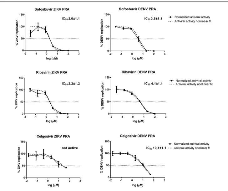
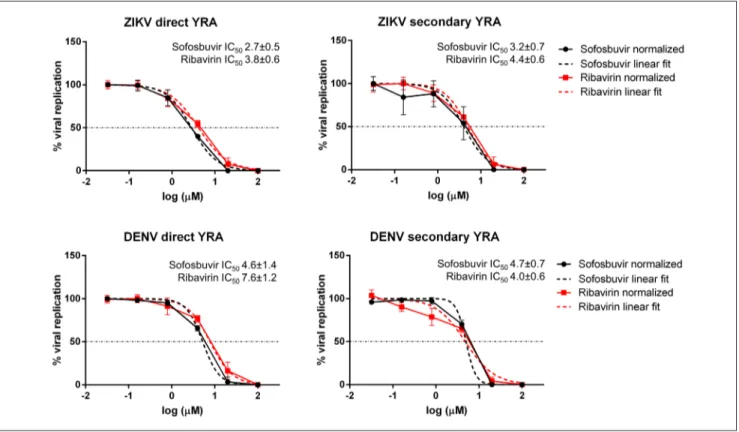
We developed a robust, easy-to-perform and fast flavivirus immunodetection assay, which allows the quantification of ZIKV and DENV viral antigen in infected cells. The system uses a specific monoclonal antibody which binds to the fusion loop of domain II of the envelope protein, which is conserved among flaviviruses. The protocol was thoroughly assessed in terms of precision and accuracy and validated by determining the inhibitory effect of reference compounds (i.e. sofosbuvir and ribavirin). This assay can be applied as the read-out of a direct yield reduction assay and viral stocks generated during the first replication cycle can be transferred to a second cell culture in the absence of drug, to better characterize antiviral activity exerted at steps occurring later than envelope expression (Vicenti et al., 2020a).
Some studies reported that sofosbuvir, an RNA-dependent RNA polymerase (RdRp) inhibitor licensed for the treatment of Hepatitis C Virus (HCV) infection, exerts a measurable antiviral activity against the flaviviruses ZIKV and Yellow Fever Virus (YFV), both in vitro and in animal models, as well as against DENV in
vitro. Since the flavivirus RdRp-coding non-structural protein 5 (NS5) is well conserved among flaviviruses,
2
experiments, we described for the first time sofosbuvir antiviral activity against WNV in the low micromolar range, as well as its genetic barrier through in vitro resistance selection experiments. Moreover, two collaborations, one with the Biophysics Institute of the National Research Council (Milano) and another with the Department of Biotechnology, Chemistry and Pharmacy of the University of Siena, allowed us to define the in vitro enzymatic activity of sofosbuvir using the purified WNV RdRp and to assess the role of the mutations observed during in vitro selection experiments through molecular docking experiments, respectively (Dragoni et al., 2020).
In the area of drug discovery, the HML is currently engaged in a project titled “ORIGINALE CHEMIAE in Antiviral Strategy” which was granted as a PRIN proposal (Progetti di Ricerca di Rilevante Interesse Nazionale). The project is aimed at exploiting Multi-Component Chemistry to synthesize promising broad-spectrum antivirals, which represent an attractive option to treat new emerging viral diseases. The project consists in a network of laboratories working in antiviral drug discovery and development from different Italian Universities (Tuscia, Parma, Roma Tor Vergata, Perugia, Siena and Roma Sapienza). The HML task in this project is to define the antiviral activity of candidate molecules in in vitro standardized virus-cell systems, against DENV, WNV, ZIKV, HIV-1 and the newly discovered SARS-CoV-2.
Italy has been one of the most and earliest affected countries by the CoV-2 pandemic. To trace SARS-COV-2 evolution, an Italian network named SCIRE (SARS-CoV-2 Italian Research Enterprise) was composed, consisting of 14 clinical centers, including the HML. The main objective of the SCIRE group was to characterize the COVID-19 Italian outbreak by full-length SARS-CoV-2 genome analysis during the first wave of the pandemic. Although the virus has been remarkably stable in its genetic make-up so far, molecular surveillance is warranted to follow the epidemic and deliver knowledge on the correlation between virus variants and clinically relevant properties, such as transmission rates, disease severity, response to treatment (Lai et al., 2020).
The introduction and continuous progress of antiretroviral therapy (ART) has brought a dramatic improvement of the quality and duration of life of HIV positive people, transforming HIV-1 infection from a fatal disease to a chronic manageable condition. However, ART is not able to clear the infection, since HIV-1 is able to persist in infected cells for several years. Therefore, strategies to eradicate or control HIV-1 without ART are a high priority. In recent years, most efforts have been focused on the so-called “Shock and kill” strategy. Few recent studies suggest that Maraviroc (MVC), the first approved anti-HIV-1 agent targeting a cellular factor, may exert a latency reversing activity in addition to its antiviral activity, hence representing a unique drug capable of combining the ability to awaken the latent provirus and block new infections. However, since the potential of MVC as a latency reversing agent was based on limited published data (López-Huertas et al., 2017; López-Huertas et al., 2020; Madrid-Elena et al., 2018), we investigated MVC ability to mediate HIV-1 induction in three cell line models, as well as in ex vivo CD4 T cells collected from six patients with suppressed viremia (Vicenti et al., 2020b).
3
1. Introduction – Flaviviruses
1.1 General overview and epidemiology
The Flaviviridae family includes four different genera of spherical and enveloped viruses with unsegmented positive-strand RNA. Among them, the Flavivirus genus includes more than 70 viruses, prevalently transmitted by insect vectors, mainly the Aedes and Culex mosquitoes; thus, they are included in the arbovirus (arthropod-borne) ecological group (Lindenbach et al., 2007; Payne, 2017). Flaviviruses include clinically relevant human pathogens such as Dengue (DENV), West Nile (WNV), Yellow Fever (YFV) and Zika (ZIKV) viruses. In the last years, these pathogens have dramatically increased in their incidence, disease severity and/or geographic range (www.who.int). This epidemic potential is related to many factors, such as adaptation of insect vectors, climatic change, deforestation and globalization. As a matter of fact, it is estimated that half of the global population may be at risk of infection with one or more of these viruses (Huang et al., 2014; Pierson and Diamond, 2020).
WNV was first isolated in 1937 (Smithburn et al., 1940) and since then, only few sporadic infections, characterized by a typically self-limited and minor illness, were reported in regions of Africa, the Middle East, Asia and Australia. However, in 1999 a WNV strain was responsible for a large and dramatic outbreak in New York City, that caused severe neurologic disease in seven humans in the New York area, as well as a large number of avian and equine deaths, spreading throughout the USA in the following years. Nowadays, WNV is endemic in Africa, Europe, Asia, north America, Australia, and the Middle East, with increasing number of human cases reporting severe diseases (Clark and Schaefer, 2020; Pierson and Diamond, 2020; www.who.int). Up to nine different WNV genetic lineages have been identified (Fall et al., 2017). The strains most often involved in human outbreaks, and thus clinically relevant, belong to lineages 1 and 2. Viruses from lineage 1 are divided in two clades, Clade 1a and Clade 1b, and they have been mostly isolated in Africa, Europe, Middle East, Asia, Oceania and north America (Fall et al., 2017), with Clade 1a more frequently associated to neurological outcome. Lineage 2 isolates appear to be less virulent to humans than lineage 1 and are found prevalently in Sub Saharan Africa, even though outbreaks both in animals and humans have been reported in central and eastern Europe (Donadieu et al., 2013; Magurano et al., 2012).
Since its discovery in 1947 (Dick et al., 1952), ZIKV remained confined to the equatorial zone across Africa and Asia, causing sporadic mild-febrile illness in a small number of humans. However, in 2007 a severe human outbreak was reported in Yap Island, followed by another large outbreak in French Polynesia and other Pacific Islands (Song et al., 2017). Later on, ZIKV rapidly spread to Brazil and other regions of the Americas, resulting in millions of infections. By March 2016, ZIKV transmission had been reported in 34 south and central American countries and territories (Faria et al., 2016; Fauci and Morens, 2016; Pierson and Diamond, 2020). Differently from previous outbreaks, ZIKV infection caused unique clinical features, including congenital malformations and other neurological disorders, leading the World Health Organization (WHO) to declare ZIKV “a public health emergency of international concern” (Hastings and Fikrig, 2017; WHO Report, 2016). Phylogenetic studies have revealed that ZIKV has evolved into African and Asian lineages (Liu et al., 2017). The African lineage strains circulated in central Africa, Senegal, Uganda and Nigeria and they were mainly detected from samples of enzootic vectors. The Asian lineage was mainly isolated throughout southeast Asia as Micronesia, Cambodia and French Polynesia. Following the introduction of ZIKV into the Americas, a new lineage within this cluster (lately defined as American strain) emerged, which has been responsible for the recent epidemics (Weaver et al., 2016). Interestingly, compared with the pre-epidemic strains from Asian lineage, the American epidemic strain has undergone the A188V substitution of NS1 protein, likely responsible for enhanced pathogenicity and increased disease severity (Liu et al., 2017; Pierson and Diamond, 2020; Xia et al., 2018). In 2019, all over the Americas about 35,000 ZIKV infections were reported (www.paho.org).
DENV is endemic in more than 100 countries of the southeast Asia, the Americas, the western Pacific, Africa and the eastern Mediterranean regions (www.who.int). Over the past 70 years, the number of people
4
infected has increased about 30-fold, making DENV the most prevalent arthropod-borne viral disease in the world. DENV causes an estimated 390 million total infections, 100 million clinically apparent cases and 500,000 presentations of severe dengue per year worldwide, and frequent DENV outbreaks have progressively increased in recent years (Bhatt et al., 2013; Maria G. Guzman and Harris, 2015; Pierson and Diamond, 2020). DENV is classified into four serotypes (DENV-1, DENV-2, DENV-3 and DENV-4) sharing limited homology (around 60–75%) at amino acid level. Within the same serotype, viruses differ about 3% at amino acid level, 6% at nucleotide level and are phylogenetically divided into genotypes and clades. Genetic variations between serotypes and clades are important determinants of differential viral fitness, virulence, and epidemic potential (M.G. Guzman and Harris, 2015; Weaver and Vasilakis, 2009). All four DENV serotypes circulate together in tropical and subtropical regions; however, the serotype 2 seems to be associated with increased disease severity (Chen et al., 2007; Vaughn et al., 2000).
1.2 Genome organization and structure
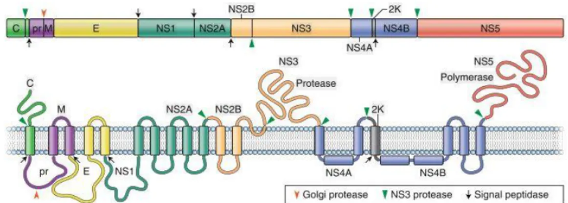
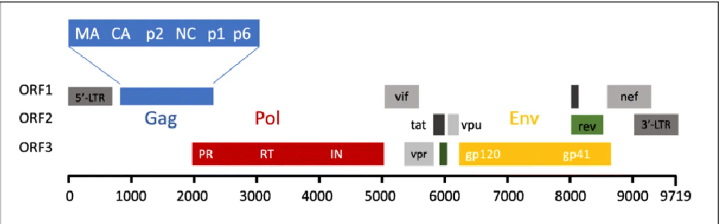
Flavivirus virions are around 50 nm in diameter, surrounded by a lipid envelope with surface proteins arranged in icosahedral-like symmetry and containing a single genomic RNA of positive-sense polarity of nearly 11 kb in length. The flavivirus genome (Fig 1) contains a single open reading frame, which is flanked by 5’ and 3’ untranslated regions (UTR), encoding a large polyprotein that is post-translationally cleaved by host and viral proteases (Lindenbach et al., 2007; Pierson and Diamond, 2020).
Fig 1. Flavivirus genome organization and structure. The genome is translated as a polyprotein, which is subsequently cleaved by viral and host cell proteases, which are indicated in the figure. (Adapted from Pierson and Diamond, 2020) The non-coding 5′UTR of flaviviruses is around 100 nucleotides in length and is not well conserved among different flaviviruses. It contains a methyl-G-cap structure and two conserved stem-loop regions with secondary structures within this region, modulating viral RNA synthesis and translation. The non-coding 3′UTR ranges from 400 to 700 nucleotides in length, depending on the virus species, but sharing similar patterns of conserved sequence and structures among flaviviruses. 3′UTR can be divided into three domains and is important for the interaction with host and viral proteins (Lindenbach et al., 2007; Ng et al., 2017).
The N-terminal region of the polyprotein encodes for the three structural proteins, capsid (C), precursor membrane (prM) and envelope (E) which are present in the virion particle, while the C-terminal region includes the seven non-structural proteins (NS1, NS2A, NS2B, NS3, NS4A, NS4B and NS5) which accomplish essential steps during infection but are not present in the viral particle (Laureti et al., 2018; Lindenbach et al., 2007; Pierson and Diamond, 2020). The NS proteins are involved in the post-processing of structural proteins and are essential in viral replication, virion assembly, and defense against the host immune response (Lindenbach et al., 2007; Morrison et al., 2012).
The E protein is the major surface glycoprotein (≈53 kDa) and is involved in viral entry, pathogenesis and immune response. The mature virion is constituted by ninety dimers of the E glycoprotein arranged in a
5
herringbone pattern with icosahedral-like symmetry (Roby et al., 2015). The E protein is a three-domain structure, which is modified post-translationally by the addition of one or two asparagine-linked carbohydrates. Once glycosylated, it is involved in cell attachment. The folding of the E protein in the endoplasmic reticulum (ER) is facilitated by interactions with the structural prM protein shortly after synthesis (Mukhopadhyay et al., 2005; Pierson and Diamond, 2020; Rey et al., 2017; Zhang et al., 2004).
Flavivirus prM is a membrane glycoprotein of approximately 19–21 kDa mainly acting as a chaperone to promote folding and assembly of the E proteins (Konishi and Mason, 1993; Lorenz et al., 2003). Together with the E protein, prM forms an integral part of the flavivirus envelope that, in the mature form (M), is combined under a well-organized icosahedral architecture. Cleavage of prM to M is mediated by a host furin-like serine protease during the transit of immature virions through the trans-Golgi network. This step is required for the formation of infectious mature virions (Li et al., 2008). Since the Golgi compartment has a pH around 5.5, to avoid the premature fusion with the host membranes of the newly assembled virions, the prM peptide is not readily released from the virus surface, but it remains bound to the E protein, guaranteeing correct release of new virions (Yu et al., 2008; Zheng et al., 2014).
The nucleocapsid (N) viral core is formed by C protein subunits complexed with the single-stranded RNA viral genome. C protein is a small (14 kDa) cytosolic protein with structurally conserved α-helices, able to bind either viral nucleic acids or host lipids and it is necessary for the incorporation of the viral genome into the virion (Jones et al., 2003; Pierson and Diamond, 2020). C protein incorporation into the virion is regulated further by the coordinated cleavage of the polyprotein by the viral NS2B–NS3 serine protease.
Among non-structural proteins, NS1 is highly conserved with a molecular weight of 46-55 kDa, depending on the extent of glycosylation. The glycosylation of NS1 is fundamental for efficient secretion, virulence and viral replication (Somnuke et al., 2011). Indeed, in vivo experiments have shown that loss of N-linked glycans on NS1 results in attenuated DENV and WNV infection in mice (Pryor and Wright, 1994; Whiteman et al., 2010). NS1, in all its forms, is a multifaceted protein that hijacks the host replication system and interferes with the host immune response (Muller and Young, 2013). Indeed, soluble NS1 interacts both with the complement system and Toll-like receptor 4 (TLR4) to guarantee the survival of secreted virus progeny (Conde et al., 2016) and to initiate the inflammation process, respectively. This process further induces peripheral blood mononuclear cells (PBMCs) and macrophages to activate the inflammatory response (Muller and Young, 2013).
NS2 consists of two subunits, NS2A and NS2B. NS2A is a relatively small (≈22 kDa) protein containing several transmembrane domains and it is an activator of NS1. Besides its apparent roles in RNA replication and virus assembly, DENV-2 and WNV NS2A have also been shown to act as an interferon (IFN) antagonist by inhibiting IFN signaling during infection (Liu et al., 2006; Muñoz-Jordán et al., 2003). NS2B is a small membrane-associated protein (14 kDa) that forms a stable complex with NS3 and acts as a cofactor for the NS2B-NS3 serine protease (Clum et al., 1997; Lindenbach et al., 2007).
The NS4A and NS4B are small proteins (16 kDa and 27 kDa, respectively) participating to the virus replication complex formation on the ER membrane. NS4A consists of four transmembrane helices and an N-terminal cytosolic region. NS4A induces the rearrangements of ER membranes and interacts with NS1 (McLean et al., 2011; Miller et al., 2007), while NS4B colocalizes with NS3 and viral dsRNA in ER-derived membrane structures presumed to be sites of RNA replication (Lindenbach et al., 2007).
NS3 is a large (≈70 kDa) multifunctional protein which is well-conserved among the Flavivirus genus, with a sequence identity of approximately 65% among WNV, DENV and ZIKV (Lindenbach et al., 2007; Weber et al., 2015). NS3 has protease, helicase, nucleoside triphosphatase (NTPase) and RNA triphosphatase (RTPase) enzymatic activities. The N-terminal region of the NS3 protein constitutes the serine protease domain (NS3pro). Proper folding and catalytic activity of the NS3pro domain require NS2B as cofactor. NS3hel comprises three subdomains and harbours the enzymatic activities of NTPase and RTPase as well as dsRNA
6
unwinding activity (Wengler and Wengler, 1993, 1991). Some evidences suggest that one of these subdomains mediates the interaction between NS3 and NS5; disruption of this interaction could affect viral replication (Brooks et al., 2002; Tay et al., 2015). The RNA helicase is responsible for the unwinding of dsRNA intermediates in order to release the newly generated viral genome and to make the negative strand available as template for another round of viral genome synthesis. Moreover, NS3hel removes secondary structures from the viral RNA, especially in the 5’ and 3’ untranslated regions, facilitating the RTPase mediated capping. NTPase hydrolysis provides the chemical energy to power the translocation and unwinding processes, although the precise mechanism coupling these two activities remains elusive (Wang et al., 2009; Wengler and Wengler, 1991).
NS5 is the largest (103 kDa) and most conserved of the flavivirus proteins with a sequence identity of approximately 68% among WNV, DENV and ZIKV. It contains an N-terminal methyltransferase (MTase) and a C-terminal RNA-dependent RNA polymerase (RdRp), coupled via a short linker (Duan et al., 2019; Dubankova and Boura, 2019). The MTase domain (residues 1-≈265, 30 kDa) is responsible for capping the viral RNA through the guanylyl transferase and the N7 and 2’-O-ribose methylation activity, both of which are required to increase the stability and to prevent degradation by 5′–3′ exoribonucleases of the newly synthesized RNAs (Devarkar et al., 2016; Züst et al., 2011). Additionally, the 2′-O-methylation protects viral RNA from being recognized by host cell sensors that stimulate the production of IFNs (Hyde and Diamond, 2015). The interaction of the MTase and the RdRp domains affects the replication activity, since full-length NS5 has higher polymerase activity than the RdRp alone for efficient viral replication (Duan et al., 2019; Potisopon et al., 2014; Saw et al., 2019).
Flavivirus RdRp carries out new RNA synthesis from the 3′ end of the viral templates without any primer (de
novo initiation). The structure of the RdRp is well conserved among flaviviruses (Dubankova and Boura,
2019; Godoy et al., 2017; Lu and Gong, 2017; Malet et al., 2007; Yap et al., 2007). RdRp (residues ≈272–900, 73 kDa) is divided in seven conserved motifs (from A to G) and it adopts a classic “right-hand” structure consisting of three subdomains with fingers, palm and thumb. Together, these regions are organized in a flat structure with three channels (the template entry, dsRNA, and the NTP entry channels) (Duan et al., 2019; Dubankova and Boura, 2019; Malet et al., 2007). Motifs A-B-C-D are located in the highly conserved palm domain and are important for dNTPs binding and catalysis; moreover, motif C, as well as motif E, interact with the backbone of the RNA product. Motif F, located in the finger domain, consists of 3-4 sub-motifs depending on the viral species and it is involved in the stabilizing of the nascent base pair. Motif G is proposed to regulate access of the ssRNA substrate to the template channel and/or RdRp translocation (Dubankova and Boura, 2019; Malet et al., 2007; Šebera et al., 2018; Yap et al., 2007; Zhao et al., 2017). In addition, a priming loop identified in the thumb subdomain is thought to play a major role in both ensuring correct de novo initiation and in controlling the conformational changes during the RdRp activity (Duan et al., 2019; Sahili and Lescar, 2017). RdRp appears to be responsible for the NS3-NS5 interaction in several flaviviruses, however the exact function of cooperation between these two enzymatic proteins is not completely understood (Duan et al., 2019; Lindenbach et al., 2007; Tay et al., 2015).
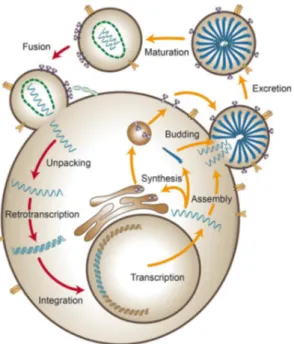
1.3 Flavivirus replication cycle
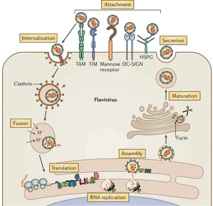
The flavivirus replication cycle (Fig 2) initiates with the stable attachment of the virion to the surface of the target cells, mainly keratinocytes, dermal fibroblast and Langerhans cells of the epidermis, through the interaction between viral surface E protein and host attachment/receptor molecules (Neufeldt et al., 2018). Host proteins defined as receptors are essential for the entry of viruses, since they catalyze conformational events. Although several host factors that increase the efficiency of flavivirus binding and infection of cells have been identified, they are not required to trigger the structural transitions that lead to membrane fusion. Therefore, these are defined as attachment factors (Pierson and Diamond, 2020). Indeed, a number of attachment factors/receptors have been identified for flaviviruses, including αvβ3 integrin, GRP78/BiP, CD14 or a closely related molecule (Mukhopadhyay et al., 2005). The most extensively studied attachment
7
factor is the C-type lectin dendritic cell-specific ICAM-grabbing non-integrin (DC-SIGN), mainly present at the surface of dermal dendritic cells and macrophages.
Fig 2. Flavivirus replication cycle. DC-SIGN: C-type lectin; TIM: T cell immunoglobulin mucin domain protein 1; TAM: tyrosine protein kinase receptor 3 (TYRO3)–AXL–MER; HSPG: heparan-sulfate proteoglycans. (Adapted from Neufeldt et al. 2018).
In addition, highly sulfated glycosaminoglycan, such as heparan-sulfate proteoglycans, act as attachment factors and have the function to concentrate viral particles at the target cell surface before their interaction with primary receptors. Recent studies show that several cellular receptors, as protein of TIM (T cell immunoglobulin and mucin domain) and TAM (TYRO3, AXL and MER) receptor families can mediate flavivirus entry through interactions occurring only between negatively charged lipids such as phosphatidylserine in the viral membrane (Diamond and Pierson, 2015; Hamel et al., 2015; Meertens et al., 2012; Mercer and Helenius, 2010; Pierson and Diamond, 2020).
After attachment, flaviviruses are internalized via clathrin-coated pits and transferred to a pre-lysosomal endocytic compartment. The low pH environment triggers viral E protein rearrangement, driving the fusion of the viral lipid envelope with cellular endosome membranes. After membrane fusion, the viral genomic RNA is released into the cytoplasm where the viral replication starts in vesicle packets, which contain the small hydrophobic NS proteins, dsRNA, nascent RNA and, presumably, some host factors (Payne, 2017). The penetration of the capped ssRNA(+) viral genome into the cytoplasm allows for the direct translation of the viral polyprotein on the rough ER. Replication occurs on virus-induced invaginations of the ER: NS1, prM and E are translated into the ER lumen, the transmembrane domains of NS2A, NS2B, NS4A and NS4B are translated into the ER membrane, whereas C, NS3 and NS5 remain in the cytoplasm. NS5 mediates the beginning of replication with the synthesis of a full genome-length ssRNA(-), followed by the formation of a dsRNA replication intermediate, which then serves as a template for the synthesis of additional ssRNA(+). Concomitantly, NS3 helicase interacts with NS5 to unwind the dsRNA. The progeny ssRNA (+) is subsequently capped at its 5′ end and methylated to form the cap RNA structure. Following RNA synthesis, newly copied RNA molecules are either recycled for translation and replication or alternatively, extruded from the vesicle to bud out on ER membranes and packaged into nascent virions. The newly synthesized
8
immature flavivirus particles are produced by budding of the C protein and the associated genomic RNA, which forms the nucleocapsid into ER-derived membranes and is studded with prM and E proteins. These immature particles traffic along the secretory pathway across the Golgi complex, where undergo the cleavage of the pr portion from M protein by a trans-Golgi resident furin-like protease, that promotes particle maturation prior to their release from the infected cell (Arakawa and Morita, 2019; Payne, 2017; Pierson and Diamond, 2020).
1.4 Transmission
The evolution and the epidemiological characteristics of flaviviruses are associated to a combination of limitations imposed by the arthropod vector, the vertebrate hosts, the ecology and the influence of human commercial activities.
DENV and ZIKV transmission is mainly associated with the mosquito vector Aedes aegypti and Aedes
albopictus; WNV is mostly transmitted by the members of the Culex pipiens spp (Ciota, 2017; Huang et al.,
2014).
The African ZIKV lineage is thought to be maintained via the enzootic transmission cycle primarily between non-human primate and mosquitoes, with humans as incidental hosts. However, the Asian ZIKV lineage has gained the ability to sustain transmission in a human-endemic cycle (sub-urban transmission cycle), thus allowing humans to serve as the carrier, multiplier, and source of ZIKV for uninfected mosquitoes (Althouse et al., 2016). Differently, DENV is the only known arboviruses that has fully adapted to humans, having lost the need for an enzootic cycle for maintenance (Payne, 2017; www.who.int). WNV enzootic cycle is between mosquitoes and birds, the latter regarded as “amplifier hosts” and with mammals serving as “dead-end” hosts, since generally they do not develop high levels of virus in their bloodstream (Pierson and Diamond, 2020).
Despite vector-borne transmission is the favorite route for ZIKV, DENV and WNV infections, transmission through transplanted organs, transfused blood, contact with body fluids from a highly viremic patient, transplacental transmission and occupational transmission have been observed (Iwamoto et al., 2003; Julander et al., 2006; Musso et al., 2017; Swaminathan et al., 2016; Williamson et al., 2017; Wiwanitkit, 2009). ZIKV sexual transmission is possible from both asymptomatic and symptomatic infections and the duration of infectivity of genital fluids is unknown (Hastings and Fikrig, 2017; Musso et al., 2015). Recently, ZIKV vertical transmission has been extensively investigated and a high risk of fetal injury connected to ZIKV infection during the first trimester of pregnancy has been evidenced (Cauchemez et al., 2016; Honein et al., 2017).
1.5 Clinical manifestations and pathogenesis
Although flavivirus infections in humans are asymptomatic in the 80% cases, clinical manifestations range from influenza-like symptoms, frequently misdiagnosed, to severe conditions leading eventually to death. After the bite of an infected mosquito, the duration of the incubation period lasts a few days (average 4 to 7 days) (Gubler, 1998; Sejvar, 2016; Slavov et al., 2016).
It is estimate that 1 in 4 DENV infections are symptomatic, showing mild to moderate acute febrile illness. However, approximately 5% of symptomatic cases of DENV infection progress to a more severe disease, whose clinical progression can be differentiated into three phases: febrile phase, critical phase and recovery phase. The febrile phase, also named Dengue Fever, may cause, over the classical flu-like symptoms, transient maculopapular rash and mild-hemorrhagic illness. The critical phase occurs at the end of febrile phase before the appearance of specific antibodies, and could be associated to Dengue Hemorrhagic fever (DHF) which has four severity grades, with the more severe grades (III and IV) classified as dengue shock syndrome (DSS) (Rodriguez-Roche and Gould, 2013). Common manifestations include skin
9
hemorrhages such as petechiae, purpuric lesions and ecchymosis. Concomitantly, severe plasma leakage can lead to shock or fluid accumulation with respiratory distress, severe bleeding, which may further develop into multi-organ failure with fatal outcome (Kalayanarooj, 2011). Although the appearance of DHF/DSS might occur at any DENV exposure, it seems to be more frequent in secondary DENV infections, particularly in children or in newborns who are partially protected by maternal antibodies (Jain and Chaturvedi, 2010).
Symptomatic ZIKV clinical manifestations occur as acute onset of fever lasting for several days to a week, along with maculopapular rash in most of the patients. Other commonly reported symptoms include myalgia, arthralgia, headache and conjunctivitis. Although severe disease requiring hospitalization is uncommon, during the recent outbreak in the Americas, ZIKV infection has been associated to severe neurological complications, such as (i) the Guillain-Barré syndrome (GBS), a serious and life-threatening neurological disorder characterized by progressive muscular weakness, encephalitis and myelitis in adults and (ii) microcephaly and other severe fetal brain defects in fetuses and neonates caused by maternal ZIKV infection (Grossi-Soyster and Desiree La Beaud, 2017; Kazmi et al., 2020).
WNV infections cause disease in approximately 20% of infected humans; the vast majority of WNV symptomatic patients develops an acute, systemic febrile illness (West Nile fever, WNF) and less than 1% develops neuroinvasive disease including aseptic meningitis, encephalitis, or an acute poliomyelitis-like syndrome. The WNF is the predominant clinical syndrome and generally occurs with the abrupt onset of fever, headache, fatigue, myalgia and rash. The rash may be transient and appears to be more frequently seen in WNF than in more severe illness manifestations (Bai et al., 2019; Zannoli and Sambri, 2019).
Flavivirus virions released after a mosquito bite are able to infect a large variety of human target cells such as fibroblasts, keratinocytes, dendritic cells, monocytes, and endothelial cells.
DENV and ZIKV both initially infects cells in dermis and epidermis, such as immature Langerhans cells and keratinocytes. Infected cells migrate to the lymph nodes, triggering the recruitment of monocyte-macrophage lineage cells. As a result, the number and variety of infected cells increase and disseminate throughout the lymphatic system (Martina et al., 2009).
Concerning DENV infection, mononuclear cells are stimulated to produce cytokines and chemokines that provide an essential protective role during DENV infection. However, this “storm” of inflammatory cytokines and other inflammatory mediators acts on the endothelium and alters normal fluid barrier functions, leading to increased plasma leakage (Harapan et al., 2020; Martina et al., 2009). In addition, NS1 released from infected cells is proposed to modulate complement signaling cascade, that triggers cellular reactions further stimulating the production of inflammatory cytokines (Avirutnan et al., 2006; Diamond and Pierson, 2015). In addition to innate immunity, cellular and humoral immunity play an essential role too. During the infection, T cells and cross-reactive memory T cells are produced, as well as antibodies targeting the principle DENV epitopes (i.e. E, NS1 and pre-M proteins). However, whether these immune responses protect against or exacerbate subsequent infections is still controversial. Indeed, if the antibodies are not completely neutralizing against the new DENV strain, they can facilitate viral entry into Fc receptor-positive cells during a subsequent infection. Such phenomenon, defined as antibody-dependent enhancement (ADE), is associated with both increased DENV infectivity and the suppression of host immune responses. Indeed, is likely that severe dengue may occur in those experiencing a secondary infection with heterotypic strain of DENV and in infants who are born to dengue-immune mothers with primary anti-DENV antibody responses (Harapan et al., 2020; Valentine et al., 2020).
Following infection, ZIKV triggers innate immunity responses that exert antiviral and pro-inflammatory effects. The over activation of the immune and inflammatory pathways, such as T- and B-cells mediated immunity, leukocyte-mediated immunity and cytokine production, may attract T cells and other leukocytes to the site of infection, leading to tissue damage (Wang et al., 2017). The humoral response may drive the predisposition to severe illness too; indeed, it was observed in cell culture that the rates of ZIKV infection
10
can be enhanced by the reactive anti-DENV antibodies (Priyamvada et al., 2016). In addition, cross-reactive human anti-ZIKV antibodies can enhance DENV infection (ADE activation) in cell culture and in mice (Stettler et al., 2016) and also in humans, as described recently (Katzelnick et al., 2020).
The overstimulation of the immune system and the consequent cascade of pro-inflammatory cytokines play a role in the pathogenesis of GBS; indeed, it was suggested that high levels of CXCL10 in ZIKV patients may contribute to neuronal damage, potentially targeting peripheral nerves (Maucourant et al., 2019; Naveca et al., 2018). The increase of microcephaly in infants caused by maternal ZIKV infection, suggests that ZIKV is capable of bypassing the placental barrier and infect human placental macrophages, resulting in the disruption of the placenta by strong activation of antiviral immune response (Wen et al., 2017). Moreover, ZIKV has the capacity to infect human neural progenitor cells and the microglia triggering their apoptosis (Ferraris et al., 2019; Zheng et al., 2015). Activation of microglia leads to the production of pro-inflammatory cytokines and cytotoxic molecules, such as nitric oxide, that contribute to neuronal damage during the fetal brain development (Maucourant et al., 2019).
Following an infectious mosquito bite, WNV replicates locally at the injection site in the keratinocytes and Langerhans cells of the epidermis. The local virus replication is enhanced due to the immune modulation of the host response by the mosquito saliva. It has been hypothesized that infected Langerhans cells migrate to the draining lymph nodes in which the virus replicates, further spreading the infection through the lymphatic system. Following WNV infection, a cascade of proinflammatory cytokines are upregulated as part of the innate immune response. However, overexpression and continuous upregulation of inflammatory cytokine genes may be detrimental by enhancing the severity of infection and/or inflammation. The exact mechanism of neuroinvasion by WNV is still unknown, despite several routes of entry have been proposed. Likely, free virus particles are able to across the disrupted blood-brain barrier (BBB) through a “transudative” mechanism, due to the increased vascular permeability caused by pro-inflammatory cytokines and chemokine (Suen et al., 2014). Alternatively, entry might occur via the “Trojan horse” mechanism, mediated by infected leukocytes trafficking into the central nervous system (CNS) through the leaking BBB (Lim et al., 2011), or by the “transneural” mechanism consisting in the virus migration following motor and sensory nerves from the point of entry (i.e. peripheral somatic nerves into the CNS and from the olfactory nerve into the CNS) (Habarugira et al., 2020). Following invasion of CNS, the virus directly infects neurons, and less frequently astrocytes, leading to neuronophagia and cell death. While nearly all brain regions may be affected, WNV appears to have a specific neurotropism for neurons in the basal ganglia, thalamus, and brainstem (Stonedahl et al., 2020).
1.6 Current status of antivirals and vaccines development 1.6.1 Antivirals development
Clinical availability of effective antivirals for the management of severe flavivirus diseases is an unmet medical need. Despite the increase of incidence of viral diseases, neither specific treatments nor immunoprophylaxis are currently available and the clinical management of symptomatic patients remains based on supportive care (i.e. intravenous infusion of fluids, respiratory support, and prevention of secondary infections, as suggested by WHO and CDC current guidelines) (www.who.int; www.cdc.gov). Several promising drug candidate molecules have been reported via high-throughput compound library screening, by de novo design targeting viral or host proteins.
The most promising viral targets for de novo design are the NS5 RdRp and the NS3 protease, and, to a lesser degree, E-glycoprotein, C, NS4B, NS3 helicase and NS5 MTase (Boldescu et al., 2017). A different therapeutic approach is targeting cellular factors required for the viral life cycle. Targeting a cellular factor rather than a viral protein is an attractive solution, since host cellular proteins are less prone to mutations, as opposed to the high rate of mutations of viral proteins, hence overcoming the emergence of resistance mutations. On the other hand, targeting a cellular host factor has a higher potential for the development of side effects. Options for successful host targeting agents include blocking a function which is redundant for
11
the cell but essential for the virus or selectively impairing a cellular pathway only in virus-infected cells. Examples of molecules that target host factors exerting antiviral activity on flavivirus are mycophenolic acid (MPA) and cyclosporines, inhibiting the cellular enzyme inosine monophosphate dehydrogenase and cyclophilin A (CyPA), respectively (Barrows et al., 2016; Diamond et al., 2002; Morrey et al., 2002; Qing et al., 2009), and lovastatin a cholesterol synthesis inhibitor, thought to limit membrane mobilization required for the assembly of the viral replication complex (Españo et al., 2019; Mackenzie et al., 2007; Martínez-Gutierrez et al., 2011). A wide range of host factor involved in virus infection cycle are currently under investigation for de novo design of antiviral compounds (Brai et al., 2019, 2016; DeWald et al., 2020; Giovannoni et al., 2020; Yang et al., 2020)
Since the discovery and approval of new drugs takes a long time, an increasing trend is to take advantage of the cost- and time-saving benefits of drug repurposing. There are some relevant examples of drug repurposing in the flavivirus antivirals field. Ribavirin, a synthetic guanosine nucleoside analogue and one of the first broad-spectrum antivirals licensed by Food and Drug Administration (FDA) against several viruses (Fernandez et al., 1986; Khan et al., 1995; Lau et al., 2002), has been shown inhibitory activity in vitro against ZIKV (Kim et al., 2018; Vicenti et al., 2018), DENV (Koff et al., 1982) and WNV (Anderson and Rahal, 2002; Jordan et al., 2000). However, ribavirin was not effective for treating WNV disease (Chowers et al., 2001) and the possible use against ZIKV infection is contraindicated in pregnancy due to its teratogenic properties. With the same strategy, the anti-HCV nucleoside analogue targeting RdRp, sofosbuvir, has shown anti-ZIKV activity in vitro (Sacramento et al., 2017; Vicenti et al., 2018) and in animal models, preventing the vertical transmission of ZIKV in pregnant mice (Mesci et al., 2018), as well as anti-DENV activity in vitro (H. T. Xu et al., 2017), and anti-YFV activity both in vitro and in vivo (de Freitas et al., 2019). Exploiting a similar mechanism of action, BCX4430, an adenosine analogue originally designed as an anti-HCV agent, exerts antiviral activity against a wide range of RNA viruses including flaviviruses and filoviruses such as Ebola virus. The broad-spectrum potency of BCX4430 has been proved against ZIKV both in vitro and in vivo and against WNV in vitro (Eyer et al., 2017; Julander et al., 2017a).
1.6.2 Vaccines development
More than 80 years ago, the development of YFV 17D live-attenuated vaccine was considered a landmark in the history of flavivirus vaccines (Barrett, 2017). Effective anti-flavivirus vaccines are also available for active immunization against Japanese encephalitis and tick-borne encephalitis virus (Heinz and Stiasny, 2012), even if with some safety concerns (Chong et al., 2019).
To date, there are several open clinical trials testing a range of ZIKV vaccine candidates, including DNA vaccines, peptides, mRNA vaccines, purified inactivated vaccines and recombinant viral-vector vaccines (www.who.int; Chong et al., 2019). One of the most important goals of a ZIKV vaccine is to prevent the congenital ZIKV syndrome. Currently, some progress has been documented on nonhuman primates as preclinical pregnancy models to test vaccine efficacy (Nguyen et al., 2017; Waldorf et al., 2016).
Despite the lack of WNV vaccines for humans, several vaccines have been successfully developed and licensed for veterinary use. Among them, two are whole inactivated WNV equine vaccines (West Nile Innovator by Pfizer and Vetera by Boehringer Ingelheim). Another commercialized vaccine is the recombinant canarypox-vectored vaccine, expressing the prM and the E protein of the NY99 strain (Recombiteck Equine West Nile Virus Vaccine by Merial-Sanofi Aventis) (Brandler and Tangy, 2013). Among the WNV human vaccine candidates, progress has been made in the DNA-delivered subunit and chimeric vaccines (Chong et al., 2019; Habarugira et al., 2020). Two live chimeric/recombinant vaccines and one DNA-vectored vaccine entered phase I clinical trial, while one YFV-17D backbone expressing WNV prM/E recombinant vaccine entered phase II clinical trial (Amanna and Slifka, 2014; Habarugira et al., 2020).
12
Considering the severe clinical outcomes following DENV infections, huge efforts have been made in its vaccine field. On 1st May 2019, FDA has approved Dengvaxia® (CYD-TDV) produced by Sanofi Pasteur, as the first dengue vaccine, already licensed in several countries including European Union, Latin America and Asia. CYD-TDV was constructed using recombinant DNA technology by replacing the sequences encoding prM and E proteins of the YFV 17D vaccine with those encoding the homologous prM and E gene sequences of the four DENV serotypes. The indication of CYD-TDV is for individuals aged from 9 to 45, living in DENV endemic areas and it is not suggested for individuals not previously infected by any DENV serotype or for whom this information is unknown. As a matter of fact, its use has some significant controversies, since an increased risk of severe DENV infection was showed during primary infection of dengue-naive individuals following vaccination; such phenomenon may be explained by ADE mechanism. Indeed, serological studies demonstrated that individuals that were DENV-seropositive at the time of vaccine administration experienced benefit from CYD-TDV, whereas DENV-naive individuals were at increased risk for severe disease over this interval. For this reason, research to find other possible dengue vaccines is still underway and to date, two other live-attenuated tetravalent DENV vaccines are in advanced stages of clinical trials (Chong et al., 2019; Harapan et al., 2020; Pierson and Diamond, 2020).
1.7 Methods to define antiviral activity
Assessment of antiviral effects in vitro is a key approach for the screening of either de novo or repurposed candidate compounds. Among the variety of methods that have been developed (Gong, 2013), cell-based assays are the most predictive methods to define antiviral activity. Several cell-based assays have been developed and can be classified into: i) assays using live viruses, ii) assays that use sub-genomic viral replicons (VRPs), containing a subset of viral genes that are required for replication, and iii) assays using virus-like particles (VLPs), containing viral E protein and prM glycoproteins but no viral RNA (Boldescu et al., 2017).
Assays using live viruses are the reference standard for antiviral screening but with some drawbacks, as the need of high-level biosafety containment facilities, dedicated personnel training, high costs and times to execution. In contrast, VRP and VLP assays can overcome safety concerns and are prevalently based on convenient readouts, such as luminescence and fluorescence. However, since they do not simulate the complete virus life cycle, they are not amenable for the screening of compounds with unknown targets; moreover, VRP and VLP assay results must be validated carefully to avoid false-positive hits.
Assays using live viruses are based on several types of readouts, each characterized by a different degree of accuracy, complexity and cost. Indeed, following virus infection, measurement of virus replication can be performed in different ways, such as quantitative polymerase chain reaction (qPCR) (Gong et al., 2013; Vicenti et al., 2018), microscopy monitoring of cytopathic effect (H. T. Xu et al., 2017) and immunofluorescence-based assays, such as the fluorescence focus assay and the most advanced fluorescence-activated cell sorting assay (Kraus et al., 2007; Payne et al., 2006). Despite these advancements, the classical plaque reduction assay (PRA) is still considered as the gold standard for antiviral screening of compounds against viruses causing cytopathic effect and it is also commonly used for antibody titration in plaque reduction neutralization tests (Cordeiro, 2019). Nevertheless, it has several drawbacks including high labor, long-turnaround time and low throughput, making it not suitable for the analysis of large numbers of compounds or sera. Consequently, the development of accurate, easy-to-perform, and fast cell-based assays is highly valuable to test candidate inhibitors of flavivirus replication.
13
2. Introduction – SARS-CoV-2
2.1 General overview and epidemiology
Coronaviruses (CoVs) are positive sense, single stranded, enveloped RNA viruses with a propensity to cross species barriers and causing disease in humans and animals (Chan et al., 2013). Within the order of
Nidovirales and the suborder of Coronavirineae, lies the family Coronaviridae. The latter is further specified
into the subfamily of Orthocoronavirinae, which consists of four genera: the alpha, beta, gamma and delta CoV (Fehr and Perlman, 2015). Whereas alphacoronaviruses and betacoronaviruses exclusively infect mammalian species, gammacoronaviruses and deltacoronaviruses have a wider host range that includes avian species (V’kovski et al., 2020).
Prior to the recent outbreaks, CoVs were only though to cause mild, seasonal, self-limiting respiratory infection in humans associated with symptoms of the ‘common cold’. Two of these human coronaviruses (HCoVs) are alphacoronaviruses, HCoV-229E and HCoV-NL63, while the other two are betacoronaviruses, HCoV-OC43 and HCoV-HKU1. HCoV-229E and HCoV-HKU1 were isolated nearly 50 years ago (Hamre and Procknow, 1966), while the last two were recently identified (Fehr and Perlman, 2015). In contrast to the mentioned CoVs, Severe Acute Respiratory Syndrome Coronavirus (SARS-CoV), Middle East Respiratory Syndrome Coronavirus (MERS-CoV) and the newly emerged SARS-CoV-2, all belonging to the betacoronavirus genera, subgenus Sarbecovirus, Merbecovirus and Sarbecovirus, respectively, are highly pathogenic and able to cause severe respiratory infection among humans (Gorbalenya et al., 2020; V’kovski et al., 2020).
During November 2002 an epidemic of pneumonia caused by SARS-CoV occurred in the Guangdong province of China and rapidly spread around the globe. Overall, SARS-CoV infected 8,098 people and caused 774 fatalities in 29 different countries by the end of the epidemic (Zumla et al., 2016).
Later, during April-June 2012, in Saudi Arabia several patients developed severe pneumonia and following analyses revealed that MERS-CoV caused the outbreak. The spread of MERS-CoV continued beyond the Middle East, causing further reports of infected individuals. Until 2020, 2,468 cases and 851 fatalities had been reported globally (Killerby et al., 2020).
During December 2019, several patients with atypical pneumonia were reported by local health facilities in Wuhan, China. The patients were found epidemiologically linked to the seafood market in Wuhan. Later, the infectious agent was confirmed and reported as a novel CoV, SARS-CoV-2, the causative agent of COVID-19 (Zhu et al., 2020). The incidence of COVID-19 grew dramatically in China and the virus rapidly spread to more than 200 countries since late February 2020. On March 11, 2020, the WHO declared the COVID-19 outbreak a global pandemic and as of 1 November 2020, nearly 46 million cases and 1.2 million deaths have been reported globally, with global death-to-cases ratio 2.6%, with these numbers still on the rise in most countries (www.who.int).
As SARS-CoV and MERS-CoV, SARS-COV-2 is a zoonotic pathogenic CoV derived from a spillover transmission from animal to human. While for SARS-CoV and MERS-CoV the primary host (bat) and the intermediate hosts have been identified (civets and camels respectively) (Krishnamoorthy et al., 2020), the source, reservoir, and cause of transmission of SARS-CoV-2 are still not well understood. However, given the similarity of SARS-CoV-2 to bat SARS-CoV-like CoVs, it is likely that bats served as reservoir hosts (Andersen et al., 2020; Krishnamoorthy et al., 2020), whereas pangolins as the probable intermediate hosts, since common features with CoVs of these animals have been identified (Lam et al., 2020; Zhang et al., 2020).
The SARS-CoV-2 whole genome share about 82% sequence identity with SARS-CoV and MERS-CoV and >90% sequence identity for essential enzymes and structural proteins (Naqvi et al., 2020).
Several nomenclatures have been introduced for SARS-CoV-2, including Nextstrain, Global Initiative on Sharing Avian Influenza Data (GISAID) and Phylogenetic Assignment of Named Global Outbreak LINeages
14
(PANGOLIN) (Alm et al., 2020; Rambaut et al., 2020). While Nextstrain and GISAID clade nomenclatures aim at providing a general categorization of globally circulating diversity, the lineages are meant to correspond to outbreaks.
To date, around 171,000 genome sequences have been submitted to GISAID. Sequences can be divided into 2 lineages (A and B, further specifications are given by a numerical value e.g. lineage A.1 or lineage B.2), 5 clades (19B, 19A, 20A, 20C and 20B) and 7 clades (S, L, O, V, G, GH and GR), according to the PANGOLIN, NextStrain and GISAID classification, respectively (Alm et al., 2020).
As other RNA viruses, even if with lower frequency, SARS-COV-2 introduces mutations in RNA sequence which can be positively selected because advantageous for the host pathogenesis. Among the variants detected, strains containing the spike D614G mutation became predominantly in the pandemic, dominating the large outbreaks in Europe and later in the Americas. Interestingly, the G614 variant seems to be associated with greater infectivity as well as higher viral loads. However, evidences of G614 variant on disease severity and on association with hospitalization status were not yet found (Eaaswarkhanth et al., 2020; Korber et al., 2020).
In the early summer 2020, a novel SARS-CoV-2 variant, characterized by the spike A222V mutation, has been rapidly spreading within Europe. Further data about this variant are still under research.
2.2 Genome organization and life cycle
SARS-CoV-2 virions are spherical, with a diameter of 70-100 nm conforming to the typical CoVs diameter (Kumar et al., 2017; Neerukonda and Katneni, 2020; Park et al., 2020). A prominent feature of the CoV virions is the club-shaped spike projections emanating from the surface of the virion, which prompt the name by resembling the appearance of a solar corona. CoVs have helically symmetrical nucleocapsids, which is uncommon among positive-sense RNA viruses, but far more common for negative-sense RNA viruses (Fehr and Perlman, 2015).
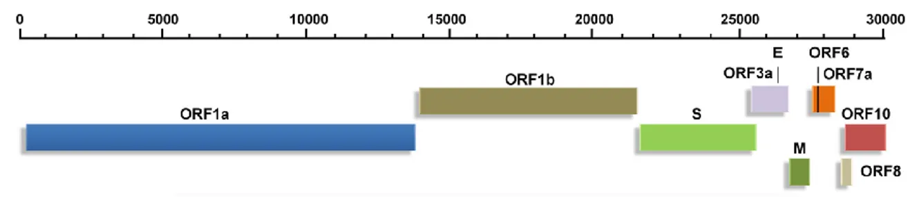
The genome of SARS-CoV-2 (Fig 3) is a single‐stranded positive‐sense RNA of about 29.9 kb and its structure follows the specific gene characteristics of known CoVs: (i) a highly conserved genomic organization with a large replicase gene, (ii) expression of many non-structural genes by ribosomal frameshifting, (iii) several enzymatic activities encoded within the large replicase-transcriptase polyprotein, (iv) expression of downstream genes by synthesis of 3’ nested subgenomic mRNAs. The genome is flanked by two UTRs, similar to those of other betacoronaviruses, with nucleotide identities of ⩾ 83.6% (Y. Chen et al., 2020; Changtai Wang et al., 2020).
Fig 3. Genomic organization of SARS-CoV-2. (Adapted from Kumar et al., 2020)
The SARS-CoV-2 genomic RNA contains a 5′‐cap structure and a 3′‐poly‐A tail, that allow immediate translation upon viral entry, to produce two coterminal replicase polyproteins from two large open reading frames (ORF1a and ORF1b), by utilizing a ribosomal frameshifting mechanism. Replicase polyproteins are subsequently cleaved by the action of two viral cysteine proteases, nsp3-PLpro and nsp5-Mpro, into the individual nsps, nsp1-11 and nsp1-16, respectively, which are necessary for the formation of the replication‐transcription complex (RTC). Of note, nsp12 encodes for the RdRp, nsp13 for the viral helicase and nsp14 for the 3’-5’ exonuclease with proofreading activity. The latter seems to have a peculiar role in the preservation of the CoV genome, which is larger in respect to other RNA viruses: the maintenance of
15
such genome may be related to the special features of the CoV RTC, in particular for the presence of the 3′‐5′ exoribonuclease (Y. Chen et al., 2020; Naqvi et al., 2020; Neerukonda and Katneni, 2020). The accessory and structural proteins constitute only the remaining 10kb of viral genome. Structural proteins include spike (S), E, M and N proteins.
The S glycoprotein plays a significant role in pathogenesis, by binding to the host cell through its receptor binding domain (RBD), which represents the most variable part of the CoV genome. It can be divided into S1 and S2 subunits, with the S1 domain containing the RBD that specifically engages the host cell receptor, thereby determining virus cell tropism and pathogenicity, while the transmembrane S2 domain containing the fusion peptide, which mediate the fusion of viral and cellular membranes upon extensive conformational rearrangements (Naqvi et al., 2020; Qing and Gallagher, 2020). A notable feature of SARS-CoV-2 is the polybasic cleavage site (RRAR) at the junction of S1 and S2, with the additional insertion of a leading proline, thus constituting the PRAAR site. This site seems to have a role in determining viral infectivity and host range and interestingly, such polybasic cleavage site has not been observed in related lineage B betacoronaviruses (Andersen et al., 2020; Coutard et al., 2020).
The E protein plays a significant role in the viral morphogenesis, pathogenesis, assembly and budding. This protein has a N-terminal ectodomain and a C-terminal endodomain and it is also act as ion-channelling viroporin, assembling into the host membrane to arrange protein-lipid pores involved in iontransport (Bianchi et al., 2020; Schoeman and Fielding, 2019).
The M protein has three transmembrane domains and it functions in concurrence with E, N, and S proteins, playing a major role in providing a distinct shape to the virus. M proteins are the most abundant viral proteins of CoVs (Naqvi et al., 2020).
The N protein constitutes the only protein present in the nucleocapsid. It plays an important role in the packaging of viral RNA into the ribonucleocapsid, by interacting with the viral genome, nsp3 and M protein during assembly. The heavy phosphorylation of N protein triggers structural changes enhancing the affinity for viral versus non-viral RNA. N protein of SARS-CoV-2 is highly conserved, sharing ~90% sequence identity with that of SARS-CoV (Cong et al., 2019; Naqvi et al., 2020).
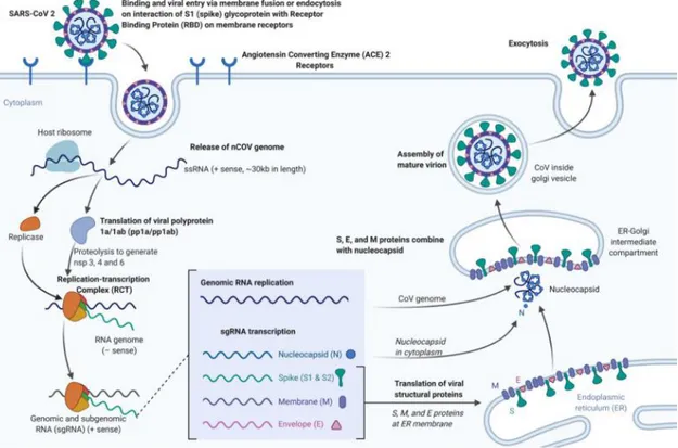
SARS-CoV-2 has an RBD that binds with high affinity the angiotensin-converting enzyme 2 (ACE2) that can be found in humans, ferrets, cats and other species. ACE2 is a type I membrane glycoprotein which is mainly expressed in lungs, heart, intestines, and kidneys (Andersen et al., 2020; Hoffmann et al., 2020; Neerukonda and Katneni, 2020) and its binding is required as the initial step of CoV infection (Fig 4). Following receptor binding, the proteolytic cleavage of CoV S proteins by host cell-derived proteases is essential to permit fusion. The protease used by SARS-COV-2 to gain access to host cell cytosol, is the TMPRSS2, a cell-surface serine protease expressed mainly in the human respiratory tract; the S cleavage operated by TMPRSS2 represents a key event in SARS-CoV-2 replication cycle, because inhibition of TMPRSS2 is sufficient to prevent SARS-CoV-2 entry in lung cell lines and in primary lung cells (Hoffmann et al., 2020; V’kovski et al., 2020). As previously described, a peculiar feature of the SARS-CoV-2 S protein is the acquisition of a polybasic cleavage site (PRRAR) at the S1–S2 boundary, whose cleavage results in enhanced infection and it has been proposed to be a key event in SARS-CoV-2 evolution. Indeed, this pre-processing of the SARS-CoV-2 S protein may contribute to the expanded cell tropism and zoonotic potential and might increase transmissibility (Neerukonda and Katneni, 2020; V’kovski et al., 2020).
Following fusion and endocytosis, the SARS-CoV-2 genomic RNA is immediately translated. The newly formed nsps are assembled into the RTC, which begins to generate anti-sense genomic copies functioning as templates for genomic and sub-genomic RNAs. The nsps also promote membrane rearrangement of rough ER membranes, in which replication and transcription processes occur.
16
Fig 4. Binding, viral entry and replication cycle of SARS-CoV-2. (Adapted from Cascella et al., 2020)
Structural proteins translated from sub-genomic RNAs are inserted into the ER and pass along the secretory pathway to the ER-Golgi intermediate compartment. There, the newly synthesized genome forms a complex with the N protein to generate virions. Following assembly with the other structural proteins, virions are transported to the cell surface in vesicles and released outside the cell through exocytosis (Hoffmann et al., 2020; Neerukonda and Katneni, 2020; V’kovski et al., 2020).
2.3 Transmission
SARS-CoV-2 is mainly transmitted through symptomatic humans, while the role of asymptomatic people in the transmission is still debated (Gao et al., 2020; He et al., 2020; Oran and Topol, 2020), by respiratory droplets and aerosols and/or contact of mucosae with virus-contaminated fomites. Therefore, environmental factors, including temperature, humidity, stability on fomites, ventilation and filtering systems, could have a significant influence on the infection (Azuma et al., 2020; Y. Chen et al., 2020; Sungnak et al., 2020). Adequate control of these environmental factors and proper human behavior play a significant role in preventing the spread of COVID-19.
Interestingly, viral RNAs or live infectious viruses have been detected in feces and urine of patients with COVID-19. Binding of SARS-CoV-2 to the ACE2 is a vital pathway for the virus entry into human cells, whose presence is not limited to the respiratory mucosa, but is also extended to the esophageal epithelium as well as in the enterocytes from ileum and colon (Barbosa da Luz et al., 2020; Sun et al., 2020; Wang et al., 2020; Xiao et al., 2020). Therefore, a fecal-oral route of transmission for SARS-CoV-2 may be hypothesized; to date, however, there have been no published reports of transmission of SARS-CoV-2 through feces or urine. Some studies have also reported detection of SARS-CoV-2 RNA in either plasma or serum. However, the role of bloodborne transmission remains uncertain; anyway, low viral titers in plasma and serum suggest that the risk of transmission through this route may be low (Chang et al., 2020; Wang et al., 2020). There is no evidence for intrauterine transmission of SARS-CoV-2 from infected pregnant women to their fetuses, neither of breastfeeding transmission, even if viral RNA fragments have been found in a few breast milk samples, but no viable virus. As a matter of fact, WHO recommends that mothers with suspected or
17
confirmed COVID-19 should be encouraged to initiate or continue to breastfeed (www.who.int). Moreover, clinical course of COVID-19 in most women is not severe, and the infection does not significantly influence the pregnancy. Indeed, in most cases the disease does not threaten the mother, and vertical transmission has not been clearly demonstrated (Di Toro et al., 2020).
On this basis, to prevent SARS-CoV-2 diffusion, the WHO suggests physical distancing policy, the use of face masks in public places and frequent hand hygiene (www.who.int). Indeed, a drastic reduction of social contacts and total lockdown periods have been implemented in many countries with outbreaks of SARS-CoV-2, leading to rapid reductions in basic reproduction number (R0), defined as the average number of secondary infections produced by a typical case of an infection in a population where everyone is susceptible. In absence of measures preventing transmission, the estimate for the R0 is between 2 and 3, and the median incubation period is 6 days (range 2-14 days).
2.4 Clinical manifestations and pathogenesis
The clinical spectrum of COVID-19 can vary from asymptomatic (40-45% of infections) and mild symptomatic, to an adverse clinical condition characterized by severe respiratory failure, requiring mechanical ventilation and hospitalization (He et al., 2020; Oran and Topol, 2020). The most important risk factors for a severe disease are age, hypertension, diabetes, immunodeficiency and chronic cardiovascular and pulmonary diseases. Countries throughout the world have reported different mortality rate, ranging from 1 to 10%, with the incidence of mortality rising after the sixth decade of life (www.who.it; John Hopkins University https://coronavirus.jhu.edu/data/mortality).
Mild COVID-19 illness symptoms are those typical of an upper respiratory tract viral infection, including mild fever, dry cough sore throat, nasal congestion, malaise, headache and muscle pain. Moreover, nausea, vomiting, abdominal pain and diarrhea may occur. As the disease become more severe, patients display symptoms such as dyspnea, respiratory distress and hypoxia with oxygen saturation levels under 90%. Patients with sudden onset of respiratory failure or impaired lung function can experience SARS and require hospitalization and mechanical ventilation. Critically ill patients have systemic symptoms characterized by sepsis, septic shock, and multiple organ dysfunction syndromes (Neerukonda and Katneni, 2020).
As we currently know, severe COVID-19 symptoms are a consequence of dysregulated immune responses. Indeed, the rapid and uncontrolled viral replication of SARS-CoV-2 is able to evade the host innate immune response during its initial steps. Consequently, the quick activation of the cell-mediated response provokes an increased pro-inflammatory status with a massive release of cytokines, causing acute lung injury and contributing to the clinical manifestation of SARS (Khalaf et al., 2020). Indeed, in severe COVID-19 cases, as opposed to mild cases, an aberrant recruitment of inflammatory macrophages and the infiltration of T lymphocytes and neutrophils have been measured in the lungs.
The accumulating evidence of dysregulated pro-inflammatory responses during SARS-CoV-2 infections has led to the use of immune modulators to inhibit hyperactivated pathogenic immune responses, such as corticosteroids and tolicizumab (V’kovski et al., 2020). Corticosteroids are used to mitigate the host inflammatory response which contributes acute respiratory distress syndrome in severe pneumonia cases. However, these agents are responsible for some adverse effects, that include delayed viral clearance and enhanced risk of secondary infection (McCreary and Pogue, 2020; Neerukonda and Katneni, 2020). Tocilizumab is an approved humanized monoclonal antibody that inhibits IL-6 receptor, originally approved for the treatment of rheumatoid arthritis. IL-6 is a key driver of dysregulated inflammation that contributes to the pulmonary pathology and the organ damage observed in COVID-19 patients. Theoretically, antibodies targeting its receptor could reduce the IL-6 signal transduction and downstream inflammation, thus improving clinical outcomes (Neerukonda and Katneni, 2020; Xu et al., 2020).