Contents lists available atScienceDirect
Interdisciplinary Neurosurgery
journal homepage:www.elsevier.com/locate/inatNeurosurgical Techniques
Symptomatic cerebral vasospasm after glioblastoma resection and
carmustine wafers implantation. A case report
☆,☆☆
Vitaliano F. Muzii
a,⁎, Anna Vaiano
a, Sandra Bracco
b, Biagio R. Carangelo
c aDepartment of Medicine, Surgery, and Neurosciences, Section of Neurosurgery, University of Siena, Siena, ItalybDepartment of Neurological and Sensorineural Sciences, Unit of Neuroimaging and Neurointervention, Azienda Ospedaliera Universitaria Senese, Siena, Italy cDepartment of Neurological and Sensorineural Sciences, Unit of Neurosurgery, Azienda Ospedaliera Universitaria Senese, Siena, Italy
A R T I C L E I N F O Keywords: Cerebral vasospasm Glioblastoma Carmustine wafer A B S T R A C T
Local chemotherapy with carmustine-impregnated wafers showed safe and effective in the treatment of ma-lignant glioma, with infrequent, though sometime serious, adverse effects.
We report a rare case of cerebral vasospasm following glioblastoma removal with carmustine wafers im-plantation in a 57-years-old man. After surgery, the patient awoke with aphasia, due to vasospasm of the left middle cerebral artery. Intra-arterial infusion of nimodipine was performed, with rapid vasospasm resolution and quick recovery.
Cerebral vasospasm is an extremely rare adverse effect after carmustine wafers implantation in glioma sur-gery, with only one case reported. In our case, intra-arterial nimodipine was rapidly effective. Although rare, such a potentially disastrous complication should be considered when a new neurological deficit unexpectedly occurs after carmustine wafers implantation, and vascular investigation should be undertaken.
1. Introduction
For the last ten years, local chemotherapy with carmustine-im-pregnated wafers (Gliadel®, Eisai Inc., Woodcliff Lake, NJ, USA) in the treatment of malignant glioma has demonstrated an improvement of median overall survival with acceptable safety profile [1–3].
Carmustine wafer, which is designed to release biodegradable 1,3-bis-2-chloroethyl-1-nitrosurea (BCNU), has the advantages of bypassing the blood-brain barrier, delivering BCNU directly to peritumoral tissue, and avoiding systemic toxicity [4,5]. Carmustine wafers are placed along the wall of the surgical site, and the chemotherapeutic agent is released continuously, diffusing into the parenchyma over approxi-mately 3–8 weeks, with peak release in the first 2 weeks [5]. However, several adverse events have been associated with Gliadel® implanta-tion, the most common of which include seizures, brain edema, and intracranial hypertension [3]. Other uncommon adverse events are reported in the literature, including wound healing defects, intracranial infections, cerebral hemorrhage, cerebrospinal fluid leakage, hydro-cephalus, and cyst formation [1,2,4,6]. According to the current lit-erature, only one case of carmustine wafer-related vasospasm with cerebral infarction has been described [7].
We report a case of symptomatic vasospasm after implantation of
carmustine wafers in a patient treated for left frontotemporal glio-blastoma (GBM).
2. Case report
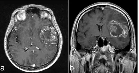
A 57-year-old man was admitted to ER for sudden onset of confu-sion, amnesia and insomnia. Magnetic resonance imaging (MRI) re-vealed a mass located in the left temporal lobe and insula with perile-sional edema, showing inhomogeneous ring enhancement (Fig. 1).
The patient was referred to our department and underwent surgery for tumor resection via left frontotemporal craniotomy. The resection was extended to the deep area of the frontal lobe and the insula, which appeared infiltrated. Sylvian arteries were preserved, except some small branches supplying the tumor. We obtained CT-confirmed subtotal re-section (> 95%) of the tumor (Fig. 2a). Eight carmustine wafers were placed on the wall of the resection cavity and secured with Tabotamp strips.
Histopathological examination revealed typical findings of WHO grade IV glioma. On postoperative day one, once sedation was inter-rupted and patient awoke, he showed moderate aphasia, with no other neurological deficit. CT and CT-angiography (CTA) were performed and revealed persistence of brain edema around the surgical cave, without
https://doi.org/10.1016/j.inat.2018.05.016
Received 21 November 2017; Received in revised form 10 April 2018; Accepted 28 May 2018
☆Declaration of interest: none.
☆☆This research did not receive any specific grant from funding agencies in the public, commercial, or not-for-profit sectors. ⁎Corresponding author at: S. Maria alle Scotte University Hospital, V.le Mario Bracci, 16, 53100 Siena, Italy.
E-mail address:[email protected](V.F. Muzii).
Interdisciplinary Neurosurgery 14 (2018) 24–27
2214-7519/ © 2018 The Authors. Published by Elsevier B.V. This is an open access article under the CC BY-NC-ND license (http://creativecommons.org/licenses/BY-NC-ND/4.0/). T
postoperative bleeding (Fig. 2a). However, vasospasm was shown at the M2 segments of the left middle cerebral artery and M3 insular branches (Fig. 2b), which was confirmed by digital subtraction angiography (DSA) (Fig. 3a). In the same session, 5 mg nimodipine were infused in 30 min in the left internal carotid artery through a 5F catheter [8], with increased size of M2 and better visualization of insular branches (Fig. 3b). Oral nimodipine (60 mg 6 times daily) was continued for 7 days and then tapered off. Aphasia resolved in two days. Transcranial Doppler monitoring showed vasospasm disappearance after 72 h. The patient was discharged on the 18th postoperative day and underwent radiotherapy and temozolomide chemotherapy. At last follow-up, 15 months after operation, he is free of symptoms and tumor recurrence on MRI (Fig. 4).
3. Discussion
Implantation of carmustine-impregnated wafers (Gliadel®) has de-monstrated a survival benefit in patients with malignant glioma [1–3]. However, a number of adverse reactions have been commonly reported [1–4,6], while vasospasm was described in only one case report before [7].
The present case is, at our knowledge, the second reported case of
symptomatic cerebral vasospasm associated with carmustine wafers implantation, confirming vasospasm as a possible rare side effect. The mechanism of vasospasm induced by Gliadel® remains unclear, al-though the vasoactive effect of carmustine has been already described in different situations. Shingleton et al. [9] reported that high-dose intravenous carmustine (800 mg/m2) has been associated with retinal artery narrowing and obstruction. With carmustine wafers the local concentration at the implantation site is 1200 times higher than con-centration achieved by systemic administration [5]. Possibly, local toxicity of carmustine or foreign body reaction to the wafer material may trigger local inflammatory response leading to vasospasm [7]. More specifically, in a rabbit lung model, BCNU inhibited glutathione reductase activity, resulting in increase of induced vasospasm, due to hydrogen-peroxide-induced prostanoid formation and calcium shift [10].
Vasospasm and cerebral infarction are rare complications of brain tumor surgery in general. In a large series of 470 skull base tumors, the incidence of vasospasm was 1.9% only [11]. In a systematic review of cerebral vasospasm following tumor resection, < 50 cases were found, and most of them were benign lesions [12], while only one case related to GBM is reported [13]. These data reinforce the hypothesis that, in our case, local carmustine may have played a causative role. In fact,
Fig. 1. Preoperative contrast-enhanced MRI. Axial (a) and coronal (b) images, compatible with high grade glioma.
Fig. 2. Postoperative CT. a) Contrast-enhanced image showing normal postoperativefindings, with carmustine wafers lying on the wall of surgical cavity, and small enhancing remains in the posterior lateral border; b) CT-angiography revealed vasospasm of the left middle cerebral artery (M2-M3 branches).
V.F. Muzii et al. Interdisciplinary Neurosurgery 14 (2018) 24–27
while intraoperative mechanical arterial manipulation is perhaps one of the most important surgical factors for vasospasm, it is mainly con-sidered for skull base tumor surgery [11]. Increased tumor vascularity with blood spillage into the cisternal space has also been advocated as a cause of vasospasm [12]. However, in our patient there was no post-operative hemorrhage. Moreover, while vasospasm after tumor resec-tion is usually delayed several days after surgery [7,11–13], in our case it occurred < 24 h after operation, thus suggesting a direct vasoactive effect on the vessels.
In the previously reported case [7], vasospasm and cerebral in-farction appeared 12 days after surgery, while in our case neurological impairment and vasospasm occurred the day after operation. A possible explanation of this difference is that, in our case, the sylvian vessels had
been exposed and were close to wafers, whereas in the other case len-ticulostriate arteries were covered by some residual tumor. Therefore, carmustine might have taken a longer time to reach the vessels by diffusing through the intervening tissue. Moreover, in our patient, prompt and aggressive endovascular treatment of vasospasm prevented eventual cerebral infarction, leading to complete neurological recovery and good outcome.
4. Conclusions
We described a unique case of symptomatic vasospasm associated with implantation of carmustine-impregnated wafers in the treatment of GBM. Our aim is to contribute to the experience of using Gliadel®, by remarking the importance of an uncommon but harmful adverse drug reaction like vasospasm. Based on our experience, in case of sudden onset of new focal neurological deficit after carmustine wafers im-plantation, with“normal” CT scan, vasospasm should be suspected and vascular investigation prompted. In our case, intra-arterial infusion of nimodipine, followed by oral administration, led to rapid reversal of vasospasm and full neurological recovery.
The patient has consented to submission of this case report to the journal.
References
[1] M. Westphal, D.C. Hilt, E. Bortey, P. Delavault, R. Olivares, P.C. Warnke, et al., A phase 3 trial of local chemotherapy with biodegradable carmustine (BCNU) wafers (Gliadel wafers) in patients with primary malignant glioma, Neuro-Oncology 5 (2003) 79–88.
[2] H.C. Bock, M.J. Puchner, F. Lohmann, M. Schütze, S. Koll, R. Ketter, et al., First-line treatment of malignant glioma with carmustine implants followed by concomitant radiochemotherapy: a multicenter experience, Neurosurg. Rev 33 (2010) 441–449. [3] A. Gutenberg, C.B. Lumenta, W.E. Braunsdorf, M. Sabel, H.M. Mehdorn,
M. Westphal, et al., The combination of carmustine wafers and temozolomide for the treatment of malignant gliomas. A comprehensive review of the rationale and clinical experience, J. Neuro-Oncol. 113 (2013) 163–174.
[4] K. Sato, M. Dan, Daisuke Yamamoto, Y. Miyajima, A. Hara, T. Kumabe, Chronic phase intracranial hemorrhage caused by ruptured pseudoaneurysm induced by carmustine wafer implantation for insulo-opercular anaplastic astrocytoma: a case report, Neurol. Med. Chir. (Tokyo) 55 (2015) 848–851.
[5] L.K. Fung, M.G. Ewend, A. Sills, E.P. Sipos, R. Thompson, M. Watts, et al., Pharmacokinetics of interstitial delivery of carmustine, 4-hydroperoxycyclopho-sphamide, and paclitaxel from a biodegradable polymer implant in the monkey brain, Cancer Res. 58 (1998) 672–684.
[6] P. De Bonis, C. Anile, A. Pompucci, A. Fiorentino, M. Balducci, S. Chiesa, et al., Safety and efficacy of Gliadel wafers for newly diagnosed and recurrent
Fig. 3. Postoperative DSA. Antero-posterior (a) and lateral (b) view showing narrowing of M1 an M2 braches of the left middle cerebral artery with some lacking or narrowed M3 insular branches (circle, arrowheads); b) after intra-arterial nimodipine infusion, vasospasm is reduced and insular branches are better visualized.
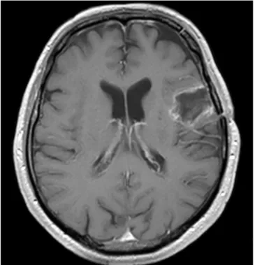
Fig. 4. Follow-up MRI. Fifteen months after surgery, there is no evidence of recurrence on contrast-enhanced MRI.
V.F. Muzii et al. Interdisciplinary Neurosurgery 14 (2018) 24–27
glioblastoma, Acta Neurochir. 154 (2012) 1371–1378.
[7] M. Nakada, S. Tanaka, M. Oishi, K. Miyashita, K. MisaKi, M. Mohri, et al., Cerebral infarction related to carmustine wafers in glioblastoma: a case report, NMC Case Report Journal 1 (2015) 36–39. Available from:https://www.jstage.jst.go.jp/ article/nmccrj/2/1/2_2014-0080/_pdf.
[8] A. Biondi, G.K. Ricciardi, L. Puybasset, L. Abdennour, M. Longo, J. Chiras, et al., Intra-arterial nimodipine for the treatment of symptomatic cerebral vasospasm after aneurysmal subarachnoid hemorrhage: preliminary results, AJNR Am. J. Neuroradiol. 25 (2004) 1067–1076.
[9] B.J. Shingleton, D.C. Bienfang, D.M. Albert, W.D. Ensminger, W.F. Chandler, H.S. Greenberg, Ocular toxicity associated with high-dose carmustine, Arch. Ophthalmol. 100 (1982) 1766–1772.
[10] W. Seeger, N. Suttorp, F. Schmidt, H. Neuhof, The glutathione redox cycle as a
defense system against hydrogen-peroxide-induced prostanoid formation and va-soconstriction in rabbit lungs, Am. Rev. Respir. Dis. 133 (1986) 1029–1036. [11] G.K. Bejjani, L.N. Sekhar, A.M. Yost, W.O. Bank, D.C. Wright, Vasospasm after
cranial base tumor resection: pathogenesis, diagnosis, and therapy, Surg. Neurol. 52 (1999) 577–583.
[12] N.M. Alotaibi, G. Lanzino, Cerebral vasospasm following tumor resection, J Neuro Intervent Surg 5 (2013) 413–418. Available from:https://doi.org/10.1136/ neurintsurg-2017-012993.
[13] P.Y.M. Woo, K.W.M. See, J.K.H. Chow, Y. Chan, H.T. Wong, K.Y. Chan, Hypertensive-nimodipine therapy for middle cerebral artery vasospasm after re-section of glioblastoma multiforme: a case report and literature review, Open Journal of Modern Neurosurgery 5 (2015) 76–83. Available from:https://doi.org/ 10.4236/ojmn.2015.53013.
V.F. Muzii et al. Interdisciplinary Neurosurgery 14 (2018) 24–27