UNIVERSITY OF CATANIA
DEPARTMENT OF CHEMICAL SCIENCES
International PhD in Chemical Sciences, XXIX cycle
Angela Margherita Aura
SURFACE PLASMON RESONANCE IMAGING
BIOSENSORS FOR THE DETECTION OF
PATHOGENS AND TOXINS IN FOOD
PhD Coordinator:
Prof. S. Sortino
Tutor:
Prof. G. Spoto
CONTENTS
ABSTRACT 4
1. INTRODUCTION 5
2. CONVENTIONAL METHODS FOR THE DETECTION OF FOODBORNE
PATHOGENS AND TOXINS 6
3. BIOSENSORS FOR THE DETECTION OF FOODBORNE PATHOGENS
AND TOXINS 7
4. SURFACE PLASMON RESONANCE IMAGING (SPRI) 9
4.1. SPRI principles 9
4.2. SPR biosensing formats 15
4.3. Functionalized gold nanoparticles (AuNPs) for biosensing 17
5. STAPHYLOCOCCUS AUREUS 19
6. LISTERIA MONOCYTOGENES 23
7. ANTIFOULING MATERIALS FOR SURFACE PLASMON RESONANCE BIOSENSING 26
8. PURPOSE OF THE PERFORMED RESEARCH ACTIVITY 33
9. MATERIALS AND METHODS 35
9.1. Materials and reagents 35
9.2. Surface Plasmon Resonance Imaging Apparatus and measurements 36
9.3. PNA probes and synthetic oligonucleotides sequences for the development
of SPRI-based genosensors for the detection of Staphylococcus Aureus and
Listeria monocytogenes in food 38
9.4. SPRI-based genosensors for the detection of Staphylococcus Aureus in food 39
9.4.1. Staphylococcus Aureus PNA probe surface immobilization 39
9.4.2. Staphylococcus Aureus ssDNA hybridization experiments 39
9.4.4. Staphylococcus Aureus and bovine gDNA hybridization experiments 40
9.4.5. Synthesis and functionalization of gold nanoparticles for Staphylococcus Aureus
genomic DNA detection 40
9.5. SPRI-based genosensors for the detection of Listeria monocytogenes in food 41
9.5.1. Listeria monocytogenes PNA probe surface immobilization 41
9.5.2. Listeria monocytogenes gDNA hybridization experiments 41
9.5.3. Synthesis and functionalization of gold nanoparticles for Listeria monocytogenes
genomic DNA detection 41
9.6. SPRI-based immunosensors for the detection of Staphylococcal enterotoxin A (SEA)
in food 42
9.6.1. SPRI sensor surface functionalization with methyl- and carboxy-thiol PEGylation reagents 42
9.6.2. SPRI sensor surface functionalization with n-dodecil mercaptano (NDM) and
graphene oxide (GO) 42
9.6.3. SPRI sensor surface functionalization with DTSP 43
9.6.4. SPRI sensor surface immobilization of Anti Staphylococcus aureus enterotoxin A
2S7-F12 (Anti-SEA F12) 43
9.6.5. SPRI detection of staphylococcal enterotoxin A 43
9.6.6. SPRI sandwich detection with Anti Staphylococcus aureus enterotoxin A
2S7-H5 (Anti-SEA H5) 44
9.6.7. Synthesis and functionalization of gold nanoparticles for Staphylococcal
enterotoxin A detection 44
9.6.8. SPRI sandwich detection with Anti Staphylococcus aureus enterotoxin A
2S7-H5 (Anti-SEA H5) and Anti Staphylococcus aureus enterotoxin A 2S7-H5
(Anti-SEA H5B) 45
9.7. Antifouling surfaces development for SPRI biosensing 46
9.7.1. SPRI sensor surface functionalization with 11-mercaptoundecanoin acid 46
9.7.2. SPRI sensor surface functionalization with Methyl- and Carboxy-Thiol
PEGylation reagents 46
9.7.3. SPRI sensor surface immobilization of Anti Staphylococcus Aureus Monoclonal Antibody 46
9.7.4. SPRI sensor surface functionalization with Methoxy PEG disulfide (mPEG disulfide)
and PEG NHS ester disulfide (NHS PEG disulfide) 47
9.7.5. SPRI sensor surface functionalization with PEG NHS ester disulfide
(NHS PEG disulfide) and backfilling with Methoxy PEG disulfide (mPEG disulfide) 47
9.7.6. Milk treatment 47
10. RESULTS AND DISCUSSION 49
10.1. SPRI-based genosensors for the detection of Staphylococcus Aureus and
Listeria monocytogenes in food 49
10.2. SPRI-based immunosensors for the detection of staphylococcal enterotoxin A (SEA)
in food 73
10.3. Antifouling surfaces development for SPRI biosensing 87
11. CONCLUSIONS AND FUTURE PERSPECTIVES 103
RESEARCH PRODUCTS 105
ACKNOWLEDGEMENTS 108
ABSTRACT
The detection of pathogens and toxins in food represents an essential requirement for food quality control. Standard methods for pathogen detection rely on laborious and time-consuming growth of pathogens in different culture media followed by biochemical or serological identification. Such methods often operate with poor sensitivity and selectivity. In recent years, efforts have been made to provide rapid, reliable and sensitive detection platforms for foodborne pathogens detection. The demand for more rapid, sensitive and accurate methods has been push forward by the implementation of the Hazard Analysis and Critical Control Points (HACCP) protocols. In this context, biosensing platforms provide promising alternatives for the detection of pathogens and toxins with good selectivity and sensitivity. In particular, optical biosensors based on Surface Plasmon Resonance Imaging (SPRI) are attractive because they allow the sensitive detection of analytes from food matrices in real-time. SPRI can assays crude samples without purification and can exploits antibodies or single-stranded DNA (ssDNA) probes for the specific detection of pathogens and toxins with high sensitivity.
My research activity has been aimed at developing SPRI biosensors able to detect pathogens and toxins in food matrices in a rapid, specific and sensitive way. In this perspective, specific oligonucleotide sequences and antibodies have been used for the detection of DNA and bacterial toxins, respectively. SPRI biosensor sensitivity benefited of the use of properly functionalized gold nanoparticles (AuNPs). The combination of the SPRI sensing apparatus with microfluidics devices reduces the amount of sample needed for the analysis and provides an efficient environment for the detection.
1. INTRODUCTION
“Food safety is a hidden, and often overlooked, problem.”1
The detection of pathogens and toxins in food represents a challenging task with implications in food safety and quality control. Pathogens, which include viruses, toxins, parasites, bacteria and bacterial toxins, can contaminate food at any point of the food chain: through the production site, at the slaughterhouse or packing plant, in manufacturing, processing and retailing of food, domestic preparation and storage.2 According to World Health Organization (WHO), foodborne illnesses are diseases caused by agents that enter the body through the ingestion of food.3
The occurrence of foodborne diseases has increased over years and represents a worldwide public health significant issue. Foodborne pathogens responsible for the majority of disease out-breaks are: Listeria monocytogenes (Listeria m.), Escherichia
coli O157:H7, Staphylococcus aureus (S. aureus), Salmonella enterica, Bacillus cereus, Vibrio spp., Campylobacter jejuni, Clostridium perfringens, and Shiga
toxin-producing Escherichia coli (STEC).4 Some foods, such as milk, cream or meat must be pathogen-free, with a specific attention to Listeria m. and S. aureus.5 As stated by Commission Regulation (EC) No 2073/2005 of 15 November 2005, “Foodstuffs should not contain microorganisms or their toxins or metabolites in quantities that present an unacceptable risk for human health”.6 Unfortunately, according to estimates provided by the Centers for Disease, Control and Prevention (CDC) for foodborne illnesses, every year 48 million U.S. citizens gets sick, 128,000 are hospitalized and 3,000 die as a consequence of foodborne diseases. The top five pathogens causing domestically acquired foodborne illnesses and deaths are listed in Table 1 and Table
Table 1. Top five pathogens contributing to domestically acquired foodborne illnesses (Ref. 7). Pathogen Estimated number of illnesses 90% Credible Interval %
Norovirus 5,461,731 3,227,078–8,309,480 58 Salmonella, nontyphoidal 1,027,561 644,786–1,679,667 11 Clostridium perfigens 965,958 192,316–2,483,309 10 Campylobacter spp 845,024 337,031–1,611,083 9 Staphylococcus aureus 241,148 72,341–529,417 3 Subtotal 91
Table 2. Top five pathogens contributing to domestically acquired foodborne illnesses resulting in
death (Ref. 7).
Pathogen Estimated number of deaths 90% Credible Interval %
Salmonella, nontyphoidal 378 0–1,011 28 Toxoplasma gondii 327 200–482 24 Listeria monocytogenes 255 0–733 19 Norovirus 149 84–237 11 Campylobacter spp. 76 0–332 6 Subtotal 88
Progress in microbiological safety of food have been extensively driven by public demand due to disease outbreaks. Nevertheless, despite efforts paid from scientists, governments and industry, foodborne disease will continue to be a major issue in public health, with implications for both the social welfare of populations and for national economies.8 Consequently, there is a need for new methods for the efficient and rapid detection of pathogens in food in order to minimize their spread and outbreaks.
2. CONVENTIONAL METHODS FOR THE DETECTION OF
FOODBORNE PATHOGENS AND TOXINS
Conventional methods for detecting foodborne bacterial pathogens rely on microbiological analysis, followed by biochemical, serological, or molecular tests for the pathogen identification. Such methods include colony counting, polymerase chain reaction (PCR) and immunological methods.9
Colony counting method is considered as the “gold standard” for pathogen detection.10 It is labor-intensive and time-consuming (typically more than 7 days)
because it is linked to the ability of microorganisms to grown in different culture media.11 In addition, contamination from fast growing microorganisms may interfere with the detection.12,13
PCR methods exploit the nucleic acid amplification to enhance the sensitivity of the detection and have been extensively used for the identification of pathogens in food matrices.14 However, PCR methodologies involve the extraction and purification of the pathogen DNA or RNA with protocols that are depending from the food sample properties. An incorrect amplification of contaminants sequences produces false positive results, while amplification of positive samples produces high concentrations of target molecules that can certainly lead to cross-contamination of negative samples.15,16 On a large scale basis, routine PCR-based detection of microbes is expensive and is usually performed by skilled personnel only.17
Immunological methods are widely used to detect bacterial cells, spores, viruses and toxins18 and are based on the specific binding of an antibody to an antigen.12,19,20,21,22 Enzyme-linked immunosorbent assay (ELISA)23 is among the immunological-based methods used for the detection of pathogens and toxins.24,25,26,27 Immunological assays are usually reliable, but an enrichment step is needed to increase the bacteria count in the food sample, thus enhancing the sensitivity but also increasing the time required for the analysis.28
3. BIOSENSORS FOR THE DETECTION OF FOODBORNE
PATHOGENS AND TOXINS
Over the last decade, biosensors have been shown to offer promising alternatives for pathogenic microorganisms detection in food.29,30,31,32,33
A biosensor is an analytical device that integrates a biorecognition element (or bioreceptor) with a suitable transducer. The bioreceptor is a crucial element in
biosensing. It defines the selectivity of the biosensor: i.e. its ability to detect only the target biomolecule and not interfering molecules. Antibodies, enzymes, phages, aptamers or single-stranded nucleic acid sequence (ssDNA, RNA) may act as the biorecognition element.
Transducers convert the biorecognition event in a detectable signal. The transduction mechanism may be optical, electrochemical, thermometric, piezoelectric, magnetic, micromechanical or a combination of them.34,35,36
Biosensors are classified on the basis of the transduction method or according to the bioreceptor involved (catalytic biosensors if an enzyme is used or affinity-based
biosensors if proteins, nucleic acids, membranes, whole cells are employed for the
biomolecular recognition).37 Antibodies and nucleic acids represents the main biorecognition elements used in biosensing.38 When antibodies or antibody fragments are used as the recognition element, the device is called immunosensor, while biosensors using nucleic acids sequences are named genosensor or DNA or RNA sensors.39
Antibodies are usually immobilized on a solid surface to obtain an immunosensor.40 The interaction between an antigen and an antigen-specific antibody can be illustrated as a lock and key fit. An antigen-specific antibody binds its unique antigen, often with high specificity and for this reason antibodies can be used as specific probes for the recognition of specific analytes that are present even at extremely low concentration and in a mixture with other species.41
Genosensors identify target nucleic acids by matching the complementary base pairs that are often the genetic components of an organism. Those biosensors can be simple, rapid, and inexpensive38 and are widely used in pathogen detection.42,43,44
Biosensors are used to detect a wide range of targets ranging from small molecules to viruses and bacteria. General characteristics include accuracy, near real-time assay,
sensitivity, specificity, reproducibility, robustness and ease of use. False-positive and false-negative results should be very low, ideally zero, particularly for application in food industry where false-negatives can bring expensive recall and the loss of public confidence, whereas false-positive could increase production costs.45
Two different approaches can be implemented for bacterial biosensing. They are based on the detection of bacterial components released after the bacterial lysis or processing-free methods targeting components at the bacteria surface.37
Among biosensors developed over the past decade for the detection of foodborne pathogens, optical and electrochemical biosensors are today the most commonly used. Optical biosensors, detecting light absorption, luminescence, reflectance, Raman scattering or changes in media refractive index, are powerful alternatives to conventional analytical techniques and have received attention for bacterial pathogen detection.46,47,48 In particular, optical biosensors based on SPRI are used for real-time and label-free monitoring interactions between biomolecules, with high sensitivity and selectivity.49 SPR biosensors have been employed to investigate the presence of pathogens and toxins in food50, particularly to detect S. aureus and Staphylococcal enterotoxins51,52, Salmonella53,54, Listeria m.55,56, Escherichia coli O157:H757,58 and tetrodotoxin59.
4. SURFACE PLASMON RESONANCE IMAGING (SPRI)
4.1. SPRI principles
The surface plasmon resonance physical phenomenon has been observed for the first time by Wood60 and since then SPR has become one of the fastest-growing biosensing analytical tool.61
SPR exploits the evanescent electromagnetic (EM) field generated at the surface of a thin metal layer when properly irradiated with an incident light beam. EM field is
generated by electron charge density fluctuations (surface plasmons62) occurring at metal/dielectric interfaces.
Free electrons in metals can be treated as a high density plasma (density about 1023 cm-3). Electron density fluctuations propagate through the volume of a metal with a characteristic frequency given by (eq.1):
e p m ne h 2 4
π
ω
= h eq.1where ωp is the oscillation frequency of the plasmon, n is the free electron density, e
and me are the electron charge and the effective mass of an electron respectively.
Electron fluctuations are confined at the boundary and vanishes both sides of the metal surface. Surface plasmon waves are p-polarized and are described by a wave vector kx
parallel to the metal surface having magnitude (eq.2):
2 1 2 1
ε
ε
ε
ε
ω
+ = c kx eq.2where ε1= ε’1 + iε’’1 is the complex dielectric constant of the metal and ε2 is the
dielectric constant of the medium.
The above mentioned surface charge fluctuations are accompanied by a mixed transversal and longitudinal electromagnetic field (E), which disappears at an infinite distance from the metal surface and has its maximum at the metal surface. The frequency ωp of the longitudinal oscillations of the surface plasmonis tied to its wave vector kx by a dispersion relation ωp(kx) (Figure 1).61
Figure 1. Surface plasmon dispersion ω(kx) on gold surface. The vertical axis is scaled as ω(eV). The
straight solid line in figure shows the light line. kx=
ω
/cε
2 (Ref. 61).For plasmon excitation by a photon to take place, the energy and the momentum must be conserved during the photon-plasmon coupling. This requirement is met when the wave vector of the electromagnetic radiation klight and the plasmon kx equal in
magnitude and direction. The direction of the wave vector is the direction of the wave propagation, while its magnitude depends on the dielectric constant of medium at the interface. The wave vector (kx) associated with surface plasmons is always larger than
wave vector of light (klight) having the same energy and travelling through the medium
ε2 whereby the surface plasmon dispersion relation never interests the dispersion
relation of electromagnetic radiation (Figure 1). As a result, surface plasmon cannot couple with freely propagating light beam irradiating the metal surface. In order to couple photons at a given energy with surface plasmons, the wave vector has to be increased by Δkx (Figure 1). A similar increase can be obtained either by passing the
light through a medium with a refractive index greater than that of the dielectric medium at the boundary at which the surface plasmon is to be excited or by using diffraction effects. Thus, prism, grating, fiber-optic or waveguide couplers are needed for SPR-based sensors. Prism couplers are the most common setup for excitation of surface plasmons. They exploit the attenuated total reflection effect with the Kretschmann configuration (Figure 2) that increases the wave vector of the radiation
travelling through a prism coupler (dielectric constant εpr) with an incidence angle θ by
pr
ε sin θ. Under similar conditions, the wave vector magnitude matching condition is obtained when (eq.3)
2 1 2 1 sin
ε
ε
ε
ε
ω
θ
ω
ω
+ = c c pr eq.3and a drop in the intensity of the light reflected by the prism under the attenuated total reflection condition is observed (Figure 2). Any change of the value of ε2 caused by
chemical or physical effects modifies the matching condition and a shift in the energy position of the minimum of the reflected light (reflectance dip) is observed.63
Figure 2. From Ref. 63. Configuration of the Kretschmann geometry.
The surface plasmon wave is therefore highly sensitive to changes of the dielectric constant (simply related to the refractive index) of the medium at the metallic interface.61 This distinguishing property is the basic principle which makes the surface plasmon resonance useful as biosensor.64
Conditions for surface plasmon resonance are obtained in the infrared and visible wavelength region for air/metal and water/metal interfaces.
Many points can be considered for the choice of a metal for SPR:
• The metal should have a low imaginary part of the refractive index to reduce the dissipation and, consequently, to obtain a more narrower reflectance dip. Silver and gold are noble metals that satisfy this condition,
while aluminum has a large imaginary part and exhibits wider dips compared to silver and gold.
• The chemical purity of the surface since oxides and sulfides formed after the metal interaction with the atmosphere can interfere with the SPR phenomenon.
• The compatibility of the metal reactivity with the surface chemistry needed for the SPR experiment.61
An SPR biosensor is able to measure the binding between target analyte molecules and receptor molecules immobilized on the gold surface. During the receptor/analyte binding event, the shift of the dip in the spectrum of the reflected light is monitored over time and information on kinetics of biomolecules interactions is gathered.65
SPR allows to investigate interactions between antigens and antibodies, nucleic acid sequences and their complementary strands, and substrates and enzymes with no need for labeling of the interacting components. The kinetic events at the metal surface, displayed as a sensorgram, can be investigated by monitoring the SPR signal change as a function of time. The time interval during which the analyte interacts with the surface bound receptor defines the “association phase” while the time interval following the analyte interaction is termed “dissociation phase”. In the association phase there is a simultaneous association and dissociation equilibrium. A steady-state condition is reached when the association rate equals the dissociation rate. Under ideal experimental conditions, only dissociation should take place during the dissociation phase. In order to perform replicate SPR experiments any bound analyte molecule should be removed with no disruption of the receptor structure and activity. So, a typical SPR experiment involves several steps displayed in Figure 3.61
Figure 3. From Ref. 61. Pictorial description for the receptor-analyte interaction: the analyt is captured
by receptors immobilized on the sensor surface. A sensorgram with the steps of an analysis cycle is also shown.
Different configurations are used to carry out SPR experiments: angle modulation66, wavelength modulation67 and intensity modulation also known as SPRI, or SPR microscopy.68
SPRI was first introduced by Rothenhäusler and Knoll in the 198869. It combines typical advantages of the traditional SPR in terms of real time analysis, no label requirements and high sensitivity, with those associated to an imaging system such as the easy coupling with microfluidic devices, the high spatial resolution and the possibility to adopt an array-compatible detection approach.61
SPRI uses optical detectors to measure differences in the intensity of the reflected light (expressed as percent reflectivity %R) at a fixed angle and wavelength.70 In particular, SPRI platforms based on a Kretschmann geometry use a p-polarized light beam to illuminate a prism/thin gold film assembly and the intensity of the reflected light is detected with a 2D detector (CCD) to obtain an SPR image. The spatially resolved surface functionalization of the metallic SPRI surface permits the detection in real time of parallel interaction events.49 In this case the spatial contrast comes from the heterogeneity of the dielectric constant owing to differences in the refractive index
or film thickness near the surface at different positions across the surface. Difference images obtained by the subtraction of a reference image from a post binding image provide visual confirmation of the binding events. The SPRI system allows to make simultaneous and independent measurements on different locations of the same sensor surface.63 Moreover, the strength of SPRI is the possibility to observe visually the sensor surface in real time, along with other advantages related to optical sensitivity and instrumentation simplicity.61
In recent years, the coupling of microfluidic devices, often made in poly(dimethylsiloxane) (PDMS), with SPRI have been show to constitute a powerful tool for the simultaneous monitoring of interactions of arrayed biomolecules. The use of microfluidic devices allows to overcome mass-transport rate limitation thanks to the reduced sample volume involved. In addition, microscale conditions induce the laminar flow of fluids and optimize the way in which liquids are put in contact to the SPRI sensor surface.71
4.2. SPR biosensing formats
Various detection formats have been used in SPR biosensing.72 The format is chosen according to the size of target analyte molecules, the binding characteristics of biomolecular recognition elements investigated, the range of concentration of the analyte to be measured, and sample matrix.73
The most frequently employed formats are the direct, sandwich or binding inhibition formats (Figure 4).
Figure 4. Modified from Ref. 74. Main detection formats used in SPR biosensors: direct, sandwich and
binding inhibition detection format.
The direct detection format is based on a direct interaction between the analyte and the biomolecular recognition element (e.g., antibody or oligonucleotide) immobilized on the SPRI sensor surface. The interaction causes a change in the local refractive index which is directly proportional to the concentration of analyte. The sandwich assay format involves two steps. Firstly, the sample is put in contact with the sensor and analyte molecules bind the biomolecular recognition element (e.g., antibody or oligonucleotide) immobilized on the sensor surface. Then a labeled secondary biorecognition element is bound to the previously captured analyte further increasing the number of bound biomolecules and thus also the sensor response. The binding inhibition detection format involves a competitive reaction of analyte in solution and analyte immobilized on the sensor surface with free antibodies. In particular, a sample is mixed with respective antibodies and analyte in the sample bind to antibodies thus blocking their binding sites. Then the mixture of analyte with antibodies is brought in contact with the sensor surface coated with analyte molecules, so that the unoccupied antibodies can bind to analyte molecules immobilized on the sensor surface. In this
format, the SPR biosensors determine the concentration of target analyte from the concentration of unbound antibodies.74
4.3. Functionalized gold nanoparticles (AuNPs) for biosensing
AuNPs have gained remarkable interest thanks to their optical properties which can be exploited in innovative applications in sensing75,76 or imaging.77,78
AuNPs can be produced in a range of shapes, such as nanospheres, nanorods, nanobelts, nanocages, nanoprisms, nanostars. The final size and shape of AuNPs affects the nanoparticles chemical, optical, and electromagnetic properties. For instance, metallic gold is golden yellow, whilst gold nanoparticles dispersions are red.79 The intense red color of AuNPs is consequence of the resonant interaction between incident light and localized collective oscillations of free electrons in the particles, known as localized SPR (LSPR). AuNPs with diameters of about 10-100 nm or aggregated AuNPs generate color changes of the colloidal dispersion from red to blue.80
Various synthetic approaches can be used to obtain AuNPs, such as chemical reduction81, photochemistry,82 sonochemistry,83 seeding growth84, radiolysis85, but the most widely employed method is the Turkevich-Frens method.86,87,88 In this case, citrate ions are used as both reducing and capping agents, in order to electrostatically stabilize nanoparticles against aggregation by exploiting the negative surface charge of the citrate layer.89,90 The formation of stable colloidal suspensions having precise particles size and shape is obtained if the right synthetic strategy in conjunction with an appropriate awareness of the close correlation between the surface of the particles and the stabilizing agent are combined.91 Production volume, temperature, stirring rate and citrate concentration affect the physical properties of the nanoparticles, namely size, size distribution, zeta potential and optical characteristics. In particular, AuNPs
with average diameters from 20 to 80 nm can be synthesized by modulating the amount of citrate added.92
AuNPs are widely employed in the developing of DNA-based biosensors, precisely because the nanoparticles can be obtained with definite size, shapes, and optical properties. In addition AuNPs dispersions have extremely high extinction coefficients (2.7 × 10⁸ M−1 cm−1 at ~ 520 nm for 13 nm spherical AuNPs), while their large surface area enables to load hundreds of capture probe whereas the three-dimensional architecture of the probe reduces steric hindrance and encourage target-probe hybridization.
Basic approaches used to functionalize AuNPs with biomolecular systems for DNA detection rely on ligand-like binding to AuNPs surface, electrostatic interactions between positively charged biological molecules and negatively charged AuNPs, covalent binding between the biological molecule and the stabilizing shell around the AuNPs and affinity-based binding.80
AuNPs surface modification with biomolecules must not affect the AuNPs properties and the biorecognition activity of the immobilized biomolecular system, which in turn is employed for the detection of specific target analytes.93,94
DNA-functionalized AuNPs fulfill a significant role in biosensing and nanobiotechnology95 and they are used in different molecular diagnostic and therapeutic applications, such as gene regulation,96,97 drug delivery,98,99 cell imaging,100,101 array-based electrical detection,102 atomic force microscopy based detection,103 ion colorimetric detection,104,105 nucleic acids detection.106,107,108,80
Recently, the use of properly DNA-functionalized AuNPs in sandwich detection formats has been exploited to obtain an ultrasensitive SPRI detection of genomic DNA (gDNA).80,109 The enhancement of the SPRI detected signal is due to:
• an increase in the intensity of the local electromagnetic field, obtained as a consequence of the interaction between electromagnetic fields associated to LSPR and propagating SPR;
• the bulk refractive index of the AuNPs that is significantly higher than that of the biomolecules.110
AuNPs have been used in the sensing of bacteria111,112 including S.
aureus113,114,115,116, Escherichia coli117,118,119,120, Campylobacter jejuni121, Listeria m.122,123,124. Recently, new gene-independent amplification detection methods have
been developed by modify the surface of AuNPs with antibodies.125 Such systems have been used to detect Listeria m.,126 Escherichia coli O157:H7,127 S. aureus,128
Salmonella.129
5. STAPHYLOCOCCUS AUREUS
S. aureus is responsible for a wide range of infections in humans and different other
animal species.130 The name of the organism is derived from Greek words staphyle (a bunch of grapes) and coccus (grain or berry).131
S. aureus is a Gram-positive, facultative anaerobic, catalase-positive,
oxidase-negative, non-motile microorganism, which creates smooth, convex, lustrous, circular colonies reaching a size of 0.5-1.5 μm in diameter and growing in an irregular three-dimensional bunch of grapes-like clusters of cells.132
Pathogenicity of S. aureus is related to its ability to produce staphylococcal enterotoxins (SEs) and other virulence factors that cause gastroenteritis in humans.133 While the microorganism can be inactivated through heat treatment of the food, enterotoxins survive the heat treatment and can cause staphylococcal food poisoning.134 SEs belongs to the superantigene protein family, which induces a polyclonal response by direct binding to class II major histocompatibility complex
proteins and T-cell without being internalized and processed like a normal antigen. Such toxins may be involved in modulating the host immune response and may contribute to evasion of hosts defenses and bacterial persistence.135 SEs are single-chain proteins with weights ranging from 26 to 30 kilo Dalton. They are highly resistant to heat, freezing and drying. They are also resistant to proteolytic enzymes, such as pepsin or trypsin. For this reason they are able to transit the gastrointestinal tract after ingestion.136
There are five classical enterotoxin serotypes: SEA, SEB, SEC1,2,3, SED, and SEE. More recently SEG, SEH, and SEI have been also described. All of them exhibit emetic activity. There are also SE-like enterotoxin serotypes, SElJ-SElU that have not been confirmed to exhibit emetic activity. SE serotypes are similar in composition and biological activity, but are different in antigenicity and are identified serologically as separate proteins.137
SEs are considered the main causative agents of staphylococcal food poisoning. The detection of SEs in food matrices, such as milk, cheese and meat represents a challenging analytical task for the low concentrations of the toxin together with high concentrations of other possibly interfering proteins in the sample that may produce false results.138 SEA, SEB, SEC and SED are the most frequent toxins in foods, and SEA is the most commonly recovered from food poisoning outbreaks139 because of its high thermal and proteolytic stability.140 The minimum level of enterotoxin that causes gastroenteritis in humans is approximately 1 ng/g or ng/mL of food.141 SEA is a single-chain protein having 233 amino acid residues with a molecular weight of 27 078 Da. It is responsible for animal (mastitis) and human (emesis, diarrhea, atopic dermatitis, arthritis, and toxic shock) syndromes.142
Origins of staphylococcal food poisoning differ widely among countries due to differences in food consumption and eating habits.143 The main source of
contamination are humans, mostly food handlers carrying enterotoxin-producing S.
aureus in their noses or on their hands. In fact, S. aureus is commonly found in nasal
passages, throat, hair and skin of carriers.144 Three categories of S. aureus carriers have been recognized: persistent, intermittent (occasional) and never-carriers. Persistent carriers are infected with the same staphylococcal strain for months or even years and are estimated to be 20%-35% of humans. Intermittent carriers represent 40%-70%, whilst never-carriers are uninfected and represent 10%-40% of the population.145 Contamination is largely associated with inappropriate handling of cooked or processed foods, followed by storage under conditions that permit growth of
S. aureus and production of the enterotoxins. Air, dust, and food contact surfaces can
also serve as vehicles in the transfer of S. aureus to foods.139 Furthermore, in food such as raw meat, sausages, raw milk, and raw milk cheese, contaminations from animal origins are more frequent and due to animal carriage or to infections (e.g., mastitis).137 Foods that have been frequently incriminated for staphylococcal poisoning comprise meat and meat products, poultry and egg products, milk and dairy products, salads, bakery products, particularly cream-filled pastries and cakes, and sandwich fillings.139 In particular, milk and dairy products can contain S. aureus due to direct contact with contaminated sources in the dairy farm environment, excretion from the mammary gland of a cow with mastitis and poor sanitation, such as coughing or sneezing and not washing hands when handling milk storage equipment, during or after milking.146
For dairy industry, the microbial quality of milk is an important challenge because: • outbreaks of disease in human beings have been attributed to the
consumption of unpasteurized and pasteurized milk;
• unpasteurized milk is consumed directly by dairy producers, farm employees, and their families, neighbors, and raw milk supporters;
• several types of cheeses are manufactured from unpasteurized milk and consumed by a large segment of the population;
• pasteurization may not destroy all foodborne pathogens in milk.147
In particular, the presence of S. aureus above 103 UFC in milk increases the risk of staphylococcal toxin production more resistant to heat processes of pasteurization.148 The growth of S. aureus and the production of enterotoxins in dairy products is affected by different conditions, such as temperature, pH, salt and water activity and competitive microorganisms.149,150
Many instances of staphylococcal diseases or food poisoning outbreaks due to ingestion of contaminated dairy products or milk have been reported.151,152,153 For example, in 1985, chocolate milk was the origin of a staphylococcal food poisoning in the Kentucky, USA. The chocolate milk was contaminated and stored at too high temperature for 4-5 hours before pasteurization. Pasteurization killed the staphylococci but had no effect on the SEs.154 In June 2000, in Japan, consumption of low-fat milk produced from skim milk powder contaminated with SEA caused a large scale outbreak.155 At the end of 2009, soft cheese made from unpasteurized milk was found to be the common source of the outbreaks in France.156
S. aureus ability to form biofilms can cause enduring contamination of food
processing.157 Biofilms are very ordered bacterial cellular structures due to irreversible attachment, proliferation, and production of an extracellular polymeric substance (EPS) on solid surfaces. EPS, made of polysaccharides, proteins, extracellular DNA (eDNA) and phospholipids, enables the bacterial adhesion to surfaces, thus protecting the microorganism from antimicrobial agents.158 As a result, biofilms lower the efficacy of sanitizers, cause economic losses to industry, contaminate food and can enhance the level of antimicrobial resistance.159 Antimicrobial resistance is a complex, many-sided, critical global problem.160 Recently, the European Food Safety Authority
has underlined the increasing concern for public health caused by the presence of
Methicillin Resistant Staphylococcus Aureus (MRSA) in food producing animals,
whereby has recommended the importance to detect and quantify MRSA.161 MRSA resistance to antibiotic treatments162 is due to the presence of the mecA gene, which encodes an altered penicillin binding protein with decreased affinity for betalactam antibiotics.163
Animal food products such as meat, meat products and milk may be contaminated by MRSA through slaughter or milking of colonized/infected animal.164 The MRSA colonization in food has an effect not only on food production but also on health of people that work with animals or that consume contaminated food.165,166,167
In the last years, different cases of MRSA in food have been food, such as in meat,
168,169,170,171,172, milk and dairy products,168,169,173,174,175,176 chicken and turkey
meat,177,178 pork.179 This aspect represents an important risk factor for the spread of staphylococcal infections, whereby the tuning of new methods for detecting S. Aureus, especially MRSA, became essential.180
6. LISTERIA MONOCYTOGENES
Listeria m. is the only species of the genus Listeria to be pathogenic to humans.181
It is a ubiquitous Gram positive, facultative anaerobic, motile, non-spore forming bacteria, typically 0.5-2μm in length and 0.5μm in diameter, present frequently in food, environment and animals.182,183 The microorganism can grow and survive at low temperature (4°C) and pH (4.4) values and at salt concentrations of up to 14%. As a result, low temperature, extreme pH and high salt concentration conditions could be unsuccessful for food preservation.184 The consumption of food contaminated with
Listeria m. causes listeriosis185, a serious infection that may cause sepsis, meningitis
immunocompetent people, while invasive listeriosis infections leading to septicemia, meningitis, and abortion is usually observed in immunocompromised individuals or pregnant women.182 However, also healthy individuals could be affected by gastroenteritis when they consume large amounts of the bacteria.181 The pathogenesis is due to the capacity of Listeria m. to cross the intestinal barrier, the blood brain barrier and the materno-fetal barrier.188
Infection by Listeria m. is mediated by virulence factors, among which the most important is listeriolysin O (LLO). It is found only in virulent strains of the species and it is a secreted protein. Its detection is indicative for the presence of Listeria m. in a food sample.189 LLO is a pore-forming toxin and it is one of the 20 pore-forming toxins of cholesterol-dependent cytolysins (CDCs). CDCs are secreted as water-soluble monomers, which bind to the cholesterol of host membranes, oligomerize and form large pores.190 Oligomerization of CDCs seems to depend on the sequential addition of monomers or multimers. Sometimes oligomerization is not complete, generating arc-like structures that have been suggested to still be active pores.191
The Listeria m. intracellular lifecycle starts with the entry of the pathogen into a host cell. Then, it is internalized by multiple phagocytic receptors, such as complements, immunoglobulins and scavenger receptors, in phagocytic cells and by several virulence factors in non-phagocytic cells. After internalization, the bacteria is located into an endosome, called the primary vacuole. This vacuole is quickly disrupted by the LLO, encoded by the hly gene. At an early stage of infection, wild type bacteria were located within a vacuole, and then they proliferate in the cytosol. Instead, if the hly is interrupted by the insertion of a transposon or deleted, the bacteria remained trapped in the vacuole, unable to divide. LLO-deficient strains were also nonvirulent in vivo, showing the crucial role of this toxin in pathogenesis.192
Since the hlyA gene is only present in the pathogenic species, it is a suitable target for the detection of Listeria m. using nucleic acid-based methods.189 The hlyA is 1717 base pairs (bp) long and only a single copy of this gene is present in the genome of the pathogen.193
Listeria m. forms biofilm difficult to remove from food processing apparatus and
bacteria in biofilms are often more resistant to sanitizers than planktonic cells.194 The biofilm formation relies on variations in transcriptional regulation, metabolism, and flagellum and peptidoglycan biosynthesis. The formation of the biofilm is based on an irreversible attachment to a surface; then a protective extracellular polymeric substance (EPS) matrix is produced, developing a mature biofilm. The EPS surrounds the cells, helping them to resist environmental stresses.195
The occurrence of Listeria m. in food has significant economic implications such as food market withdrawal and declining sales.196 Listeria m. is commonly found in processed seafood and meat, ready-to-eat (RTE) food and dairy products.197 The European Union has notified an increase of human cases of listeriosis of 8.6 % between 2012 and 2013198 and 30% between 2013 and 2014, confirming a statistically significant increasing trend of listeriosis over 2008-2014.199 Recent Listeria m. outbreaks have involved frozen vegetables,200 raw milk,201 packaged salads,202 soft cheese,203 creameries products,204 caramel apples,205 dairy products.206 According to the European commission regulation 2073/2005 on microbiological criteria for foodstuffs, the concentration of Listeria m. must be kept below 100 cfu/g.207
Food contact surfaces, like stainless steel (SS) or polyethylene (PE) are the main ways of cross-contaminations with Listeria m. and have been associated with many outbreaks linked to the food industry. Organic residues on these surfaces rise the persistence of the pathogen.208
In order to guarantee food-safety and to preserve humans from infections, many efforts have been paid to reduce bacterial aggregation, biofilm formation and persistence of Listeria m. on industrial surfaces and food.209
Listeria m. is usually susceptible to antimicrobial agents against Gram positive
bacteria, but some reports underline antimicrobial resistance originating from food-producing animals, food processing environments, and food (e.g. the food chain). In particular, food chain impacts on antimicrobial resistance through horizontal exchange of genes, induction of antimicrobial resistance-stress responses and frequent exposure to disinfectants in the processing environments.182 Usually, β-lactam antibiotics, such as penicillin or ampicillin, alone or in combination with an aminoglycoside (e.g. gentamicin) are administrated in case of listeriosis in immunocompromised patients. For patients allergic to β-lactams, trimethoprim and a sulfonamide (e.g. sulfamethoxazole) are used. The diffusion of antibiotic resistance in Listeria m. may have noteworthy future clinical repercussions for the treatment of listeriosis. Antimicrobial susceptibility of Listeria m. is little known, above all for strains isolated from food and food processing environments, indicating the need of monitoring the spreading and diffusion of antimicrobial resistance.210
7. ANTIFOULING MATERIALS FOR SURFACE PLASMON
RESONANCE BIOSENSING
The non-specific adsorption of molecules from complex samples (e,g, milk, juice, blood plasma) on the solid surface of biosensors generates responses unrelated to the analyte binding. Such effect is referred to as “fouling”.211,212
The Vroman effect helped in describing the adsorption of proteins on a solid surface on the basis of a thermodynamically favored displacement of low molecular
weight proteins already adsorbed on the surface by higher molecular weight proteins.213
Proteins adsorption on a surface leads initially the exclusion of water molecules from both the surface and the protein, with a reduction of the free energy barrier due to dehydration entropic effects. The surface hydration depends on physicochemical properties and surface packing of materials.214 Further water molecules exclusion is triggered by protein-surface interactions involving hydrogen bonding, van der Waals and electrostatic interactions, with a reduction of the Gibbs free energy.215
The surface hydration is considered as the main driving factor in the non-specific adsorption of proteins.216 Water molecules can interact with antifouling molecules through hydrogen bonds, generating a ‘‘hydrated layer” on the surface with antifouling capabilities.217
The use of surfactants (e.g. Tween) or additives (e.g. bovine serum albumin, BSA) has only limited effect on the reduction of hydrophobic and/or electrostatic interactions between the sample components and the surface.211
Surfaces able to resist to the adsorption of proteins hold the following molecular properties:
• comprise polar functional groups; • contain hydrogen-bond acceptors; • do not contain hydrogen-bond donors; • are electroneutral.218,219
Antifouling materials can be generally classified into hydrophilic materials, such as poly(ethylene glycol) (PEG) and zwitterionic compounds. PEG and PEG-derived materials are able to resist protein fouling because favor the formation of a strong hydration layer through hydrogen bonding. Zwitterionic compounds are hopeful
alternatives to PEG reagents thanks to their ability to bind water molecules more strongly than PEG.220
Mixed self-assembled monolayers (SAMs) of alkane monothiols ending with tri and hexa ethylene glycol units have been used for SPR biosensing.221 The length of the PEG chain, the chemical functionality and the concentration of functional groups available for the binding of the receptor can be easily modulated.
Surface monolayers containing a mixture of hydrophobic (methyl-terminated) and hydrophilic [hydroxyl-, maltose-, hexa(ethylene glycol)-terminated] alkanethiols can be used to modulate the degree of adsorption.222 In the mixture one of alkanethiols forming SAM contains functional groups able to bind the probe molecule while the other carries hydrophilic end groups, such as hydroxyl or PEG, and limit non-specific adsorption of undesired biological entities.223 SAMs with short ethylene glycol oligomers ([CH2CH2O]n, n=2-7; OEG, oligo(ethylene glycol)) effectively resist
non-specific adsorption of proteins.224
Experimental and theoretical studies on OEG- and PEG-terminated SAMs show that the protein adsorption-resistance is due to several factors, such as the packing density and the hydrophilicity of the chains, the nature of the surrounding environment and the temperature.225 In particular, antifouling capabilities of OEGn SAMs and PEG coatings depend on the correlation between the ability to bind interfacial water molecules and the angular distribution of water dipole moments. Lone pairs of oxygen atoms in the ethylene glycol repeating unit highly interact with the electron-poor regions of water molecules, providing an orientation of the interfacial water molecules with oxygen atoms pointing away from the PEG chain. The enthalpy required to break up the hydrogen bonding of the interfacial hydration layer is high enough to disfavor the adsorption of proteins.226
Several grafting approaches have been developed to enhance the antifouling capabilities of PEG. For example, “grafting to” approaches can be used to covalently link PEG to surfaces in order to form linear polymer brushes, while “grafting from” techniques can be used to polymerize PEG methacrylates, by forming linear polymer with PEG side chains.227 Graft density and molecular weight (MW) of PEG affect the conformation of PEG chains on surfaces. PEG chains are settled in the “brush” or “mushroom” conformation if a high or low graft density is obtained, respectively. High graft density can be obtained with “grafting from” approaches.228
The reduced protein adsorption on high molecular weight PEG is due to steric repulsion in the polymer. The adsorption of protein on the polymer generates a compression of the chains and is entropically disfavored. The compression also causes the transfer of water molecules to the bulk, so that the polymer is desolvated and an enthalpic penalty occurs. These factors do not affect monolayer with short oligomers of the ethylene glycol groups, because the short chains probably cannot accommodate a high degree of solvation and conformational dynamics.229
Gold surfaces can be functionalized with PEG/OEG-based compounds through a single-step self-assembly, leaving active functional groups on the surface for later immobilization. Thanks to their easy preparation, PEG-based antifouling materials are often used in SPR biosensing. PEG/OEG-based compounds resist protein adsorption in both single-protein systems, such as human serum albumin (HSA) and fibrinogen, as well as complex systems, such as serum and plasma.217 However, PEG suffers rapid autoxidation, particularly in the presence of oxygen and transition metal ions, which limit its use for long-term stability in antifouling coatings.230 In order to beat this limitation, several modifications of the PEG/OEG chains (the chain length, side chain morphology, polymerization density, and terminal group features) have been studied.217
Zwitterionic molecules have been shown to operate as antifouling coating materials able to provide ultralow protein adsorption (range of <0.3 ng/cm2).231 Zwitterionics bring an equivalent fraction of positive and negative charges, retaining thus the overall charge neutrality required for antifouling surfaces.
Zwitterionic molecules are commonly considered substitutes of PEGs in preventing non-specific protein adsorption. From a chemical point of view, while PEG polymers usually show the same repeating unit, zwitterionic polymers exhibit different monomeric units. In particular, the chemical diversity involve:
• type of ionic groups in the structure (e.g. carboxylate, sulfonate or phosphate as anionic moieties, and quaternary ammonium, phosphonium, pyridinium or imidazolium as cationic moieties);
• the nearness of the oppositely charges within the same monomeric unit or the separation of the oppositely charges onto different side chains;
• structures that can switch between zwitterionic and non-zwitterionic forms or that have a charged biologically active molecule as a part of the zwitterionic component.232
The stimulus to employ zwitterion materials as antifouling agents derived from the study of the external surface of the mammalian cell membrane with phospholipids bringing zwitterion headgroups.233
Similarly to PEG, zwitterionics form a closely bounded water molecules layer, which represents a physical and energetic barrier for protein adsorption on the surface.231 Zwitterionics can bind with water molecules more strongly than conventional hydrophilic materials, through an electrostatically induced hydration. Moreover, it is believed that the bilayer of plasma lipids formed on the surface of zwitterionic materials represents a further protection against non-specific adsorption. Nevertheless, this explanation do not account for the antifouling properties in fluids
derived from plasma lipids. Then, like PEG, mobility and flexibility of zwitterionic materials provide protein resistance.215
Polyzwitterionic materials can be categorized in:
• polybetaines, having a positive and a negative charge on the same monomer unit. Examples are 2-methacryloyloxylethyl phosphorylcholine, sulfobetaine methacrylate and carboxybetaine methacrylate;
• polyampholytes, carrying 1:1 positive and negative charge ratio on two different monomer units. Examples are mixed charge complex and natural amino acids.214
The sulfobetaine monomer, having a quaternary ammonium cation and a sulfonate anion on the same side chain is easier to handle than phosphorylcholine. Carboxybetaine methacrylate holds side chains made of cationic quaternary ammonium and anionic carboxylate functional groups. Carboxybetaine differs from sulfobetaine because of the negatively charged carboxylic acid groups. Poly(carboxybetaine) integrates the ultralow fouling with bio-recognition elements.234
Surface modification with zwitterionic materials includes several approaches: • Surface coating: initiated chemical vapor deposition (iCVD), adsorption and
SAMs.
• Surface segregation: self-assembly process of amphiphilic block copolymers.
• Surface grafting: inclusion of polymer chains onto a solid surface with good long-term stability through ‘‘grafting to” and ‘‘grafting from” approaches, which involve the direct graft of preformed end-functionalized polymer chains onto surfaces and the graft polymerization of monomers from surfaces, respectively. “Grafting from” techniques include conventional radical polymerization and living radical polymerization (atom transfer
radical polymerization (ATRP) and reversible addition-fragmentation chain-transfer polymerization (RAFT)). 235
Controlled/living radical polymerization approaches are more advantageous than conventional polymerization for the preparation of brushes and bioconjugates.236
SPR specificity is a valuable task for biosensing, particularly in real-sample analysis. In general, two kinds of non-specific adsorption could affect the detection: the binding of non-specific proteins, cells, and bacteria on the sensor surface, due to the great amount of active binding sites, which gives non-specific signals; the specific and non-specific adsorption on sensor surface, which originates a high baseline and scarce immobilization of the bioreceptor. The reference method and antifouling applications are the only approaches able to beat these limitations, even if the reference method is not reliable in complicated real samples.217
8. PURPOSE OF THE PERFORMED RESEARCH ACTIVITY
The development of analytical methodologies capable to detect, with high sensitivity and specificity, potentially harmful pathogens and toxins present in food, represents a field of research whose effects have an important and considerable social impact, both for the requirements deriving from the protection of consumers' interests and health, and economic implications of the food industry.
In this context, the purpose of the research activity I performed during my PhD course was to develop SPRI-based biosensors for the ultrasensitive detection of S.
Aureus and Listeria m. in food samples.
In particular, three different aspects have been investigated:
1) the development of SPRI genosensors for the detection of S. Aureus and Listeria
m. genomic DNA (gDNA);
2) the development of SPRI immunosensors for the detection of SEA; 3) the tuning of SPRI sensor surfaces able to resist non-specific adsorptions.
SPRI capabilities for genosensing have been explored through the in situ gDNA hybridation to peptide nucleic acid (PNA) probes immobilized on SPRI sensor surface. An appropriate design of the surface geometry has been taken into account.
At the same time, specific antibodies have been used to produce immonosensors able to detect very low concentrations (pM) of the toxins in complex food matrices (e.g. milk). Different antibody immobilization strategies have been studied in order to immobilize a suitable number of biorecognition elements as well as to minimize non-specific binding events.
Properly functionalized AuNPs have been employed to amplify SPRI signals thus enhancing the biosensors sensitivity.
The development of SPRI surfaces able to resist fouling started from the requirement of detecting pathogens and toxins in real food matrices.
9. MATERIALS AND METHODS
9.1. Materials and reagents
Reagents were obtained from commercial suppliers and used without further purification. Trisodium citrate dihydrate, tetrachloroauric(III) acid, ethanol, dimethyl sulfoxide, sodium hydroxide solutions (10M in water), 11-mercaptoundecanoic acid (MUA), N-Ethyl-N’-(3-Dimethylaminopropyl)Carbodiimide (EDC), N-Hydroxy-succinimide (NHS), ethanolamine hydrochloride, dithiobis(N)succinimidylpropionate (DTSP), n-dodecil mercaptano (NDM), graphene oxide (GO), Tween 20, bovine serum albumin (BSA), methoxypolyethylene glycol amine (mPEG 5000), tris(hydroxymethyl)aminomethane hydrochloride (TRIZMA HCl), 11
-mercaptoundecanoic acid (MUA), Staphylococcal enterotoxin A (SEA) from
Staphylococcus aureus were purchased from Sigma-Aldrich (Italy). Methyl- and
carboxy-thiol PEGylation reagents [2,5,8,11-tetraoxatridecane-13-thiol or Methyl-PEG4-Thiol or MT(PEG)4;
1-mercapto-3,6,9,12,15,18,21,24,27,30,33,36-dodecaoxanonatriacontan-39-oic acid or Carboxy-PEG12-Thiol or CT(PEG)12] were
purchased from Thermo Scientific. Streptavidin was purchased from Invitrogen. Phosphate buffered saline (PBS) solutions at pH 7.4 (137mM NaCl, 2.7mM KCl, phosphate buffered 10mM) were obtained from Amresco (Italy). Anti Staphylococcus
aureus enterotoxin A 2S7-F12 (Anti-SEA F12), anti Staphylococcus aureus
enterotoxin A 2S7-H5 (Anti-SEA H5) and anti Staphylococcus aureus enterotoxin A 2S7-H5B (Anti-SEA H5B) were purchased from Acris Antibodies. Anti
Staphylococcus Aureus Monoclonal Antibody (Anti-S. Aureus) was purchased from
LSBio, LifeSpan BioSciences, Inc. Oligonucleotides were purchased from ThermoFisher Scientific, Inc. PNA probes were purchased from Panagene.
Staphylococcus aureus subsp. Aureus genomic DNA ATCC-700699D-5 (S. Aureus
ATCC-BAA-679D-5 (Listeria m. gDNA) were purchased from LGC Standards. Bovine Genomic DNA Female was purchased from AMS Biotechnology. SPRI gold chips were purchased from Xantec Bioanalytics. Gold nanoparticles conjugated with streptavidin were purchased from Nanopartz (AuNPs-SA NP). Methoxy PEG disulfide (mPEG) and PEG NHS ester disulfide (NHS PEG disulfide) were purchased from Polypure. Ultra-pure water (Milli-Q Element, Millipore) was used for all the experiments.
9.2. Surface Plasmon Resonance Imaging Apparatus and measurements
SPRI experiments were carried with an SPR imager apparatus (GWC Technologies, USA), equipped with a white light source and an SF-10 prism. A narrow bandpass filter (830 nm) was placed before the CCD camera used as a detector for SPR images. SPR images were analyzed by using the V++ software (version 4.0, Digital Optics Limited, New Zealand).
Data provided by the instrument were expressed as pixel intensity units (PIUs) on a 0-255 scale and were converted to the real change in reflectivity Δ%R, that is an absolute physical unit of measurement. To convert SPR signal change to Δ%R the polarizer adjuster was rotated 90° in order to change the polarization of the light from “p” to “s”. There is no SPR response to s-polarized light, so all of the light is reflected off the gold surface, and the CCD image for these regions is white. The light energy reaching the prism was reduced by using a neutral density filter in front of the polarizer to not saturate the detector. After having acquired the s-polarized image, the V++ software generated a text window containing reflectivity data for each ROI on the s-polarized image. These values are used for the adjustment of data to Δ%R using the following formula (eq.4):
(
)
sp o
o R=100⋅I ⋅0.85⋅ 1−d /I
where d is the density of the filter used, and Ip and Is refer to the reflected light intensity detected using p- and s-polarized light, respectively.
Kinetics data were obtained by plotting the difference in percent reflectivity (Δ%R) from selected regions of interest (ROIs) of the SPR images as a function of time.
SPRI experiments were carried by coupling SPR imager apparatus with a microfluidic device: microfluidic channels were fabricated in poly(dimethylsiloxane) (PDMS) polymer as described elsewhere.237 Briefly, PDMS microchannels were created by replication from a master in polyvinyl chloride (PVC), with a pattern of six (80 μm depth, 1.4 cm length, 400 μm width) parallel channels, featuring circular reservoirs (diameter 400 μm) at both ends of each channel. PEEK tubes (UpChurch Scientific) were inserted in such reservoirs in order to connect the PDMS microfluidic cell to an Ismatec (Ismatec SA, Switzerland) peristaltic pump. The microfluidic device was assembled by fixing the PDMS mold on the SPRI gold chip surface (Figure 5). A refractive index (1.7200±0.0005) matching liquid (Cargille Laboratories, Inc.) was used to obtain the optical contact between the gold chip and the prism.
Figura 5. Representative SPRI image. The contrast was generated by the continuous flowing of PBS
9.3. PNA probes and synthetic oligonucleotides sequences for the development of SPRI-based genosensors for the detection of Staphylococcus Aureus and Listeria
monocytogenes in food
Sequences of PNA probes and synthetic oligonucleotides (Table 3) used for the selective detection of DNA target sequences from S. Aureus and Listeria m. were selected from sequences of the S. Aureus gene encoding the SEA and Listeria m. gene encoding the LLO, respectively.
Table 3. PNA and oligonucleotide sequences used for the experiments.
Staphylococcus Aureus sequences
PNA Sau 5’-(AEEEA)
2-TTATGGCTAGACG-3’
PNA Ctr 5’-(AEEEA)
2-CTCTACTAATTCC-3’
Target Sau 5’-ACCGTTTCCAAAGGTACTGTATTTTGTTTACCGTCTAGCCATAA-3’ DNA oligo Sau 5’-TACAGTACCTTTGGAAACGGT-Biotin TEG-3’
Listeria monocytogenes sequences
PNA Lm 5’-(AEEEA)2-CCTAAGACGCCAATCGAA-3’
Target Lm 5’-CGATTTCATCCGCGTGTTTCTTTTCGATTGGCGTCTTAGG-3’
DNA oligo Lm 5’-GCGGATGAAATCG-Biotin TEG-3’
AEEEA={2-[2-(2-amino-ethoxy)-ethoxy]-ethoxy}-acetic acid
In order to check for the specificity of the DNA target detection, a PNA unrelated sequence (PNA Ctr) was used as the negative control for S. Aureus detection, whereas
S. Aureus gDNA was employed as the negative control for Listeria m. detection.
Figure 6 shows the region of S. Aureus Strain Mu50 (ATCC 700699D-5) gDNA
Figure 6. Target Sau sequence within the gDNA of S. Aureus Strain Mu50 (Ref. 238).
The S. Aureus Mu50 is a MRSA strain with vancomycin resistance239 isolated in 1997.240
9.4. SPRI-based genosensors for the detection of Staphylococcus Aureus in food
9.4.1. Staphylococcus Aureus PNA probe surface immobilization
PNA probes were immobilized on gold sensors modified with DTSP (Figure 7), through an amine-coupling reaction between the N-hydroxysuccinimidylester ends of DTSP and the N-terminal group of the {2-[2-(2-amino-ethoxy)-ethoxy]-ethoxy}-acetic acid (AEEEA) linker of PNA probes. The probe immobilization was performed by using 0.1 μM PNA probes solutions in PBS (flow rate 10μL/min).
N O O O O S N O O O O S Figure 7. DTSP.
9.4.2. Staphylococcus Aureus ssDNA hybridization experiments
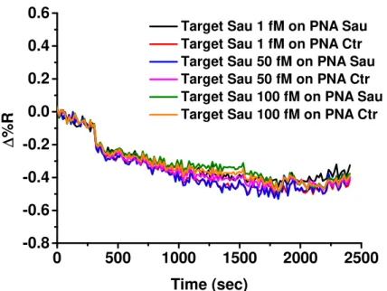
The SPRI hybridization experiments were performed at room temperature by using 300 μL of ssDNA solutions at concentrations 1, 50 and 100 fM which were adsorbed (flow rate 10 μL/min) on the PNA probes functionalized surfaces.
9.4.3. Staphylococcus Aureus gDNA hybridization experiments
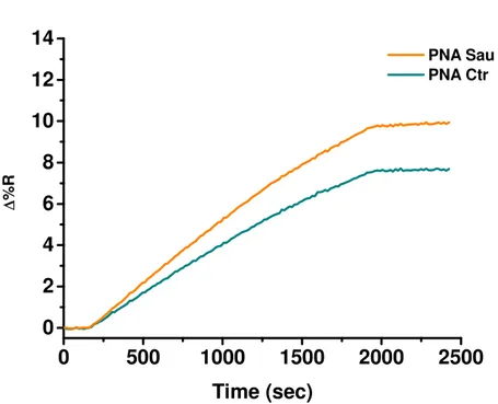
The SPRI hybridization experiments were performed at room temperature by using 300 μL of gDNA solutions at concentrations 1, 50 and 100 fM which were allowed to flow (flow rate 10 μL/min) on the PNA probes functionalized surfaces. Before SPRI analyses gDNA samples were fragmented by sonication (ELMA Transsonic T480/H-2) for 2 minutes and by vortexing (IKA Vortex GENIUS 3) for 1 min and then they were heated to 95°C for 5 minutes to denature the DNA strands into single-stranded state.
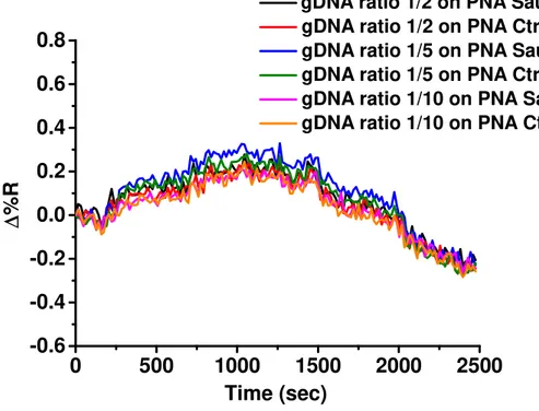
9.4.4. Staphylococcus Aureus and bovine gDNA hybridization experiments
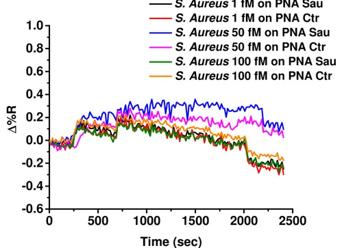
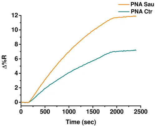
The SPRI hybridization experiments were performed at room temperature by using mixtures of bacterial and bovine gDNA. The total concentration of bacterial and bovine gDNAs was 5 ng/mL with different ratios between bovine and bacterial gDNA. Before SPRI analyses gDNA mixtures were fragmented by sonication (ELMA Transsonic T480/H-2) for 2 minutes and by vortexing (IKA Vortex GENIUS 3) for 1 min and then they were heated to 95°C for 5 minutes to denature the DNA strands.
9.4.5. Synthesis and functionalization of gold nanoparticles for Staphylococcus
Aureus genomic DNA detection
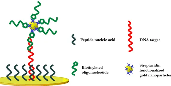
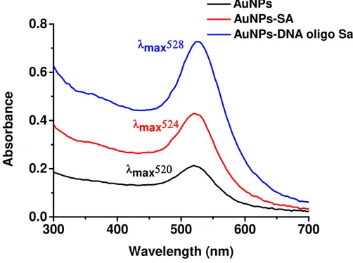
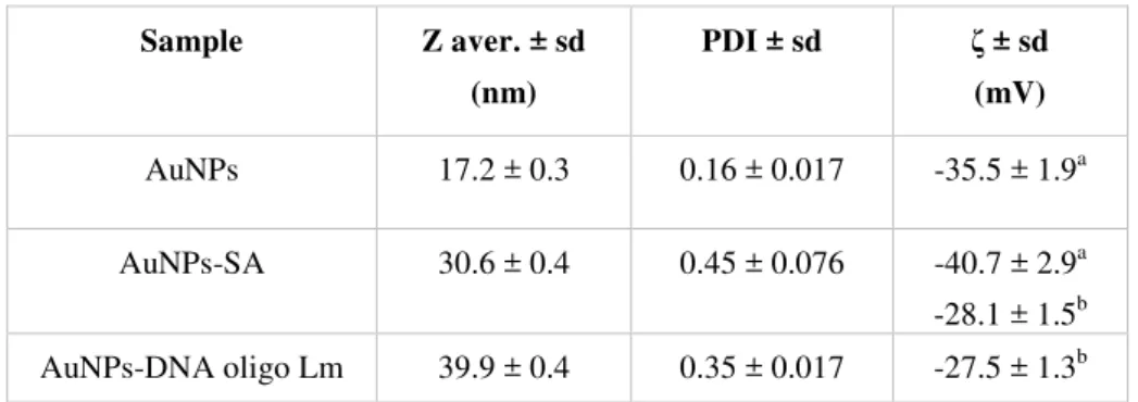
AuNPs were synthesized by citrate reduction of HAuCl4·3H2O according to
methods elsewhere described241 and were functionalized with streptavidin. Streptavidin (like avidin) is a tetrameric protein with a high-affinity biotin-binding site (Kd ~ 10-15 M-1) for each subunit.242 Consequently, AuNPs were conjugated to the biotinylated DNA sequence according to procedures elsewhere described.110
Gold nanoparticles were characterized by UV-vis spectroscopy (NanodropTM 1000) and Dynamic Light Scattering (DLS) and Zeta Potential (ζ) measurements (Zetasizer Nano Series, Malvern Instruments).