Università degli Studi di Catania
Dipartimento di Medicina Clinica e
Sperimentale
Dottorato di Ricerca in Biomedicina Traslazionale
XXX Ciclo
Rosalia Emma
Evidence for enhanced oxidative stress in
smokers with severe asthma
Tesi di Dottorato
Coordinatore: Chiar.mo Prof. Lorenzo Malatino Tutor: Chiar.mo Prof. Riccardo Polosa
Index
Abbreviations……….4
Abstract………..5
1. Introduction………7
1.1 Definition and epidemiology of severe asthma………...…8
1.1.1 Prevalence of severe asthmatic smokers………..…..…12
1.2 Development of severe asthma in smokers………...……13
1.3 Clinical outcomes in smokers with severe asthma………15
1.4 Asthma and Smoking: biological mechanisms………….19
1.4.1 Immunological phenotypes………….……….…19
1.4.2 Oxidative stress in asthmatic smokers………….22
1.5 U-BIOPRED project……….27
2. Aim of the Study……….30
3. Methods………..…….……31
3.1 Patients………..31
3.2 Protocol and sample collection……….32
3.3 8-iso-PGF2α assessment………...33
3.5 Statistical analysis……….37
4. Results……….38
4.1 Clinical parameters………38
4.2 Lipid peroxidation in SAs/ex compared to SAn…………43
4.3 mRNA-expression of pro-/anti-oxidant enzymes in induced sputum……….45
4.4 mRNA-expression of pro-/anti-oxidant enzymes in bronchial biopsy………..46
4.5 mRNA-expression of pro-/anti-oxidant enzymes in bronchial brushing………...47
4.6 Correlation analysis………...48
4.7 Fractional exhaled NO (FeNO)……….49
5. Discussion……….. 51
5.1 Oxidative stress in severe asthmatic smokers/ex smokers vs non-smokers………51
5.2 Pro-oxidant enzymes……….52
5.3 Anti-oxidant enzymes………...55
5.4 Limitations of the study………57
Abbreviations
8-iso-PGF2α : 8-Isoprostaglandin F2α BB: Bronchial Biopsy BBr: Bronchial Brushing CAT: catalase CS: Cigarette Smoke FC: Fold change GPX1: glutathione peroxidase 1 IS: Induced SputumLOD: limit of detection NOS: nitric oxide synthase NOX2: NADPH oxidase 2 RNS: reactive nitrogen species ROS: reactive oxygen species SA: severe asthma/asthmatic SAn: severe asthma non smokers
SAs/ex: severe asthma smokers/ex-smokers SOD: superoxide dismutase
U-BIOPRED: Unbiased BIOmarkers for the PREDiction of Respiratory Disease Outcomes
Abstract
BackgroundAs part of the U-BIOPRED project, extensive clinical and biomarker information was collected from cohorts of adult severe asthma smokers/ex-smokers (SAs/ex) and severe asthmatic non-smokers (SAn). Oxidative stress in SAs/ex vs. SAn patients was
investigated by analysing urinary 8-iso-PGF2α and the mRNA
expression of the main pro-oxidant (NOX2; NOSs) and anti-oxidant (SODs; CAT; GPX1) enzymes in the airways, in relation to clinical outcomes.
Methods
Urine and induced sputum (IS) were obtained from severe asthma patients. A bronchoscopy to obtain bronchial biopsy (BB) and brushing (BBr) was performed in a subset of patients. Urinary 8-iso-PGF2α was quantified using mass spectrometry. IS, BB and
Results
Urinary 8-iso-PGF2α was increased in SAs/ex compared to SAn
(p< 0.001) and in current smokers vs ex-smokers and non-smokers (p= 0.004). Sputum mRNA expression of NOX2 were increased in SAs/ex compared to SAn (three probe-sets with p< 0.05). The mRNA expression of antioxidant enzymes was similar between the two severe asthma cohorts in all airway samples. NOS2 mRNA expression was decreased in BBr of SAs/ex compared to SAn (p= 0.029). NOS2 mRNA expression in BBr correlated with FeNO (p< 0.001). FeNO was lower in current-smokers than in ex-current-smokers (p= 0.007) indicating an effect of active smoking.
Conclusions
The data suggests evidence of greater systemic oxidative stress in SAs/ex with significant effects on the mRNA expression of NOX2 and NOS2 in the airways of severe asthmatic patients.
1. Introduction
Asthma is a life-long chronic inflammatory disease of the airways characterised by variable and recurring symptoms, airways hyperresponsiveness and airflow obstruction that is often reversible either spontaneously or with treatment. According to the World Health Organization (WHO), 235 million people suffer from asthma, but it is under-diagnosed and under-treated with substantial burden to individuals and families. It is a complex and heterogeneous disorder that vary in severity, onset, risk factors, triggers, response to treatments, genetics/epigenetics and natural history (1). The aetiology of asthma is not completely understood but it is know that a gene-environment interaction play an essential role (2). Cigarette smoke is a key factor implicated in modulation of asthma. Data on asthmatic smokers suggest marked impairment in asthma control, accelerated decline in lung function, increased airflow obstruction and increase in disease severity (3). The majority of asthmatics have a mild and controlled disease, whereas others exhibit a more severe condition with poor asthma control, frequent severe exacerbations and accelerated loss of lung function, despite intensive treatment.
Severe asthma is a serious health problem, which has been subject of intensive research over the last years. The Unbiased BIOmarkers in PREDiction of respiratory disease outcomes (U-BIOPRED) is one of the research projects with the aim of improving the management of severe asthma.
1.1 Definitions and Epidemiology of Severe Asthma
Severe asthma is a heterogeneous disease with varying in pathophysiological characteristics (4,5), and several definitions have taken place over the past decade [Table. 1].
The first definition were published in 1999 by European Respiratory Society (ERS) Task Force, which defined difficult asthma as ‘‘asthma remaining uncontrolled despite high dose inhaled corticosteroids (ICS) with or without systemic corticosteroids (OCS)’’, and uncontrolled asthma as ‘‘persistent asthma symptoms or recurrent exacerbations’’(6).
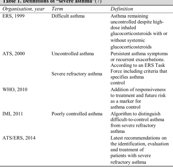
Table 1. Definitions of “severe asthma”(7)
Organisation, year Term Definition
ERS, 1999 Difficult asthma Asthma remaining
uncontrolled despite high-dose inhaled
glucocorticosteroids with or without systemic
glucocorticosteroids ATS, 2000 Uncontrolled asthma
Severe refractory asthma
Persistent asthma symptoms or recurrent exacerbations. According to an ERS Task Force including criteria that specifies asthma
control
WHO, 2010 Addition of responsiveness
to treatment and future risk as a marker for
asthma control
IMI, 2011 Poorly controlled asthma Algorithm to distinguish difficult-to-control asthma from severe refractory asthma
ATS/ERS, 2014 Latest recommendations on
the identification, evaluation and treatment of
patients with severe refractory asthma
ERS: European Respiratory Society; ATS: American Thoracic Society; WHO: World Health Organization; IMI: Innovative Medicines Initiative.
A year later, the American Thoracic Society (ATS) defined refractory asthma on the base of two major criteria (continuous of high dose of ICS or OCS for >50% of the time during the previous year) with two out of seven minor criteria: requirement of additional controller medications, aspects of disease stability, exacerbations and lung function (8). In 2010, the World Health Organization (WHO) added the “responsiveness to treatment”
and “future risk” as indicators of asthma control. The WHO distinguished three type of severe asthma: 1) untreated severe asthma; 2) severe difficult-to-control asthma; 3) severe refractory asthma (9). One year later the Innovative Medicines Initiative (IMI) published an algorithm in order to distinguish difficult-to-control asthma from severe refractory asthma. This algorithm included factors that can make asthma difficult to control, such as non-adherence to treatment, inadequate inhalation technique, continuous exposure to environmental triggers and comorbidities. The latest definition of severe asthma was given by a task force of ERS and ATS in 2014: asthma which requires treatment with high dose inhaled corticosteroids (>1000 mcg of fluticasone equivalent) plus a second controller and/or systemic corticosteroids, to prevent it from becoming “uncontrolled” or which remains “uncontrolled” despite this therapy (10).
Given the different definitions of severe asthma, it has been hard to determine the prevalence. In literature, severe asthma prevalence has been estimated around 5-10% of the total asthma population (11,12). However, this is a rough estimate, which varies from country to country due to access to, quality of
healthcare and adherence to treatment prescription. According to recent estimates reported by ERS, severe asthma affects 1.5 million of people in Europe (13). A recent database analysis based on 2014 ERS/ATS guidelines was conducted in Israel, where severe asthma prevalence was 4.65% of all asthmatics, and 33% of these was uncontrolled severe asthma (14). In the Netherlands the 17.4% of the total asthmatic population had difficult to control asthma according to the IMI criteria, but only 3.6% of these patients were adherent to their high dose ICS prescription (15). Thus, this suggested that the prevalence of truly severe refractory asthma could be lower than estimated in the past years.
Severe asthma, despite represent a small percentage of asthma patients, is responsible for a large part of asthma-related costs. In UK, it is estimated that 50% of all annual healthcare asthma costs come from the most severe asthmatic population (16). But, the severe asthma burden goes far beyond the healthcare costs: severe asthmatics patients often experienced work-related disability (partial or total lost-day, early disability or permanent disability) with a substantial socio-economics impact (11,16).
1.1.1 Prevalence of severe asthma smokers
The WHO has estimated in 2015 that there are 1.1 billion of smokers worldwide. The prevalence of active smoking in adults with asthma is similar to the prevalence in the general population, and ranges from 20% to 35% (17). Moreover, 20-30% of asthmatic adults are ex-smokers (18,19). As stated above, severe asthma occurs in 5-10% of the asthmatic population, but the prevalence of severe asthma current and ex-smokers is difficult to estimate. Indeed in the past years, asthmatic patients with a smoking history are often excluded from studies of severe asthma, such as the US Severe Asthma Research Program (20) and the European Network for Understanding Mechanisms of Severe Asthma (21), where smokers with or more 5 pack years use were excluded. Otherwise some studies, including the Epidemiology and Natural History of Asthma: Outcomes and Treatment Regimens (TENOR) (22), British Thoracic Society (BTS) Severe Asthma Registry (23) cohorts and U-BIOPRED research project (24), did not exclude heavier smokers. The first two study report figures of 6% and 4% for current smokers, respectively, suggesting that active smoking is lower in severe
asthmatics than in the general population (17,25). Instead, ex-smokers with asthma represent one third of patients with severe or difficult-to-treat asthma (22,23). This interesting data suggest that cigarette smoke could have a role in the progression of asthma disease.
1.2 Development of severe asthma in smokers
Epidemiological evidences about the association of active smoking and asthma development was observed in some (26–35), but not in all studies (36,37). Many studies lack consistency, due to cross-sectional and case-control design, self-reported measures or questionnaires, potential inaccuracy in asthma diagnosis, which provide significant bias (3). Nevertheless, the majority of studies suggested that cigarette smoke increase the risk of developing asthma. Rasmussen et al. investigated the development of asthma in asymptomatic teenagers over a 6 years period, and showed that active smoking was independently associated with asthma-like symptoms (OR= 2.1; 95% CI= 1.2-3.8), as well as atopy and bronchial hyperresponsiveness to methacholine (27). A strong association was also found between
smoking and the onset of asthma among non-atopic subjects (OR= 5.7; 95% CI=1.7-19.2) (26). Moreover, tobacco smoking is a risk factor for the development of asthma in the elderly (28). In non-asthmatic adult with established allergic rhinitis, smoking was significantly related to the asthma onset within a period of 10 years (OR= 2.98; 95% CI= 1.81-4.92) with a clear dose-response association (35). Furthermore, active smoking was shown to be a predictor of an accelerate decrease of FEV1 in longitudinal
studies of asthma population (38,39). The analysis of a well-characterised cohort of allergic rhinitis patients followed for 10 years, showed that smoking status and smoking duration are associated with greater severity of new-onset asthma, with a dose-response association (40). Guus et al. also showed that the number of pack years was the only independent predictor for the progression of asthma severity, with an OR of 1.4 (95% CI, 1.02-1.91) every 10 pack years smoked (41).
All these studies showed that a positive smoking history in adult subjects is predictive of new-onset asthma and increase of asthma severity, in a dose-dependent manner.
1.3 Clinical outcomes in smokers with severe asthma
Cigarette smoking interacts with asthma and induces important adverse effects on clinical, functional, prognostic and therapeutic outcomes (3). Adults and teens with asthma who are active smokers show more severe symptoms and worse quality of life compared to asthmatic non-smokers (40). Many studies have reported an accelerated decline in lung function and an increased severity of airflow obstruction in asthmatic who smoke (38,42). Moreover, asthmatic patients who smoke don't have a response to the beneficial effects of inhaled corticosteroids (ICS) or oral corticosteroids (OCS) in contrast to asthmatic who don't smoke (43–45). Asthmatic smokers or ex-smokers often require hospitalization and emergency room treatment. In these patients COPD (secondary to asthma) and rhinovirus infections are frequently diagnosed (46). The viral infections, associated to asthma and smoking are difficult to treat because tobacco smoke increases viral replication and suppresses the protective functions of airway epithelium, alveolar macrophages, dendritic cells, NK cells and adaptive immune mechanism (47).
Few studies investigated clinical and inflammatory characteristics in severe asthmatic smokers, until now (Tab. 2).
Thomson et al. studied the cigarette smoking effect on severe asthma (25). They compared patient characteristics, clinical outcomes, and biomarkers of inflammation in current smokers, ex-smokers, and never smokers within a group of 760 patients with severe asthma recruited from the British Thoracic Society Severe Asthma Registry in the UK. These researchers found that current smokers with severe asthma had worse clinical outcomes compared to never smokers and ex-smokers, such as higher Asthma Control Questionnaire (ACQ) score, implying poorer asthma control, and lower Asthma Quality of Life Questionnaire (AQLQ) score, more unscheduled health care visits, more rescue courses of oral steroids, and higher anxiety and depression scores. Severe asthmatic current smokers had reduced sputum eosinophils and lower FeNO when compared to never smokers, whereas ex-smokers had increased proportion of sputum neutrophils than never smoker, but a similar sputum eosinophils count and FeNO value. Both current and former smokers had
reduced blood specific allergy antibody levels to several common environmental allergens (25).
Table 2. Clinical and inflammatory outcomes of severe asthma smokers
Clinical outcomes
- Late onset of severe asthma (25,48,49) - Poorer asthma control (25,49,50) - Worse quality of life (25,48)
- More unscheduled health care visits (25,50) - More emergency department visits (25) - More rescue courses of OCS (25)
- Higher anxiety and depression scale scores (25) Lung function
- Lower Kco values (25)
- Severe airflow obstruction (49) Inflammatory outcomes
- Lower FeNO value (25,48)
FeNO: Fractional exhaled nitric oxide; Kco: transfer coefficient for carbon monoxide; OCS: oral corticosteroids.
An initial analysis of clinical and inflammatory characteristics of U-BIOPRED severe asthma non-smokers and smokers/ex-smokers cohorts identified few differences. The two severe asthma groups showed a similar percentage received OCS therapy and similar degree of airflow obstruction. A lower FeNO level was observed in the smokers/ex-smokers cohort. Moreover, they showed that asthma onset occurred on average 18 years later in smokers/ex-smokers with severe asthma and a positive correlation between AQLQ scores and the number of pack years (48). A further cluster analysis of asthma patients from the U-BIOPRED adult cohorts identified four distinct clusters, one of these linked to smoking history. This cluster was composed of overweight to obese, late-onset severe asthmatic smokers/ex-smokers, with relatively poor control, severe airflow obstruction (mean FEV1= 58.9% predicted), and highest sputum and blood eosinophils, with a lower proportion of atopic subjects than in the other clusters (49).
These findings showed that cigarette smoking impaired the quality of life of severe asthma subjects with worse symptoms
and health-care outcomes compared to never smokers with severe asthma.
1.4 Asthma and Smoking: biological mechanisms
The mechanisms accounting for the adverse effects of smoking in asthma have been studied over the last few years, but are not completely understood. Inflammation and oxidative stress, induced by smoking, seems to play an important role in the pathogenesis of severe asthma smokers.
1.4.1 Immunological phenotypes
It is known that cigarette smoking induces airway inflammation in non-asthmatic smokers (51–54), with increased T lymphocytes and macrophages in the airway wall, higher neutrophils count in the bronchial secretion, and infiltration of mononuclear cells and macrophages in airways (52). Moreover, increased number of eosinophils infiltrating the submucosa was found in the peripheral lung sections of smokers compared to non-smokers (55). There is now accumulating evidence that cigarette smoke/asthmatic inflammation interaction may induce important changes in the
asthma phenotype [Fig.1]. Some researchers reported a predominance of macrophages and neutrophils in mild asthma who smoke, likewise in early COPD (51,56,57) and reduced sputum eosinophil count both in mild (57) and severe asthmatic smokers (25) compared to mild and severe asthmatic non-smokers, respectively. Neutrophils contribute to mucous hypersecretion, which is an additional risk factor for the accelerated decline in FEV1 (3). Metalloproteinases (MMPs) are
also involved in the modulation of inflammation, repair, and remodelling processes, along with their intrinsic and tissue inhibitors (58). Elevated concentration of MMP-12 was found in sputum of asthmatic smokers, with a positively correlation to sputum neutrophil count, and an inversely correlation to lung function (59). Sputum MMP-9 activity/TIMP-1 and TIMP-2 ratios, and nasal epithelial MMP9/TIMP1 and MMP9/TIMP2 expression ratios were found reduced in asthmatic smokers compared to never smokers. Furthermore, the low sputum ratios in asthmatic smokers were associated with reduced post-bronchodilator FEV1, FEV1/FVC ratio and segmental airway lumen area (60). Persistent exposure to cigarette smoke may also
alter the Th1/Th2-cytokine and Th17-cytokines secretion, modifying the inflammatory response (3,51).
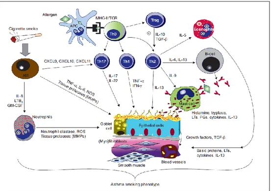
Figure 1. Inflammatory mechanisms for the mixed asthma-smoking phenotype. In allergic asthma,
initial exposure(s) of antigen-presenting cells (APCs) to allergen mainly leads to the activation of allergen-specific T-helper Th2 cells and immunoglobulin IgE synthesis. Subsequent exposures to allergen cause inflammatory-cell recruitment (e.g. eosinophils) and activation (e.g. mast cells) and mediator release. Th1-cell responses might also be responsible for epithelial apoptosis and smooth-muscle cell activation. Regulatory T-cells (Treg) are involved in the suppression of Th2-cell responses in humans through the inhibitory cytokines interleukin (IL)-10 and transforming growth factor (TGF)-b. The newly identified CD4+ T-cell subset, Th17 cells, seems to be specifically associated with the neutrophilic inflammatory events that occur during disease exacerbation and in tissue remodelling. Activated macrophages (AMs), triggered by cigarette smoke, will produce a myriad of pro-inflammatory molecules, reactive oxygen species (ROS), matrix metalloproteinase (MMPs), and chemokines involved in the mobilisation and prolonged survival of neutrophils in the lung tissue (e.g. IL-8). AMs in the smokers appear to produce less IL-10, which leads to less Th2-cells development and reduced B-cell number and lower levels of IL-4 and IL-5. This will produce fewer and less active eosinophils and lower production of IgE in smoking asthmatics. AMs are also involved in the direct stimulation of Th1 and Th17 cells that in turn promote bronchial epithelial cells and goblet cell activation to increase inflammatory responses, mucous production and tissue remodelling. Cigarette smoke might also directly activate bronchial epithelial cells to release further pro-inflammatory cytokines, chemokines, growth factors and TGF-b, which are known to modulate smooth muscle cell and fibroblast proliferation with subsequent progression to fibrosis and extracellular matrix deposition. In smoking, asthma-increased levels of thymic stromal lymphopoietin may further stimulate mast cells to release remodelling mediators, such as IL-13, independently of Th2 cells. Increased MCs, together with smoking-induced epithelial cell activation, may increase IL-8 production, contributing to further infiltration of neutrophils. Neutrophil infiltration favours the progression of the inflammatory response and lung damage of the smoker through the production of ROS, lipid mediators and tissue proteases. The persistent exposure to cigarette smoke drives additive or synergistic inflammatory and remodelling responses (e.g. chronic mucus hypersecretion) in the asthmatic airways (3). LTB4: leukotriene B4; GM-CSF: granulocyte-macrophage colony-stimulating factor; MHC: major histocompatibility complex; TCR: T-cell receptor; TNF-a: tumour necrosis factor-a; IFN: interferon-c; LTs: leukotrienes; PGs: prostaglandins (Polosa et al. Eur Respir J. 2013).
The recent severe asthma smokers/ex-smokers phenotype, identified by U-BIOPRED investigators, had a high sputum and blood eosinophil count and a differential transcript expression profile in sputum cells related to the regulation of actin cytoskeleton (ITGB1, ITGB8, FN1, DIAPH2, F2R, 332 ACTN2), and to fibronectin matrix formation (ITGB1, FN1) (49). These findings give a further confirmation of the cigarette smoke effect in severe asthma.
1.4.2 Oxidative stress in asthmatic smokers
“Oxidative stress” describes the level of oxidative damage in cells, tissues or organs, caused by reactive chemical species, derived from oxygen or nitrogen, such as reactive oxygen species (ROS) or reactive nitrogen species (RSN), respectively. ROS/RSN are oxidant metabolites that have the ability to snatch electrons from other molecules which come in contact with them (61). The sources of ROS/RSN can be exogenous, primarily tobacco smoke and air pollution, and endogenous, such as chemical reactions that occur in physiological or pathological conditions (62,63). Tobacco smoke is one of major source of
oxidants/free radicals (64). These reactive chemical species have a physiologic function, as signalling molecules, at low concentrations, but at high concentration, they are harmful to cells (61). Due to high risk of exposure to a number of oxidizing substances, the human body has developed a wide range of enzymatic and non-enzymatic antioxidant defences (65). The oxidative stress is particularly enhanced in chronic inflammatory conditions, such as chronic asthma, characterized by raised ROS/RSN generation at the site of inflammation (61). Furthermore increased levels of ROS/RSN promote the inflammatory responses (62) triggering a chain of events that lead to the maintenance and worsening of pathological conditions, such as in asthma.
The airways are normally exposed to environmental oxidative compounds, moreover in asthma patients the conditions are worsened by the persistence of activated inflammatory cells in lung parenchyma. The activation of inflammatory cells leads to
production of anion superoxide (O2•-) and nitric oxide (NO). The
(NOXs) pathway that are enzymes catalyzing the production of
O2•- by reducing oxygen, using NADPH as electron donor (66):
2 O2 + NADPH = 2 O2•- + NADP+ + H+
NOXs enzymes are expressed in a number of lung cell types, as well as epithelial, endothelial and mesenchymal cells (67). The NOX2 isoform, in particular, is referred to “phagocyte NOX”. Indeed, it is primarily present in macrophages, neutrophils and eosinophils, and its activity is normally required for phagocyte respiratory burst and regulation of cell signaling (66,67). The
O2•-, produced by NOXs, is quickly converted to hydrogen
peroxide (H2O2) by superoxide dismutase enzymes (SOD) (68).
There are three forms of SOD, present in all classes of lung cells with different expression and localization. The cytosolic SOD (SOD1; CuZnSOD) and the mitochondrial SOD (SOD2; MnSOD) are expressed in human bronchial epithelial cells and in alveolar macrophages. While, the extracellular SOD (SOD3; ECSOD) is expressed in the pulmonary vasculature and interstitium (69,70). H2O2 is not a free radical, but it is, at least, a
reactive chemical specie. For this reason, it is transformed in H2O
peroxidase (GPX) (64). CAT is localized in peroxisomes and cytoplasm of alveolar type II pneumocytes, macrophages and neutrophils (64,65). GPX family comprises 8 enzymatic isoforms and their expression levels vary depending on the tissue type. The most abundant isoform is the glutathione peroxidase 1(GPX1) (71). The H2O2, not converted in H2O, can react with transition
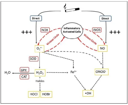
metals to generate hydroxyl radicals (•OH) or interacts with halides to form hypobromous acid (HOBr) or hypochlorous acid (HOCl), that are reactive compounds too (68). Nitric oxide (NO) is the other free radical produced by activated inflammatory cells through the degradation of L-arginine to L-citrulline by nitric oxide synthase (NOS) enzymes (72,73):
L-arginine + O2 = N-idrossiarginine = L-citrulline + NO
There are three isoforms of NOS: NOS1 and NOS3 are classified as constitutive and calcium-dependent isoforms, whereas NOS2 (iNOS) is the inducible and calcium-independent isoform (74,75). The up-regulation of iNOS leads to increased levels of NO that, in the presence of ROS, rapidly forms RNS (68) [Fig. 2]. Excess ROS and RSN lead to peroxidation of membrane lipids, depletion
of nicotinamide nucleotides, enhanced intracellular Ca2+, cytoskeleton breakage and DNA damage (64,68).
Figure 2. Generation of reactive species and antioxidant defences in the lung. O2•- = superoxide anion; H2O2= hydrogen peroxide; HOBr= hypobromous acid; HOCl= hypochlorous acid; NO= nitric
oxide; RSN= reactive nitrogen species; •OH= hydroxyl radical; Fe2+= ferrous ion; NOX= NADPH oxidase; SOD= superoxide dismutase; GPX= glutathione peroxidase; CAT= catalase; iNOS= inducible nitric oxide synthase.
Tobacco smoke is a complex mixture of thousands of different chemical species, which can damage macromolecules and biochemical pathways. Consequently, the inhalation of cigarette smoke triggers many cellular and signaling responses that contribute to maintenance and progression of the inflammatory response and disease chronicity in asthma patients who smoke.
Many oxidant compounds (ROS and RNS, quinones, aldehydes, ketones, metals) are present in cigarette smoke (76,77). These molecules may induce direct or indirect oxidative damage, through the generation of pro-inflammatory compounds
(chemokines, cytokines, prostaglandins, leukotrienes,
isoprostanes) and the modulation of intracellular pro-inflammatory pathways (MAPK, NF-kB, AP-1, Keapl-Nrf2-ARE) (62,76,78). Very little is known about the role of oxidative stress in severe refractory asthma and even less about the combined impact of cigarette smoking.
1.5 U-BIOPRED project
U-BIOPRED (79) is an European research project started in 2008 by the European Commission and the Innovative Medicines Initiative (IMI), using information and samples from adults and children to learn more about different types of asthma to ensure better diagnosis and treatment for each person.
To achieve their aim, the pan-European consortium U-BIOPRED used an innovative systems biology approach to integrate
unbiased high dimensional data obtained by different “omics” technologies from invasive and non-invasive sampling, together with clinical data. The ultimate outcome of the project is to advance the understanding of asthma and create a “handprint” of severe asthma.
Figure 3. U-BIOPRED Innovative approach to generate “phenotype handprint”.
U-BIOPRED has enrolled up to 500 adult asthmatic patients, 421 of which are severe asthmatics (severe asthma non-smokers: 311; severe asthma smokers/ex-smokers: 110).
The Polosa’s team of University of Catania, together with the 11 European academic centers, has contributed to create a detailed clinical database of severe asthmatic patients, with and without a smoking history, and a biobank of their biologic samples. Thus, the U-BIOPRED database is a unique opportunity to learn more about severe asthmatic smokers.
2. Aim of the Study
We hypothesize that cigarette smoke increases airway inflammation not only by providing additional oxidant species, but also by modulating the anti-/pro-oxidant balance.
We therefore compared the oxidative stress status in SA patients with and without a significant smoking history. We investigated the level of the lipid peroxidation marker 8-iso-PGF2α in the urine
and the mRNA expression profile of key pro-oxidant (NADPH oxidase 2, NOX2; inducible NOS, NOS2; constitutive NOSs, NOS1 and NOS3) and anti-oxidant (superoxide dismutases, SOD1, -2 and -3; catalase, CAT; and glutathione peroxidase 1, GPX1) enzymes in the airways, in particular in bronchial biopsy (BB), bronchial brushing (BBr) and induced sputum (IS) samples.
3. Methods
3.1 Patients
Participants with severe asthma were recruited from 16 clinical centres in 11 European countries (48). Severe asthma was defined according to the U-BIOBRED international consensus criteria (80). Prior to enrolment, participants with severe asthma were required to have been under follow-up by a respiratory physician for at least six months, during which time assessments had been undertaken to optimize asthma control and assess medication adherence. The Severe Asthma U-BIOPRED patients were divided in two groups:
- Severe non-smoking asthma (SAn): non-smokers and ex-smokers with a less than five pack-year smoking history, with asthma and uncontrolled symptoms defined according to GINA guidelines 22 and/or frequent exacerbations (more than two per year) despite high-dose inhaled corticosteroids (ICS ≥ 1000 µg fluticasone propionate/day or equivalent dose).
- Smokers and ex-smokers with severe asthma (SAs/ex): this group was defined as for the SAn group except that they were either current smokers or ex-smokers with a pack history of at least five years.
The study was approved by the ethics committee for each participating clinical institution, and adhered to the standards set by International Conference on Harmonisation and Good Clinical Practice. It is registered on ClinicalTrials.gov, (Identifier: NCT01982162). All participants gave signed informed consent.
3.2 Protocol and sample collection
All enrolled subjects underwent a baseline visit to assess current health status, atopy and pulmonary function. Spirometry, reversibility test (post-bronchodilator) and fraction of exhaled nitric oxide level (FeNO) at 50 ml/sec were performed. Spirometry were performed according to current ATS/ERS guidelines (81,82). Allergic status was assessed by skin prick test and measurement of total and specific IgE to six common aeroallergens. Questionnaires were administered to assess asthma
control (ACQ), health outcomes (AQLQ and HADS), upper airways symptoms (SNOT20), adherence (MARS) and sleep and daytime drowsiness (ESS). Urine samples and induced sputum (IS) were collected during the baseline visit. Some patients underwent an optional bronchoscopy visit during which bronchial biopsy (BB) and bronchial brushing (BBr) were obtained.
3.3 8-iso-PGF2α assessment
8-iso-PGF2α was extracted from spot urine samples (SAn: 305; SAs/ex: 109) using solid phase extraction and quantified via liquid chromatography coupled to mass spectrometry (LC-MS/MS) using an Acquity UPLC coupled to a Xevo TQ-S mass spectrometer (Waters, Milford, MA). Optimal extraction volumes for solid phase extraction were calculated using individual UV absorbance (λ=300 nm) measurements to minimize matrix effects and levels of 8-iso-PGF2α were normalized to urinary creatinine (83).
3.4 Microarray assessment
RNA from RNAlater-preserved IS, BB, and BBr samples were extracted using Qiagen miRNeasy kit and amplified with Nugen ovation pico WTA kit (NuGen Technologies; San Carlos, CA). The cDNA was analysed using the Affymetrix HG-U133+PM microarray platform (Affymetrix, Santa Clara, CA). CEL files were normalized, assessed for quality control to exclude technical outliers (chip image analysis, Affymetrix GeneChip QC, RNA degradation analysis, distribution analysis, principal components analysis, and correlation analysis), and re-normalized using the robust multi-array (RMA) method.
For IS, RNA was extracted from 362 samples, of which 228 had sufficient sample for microarray analysis. 15 of these samples failed QC metrics, 19 additional samples were excluded (not considered enrolled in adult cohorts), and 71 samples disqualified for squamous>30% or no differential counts available, with 123 quality samples remaining for subsequent analyses (SAn: 61; SAs/ex: 23; remainder were for non-severe asthmatics and healthy).
For BB, RNA was extracted from 116 samples and had sufficient sample for microarray analysis. Seven of these samples failed QC metrics, and 1 additional sample was excluded (not considered enrolled in adult cohorts), with 108 quality samples remaining for subsequent analyses (SAn: 40; SAs/ex: 13; remainder were for non-severe asthmatics and healthy).
For BBr, RNA was extracted from 159 samples, of which 158 had sufficient sample for microarray analysis. Six of these samples failed QC metrics, 3 additional samples were excluded (2 duplicate samples and 1 not considered enrolled in adult cohorts), with 149 quality samples remaining for subsequent analyses (SAn: 49; SAs/ex: 18; remainder were for non-severe asthmatics and healthy).
A log2-intensity threshold was established as the limit of reliable quantification (LOD) based on the 90th percentile signal of merged nonspecific probe-sets distribution in the array and by the inflection point of maximum variance with decreasing signal in a standard deviation vs. mean intensity plot across all probe sets. Probe sets with mean log2 intensity above this threshold in at least one of the 4 study cohorts were considered quantifiable and
included in subsequent analyses. The cut-offs (number of probe sets included) were 5.5 (23496), 5.0 (18697), and 4.75 (21363) for sputum, BB, and BBr, respectively.
Batch effects from RNA processing sets were observed for the sputum and BBr datasets, with the batch effect adjusted in the data matrices using linear modelling of batch (as random factor) and cohort. For the sputum dataset, 3 subjects had duplicate samples, of which the mean of the log2 intensities, after RNA processing set batch adjustment, were used in the final analysis dataset.
The probe-sets used in this study were as follow: NOX2
(203922_PM_s_at, 203923_PM_s_at, 217431_PM_x_at,
233538_PM_s_at); NOS1 (1560974_PM_s_at, 207309_PM_at,
207310_PM_s_at, 231916_PM_at, 239132_PM_at,
240911_PM_at); NOS2 (210037_PM_s_at); NOS3
(205581_PM_s_at); SOD1 (200642_PM_at); SOD2
(215078_PM_at, 215223_PM_s_at, 216841_PM_s_at,
221477_PM_s_at); SOD3 (205236_PM_x_at); CAT
(201432_PM_at, 211922_PM_s_at, 215573_PM_at); GPX1 (200736_PM_s_at).
3.5 Statistical analysis
Categorical data were summarized by counts and percentages; continuously distributed data, exhibiting approximate symmetry of distribution, were summarized using the mean (standard error; SE); continuously distributed data exhibiting skewness were summarized using the median (inter-quartile range; IQR). The latter all exhibited positive skewness and were log-transformed prior to parametric association testing. P-values were calculated by applying ANOVA to a generalised linear model with adjustment for age and gender. All analyses were considered significant with a p-value of less than 5 %. Acknowledging the modest sample size of the current exploratory study, adjustments for multiple-testing were not applied. In order to characterise further some of the observed associations, rank correlation was assessed by calculation of Kendall’s Tau. A p-value was calculated in a test of the null hypothesis of zero correlation. Analyses of clinical and inflammatory data were performed using R version 2.15.2 (R Core Team, 2012).
4. Results
4.1 Clinical parameters
The clinical and inflammatory characteristics of severe asthma patients (SAn and SAs/ex) are reported in Tables 3-7 for each examined patient subset (urine-lipidomics; IS-transcriptomics; BB-transcriptomics; BBr-transcriptomics). In brief, spirometry parameters were similar among SAn and SAs/ex. The onset of asthma occurred 17 years later in SAs/ex than in SAn although subjects had a similar degree of airway obstruction. Moreover SAs/ex had increased prevalence of gastro-esophageal reflux disease (GERD) than SAn. FeNO levels were significantly lower in SAs/ex than SAn for those subjects for whom BB and BBr transcriptomics data was available but this was not seen across all subgroups (data not show).
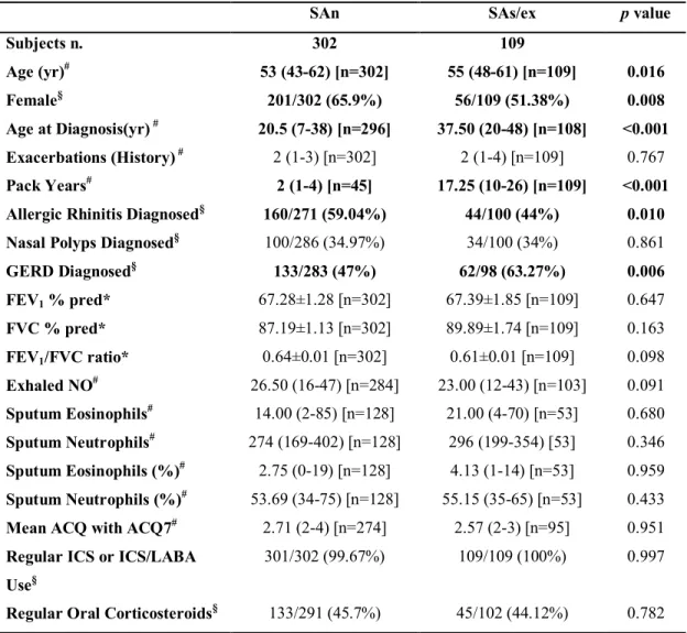
Table 4. Clinical and inflammatory characteristics of severe asthma patients present in the urinary eicosanoids subset.
SAn SAs/ex p value
Subjects n. 302 109 Age (yr)# 53 (43-62) [n=302] 55 (48-61) [n=109] 0.016 Female§ 201/302 (65.9%) 56/109 (51.38%) 0.008 Age at Diagnosis(yr) # 20.5 (7-38) [n=296] 37.50 (20-48) [n=108] <0.001 Exacerbations (History) # 2 (1-3) [n=302] 2 (1-4) [n=109] 0.767 Pack Years# 2 (1-4) [n=45] 17.25 (10-26) [n=109] <0.001 Allergic Rhinitis Diagnosed§ 160/271 (59.04%) 44/100 (44%) 0.010 Nasal Polyps Diagnosed§ 100/286 (34.97%) 34/100 (34%) 0.861 GERD Diagnosed§ 133/283 (47%) 62/98 (63.27%) 0.006 FEV1 % pred* 67.28±1.28 [n=302] 67.39±1.85 [n=109] 0.647 FVC % pred* 87.19±1.13 [n=302] 89.89±1.74 [n=109] 0.163 FEV1/FVC ratio* 0.64±0.01 [n=302] 0.61±0.01 [n=109] 0.098 Exhaled NO# 26.50 (16-47) [n=284] 23.00 (12-43) [n=103] 0.091 Sputum Eosinophils# 14.00 (2-85) [n=128] 21.00 (4-70) [n=53] 0.680 Sputum Neutrophils# 274 (169-402) [n=128] 296 (199-354) [53] 0.346 Sputum Eosinophils (%)# 2.75 (0-19) [n=128] 4.13 (1-14) [n=53] 0.959 Sputum Neutrophils (%)# 53.69 (34-75) [n=128] 55.15 (35-65) [n=53] 0.433 Mean ACQ with ACQ7# 2.71 (2-4) [n=274] 2.57 (2-3) [n=95] 0.951 Regular ICS or ICS/LABA
Use§
301/302 (99.67%) 109/109 (100%) 0.997
Regular Oral Corticosteroids§ 133/291 (45.7%) 45/102 (44.12%) 0.782
Data are presented as *mean±SE, #median (interquartile range) [n] or §n/N (%), unless otherwise
stated. GERD: gastro-esophageal reflux disease; FEV1: forced expiratory volume in one second;
FVC: forced vital capacity; ACQ: Asthma Control Questionnaire; ICS: inhaled corticosteroids; LABA: long-acting β2-agonist.
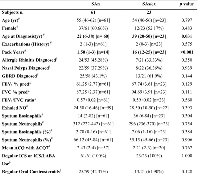
Table 5.Clinical and inflammatory characteristics of severe asthma patients present in the induced sputum-transcriptomics subset.
SAn SAs/ex p value
Subjects n. 61 23 Age (yr)# 55 (46-62) [n=61] 54 (46-56) [n=23] 0.797 Female§ 37/61 (60.66%) 12/23 (52.17%) 0.483 Age at Diagnosis(yr) # 22 (6-38) [n= 60] 39 (20-50) [n=23] 0.031 Exacerbations (History) # 2 (1-3) [n=61] 2 (0-3) [n=23] 0.575 Pack Years# 1.50 (1-3) [n=14] 16 (12-25) [n=23] <0.001 Allergic Rhinitis Diagnosed§ 24/53 (45.28%) 7/21 (33.33%) 0.350
Nasal Polyps Diagnosed§ 22/59 (37.29%) 8/22 (36.36%) 0.939
GERD Diagnosed§ 25/58 (43.1%) 13/21 (61.9%) 0.144 FEV1 % pred* 61.25±2.77[n=61] 67.74±3.61 [n=23] 0.129 FVC % pred* 87.25±2.37[n=61] 94.69±3.91 [n=23] 0.111 FEV1/FVC ratio* 0.57±0.02 [n=61] 0.59±0.02 [n=23] 0.560 Exhaled NO# 24.50 (16-46) [n=59] 28.50 (10-50) [n=22] 0.393 Sputum Eosinophils# 14 (2-82) [n=61] 36 (6-84) [n=23] 0.304 Sputum Neutrophils# 312 (222-442) [n=61] 296 (236-370) [n=23] 0.754 Sputum Eosinophils (%)# 2.70 (0-16) [n=61] 7.06 (1-16) [n=23] 0.384 Sputum Neutrophils (%)# 66.12 (45-84) [n=61] 55.15 (45-66) [n=23] 0.906 Mean ACQ with ACQ7# 2.43 (2-4) [n=57] 2.21 (2-3) [n=20] 0.767 Regular ICS or ICS/LABA
Use§
61/61 (100%) 23/23 (100%) 1.000
Regular Oral Corticosteroids§ 25/59 (42.37%) 13/21 (61.90%) 0.128
Data are presented as *mean±SE, #median (interquartile range) [n] or §n/N (%), unless otherwise stated.
GERD: gastro-esophageal reflux disease; FEV1: forced expiratory volume in 1 second; FVC: forced
vital capacity; ACQ: Asthma Control Questionnaire; ICS: inhaled corticosteroids; LABA: long-acting β2-agonist.
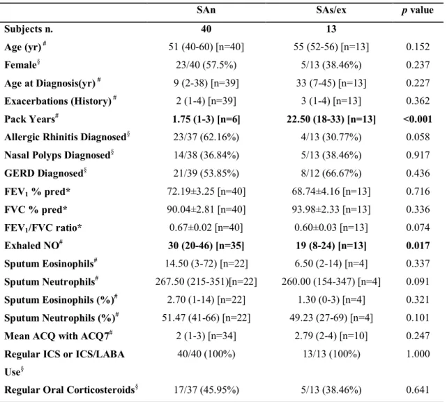
Table 6. Clinical and inflammatory characteristics of patients present in Bronchial Biopsy-transcriptomics subset.
SAn SAs/ex p value
Subjects n. 40 13 Age (yr) # 51 (40-60) [n=40] 55 (52-56) [n=13] 0.152 Female§ 23/40 (57.5%) 5/13 (38.46%) 0.237 Age at Diagnosis(yr) # 9 (2-38) [n=39] 33 (7-45) [n=13] 0.227 Exacerbations (History) # 2 (1-4) [n=39] 3 (1-4) [n=13] 0.362 Pack Years# 1.75 (1-3) [n=6] 22.50 (18-33) [n=13] <0.001 Allergic Rhinitis Diagnosed§ 23/37 (62.16%) 4/13 (30.77%) 0.058
Nasal Polyps Diagnosed§ 14/38 (36.84%) 5/13 (38.46%) 0.917
GERD Diagnosed§ 21/39 (53.85%) 8/12 (66.67%) 0.436 FEV1 % pred* 72.19±3.25 [n=40] 68.74±4.16 [n=13] 0.716 FVC % pred* 90.04±2.81 [n=40] 93.98±2.33 [n=13] 0.336 FEV1/FVC ratio* 0.67±0.02 [n=40] 0.60±0.03 [n=13] 0.074 Exhaled NO# 30 (20-46) [n=35] 19 (8-24) [n=13] 0.017 Sputum Eosinophils# 14.50 (3-72) [n=22] 6.50 (2-14) [n=4] 0.337 Sputum Neutrophils# 267.50 (215-351)[n=22] 260.00 (154-347) [n=4] 0.091 Sputum Eosinophils (%)# 2.70 (1-14) [n=22] 1.30 (0-3) [n=4] 0.321 Sputum Neutrophils (%)# 51.47 (41-66) [n=22] 49.23 (27-69) [n=4] 0.101 Mean ACQ with ACQ7# 2 (1-3) [n=34] 2.79 (2-4) [n=10] 0.247 Regular ICS or ICS/LABA
Use§
40/40 (100%) 13/13 (100%) 1.000
Regular Oral Corticosteroids§ 17/37 (45.95%) 5/13 (38.46%) 0.641
Data are presented as *mean±SE, #median (interquartile range) [n] or §n/N (%), unless otherwise
stated. GERD: gastro-esophageal reflux disease; FEV1: forced expiratory volume in 1 second; FVC:
forced vital capacity; ACQ: Asthma Control Questionnaire; ICS: inhaled corticosteroids; LABA: long-acting β2-agonist.
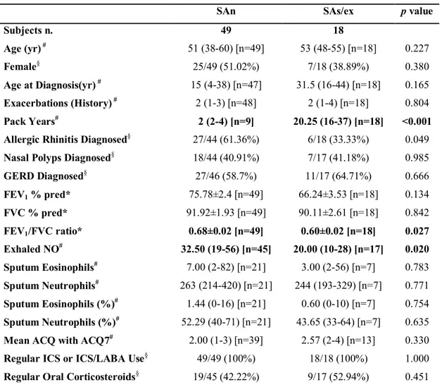
Table 7. Clinical and inflammatory characteristics of severe asthma patients present in Bronchial Brushings-transcriptomics subset.
SAn SAs/ex p value
Subjects n. 49 18 Age (yr) # 51 (38-60) [n=49] 53 (48-55) [n=18] 0.227 Female§ 25/49 (51.02%) 7/18 (38.89%) 0.380 Age at Diagnosis(yr) # 15 (4-38) [n=47] 31.5 (16-44) [n=18] 0.165 Exacerbations (History) # 2 (1-3) [n=48] 2 (1-4) [n=18] 0.804 Pack Years# 2 (2-4) [n=9] 20.25 (16-37) [n=18] <0.001 Allergic Rhinitis Diagnosed§ 27/44 (61.36%) 6/18 (33.33%) 0.049
Nasal Polyps Diagnosed§ 18/44 (40.91%) 7/17 (41.18%) 0.985
GERD Diagnosed§ 27/46 (58.7%) 11/17 (64.71%) 0.666 FEV1 % pred* 75.78±2.4 [n=49] 66.24±3.53 [n=18] 0.134 FVC % pred* 91.92±1.93 [n=49] 90.11±2.61 [n=18] 0.842 FEV1/FVC ratio* 0.68±0.02 [n=49] 0.60±0.02 [n=18] 0.027 Exhaled NO# 32.50 (19-56) [n=45] 20.00 (10-28) [n=17] 0.020 Sputum Eosinophils# 7.00 (2-82) [n=21] 3.00 (2-56) [n=7] 0.783 Sputum Neutrophils# 263 (214-420) [n=21] 244 (193-329) [n=7] 0.771 Sputum Eosinophils (%)# 1.44 (0-16) [n=21] 0.60 (0-10) [n=7] 0.754 Sputum Neutrophils (%)# 52.29 (40-71) [n=21] 43.65 (33-64) [n=7] 0.635 Mean ACQ with ACQ7# 2.00 (1-3) [n=39] 2.57 (2-4) [n=13] 0.330 Regular ICS or ICS/LABA Use§ 49/49 (100%) 18/18 (100%) 1.000
Regular Oral Corticosteroids§ 19/45 (42.22%) 9/17 (52.94%) 0.451
Data are presented as *mean±SE, #median (interquartile range)[n] or §n/N (%), unless otherwise
stated. GERD: gastro-esophageal reflux disease; FEV1: forced expiratory volume in 1 second; FVC:
forced vital capacity; ACQ: Asthma Control Questionnaire; ICS: inhaled corticosteroids; LABA: long-acting β2-agonist.
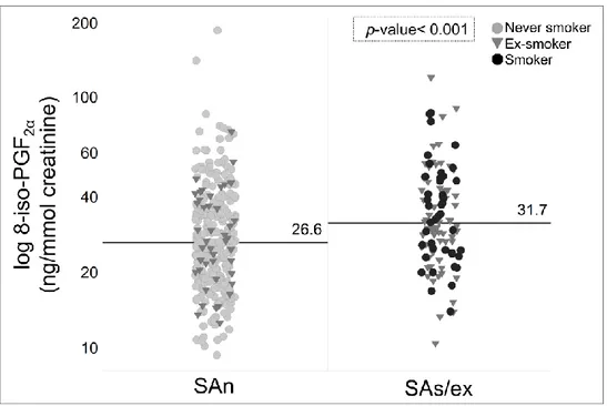
4.2 Lipid peroxidation in SAs/ex compared to SAn.
We analysed 8-iso-PGF2α, a specific biomarker of lipid
peroxidation, in spot urine samples. The median (IQR) concentration of 8-iso-PGF2α was significantly increased in the
urine of SAs/ex compared to SAn, 31.7 (24.5-44.7) vs. 26.6 (19.6-36.6) ng/mmol creatinine respectively (FC= 1.19; p< 0.001) (Fig. 4), indicating an increased level of oxidative stress in asthmatic patients who smoke.
Figure 4. Comparison of 8-iso-PGF2α in urine between SAn and SAs/ex. The marker of lipid
peroxidation is increased in SAs/ex, median (IQR)= 31.7 (24.5-44.7) compared to SAn, median (IQR)= 26.6 (19.6-36.6).
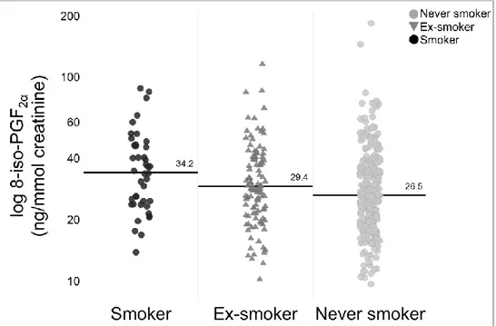
Furthermore, we assessed 8-iso-PGF2α taking into consideration
three smoking subgroups (current-, ex-, non-smokers) within the two SA cohorts (SAn+SAs/ex). A significant difference was observed between the smoking subgroups (p= 0.004) with increased median (IQR) concentration of urinary 8-iso-PGF2α in
current-smokers, 34.25 (24.4-47.7), vs ex-smokers, 29.4 (22.3-40.5), and non-smokers, 26.5 (19.6-16.6) (Fig. 5).
Figure 5.Comparison of urinary 8-iso-PGF2α between severe asthma (SA) smoking subgroups. The
median (IQR) concentration was increased in current-smokers, 34.25 (24.4-47.7), vs ex-smokers, 29.4 (22.3-40.5), and non-smokers, 26.5 (19.6-16.6). SAn: Severe Asthma non smokers; SAs/ex: Severe Asthma smokers/ex-smokers.
4.3 mRNA-expression of pro-/anti-oxidant enzymes in induced sputum
Sputum mRNA levels of the pro-oxidant enzymes NOX2 and NOS3 were examined. NOS1 and NOS2 probe sets were excluded because their mRNA expression levels were below the LOD. The mRNA levels of NOX2 were increased in SAs/ex compared to SAn. In particular the probe sets 203922_PM_s_at (FC= 1.05 p= 0.006), 203923_PM_s_at (FC =1.06; p= 0.003), 233538_PM_s_at (FC =1.06; p= 0.014) were over-expressed in SAs/ex. The NOX2 probe set 217431_PM_x_at showed log2 intensity lower than the other probe sets but was not significantly different between SAn and SAs/ex (FC= 1.00; p= 0.950). The expression of NOS3 mRNA in IS was similar between SAs/ex and SAn.
SOD3 was excluded in IS analyses because its mRNA levels were below the LOD. CAT mRNA levels were over-expressed in SAs/ex compared to SAn for the probe set 201432_PM_at (FC= 1.07; p= 0.028) but the probe set 211922_PM_s_at do not show a significant difference (p= 0.359). By contrast, mRNA expression
of SOD1, SOD2, SOD3 and GPX1 were not statistically different between SAn and SAs/ex (Table 8).
Table 8. Microarray data of pro-/anti-oxidant enzymes in induced sputum of severe asthma cohorts. Gene symbol Probe-set ID SAn (n=61)
Mean SAs/ex (n=23) Mean Change Fold p value
NOX2 203922_PM_s_at 9.89 10.35 1.05 0.006 203923_PM_s_at 9.03 9.59 1.06 0.003 217431_PM_x_at 5.96 5.97 1 0.950 233538_PM_s_at 6.57 6.96 1.06 0.014 NOS3 205581_PM_s_at 6.32 6.19 -1.02 0.115 SOD1 200642_PM_at 8.37 8.69 1.04 0.094 SOD2 215078_PM_at 9.93 9.62 -1.03 0.225 215223_PM_s_at 11.65 11.46 -1.02 0.303 216841_PM_s_at 11.40 11.20 -1.02 0.098 221477_PM_s_at 11.26 10.99 -1.02 0.121 CAT 201432_PM_at 8.41 9.01 1.07 0.028 211922_PM_s_at 7.81 7.97 1.02 0.359 GPX1 200736_PM_s_at 9.57 9.74 1.02 0.385
Probe-sets with log2 intensity >5.5 are reported in table. SAn: severe asthma non-smokers; SAs/ex: severe asthma smokers/ex-smokers; NOX2: NADPH oxidase 2; NOS3: nitric oxide synthase 3; SOD1/2: superoxide dismutase 1/2; CAT: catalase; GPX1: glutathione peroxidase 1.
4.4 mRNA-expression of pro-/anti-oxidant enzymes in Bronchial Biopsy.
All of the pro-oxidant enzymes were excluded in BB analyses because their expression levels were below the limit of detection (LOD threshold= 5). The expression of all antioxidant enzymes in BB was similar among SAn and SAs/ex (Table 9).
Table 9. Microarray data of pro-/anti-oxidant enzymes in bronchial biopsies of severe asthma
cohorts.
Gene symbol Probe-set ID SAn (n=40)
Mean SAs/ex (n=13) Mean Change Fold p value
SOD1 200642_PM_at 8.36 8.30 -1.01 0.253 SOD2 215223_PM_s_at 7.00 7.03 1.00 0.667 216841_PM_s_at 6.35 6.10 -1.04 0.075 221477_PM_s_at 6.28 6.11 -1.03 0.148 SOD3 205236_PM_x_at 6.77 7.07 1.04 0.161 CAT 201432_PM_at 6.84 6.89 1.01 0.941 211922_PM_s_at 5.73 5.68 -1.01 0.558 GPX1 200736_PM_s_at 7.04 7.19 1.02 0.437
Probe-sets with log2 intensity >5 are reported in table. SAn: severe asthma non-smokers; SAs/ex: severe asthma smokers/ex-smokers; SOD1/2/3: superoxide dismutase 1/2/3; CAT: catalase; GPX1: glutathione peroxidase 1.
4.5 mRNA-expression of pro-/anti-oxidant enzymes in Bronchial Brushing.
NOS2 mRNA levels were significantly decreased in SAs/ex compared to SAn (FC= -1.10; p= 0.029). NOS3 mRNA expression was similar between SAn and SAs/ex (FC= -1.01; p= 0.310). In addition, the mRNA expression levels of anti-oxidant enzymes were similar among the two study groups (Table 10). NOX2, NOS1 and SOD3 were excluded from the analysis because their mRNA expression levels were below the LOD.
Table 10. Microarray data of pro-/anti-oxidant enzymes in bronchial brushings of severe asthma
cohorts.
Gene symbol Probe set ID SAn (n=49)
Mean SAs/ex (n=18) Mean Change Fold p value
NOS2 210037_PM_s_at 5.36 4.87 -1.10 0.029 NOS3 205581_PM_s_at 4.97 4.88 -1.02 0.310 SOD1 200642_PM_at 9.03 8.99 -1.00 0.802 SOD2 215223_PM_s_at 7.58 7.25 -1.05 0.176 216841_PM_s_at 6.90 6.56 -1.05 0.165 221477_PM_s_at 6.84 6.60 -1.04 0.206 CAT 201432_PM_at 7.47 7.39 -1.01 0.620 211922_PM_s_at 6.20 6.24 1.01 0.681 GPX1 200736_PM_s_at 7.56 7.58 1.00 0.960
Probe-sets with log2 intensity >4.5 are reported in table. SAn: severe asthma non-smokers; SAs/ex: severe asthma smokers/ex-smokers; NOS2/3: nitric oxide synthase 2/3; SOD1/2: superoxide dismutase 1/2; CAT: catalase; GPX1: glutathione peroxidase 1.
4.6 Correlation analyses
There was no correlation between NOX2 mRNA in sputum and 8-iso-PGF2α in urine (p> 0.10). Instead, NOX2 in sputum
correlated significantly with macrophage numbers (p< 0.001) and percentages (p< 0.001) but inversely with eosinophil (p= 0.01, p= 0.008) and neutrophil (p= 0.014, p= 0.008) numbers and percentages respectively. Moreover, a strong correlation between NOS2 in BBr and FeNO was observed with a Kendall’s Tau= 0.535 (p< 0.001) (Fig. 6). The same correlation analysis was also performed by smoking status (non-smokers, ex-smokers, current smokers): we observed a significant correlation in 36 non-smokers (Kendall’s Tau= 0.551; p< 0.001); in 20 ex-non-smokers
(Kendall’s Tau= 0.394; p= 0.085); the number of current smokers was very low (only six) and no correlation analysis was possible.
Figure 6. Scatter plot of the relationship between NOS2 expression in bronchial brushing
and FeNO. There is a strong correlation between the two variables (62 non missing value; Kendall’s Tau= 0.535, p< 0.001). SAn: Severe Asthma non-smokers; SAs/ex: Severe Asthma smokers/ex-smokers.
4.7 Fractional exhaled NO (FeNO)
FeNO levels correlated with NOS2 mRNA expression in BBr. To determine whether the slightly lower FeNO in SAs/ex is an effect of current smoking, we assessed FeNO between the three separate
smoking phenotypes (current-smokers, ex-smokers and never-smokers). We observed a significant difference between the three subgroups (p= 0.012). In particular, FeNO levels were lower in SA current-smokers than SA ex-smokers (p= 0.007) and SA never-smokers (p= 0.004). In contrast, SA ex-smokers and SA never-smokers showed similar levels of FeNO (p= 0.975) (Fig. 7). Further stratification into subgroups based on oral steroid prescriptions did not alter our findings (data not shown).
Figure 7. FeNO analysis among the three Severe Asthma (SA) smoker subgroups. The
median (IQR) are: SA current smokers= 16 (10-31) ppb; SA ex-smokers= 25 (15-48) ppb; SA never-smokers= 27 (16-48) ppb.
5. Discussion
Little work has been performed to date to define the ability of tobacco smoke to modulate the oxidative stress balance in the airways of SA patients. The main findings of this paper can be summarised as follows: (i) SAs/ex showed an increased oxidative
stress compared to SAn evidenced by 8-iso-PGF2α levels in urine;
(ii) tobacco smoke was associated with increased NOX2 mRNA expression in induced sputum; (iii) the mRNA expression of inducible isoform of NOS (NOS2) in airways correlates with FeNO levels, and (iv) FeNO levels are inversely associated with active smoking.
5.1 Oxidative stress in severe asthmatic smokers/ex smokers vs. non-smokers
Oxidative stress is thought to play a critical role in the pathophysiology of SA (62). Tobacco smoke is one of the major environmental sources of oxidative stress, which can lead to greater lipid peroxidation as evidenced by the increased level of
urinary 8-iso-PGF2α in SAs/ex, particularly in current smokers.
This is in line with other studies that showed the ability of cigarette smoke to increase isoprostane levels (84,85). F2-isoprostanes have a potent smooth muscle and vascular constrictive action, which increases airway hyperresponsiveness and obstruction, and induces plasma exudation and inflammation (86). Indeed, 8-iso-PGF2α levels are elevated as asthma severity
increases (87) and further enhanced during acute exacerbations of asthma (88) and following allergen challenge in asthmatics (87).
5.2 Pro-oxidant enzymes
The NOX2 isoform (phagocyte NOX) is primarily present in macrophages, neutrophils and eosinophils and is most highly expressed in IS. NOX2 activity is normally required for phagocyte respiratory burst and regulation of cell signalling (66,67). Furthermore, the enzyme NOX2 is activated by cigarette smoke through the phosphorylation of c-Src, a tyrosine kinase protein (89) leading to a higher production of ROS (89,90). In our study, NOX2 mRNA is over-expressed in sputum of SAs/ex compared to SAn, supporting the hypothesis that exposure to
smoke in asthmatic subjects is able to amplify NOX2 mRNA
expression with a consequent elevation of O2•- production. There
was no correlation between the NOX2 over-expression in IS and 8-iso-PGF2α in urine. In all likelihood, NOX2 is just one of the
several factors contributing to the increase of oxidative stress. The positive correlation of NOX2 mRNA with macrophages and the inverse relationship with phagocytes may highlight the importance of distinct cellular phenotypes of these cells in regulating the inflammatory process in severe asthmatics who smoke (91,92). This area deserves further investigation.
NOS enzymes are important pro-oxidants that produce NO, an endogenous regulator involved in homeostatic and immunological functions with a role in asthma pathogenesis (73). NOS1 and NOS3 are the constitutive isoforms and are commonly expressed in non-immunological cells. But non-canonical expression patterns were found in some cases (75). We observed no differences in NOS3 mRNA expression between SAn and SAs/ex. This could indicate a lack of effect of cigarette smoke on NOS3. NOS2 is the inducible isoform (iNOS) that produces high levels of NO and its activity persists for many days after
induction (74,75) leading to cell death and tissue damage (74). This enzyme is mainly expressed in lung epithelium (75), so it is mainly detectable in BBr. NOS2 mRNA expression is induced by pro-inflammatory cytokines (73,93), and is increased in chronic asthma in proportion to the severity of the disease (94), and in particular it is increased by allergen provocation (95). In this study, NOS2 mRNA is under-expressed in BBr of SAs/ex. Some researchers have shown that NO generated by NOS2 is able to attenuate its own expression through the negative regulation of NF-B (96). Furthermore, we demonstrated a strong correlation between FeNO levels and BBr NOS2 mRNA expression in SA cohorts, as previously reported (97). Therefore, we hypothesise that active exposure to cigarette smoke can lead to inhibition of NOS2 mRNA expression in a negative feedback manner.
Evidence exists of lower FeNO levels in asthmatic smokers (98), particularly in asthmatic current smokers (25). Moreover, the analysis of clinical and inflammatory characteristics of the U-BIOPRED adult cohorts found lower FeNO levels in the SAs/ex cohort, indicating a possible effect of current smoking (48). Nevertheless, we did not observe a significant difference on
FeNO levels comparing only SAn vs SAs/ex (p= 0.89, data not shown). Therefore, we checked if there was a direct effect of active smoking on exhaled NO. The analysis of FeNO, in relation to the cigarette smoking status of patients (current-, ex- and never-smokers), showed decreased FeNO levels in current smokers confirming the effect of active smoking on exhaled NO. Cigarette smoke contains high levels of superoxide, which can rapidly react with NO, which may account for the reduced FeNO levels in the current smokers. Moreover the product of this reaction, peroxynitrite, is a very strong oxidant species (64,68,73). Thus, knowledge of current smoking status is important when using FeNO measurements in assessing asthma control and severity.
5.3 Anti-oxidant enzymes
SODs are the primary enzymes able to dismantle superoxide anion to form H2O2 (64). We observed no significant differences
in SOD1, SOD2 and SOD3 mRNA expression in airways of SAn and SAs/ex. In the literature, there is some contradictory evidence about the SODs enzymes in relation to asthma and tobacco
smoke. Some studies report that SOD1 has low activity and expression in asthmatic airways (64,70). SOD2 also was found to be inactivated and down regulated in asthmatic patients (70,99,100). Furthermore, the expression of SOD3 seems to be decreased in vitro by TNF-α, TGF-β and IL1-α, while it is enhanced by IFN-γ (64,101). Cigarette smoke enhanced the expression (102) and activity (103) of SODs in rat airways. Conversely, the levels of SOD were found increased in blood and saliva of subjects who smoke (104), and prolonged cigarette smoke exposure was found to increase the mRNA level of SOD2 in human bronchial epithelial cells (105). Therefore, it is conceivable that whilst SOD mRNA expression is decreased in SA, smoking enhances SOD mRNA expression, resulting in a balance between the two actions and overall no difference between SAn and SAs/ex.
CAT and GPX1 are key antioxidant enzymes for the degradation of reactive H2O2 to H2O and O2. Catalase (protein and mRNA
levels) were previously reported as decreased in the bronchiolar epithelium of smokers with COPD (106). Furthermore, the activity of CAT and GPX was previously found reduced in
asthmatic patients (64). In contrast, we observed no significant effects of smoking on CAT and GPX1 levels. Further studies are required to determine whether the decreased expression reported by Betsuyaku et al. (106) were a result of smoking or of COPD.
5.4 Limitations of the study
Due to the explorative nature of this study, there are several limitations. The major limitation is the absence of a healthy smoker and non-smokers control groups, which we cannot to establish for sure the influence of cigarette smoking and/or severe asthma. However, our purpose was to explore the CS effect in patients with the same severity degree of asthma, assuming that the observed changes are predominantly due to smoking. Another limitation is that the measurement of whole body excretion of 8-iso-PGF2α in urine cannot determine the source (airways or
systemic source). Given that the patients in this study suffer from asthma, and with the observed relations to smoking, it is however likely that airways inflammation contributes the most to increased urinary excretion of 8-iso-PGF2α; however, it would be of interest
to measure these effects in healthy smokers with normal lung function.
Few patients provided samples from each compartment within the U-BIOPRED study. Particularly, the number of SAs/ex samples is low for the sputum, BB and BBr transcriptomics set, thus we cannot divided SAs/ex group into current and ex-smokers for this subsets. Moreover, a negative result should be considered inconclusive. Due to the modest sample size of the current exploratory study, adjustments for multiple-testing were not applied. There is a good overlap, in term of patient coverage between BB, BBr and urine, and between sputum and urine. In contrast, there is little overlap between patients who had IS and BB/BBr.
We evaluated the mRNA expression of pro-/anti-oxidant enzymes, but the mRNA levels do not necessarily correlate with the production and activity of corresponding enzymes and proteins. Moreover, mRNA-expression of several enzymes could not be accurately assessed due to their low expression levels on the microarray. Although we report significant differences in oxidant gene expression of SAs/ex, the changes are small and
other mechanisms driving asthma severity in these patients may be present. Independent replications of these findings are warranted.