UNIVERSITY OF PISA
School of Graduate Studies
“Scienza del Farmaco e delle Sostanze
Bioattive
”
PhD THESIS
XXIII cycle (2008-2010)
“New agents against hypoxic tumours
counteracting invasiveness and metabolism”
CANDIDATE: Dott.ssa Carlotta Granchi
TUTOR: Prof. Filippo Minutolo
CHIM/08
DIRECTOR OF THE SCHOOL
Prof. Claudia Martini
3
AIMS OF THE THESIS 7
PREFACE 8
CHAPTER 1 Introduction 9
1.1 Tumour hypoxia 11
1.1.1 The main pathological aspects of hypoxic tumours 11 1.1.2 Hypoxia as an obstacle to medical treatment of cancer 18
1.2 LOX AND LDH-A: two key enzymes in hypoxic invasive tumours 19
1.2.1 Targeting LOX for the treatment of invasive hypoxic tumours 20 1.2.2 Exploiting tumour hypoxia: LDH-A inhibition 24
1.3 References 27
CHAPTER 2 Inhibitors of lysyl oxidase (LOX) 31
2.1 The enzyme lysyl oxidase (LOX) 33
2.1.1 Biological functions 33
2.1.2 LOX Inhibitors 37
2.2 Bioreductively activated LOX inhibitors 40
2.2.1 Hypoxia-activated cytotoxins: the nitroaromatic prodrugs 40 2.2.2 Pro-BAPNs as hypoxia-activated LOX inhibitors 43
2.3 Biological evaluation of LOX inhibitors 48
2.4 Conclusions 51
4
3.1 Glycolytic enzymes: potential targets against hypoxic tumours 59
3.2 The enzyme lactate dehydrogenase (LDH) 65
3.2.1 Introduction 65
3.2.2 hLDH isoforms 69
3.2.3 Structural and kinetic features of hLDHs 72
3.3 Plasmodium LDH (pfLDH) as an antimalarial target 73
3.3.1 pfLDH role in the fight against malaria 73
3.3.2 Unique features of pfLDH 74
3.4 LDH Inhibitors - Background 78
3.4.1 Perspective therapeutic applications 78
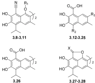
3.4.2 Gossypol and its derivatives 78
3.4.3 Naphthoic acids 87
3.4.4 N-Substituted oxamic acids 89
3.4.5 Azole-based compounds 96
3.4.6 Other LDH inhibitors 100
3.4.7 Features and perspectives of LDH inhibitors 103
3.5 N-Hydroxyindoles as LDH-A inhibitors 106
3.5.1 The N-hydroxyindole nucleus in Medicinal Chemistry 106
3.6 N-hydroxyindoles substituted with small groups 111
3.6.1 Synthesis 111
5
3.7.1 Synthesis 119 3.7.2 Enzymatic assays 123 3.8 Aryl-substituted N-hydroxyindoles 124 3.8.1 Synthesis 124 3.8.2 Enzymatic assays 1413.8.3 Molecular modeling studies 148
3.9 Triazole-based derivatives 156
3.9.1 Synthesis 156
3.9.2 Enzymatic assays 168
3.9.3 Molecular modeling studies 171
3.10 Amide and sulfonamide derivatives 172
3.10.1 Synthesis 172
3.10.2 Enzymatic assays 185
3.10.3 Molecular modeling studies 188
3.11 Cellular assays 190
3.11.1 Cytotoxicity assays 190
3.11.2 Cell-based NMR studies 193
3.12 Conclusions 198
3.13 References 199
CHAPTER 4 Experimental Section 215
6
4.1.2 Synthetic procedures and characterization data 218
7
AIMS OF THE THESIS
Tumour cells have abnormal growth rates, which arise from malfunctions in the regulatory mechanisms that oversee the cells’ growth and development. This uncontrolled growth leads to a dramatic change in the physiology of the tumour itself. In particular, the development of hypoxic zones occurs as a result of a discrepancy between oxygen consumption and oxygen supply of the rapidly growing tumour. Hypoxia, in its turn, deeply modifies the metabolism and the behaviour of the tumour cells. Warburg effect, which is a shift from ATP generation through oxidative phosphorylation to ATP generation through glycolysis, even under normal oxygen concentrations, is the best characterized metabolic switch observed in hypoxic tumours. Hypoxia is also strongly associated with tumor progression and metastatic disease and this is likely because low oxygen tension is able to increase cell invasiveness. These pathological features make tumour hypoxia a poor prognostic factor in several kinds of malignancies.
The goal of this PhD Thesis was to interfere with these two main peculiar features of hypoxic tumours, glycolytic metabolism and invasiveness, by focusing our attention on two key enzyme: lactate dehydrogenase A (LDH-A), which is essential for energy production in hypoxic conditions and lysyl oxidase (LOX), which is strictly involved in the metastasis formation deriving from hypoxic tumours. Specifically, this Thesis aimed at designing and synthesizing small organic molecules able to inhibit LDH-A, so to be developed as starvation agents against hypoxic tumours. The other aim of this Thesis was to develop hypoxia-activated prodrugs as LOX inhibitors, in order to selectively block this enzyme in hypoxic cancer cells and, consequently, reduce or even completely stop the invasiveness of hypoxic tumours.
8
PREFACE
The first Chapter gives a broad overview of tumour hypoxia and the main pathological aspects of this condition, explaining why hypoxia is considered a poor prognostic factor in many cancers and introducing LDH-A and LOX in this context. Furthermore, the general strategies for attacking hypoxic tumours by inhibition of LDH-A and LOX are presented in Chapter 1.
Chapter 2 is focused on LOX, starting with the detailed description of this enzyme and previous research in this area, which support our choice to develop hypoxia-activated prodrugs as LOX inhibitors. Synthesis and biological results for the synthesized molecules are reported.
Chapter 3 deals with the main subject of this PhD Thesis, that is, LDH-A inhibition. Background of enzyme LDH-A is reported, including kinetic and structural features of human and Plasmodium falciparum enzyme isoforms, as well as inhibitors reported in the literature. A small overview of the most representative glycolytic inhibitors is also presented. The central part of this Chapter comprises the design and the synthesis of LDH-A inhibitors developed in my PhD project, which have been subdivided into structural classes and, for each group, enzymatic assays and molecular modeling studies are discussed. The last part of this Chapter is about cellular assays performed during the period spent at the University of Illinois at Urbana-Champaign (USA).
Detailed experimental procedures for the synthesis and characterization of LOX and LDH-A inhibitors are described in Chapter 4.
CHAPTER 1
11
1.1. TUMOUR HYPOXIA
1.1.1. The main pathological aspects of hypoxic tumours
A distinctive feature of many solid tumours is the presence of hypoxic regions, which originate from an imbalance between blood supply and consumption of oxygen. This clinical consideration was made for the first time by Thomlinson and Gray about 50 years ago and was then confirmed thanks to the subsequent introduction in the 1990s of an oxygen electrode, called the “Eppendorf” electrode, that made it possible to accurately measure the oxygen pressure in human tumours [1, 2]. In order to explain this pathological aspect, we have to take into account the fact that cancer cells possess an elevated rate of growth, so their metabolic requirements surpass the vasculature ability to provide oxygen and nutrients. This pathological condition results in areas characterized by low levels of oxygen pressure (pO2 < 5 mmHg corresponding to < 1%
in the gas phase), and sometimes in necrotic regions, which have been detected in a wide range of cancers, such as brain, head and neck, cervix, breast tumours and soft tissues sarcomas. The development of this condition may be due to: a) temporary obstruction of microvessels or variable blood flow caused by structural and functional abnormalities of intratumoural vasculature (perfusion-limited or acute hypoxia); b) increased mean diffusion distances, about 100 μm or more, or adverse diffusion geometry caused by the irregular and fast tumour growth (diffusion-limited or chronic
12
Figure 1.1. The vascular network of normal tissues (on the left) versus tumour tissues (on the right) [4].
Copyright©2004 Nature Publishing Group
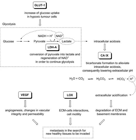
Hypoxia, in turn, acts as a promoter of tumour progression, because it provokes an overexpression of a great number of genes implicated in the cell survival, leading to many changes in tumour cellular behaviour and metabolism. In particular, the transcription factor HIF-1α (Hypoxia-Inducible Factor-1α), that is overexpressed in hypoxic tumours, starts a transcriptional program that provides many solutions to hypoxia by up-regulating a series of downstream target genes, such as Lactate Dehydrogenase A (LDH-A), Lysyl Oxidase (LOX), together with Glucose Transporter-1 (GLUT-Transporter-1), Carbonic Anhydrase IX (CA IX) and Vascular Endothelial Growth Factor (VEGF). In particular, the overexpression of two selected key enzymes, LDH-A and LOX at different oxygen concentrations and pH values in two tumour cells lines is shown in the graphs below (Fig. 1.2) [5, 6]. The x axis reports the oxygen concentration and the relative expression of these enzymes is displayed on the y axis: it is evident that, depending on the pH, the relative expression of the two enzymes is increased in both cell lines at 1% of oxygen compared with normoxia condition that is 21% of oxygen.
13
Figure 1.2. mRNA expression of LDH-A (top) and LOX (bottom) in SiHa (uterine cervix squamous cell
carcinoma) and FaDuDD cells (pharyngeal squamous cell carcinoma) under different oxygen concentrations at
various grades of acidosis. Cells were exposed to various oxygen levels at the different pH values for 24 h, then mRNA levels were determined, compared to untreated cells and plotted [5]. Copyright©2007 Elsevier B. V.
The main function of these proteins, together with other factors, is to protect cells from the hostile hypoxic environment and to help them to proliferate and to survive in adverse conditions, constituting adaptive responses to hypoxia. Consequently, tumour cells become able to overcome the nutritive deprivation by increasing their vascularization through angiogenesis, enhancing the glucose uptake, and optimizing its sugar metabolism. Moreover, tumour cells acquire the ability of invading surrounding healthy tissues in order to escape from the adverse environment (Fig. 1.3). The overexpressed proteins mentioned above are considered among the main responsible factors contributing to the typical pathological alterations associated to tumours, although other molecular pathways that may be implied, such as oncogene activation and tumour suppressor loss, actively contribute to complete this process. Indeed, it is
14
recognized that the tumour microenvironment, involving several factors having both anti- and pro-tumour effects, makes the understanding of the tumour development and progression a very complex picture [7]. All these hypoxic tumour hallmarks result in the development of a more malignant and aggressive phenotype and provide a selective growth advantage over other competing cell populations. They also confer a higher survival ability to hypoxic tumour cells. As a consequence, a direct relationship between intratumoural hypoxia and poor patient prognosis has been observed [3, 8].
Figure 1.3. The biological relationships between the proteins up-regulated by HIF-1α in hypoxic conditions.
15
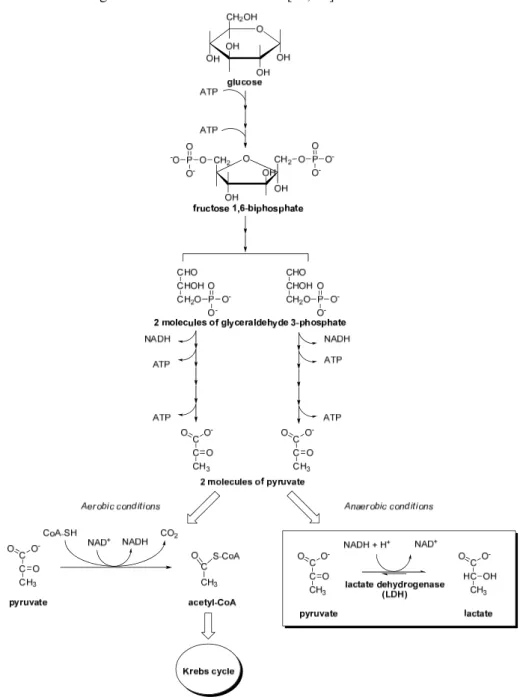
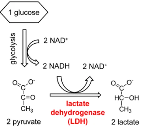
One of the most important adaptive responses to hypoxia is the metabolic switch from oxidative to glycolytic pathway. Normally, cells produce most of the ATP from glucose through the oxidative phosphorylation (OXPHOS), but many cancer cells in hypoxic conditions obtain ATP by glycolysis and the final conversion of glucose to lactate, showing low levels of OXPHOS activity. This metabolic strategy is mainly permitted by the overexpression of LDH-A, a glycolytic enzyme which is a direct target of HIF-1α [10], although other enzymes belonging to the glycolytic pathway are also up-regulated. In fact, HIF-1α was proven to induce overexpression of glucose transporters GLUT-1 and GLUT-3, as well as of glycolytic enzymes hexokinase I and II (HK-I, HK-II), the liver isoform phosphofructokinase (PFK-L), aldolase A and C (ALD-A, ALD-C), phosphoglycerate kinase 1 (PGK-1), enolase α (ENO-α), pyruvate kinase M2 (PYK-M2), 6-phosphofructo-2-kinase/fructose-2,6-biphosphatase 3 (PFKFB-3), and, of course, LDH-A [11]. As mentioned before, LDH-A converts pyruvate, the final product of glycolysis, to lactate, instead of undergoing the Krebs cycle in the mitochondria, due to the lack of oxygen that is the final electron acceptor in the electron transport chain. This reaction regenerates the cofactor NAD+ and, therefore,
permits the continuation of glycolysis (Fig. 1.4). This kind of metabolism is maintained even when cells derived from hypoxic tumours are placed under normoxic conditions, confirming that the conversion of pyruvate into lactate, in order to obtain energy in conditions of low levels of oxygen, represents not only a simple adaptive mechanism to hypoxia, but above all a hallmark of hypoxic cancers involving epigenetic transformations that deeply affect some cellular pathways. A high rate of conversion of glucose into lactate with an elevated glucose uptake are the typical traits of the glycolytic phenotype and ensure adequate ATP levels and constant supply of intermediates essential for cell growth and division [12]. ATP generation via glycolysis is a less efficient energy production process (2 ATP molecules per glucose) than oxidative phosphorylation (36 ATP molecules per glucose). Hence, cancer cells consume more glucose than normal cells for maintaining an adequate ATP supply to proliferate. The fundamental role of this glycolytic phenotype is emphasized by studies demonstrating that increased glucose uptake coincides with the transition from
pre-16
malignant lesions to invasive cancer, since the cancer cells’ dependence on glycolysis increases as malignant transformation increases [13, 14].
Figure 1.4. The glycolytic process and the two main metabolic pathways in which pyruvate is involved,
depending on the presence of oxygen. The reaction catalyzed by LDH is highlighted (bottom-right) [9]. Copyright©2010 Bentham Science Publishers
17
The increased production of lactate by malignant cells even under aerobic conditions is called aerobic glycolysis or “Warburg effect” and it has been observed only in tumour tissues. This phenomenon was first reported by Otto Warburg in the 1920s, hypothesizing that this altered glucose metabolism derives from impaired mitochondrial activity [15]. Nowadays, the “Warburg hypothesis” has proven incorrect, but an increased dependence on glycolytic activity for ATP generation in tumours even in the presence of oxygen has often been confirmed. The metabolic switch from the energetically efficient Krebs cycle to the tumour glucose utilization in glycolysis is regulated by a very wide network of pathways involving both oncogenes and tumour suppressor genes, thus playing a central role in the altered catabolic and anabolic processes of several tumours [16].
The invasive and aggressive behaviour is another typical pathological feature found in hypoxic tumours. Since metastasis is considered the main cause of cancer death, the understanding of this very complex process is an extremely challenging goal for the treatment of cancer. Metastasis is a multistep process starting with the detachment of cells form the original tumour. In order to metastasize, a malignant cell must dissociate from the primary tumour and intravasate into the vascular or lymphatic systems by degrading and migrating through the basement membrane and extracellular matrix. Once in the lymph or in the blood streams, this cell must evade the host immune system, migrate to its target site, and finally extravasate out of the circulation to implant in a healthy tissue where proliferation gives rise to cancer metastases [7, 17, 18]. A complex network of proteins of the extracellular matrix were found to be involved in metastasis formation, such as Matrix Metalloproteinases (MMPs), integrins, Connective Tissue Growth Factor (CTGF), which favour dissemination of malignant cells from a primary tumour site to a secondary site by different mechanisms. Moreover, the tumour itself becomes able to secrete a large number of growth factors and proteases, which permit to modify the local microenvironment making it more permissive for cell motility and adhesion. Even the extracellular acidosis, caused by the increased lactate production operated by LDH-A, is a factor promoting the entry of tumour cells into the blood vessels, so leading to metastasis formation. In this picture, a key role is played by another direct target of HIF-1, that is LOX, an amine oxidase of the extracellular matrix,
18
whose catalytic function contributes to the formation and the reinforcement of the ECM. This enzyme was found to be directly involved in the metastatic process. Numerous studies demonstrate that its expression is directly correlated to the invasiveness of a large number of tumours, causing disease progression and poor overall survival [19].
1.1.2. Hypoxia as an obstacle to medical treatment of cancer
Tissues possessing low levels of oxygen show resistance to radiotherapy and this is one of the principal clinical problems that compromises hypoxic cancer treatment documented as early as since 1953 [20]. The mechanism linking hypoxia to tumour radioresistance is generally known as the “oxygen enhancement effect” [21]. This phenomenoncan be explained considering that DNA damage is usually provoked by direct ionization from ionizing radiations or is induced by oxygen radicals, such as hydroxyl radical, superoxide anion etc. In the presence of oxygen, DNA double-strand breaks cannot be repaired because O2 reacts with the broken DNA forming stable
organic peroxides which are hardly repaired by the cell, leading to fatal chromosome aberrations. Basically, oxygen fixes the DNA damage, making it permanent. On the other hand, in the absence of oxygen, the damage is more easily repaired: the broken DNA can be restored to its original form, thanks to reparative processes based upon reductions by –SH containing intracellular components. This mechanism explains why tumours with low levels of oxygen show a reduced effect on response to radiotherapy, compared with normally oxygenated tissues [4, 21, 22].
Hypoxic cells are also resistant to conventional anticancer therapies and this is mainly due to their distance from blood vessels, so they are not sufficiently reached by most anticancer drugs. We can also mention other reasons that are behind chemotherapy resistance. For example, anticancer chemotherapy is usually more efficient against cells in rapid proliferation due to the mechanisms of actions commonly associated to most of the currently available treatment regimens, so it causes lower effects to hypoxic cells, which are characterized by slower rates of proliferation compared to normally oxygenated cancer cells. Furthermore, hypoxia selects cells that up-regulate genes encoding for proteins involved in drug resistance. It follows that, at present, there are not adequate anticancer drugs able to efficiently kill hypoxic cells [4, 23].
19
In addition to radiotherapy and chemotherapy resistance, the higher malignant phenotype, typical of hypoxic cancer cells, is associated with a more metastatic and aggressive behaviour, consequently predisposing to the formation of metastases, that compromise curability of tumours by surgery [24].
Since hypoxic cells are resistant to standard therapies, it is clear that hypoxia has a negative impact on cancer treatment and prognosis. Nevertheless, the unique features of hypoxic tumours that do not occur in healthy tissues, such as the metabolic switch associated to the “Warburg effect”, could be exploited in cancer-selective therapy.
1.2. LOX AND LDH-A: TWO KEY ENZYMES IN HYPOXIC
INVASIVE TUMOURS
Tumour cells become able to survive in conditions of low oxygen concentration, that is restrictive to normal cells, thanks to many pathological alterations in their metabolism and behaviour. In particular, the glucose uptake is increased and then the internalized sugar is processed through aerobic glycolysis in order to generate energy for continuously growing cells, even in the absence of oxygen. Moreover, hypoxia represents a stimulus for invasion and metastasis, because tumour cells, living in areas of nutrient and oxygen deprivation, need to invade new healthy tissues more permissive for their growth (Fig. 1.5). As a consequence, aerobic glycolysis and metastasis represents essential hallmarks of hypoxic cancer. In particular, in this PhD Thesis we focused our attention on two key enzymes, LOX and LDH-A, which play important roles in the metabolism and behaviour of tumour cells. LOX is deeply implicated in the progression of the tumour and in its spread in the surrounding tissues by metastasis formation; on the other hand, LDH-A supports the growth and the survival of hypoxic tumour cells, permitting the obtainment of ATP by glycolysis. Our interest is also justified by the almost total absence of suitable and selective inhibitors of these enzymes, so LOX and LDH-A can be considered innovative targets in the fight against invasive hypoxic tumours.
20
Figure 1.5. Tumour hypoxia-induced changes inside and outside the cancer cell. Increased glycolysis (in the
cytoplasm) and increased invasive ability of tumour cells (in the extracellular environment) are schematically represented, together with the two key enzymes responsible for this altered behaviour, LDH-A and LOX.
1.2.1. Targeting LOX for the treatment of invasive hypoxic tumours
Metastasis constitutes an intricate process involving many tumours and stromal proteins necessary for the remodelling of the extracellular matrix and, of course, LOX represents one of the key ECM components involved in metastatic disease. The LOX involvement in metastatic disease is proved by numerous studies linking LOX to tumour development and metastasis formation [17]. LOX was found to be implicated in the promotion of the metastatic process in vivo and in vitro, being up-regulated by hypoxia-inducible factor-1 (HIF-1) in hypoxic tumour tissues [5, 6, 19]. An overexpression of LOX mRNA was identified principally in MDA-MB-231 cancer cell line, which is a highly invasive and metastatic breast cancer cell line, SiHa cervical cancer cells, and in head and neck cancers, but the same association was not found in the poorly invasive and non-metastatic human breast cancer phenotype MCF-7, demonstrating that this enzyme is required for the increased invasion in tumours and its expression seems to occur mainly in distant metastatic cancer tissues compared with primary tumours. Recently, LOX expression has been correlated to disease progression,
21
metastasis and poor overall survival also in oral cancers [25, 26]. LOX stimulates cell invasion through a mechanism mediated by hydrogen peroxide, a well-established LOX by-product [27-29].
In vitro and in vivo invasion assays demonstrated that only catalytically active LOX,
secreted in the extracellular matrix, is responsible for the increased invasion ability in hypoxic tumours. In fact, since LOX is a copper-dependent enzyme, the use of a copper chelator that cannot enter the cell, such as bathocuprione disulphonate (BCS), results in a reduction of invasiveness, thus confirming that extracellular LOX involvement is the responsible for such invasion ability. Furthermore, when LOX catalytic activity was compromised by using LOX short hairpin RNA cells (shRNA), treatment with LOX antibody or administration of a LOX irreversible inhibitor such as β-aminoproprionitrile (BAPN), the invasion ability of tumour cells was significantly decreased and the formation of metastases was considerably reduced or completely blocked. This confirms that LOX is actively involved in the invasion ability of hypoxic tumour cells. This behaviour is also confirmed by the cellular shape: in air, wild-type tumour cells show a branching morphogenesis typical of invasive human cancer cells grown on collagen, which was further enhanced by hypoxia. On the other hand, the LOX shRNA cells grew in a markedly different manner, remaining more rounded in spheroid-like cell clusters, showing little branching in air or hypoxia. These results definitively indicate a role for LOX in the development of an invasive phenotype of hypoxic human tumours.
A recent study performed on a mouse model of mammary cancer proved the correlation among LOX activity, increased ECM stiffness due to collagen cross-linking, and progression to invasive disease. Pre-conditioning of mouse mammary fat pads with LOX-expressing fibroblasts promoted the growth and invasion of mammary cells and LOX inhibition with BAPN or LOX-specific antibody tempered tissues fibrosis, increased tumour latency, and decreased tumour incidence, suggesting that LOX-mediated cross-linkages likely regulate tumour progression by modifying the tumour microenvironment [7, 30].
Furthermore, LOX seems to play a critical role in premetastatic niche formation. Upon modification of the extracellular matrix and recruitment of CD11b+ myeloid cells to premetastatic sites, LOX prepares the target organs for the subsequent arrival of
22
metastatic tumour cells and enhances the establishment and growth of metastases. It appears that LOX alters tumour microenvironment and is involved in tumour invasion at different stages of this process. These findings together support the high potentiality of this perspective therapeutic target [31].
From a mechanistic point of view, the hypoxia-induced metastatic process starts with a series of adhesion and detachment interactions with the extracellular matrix by means of a cell pseudopode protrusion driven by actin polymerization. In particular, remarkable amounts of LOX are localized in proximity of the protrusion leading edge and on the many hair-like fibres sticking out from it. These events lead to invasion into the bloodstream and migration of the primary tumour cells, leaving behind a route of remodelled extracellular matrix that constitutes a sort of “highway” for the other cells, that can quite easily follow the first ones (Fig. 1.6 and 1.7). As a consequence, it is confirmed to LOX the role of an ECM remodelling enzyme during invasion that allows cell motility, but also during extravasation and colonization processes. According to these findings, LOX could be considered not only a potential marker for identifying highly invasive/metastatic and hypoxic tumours, but its inhibition could be exploited for the prevention and also the treatment of aggressive and metastatic tumours, so revealing a new good therapeutic target for several kinds of metastatic cancers [19].
23
primary tumour
hypo xia
ECM
cell protrusion & adhesion
migration & intravasation invas ion blood vessel mass ive invas ion blood vessel h igh w ay remodeled ECM LOX-enriched leading edge & hair-like fibers
LOX
involvement
“highway” formation
Figure 1.6. Metastasis early stage: ECM-remodeling and migration.
extravasation & colonization metastasis ECM LOX involvement blood vessel blood vessel remodeled ECM
Figure 1.7. Metastasis later stage: extravasation and colonization of new tissues.
The lack of suitable LOX inhibitors to be used in clinic makes lysyl oxidase inhibition a very challenging goal in the field of anti-tumour research and there are wide margins of research work to do because LOX inhibitors have been poorly studied so far. LOX involvement in metastasis opens a new research way that is potentially selective, LOX being a hypoxia-induced and extracellular target, and also innovative, due to very few treatment options for patients with metastases. For these reasons, we have developed molecules able to selectively inhibit LOX in hypoxic environments, and the results are reported in Chapter 2 of this Thesis.
24
1.2.2. Exploiting tumour hypoxia: LDH-A inhibition
The much lower levels of oxygen in tumour cells compared to those in normal tissues constitute an useful opportunity to develop a selective anticancer therapy based on this peculiarity. At present, the most commonly and used strategies that have been attempted in this field are represented by hypoxia-activated pro-drugs [32] and by gene therapies based on the inhibition of HIF-1 in hypoxic conditions [33, 34].
Another way of taking advantage from the distinctive features of tumour cells is to consider their altered carbohydrate metabolism, that is the conversion of glucose to lactate even in the presence of normal oxygen pressure. The dependency of tumour cells on glucose metabolism constitutes the weak point of hypoxic cancers and it could be therapeutically exploited by interfering with LDH-A activity. Several attempts to inhibit the glycolytic enzymes, such as hexokinase (HK), up-regulated in hypoxic tumours and specially involved in malignancy progression have been realized. However, among the enzymatic checkpoints of the glycolytic pathway, LDH-A still remains one of the most attractive targets in this context [22, 35]. LDH-A is overexpressed in hypoxic tumour environment, whereas the other main isoform LDH-B is predominant in all living cells, both normal and malignant. The data reported in literature confirm that up-regulation of LDH-A ensures an efficient aerobic glycolysis in tumour cells, whereas this enzyme is not so necessary to healthy cells in normal conditions that generally use the aerobic oxidation pathway [5, 36-38].
It was discovered that when LDH-A levels are knocked down by means of shRNAs (short hairpin RNA) in tumour cell lines that show high glucose dependency and high LDH-A expression under both normal and hypoxic conditions, LDH-A activity is severely compromised and the ability of these cell populations to proliferate shows a remarkable reduction [39]. In particular, when the oxygen pressure is limited the effect is even more pronounced, because the proliferation was completely blocked and ATP levels were much lower. This suppression of aerobic production of lactate had also a great impact on OXPHOS activity and oxygen consumption, which were both significantly increased. Consistent with these observations, cancer cells with low levels of LDH-A possess a reduced ability to grow under hypoxic conditions and their tumorigenic potential is severely compromised. In order to determine if the growth
25
decrease is due to LDH-A knockdown and, as a consequence, to the inability of tumour cells to metabolize glucose to lactate, cDNA corresponding to human LDH-A was introduced into LDH-A-silenced cells. Ectopic expression of this enzyme restores the proliferation and reestablishes the glycolytic phenotype, thus confirming the specificity of LDH-A knockdown approach. Altogether, these data suggest that the A isoform of lactate dehydrogenase plays a fundamental role in tumour maintenance, because cancer cells rely on LDH-A activity for energy obtainment even when oxygen is not a limiting factor [39].
As regards possible side effects caused by this approach, humans suffering from a complete hereditary deficiency of LDH-A due to the lack of A subunit production have been studied. These individuals show myoglobinuria, that is, the presence of myoglobin in the urine usually associated with muscle damage, only after intense anaerobic exercise (exertional myoglobinuria), without displaying any symptoms under ordinary circumstances [40, 41].
All this evidence constitutes a good target validation for LDH-A. In fact, not only does the inhibition of LDH-A prevent the Warburg effect, by forcing hypoxic tumour cells to revert to OXPHOS metabolism from the glycolytic pathway, but it also attenuates cell proliferation, supporting the growing evidence that the glycolytic phenotype is responsible of tumorigenicity of hypoxic cancer cells. Up to now, unlike other glycolytic targets for which some suitable molecules as drug candidates have been found, only shRNAs have been used to selectively inhibit LDH-A in tumours, and there are not many small molecules able to selectively block LDH-A activity (Fig. 1.8). As a consequence, antagonizing LDH-A could be considered an interesting, innovative, selective and also potentially non-toxic strategy of interfering with hypoxic cancer growth. Therefore, we focused our efforts in an attempt to develop small molecules able to selectively inhibit LDH-A, and our results are reported in Chapter 3 of this Thesis.
26
Figure 1.8. Potential metabolic targets for the treatment of cancer with relative desired effects and
compounds. For LDH-A inhibition (entry 4), only genetic techniques have been used [12]. Copyright©2008 Elsevier Inc.
27
1.3. REFERENCES
[1] Thomlinson, R.H.; Gray, L.H. The histological structure of some human lung cancers and the possible implications for radiotherapy. Br. J. Cancer, 1955, 9, 539-549.
[2] Vaupel, P.; Schlenger, K.; Knoop, C.; Höckel, M. Oxygenation of human tumors: evaluation of tissue oxygen distribution in breast cancers by computerized O2 tension measurements. Cancer Res., 1991, 51, 3316-3322.
[3] Vaupel, P.; Mayer, A. Hypoxia in cancer: significance and impact on clinical outcome. Cancer Metastasis Rev., 2007, 26, 225-239.
[4] Brown, J.M.; Wilson, W.R. Exploiting tumour hypoxia in cancer treatment.
Nat. Rev. Cancer, 2004, 4, 437-447.
[5] Sørensen, B.S.; Alsner, J.; Overgaard, J.; Horsman, M.R. Hypoxia induced expression of endogenous markers in vitro is highly influenced by pH.
Radiother. Oncol., 2007, 83, 362-366.
[6] Sørensen, B.S.; Hao, J.; Overgaard, J.; Vorum, H.; Honore, B.; Alsner, J.; Horsman, M.R. Influence of oxygen concentration and pH on expression of hypoxia induced genes. Radiother. Oncol., 2005, 76, 187-193.
[7] Allen, M.; Jones, J.L. Jekyll and Hyde: the role of the microenvironment on the progression of cancer. J. Pathol., 2011, 223, 163-177.
[8] Hsu, P.P.; Sabatini, D.M. Cancer cell metabolism: Warburg and beyond. Cell,
2008, 134, 703-707.
[9] Granchi, C.; Bertini, S.; Macchia, M.; Minutolo, F. Inhibitors of lactate dehydrogenase isoforms and their therapeutic potentials. Curr. Med. Chem.,
2010, 17, 672-697.
[10] Xie, H.; Valera, V.A.; Merino, M.J.; Amato, A.M.; Signoretti, S.; Linehan, W.M.; Sukhatme, V.P.; Seth, P. LDH-A inhibition, a therapeutic strategy for treatment of hereditary leiomyomatosis and renal cell cancer. Mol. Cancer
Ther., 2009, 8, 626-635.
[11] Marin-Hernandez, A.; Gallardo-Perez, J.C.; Ralph, S.J.; Rodriguez-Enriquez, S.; Moreno-Sanchez, R. HIF-1α modulates energy metabolism in cancer cells
28
by inducing over-expression of specific glycolytic isoforms. Mini-Rev. Med.
Chem., 2009, 9, 1084-1101.
[12] Kroemer, G.; Pouyssegur, J. Tumor cell metabolism: cancer's Achilles' heel.
Cancer Cell, 2008, 13, 472-482.
[13] Younes, M.; Lechago, L.V.; Lechago, J. Overexpression of the human erythrocyte glucose transporter occurs as a late event in human colorectal carcinogenesis and is associated with an increased incidence of lymph node metastases. Clin. Cancer Res., 1996, 2, 1151-1154.
[14] Yasuda, S.; Fujii, H.; Nakahara, T.; Nishiumi, N.; Takahashi, W.; Ide, M.; Shohtsu, A. 18F-FDG PET detection of colonic adenomas. J. Nucl. Med., 2001,
42, 989-992.
[15] Warburg, O. On the origin of cancer cells. Science, 1956, 123, 309-314. [16] a) Gatenby, R.A.; Gillies, R.J. Why do cancers have high aerobic glycolysis?
Nat. Rev. Cancer, 2004, 4, 891-899; b) Levine, A.J.; Puzio-Kuter, A.M. The
control of the metabolic switch in cancers by oncogenes and tumour suppressor genes. Science, 2010, 330, 1340-1344.
[17] Finger, E.C.; Giaccia, A.J. Hypoxia, inflammation, and the tumour microenvironement in metastatic disease. Canc. Met. Rev., 2010, 29, 285-293. [18] Chan, D.A.; Giaccia, A.J. Hypoxia, gene expression, and metastasis. Cancer
Metastasis Rev., 2007, 26, 333-339.
[19] Erler, J.T.; Bennewith, K.L.; Nicolau, M.; Dornhöfer, N.; Kong, C.; Le, Q.-T.; Chi, J.-T.A.; Jeffrey, S.S.; Giaccia, A.J. Lysyl oxidase is essential for hypoxia-induced metastasis. Nature, 2006, 440, 1222-1226.
[20] Gray, L.H.; Conger, A.D.; Ebert, M.; Hornsey, S.; Scott, O.C. The concentration of oxygen dissolved in tissues at the time of irradiation as a factor in radiotherapy. Br. J. Radiol., 1953, 26, 638-648.
[21] Moeller, B.J.; Richardson, R.A.; Dewhirst, M.W. Hypoxia and radiotherapy: opportunities for improved outcomes in cancer treatment. Cancer Metastasis
Rev., 2007, 26, 241-248.
[22] López-Lázaro, M. Role of oxygen in cancer: looking beyond hypoxia.
29
[23] Iessi, E.; Marino, M.L.; Lozupone, F.; Fais, S.; De Milito, A. Tumor acidity and malignancy: novel aspects in the design of anti-tumor therapy. Cancer
Ther., 2008, 6, 55-66.
[24] Sullivan, R.; Graham, C.H. Hypoxia-driven selection of the metastatic phenotype. Cancer Metastasis Rev., 2007, 26, 319-331.
[25] Sakai, M.; Kato, H.; Sano, A.; Tanaka, N.; Inose, T.; Kimura, H.; Sohda, M.; Nakajiama, M.; Kuwano, H. Expression of lysyl oxidase is correlated with lymph node metastasis and poor prognosis in esophageal squamous cell carcinoma. Ann. Surg. Oncol., 2009, 16, 2494-2501.
[26] Albinger-Hegyi, A.; Stoeckli, S.J.; Schmid, S.; Storz, M.; Iotzova, G.; Probst-Hensch, N.M.; Rehrauer, H.; Tinguely, M.; Moch, H.; Hegyi, I. Lysyl oxidase expression is an independent marker of prognosis and a predictor of lymph node metastasis in oral and oropharyngeal squamous cell carcinoma (OSCC).
Int. J. Cancer, 2010, 126, 2653-2662.
[27] Erler, J.T.; Giaccia, A.J. Lysyl oxidase mediates hypoxic control of metastasis.
Cancer Res., 2006, 66, 10238-10241.
[28] Kirschmann, D.A.; Seftor, E.A.; Fhong, S.F.T.; Nieva, D.R.C.; Sullivan, C.M.; Edwards, E.M.; Sommer, P.; Csiszar, K.; Hendrix, M.J.C. A molecular role for lysyl oxidase in breast cancer invasion. Cancer Res., 2002, 62, 4478-4483. [29] Payne, S.L.; Fogelgren, B.; Hess, A.R.; Seftor, E.A.; Wiley, E.L.; Fong, S.F.T.;
Csiszar, K.; Hendrix, M.J.C.; Kirschmann, D.A. Lysyl oxidase regulates breast cancer cell migration and adhesion through a hydrogen peroxide-mediated mechanism. Cancer Res., 2005, 65, 11429-11436.
[30] Levental, K.R.; Yu, H.; Kass, L.; Lakins, J.N.; Egeblad, M.; Erler, J.T.; Fong, S.F.T.; Csiszar, K.; Giaccia, A.;.Weninger, W.; Yamauchi, M:; Gasser, D.L.; Weaver, V.M. Matrix crosslinking forces tumor progression by enhancing integrin signaling. Cell, 2009, 139, 891-906.
[31] Erler, J.T.; Bennewith, K.L.; Cox, T.R.; Lang, G.; Bird, D.; Koong, A.; Le, Q.– T.; Giaccia, A.J. Hypoxia-induced lysyl oxidase is a critical mediator of bone marrow cell recruitment to form the premetastatic niche. Cancer Cell, 2009,
30
[32] Chen, Y.; Hu, L. Design of anticancer prodrugs for reductive activation. Med.
Res. Rev., 2009, 29, 29-64.
[33] Brown, J.M. Exploiting the hypoxic cancer cell: mechanisms and therapeutic strategies. Mol. Med. Today, 2000, 6, 157-162.
[34] Giaccia, A.J.; Siim, B.G.; Johnson, R.S. HIF-1 as a target for drug development. Nat. Rev. Drug Discov., 2003, 2, 803-811.
[35] Scatena, R.; Bottoni, P.; Pontoglio, A.; Mastrototaro, L.; Giardina, B. Glycolytic enzyme inhibitors in cancer treatment. Expert Opin. Investig.
Drugs, 2008, 17, 1533-1545.
[36] Koukourakis, M.I.; Giatromanolaki, A.; Sivridis, E.; Bougioukas, G.; Didilis, V.; Gatter, K.C.; Harris, A.L.; Tumour and Angiogenesis Research Group. Lactate dehydrogenase-5 (LDH-5) overexpression in non-small-cell lung cancer tissues is linked to tumour hypoxia, angiogenic factor production and poor prognosis. Br. J. Cancer, 2003, 89, 877-885.
[37] Koukourakis, M.I.; Giatromanolaki, A.; Sivridis, E.; Tumour and Angiogenesis Research Group. Lactate dehydrogenase isoenzymes 1 and 5: differential expression by neoplastic and stromal cells in non-small cell lung cancer and other epithelial malignant tumors. Tumour Biol., 2003, 24, 199-202.
[38] Pathak, C.; Jaiswal, Y.K.; Vinayak, M. Modulation in the activity of lactate dehydrogenase and level of c-Myc and c-Fos by modified base queuine in cancer. Cancer Biol. Ther., 2008, 7, 85-91.
[39] Fantin, V.R.; St-Pierre, J.; Leder, P. Attenuation of LDH-A expression uncovers a link between glycolysis, mitochondrial physiology, and tumor maintenance. Cancer Cell., 2006, 9, 425-434.
[40] Kanno, T.; Sudo, K.; Maekawa, M.; Nishimura, Y.; Ukita, M.; Fukutake, K. Lactate dehydrogenase M-subunit deficiency: a new type of hereditary exertional myopathy. Clin. Chim. Acta, 1988, 173, 89-98.
[41] Miyajima, H.; Takahashi, Y.; Suzuki, M.; Shimizu, T.; Kaneko, E. Molecular characterization of gene expression in human lactate dehydrogenase-A deficiency. Neurology, 1993, 43, 1414-1419.
CHAPTER 2
‡
“Inhibitors of lysyl oxidase”
‡ Parts of this chapter were published in: Granchi, C.; Funaioli, T.; Erler. J.T.; Giaccia, A.J.;
Macchia, M.; Minutolo, F. Bioreductively activated lysyl oxidase inhibitors against hypoxic tumours. ChemMedChem, 2009, 4, 1590-1594. (Inside Cover of Volume 4)
33
2.1. THE ENZYME LYSYL OXIDASE (LOX)
2.1.1. Biological functions
In the context of hypoxic tumours, the enzyme lysyl oxidase (LOX) plays a very important role, proving to be strictly involved in the tumour ability of invasion and metastasis, as already mentioned in Chapter 1.
Lysyl oxidase is an extracellular copper-dependent amine oxidase, which is secreted in the ECM as a N-glycosylated pro-enzyme of 50 kDa (proLOX) and then the inactive pro-enzyme is cleaved by extracellular proteases, so releasing the active mature form of LOX (32 kDa). LOX, which was discovered in 1968 [1], belongs to a larger family consisting of five members, because there are four LOX-like enzymes (LOXLs) with various degrees of similarity to LOX, thus forming a isozyme family comprising LOX, LOXL1, LOXL2, LOXL3, LOXL4, nevertheless their exact distinct roles have not been clarified yet [2, 3]. In addition to being found extracellularly, mature LOX has also been localized into the cellular nucleus, suggesting a nuclear activity, in fact it interacts with basic nuclear proteins like histone H1 [4], so intranuclear LOX is involved in modifying gene expression, although the exact intracellular role of LOX still needs a more exhaustive characterization.
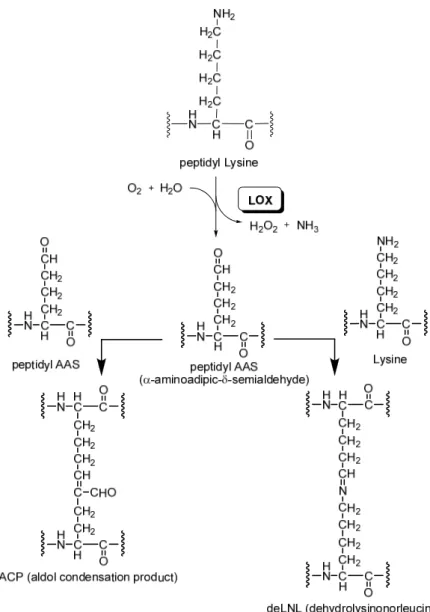
The main biochemical function of LOX is to catalyze the covalent cross-linking of collagen and elastin molecules within the extracellular matrix (ECM), so stabilizing the fibrous deposits of these proteins in the ECM. These cross-links formed by the enzyme increase tensile strength and structural integrity necessary for normal connective tissue function, embryonic development, tissue remodeling and repair of diseased and aging connective tissues. In particular, it catalyzes the oxidation of the ε-amino group of peptidyl lysine to peptidyl aldehyde (α-aminoadipic-δ-semialdehyde, AAS) by consuming oxygen and water and producing ammonia and hydrogen peroxide (Fig.
2.1). Then, the aldehyde group of AAS starts the formation of covalent cross-linkages,
condensing either with a neighbouring ε-amino group of an unmodified lysine residue to form anhydrolysinonorleucine (deLNL) or with another peptidyl AAS to obtain an aldol condensation product (ACP) [5, 6].
34
Figure 2.1. LOX-catalyzed reaction and spontaneous formation of crosslinkages from peptidyl AAS.
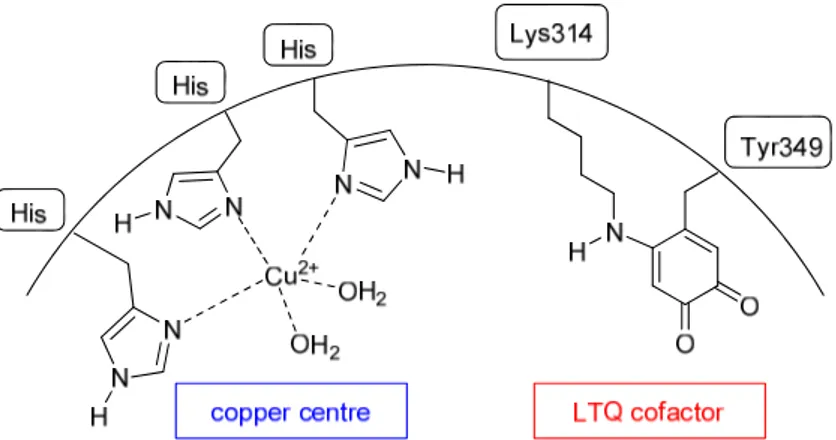
Although the X-ray structure of human LOX has never been obtained yet, it has been possible to determine the presence of a copper (II) ion in the LOX catalytically active site, which is bound to three histidine residues (not been exactly identified as of yet), and also of a covalently bound organic cofactor lysine tyrosylquinone (LTQ, Fig.
35
2.2), which is peculiar since other known amine oxidases are associated to cofactor
trihydroxyphenylalanine quinine (TPQ).
Figure 2.2. Active site of LOX. Copper atom bound to histidine residues is highlighted in blue, LTQ cofactor
in red.
Probably, the cofactor LTQ is formed within the nascent enzyme, by an autocatalytical reaction between tyrosine 349 and lysine 314, in which Cu2+ catalyzes
the oxidation of Tyr to quinone, followed by the covalent attack of the ε-amino group of lysine residue to the quinone ring, hence starting the formation of LTQ (Fig. 2.3) [7].
36
The catalytic cycle (Fig. 2.4) performed by LOX starts with the condensation of its cofactor with the amino group of the substrate peptidyl lysine, then subsequent rearrangements of the imino double bond, hydrolysis and reoxidation complete the cycle affording the aldehyde product and regenerating the initial form of LTQ. It is supposed that the copper is involved in the reoxidation of the cofactor, due also to the fact that deprivation of this element compromises the catalytic activity of the enzyme.
Figure 2.4. LTQ role in the LOX catalytic cycle.
The soluble precursors of collagen and elastin are not the only substrates of LOX, because it has been demonstrated that several basic proteins are oxidized in vitro by LOX. The selective preference for proteins with positive charges at physiological pH can be explained by the copious presence of anionic residues such as Asp and Glu in the catalytic active site (Fig. 2.5), furthermore this abundance of negatively charged sites could be exploited in the development of LOX inhibitors.
37
Arg307 Arg308 Glu311 Lys314 Glu320 Asp321 Asp325
Tyr349
Asp347 Asp352 Asp354 Asp357
LTQ
Figure 2.5. Charged aminoacid residues adjacent the LTQ cofactor (red: anionic; blue: cationic).
Beyond the deaminative oxidation of structural proteins of the ECM, LOX possesses other biological roles influencing cell behaviour, for example it has a potent chemoattractant activity for human peripheral blood monocytes and vascular smooth muscle cells, enhancing their migration with a mechanism mediated by hydrogen peroxide, a product of LOX catalytic activity [8, 9].
2.1.2. LOX inhibitors
BAPN (2.1, Fig. 2.6) has been extensively used in assays devoted to demonstrate the role of LOX in metastasis formation, because it represents one of the very few known inhibitors of lysyl oxidase and so it constitutes the reference LOX inhibitor (IC50 = 10
μM). In particular, BAPN irreversibly inhibits LOX by forming covalent bonds within the catalytically active site, probably by a condensation between BAPN pharmacophoric primary amino-group and the quinone group of LOX covalently bound cofactor lysine tyrosylquinone (LTQ) [10, 11]. Recently, the reduction of the metastatic colonization potential on MDA-MB-231 cells due to LOX inhibition by BAPN was proved in vivo by bioluminescence imaging and microcomputed tomography [12]. Unfortunately, BAPN itself cannot be developed as a drug because of its many unwanted side effects. In fact, the small and “simple” molecular structure of BAPN is able to cross many physiological barriers and interact with a variety of biological targets, thus causing many unwanted side effects. Besides, the toxicity of BAPN is ascribed also to its LOX inhibitory property, because it is considered the main responsible for a syndrome called
Lathyrism [13], a pathological condition involving several body districts, observed in
people and animals after excessive ingestion of sweet pea (Lathyrus odoratus), which contains remarkable amounts of BAPN. Usually, this syndrome manifests in intoxicated
38
children or weanling animals, for which a mature connective tissues has not yet been developed.
NH2
N
2.1
Figure 2.6. Structure of BAPN (2.1).
In literature, there is a small number of LOX inhibitors, mainly because only few useful therapeutic uses had been envisaged so far. Most of them possesses primary amino group like BAPN itself, such as taurine 2.5 and compounds 2.6-2.14 [14-23] (Fig. 2.7) or they are hydrazine-derivatives, such as isoniazide 2.2, semicarbazide 2.3 and thiosemicarbazide 2.4 [24], so they should interact with LTQ cofactor similarly to BAPN, whereas only a few most recent inhibitors show heterocyclic scaffolds (compounds 2.15-2.16). Besides, the presence of basic centers confirms the preference of LOX for cationic substrates. Unfortunately, most of these molecules not only are small and unspecific compounds, but they also have quite reactive structures and/or toxic functional groups, so they do not possess the proper “drug-like” requirements to be used as drugs, consequently they cannot be developed as anti-metastasis agents. Only the most recent LOX inhibitors such as benzazole 2.15 and pyridazinone 2.16, possessing different structures, have been patented for treating diseases as scleroderma and fibrosis, respectively.
39
HYDRAZINO- DERIVATIVES PRIMARY AMINO- DERIVATIVES HETEROCYCLIC DERIVATIVESFigure 2.7. Structures of known LOX inhibitors (2.2-2.16), divided in three different structural classes
(hydrazino-, primary amino- and heterocyclic derivatives).
At present, there are not small organic molecules able to selectively inhibit lysyl oxidase without provoking significant side effects in order to be used as anti-metastatic drugs, whereas there is a patent covering antibodies, polynucleotides, polypeptides and nucleic acids designed with the aim of blocking LOX activity in tumour tissues [25].
40
2.2.
BIOREDUCTIVELY ACTIVATED LOX INHIBITORS
2.2.1. Hypoxia-activated cytotoxins: the nitroaromatic prodrugs
The lack of selectivity towards tumour cells is the responsible of dose-limiting side effects and toxicity associated with conventional chemotherapeutic agents. One approach to improve the therapeutic index and decrease side effects is to deliver selectively the drugs to tumour sites. The design of targeted prodrugs for tumour site-specific activation exploits the unique features associated with cancers, and in particular one of the well-known strategies exploits the low oxygen tension present in many solid tumours with the aim of developing prodrugs activated by hypoxia (hypoxia selective agents, HSA) [26].
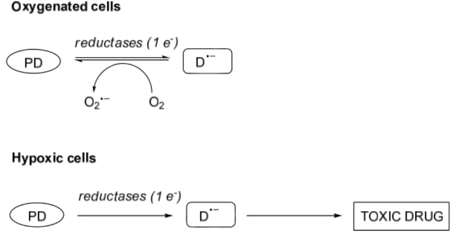
In a general sense, this hypoxia-selective mechanism is based on a reductive process, by which an inactive prodrug is activated to a cytotoxic drug upon reduction in a hypoxia-dependant manner, but the same reductive reaction could occur also in oxygenated tissues, because reducing enzymes may be also present in aerobic regions. Nevertheless, in this case the electron of the prodrug radical is rapidly transferred back to molecular oxygen, forming superoxide, which is scavenged by the enzyme superoxide dismutase to less toxic species (but it can contribute to some unwanted toxic side effects), and regenerating the initial prodrug (Fig. 2.8). This redox cycles is clearly advantageous, it ensures the cytotoxic action of the prodrug only in hypoxic areas, providing a selective treatment and minimizing the toxic side effects due to the activation of the drug in healthy oxygenated tissues. An ideal prodrug candidate has to meet some requirements to be developed as hypoxia-activated cytotoxin: it must be non toxic and stable and it must be able to diffuse efficiently through hypoxic tumour tissues but above all it must possess a reducible function, able to be converted to a potent cytotoxin when selectively activated by a specific enzyme or metabolic pathway present exclusively or predominantly in the targeted cancer cells, so exhibiting significantly greater toxicity to hypoxic compared to aerobic cells [27].
41
Figure 2.8. Mechanism of action of hypoxia selective agentsin oxygenated and hypoxic cells. PD = prodrug,
D.¯ = prodrug radical.
Hypoxia-activated prodrugs can be divided into four main classes on the basis of the structure of the group that undergoes the initial reductive process (usually called the “trigger group”): quinone, nitroaromatics, N-oxides and metal complexes. Nitroaromatic class comprises a very wide group of efficient prodrugs whose biotransformation leads to drug activation in hypoxic tumours, but albeit the interest in their utilization is great, no clinically useful compounds have been obtained yet. Nitroreductive activation is also of current interest, due to the recent possibility of introducing specific exogenous nitroreductases into tumours [28]. Hypoxia-targeted prodrugs using nitroaromatic moieties were initially developed from the nitroimidazole-based radiosensitizers Misonidazole 2.17 and etanidazole 2.18 (Fig. 2.9). They acts as mimetics of oxygen, causing fixation of radiation-induced damage to DNA or other biologically relevant biomolecules, when used in combination with radiation therapy, thus increasing the radiation sensitivity of hypoxic cells and, consequently, enhancing radiotherapeutic effect in vivo. Unluckily, they do not possess significant cytotoxicity or marked hypoxia selectivity. Moreover, they provoke neurotoxicity. Extensive studies and important progresses were made in the development of this class, starting from these first structures.
42
Figure 2.9. Structure of the radiosensitizers Misonidazole (2.17) and etanidazole (2.18).
Nitroreductase enzymes catalyze the reduction of aromatic nitro groups and usually the major metabolic products of nitroreductase-catalyzed reductions are the four-electron product hydroxylamine and the six-four-electron product amine, both of them passing by the first electron adduct of nitro group, the nitro radical anion. This first intermediate can be oxidized back by O2 to the parent nitro group, so limiting the
activation reaction to hypoxic areas, where the nitro radical anion is further reduced stepwise by irreversible reactions to hydroxylamine or amine (Fig. 2.10). The reduction processes catalyzed by nitroreductases are particularly facilitated in cells characterized by total or partial absence of oxygen because they are rich in reduced cofactors.
Figure 2.10. Reduction reactions of nitroaromatics and back-oxidation of nitro radical anion by oxygen. The electronic reduction represents a very significant electronic density change in the structure of a nitroaromatic compound, thus it can be exploited as an efficient “electronic switch” to release or generate the latent active drug [29]. Nitroaromatics have been extensively used as pro-moieties to release a large number of anticancer agents and various linking groups have been employed to attach the cytotoxic portion to the masking nitroaromatic group. However, the most investigated mechanism of activation and release of drug involves the bioreduction of the nitro group of a nitrobenzyl portion to a hydroxylamine intermediate, followed by double bond
43
rearrangements and consequent spontaneous 1,6-elimination to release the cytotoxic drug (Fig. 2.11).
Figure 2.11. Activation and release of active drug from nitro-benzyl prodrug triggered by bioreduction and
1,6-elimination.
2.2.2. Pro-BAPNs as hypoxia-activated LOX inhibitors [30]
During my PhD project, in order to selectively deliver the reference LOX inhibitor BAPN to hypoxic tumours and block LOX-induced metastasis, we used the well-known hypoxia-sensitive nitroaromatic group to mask the pharmacophoric primary amino-group of BAPN. We developed a series of inactive nitroaryl pro-drugs of BAPN, that could be selectively activated to BAPN itself by bioreductive processes in the hypoxic environment of invasive tumours. Under hypoxic conditions, the masking nitro group is readily reduced to hydroxylamine or amine. Consequently, this transformation weakens the bond between the masking moiety and BAPN portion, promoting the spontaneous removal of the masking group and allowing the release of active BAPN selectively in hypoxic tumour cells (Fig. 2.12).
O2N aromaticring HN Bioreductiveactivation Hypoxic tumour environment RHN aromaticring HN CN R = H, OH labile bond Spontaneous cleavage electron-withdrawing electron-donating
Inactive pro-BAPN BAPN
H2N
CN CN
Figure 2.12. Activation mechanism of pro-BAPNs.
Exploiting this strategy, we designed and synthesized new pro-LOX inhibitors, which are nitrobenzyl- (2.19 and 2.20), nitrobenzylcarbonyl- (2.21) [31] and nitrofuranmethyl- (2.22) [32] derivatives of BAPN (Fig. 2.13). In these preliminary series of prodrugs, the nitrogen of the pharmacophoric primary amino group of BAPN
44
was linked to nitroaromatic moieties by a methyl (2.19, 2.20 and 2.22) or a methoxycarbonyl (2.21) unit.
Figure 2.13. Structures of pro-BAPNs (2.19-2.22).
Synthesis of compounds 2.19-2.22 was accomplished as illustrated in Scheme 2.1. Compounds 2.19, 2.20 and 2.22 were prepared starting from the correspondent commercially available aldehyde precursors (2.23, 2.26 and 2.30), whose condensation with an excess of β-aminopropionitrile in anhydrous dichloromethane afforded imine derivatives 2.24, 2.27 and 2.31, respectively. Then, reduction of the imine double bond with sodium borohydride at 0 °C afforded compounds 2.25, 2.28 and 2.32, and final treatment with Et2O.HCl converted them into their corresponding water-soluble
hydrochloride salts 2.19, 2.20 and 2.22 [33]. Carbamate derivative 2.21 was simply obtained by a single step involving p-nitrobenzylchloroformate 2.29 and BAPN in DCM. Nitrofurane analogue 2.22 required a very slow addition of NaBH4 to the
intermediate 2.31 and strict control of the temperature to avoid the possible undesired reduction of the highly electron-deficient furan ring.
45
2.19 2.22 CHO O2N CHO NO2 O O2N CHO O2N N H N N H N NO2 N N O O2N H O2N N H N N H N N H N O O2N O2N O Cl O 2.20 2.21 2.23 2.24 2.25 2.26 2.27 2.28 2.29 2.30 2.31 2.32 a a a a b b b c c c NO2Scheme 2.1. a) β-aminopropionitrile, dry DCM, RT; b) NaBH4, dry MeOH, 0 °C.; c) Et2O.HCl, dry MeOH,
RT.
It has often been observed a direct association between the ease of activation of bioreductive prodrugs to their electrochemical reduction potential values [34, 35]: usually, a higher reductive potential value corresponds to a ready activation and, consequently, to a greater cytotoxicity in hypoxic cells. For this series of pro-BAPNs, cyclic voltammograms were recorded in iso-osmotic saline phosphate buffer solutions at pH 7.2, so measurements of their reduction potentials were obtained in order to evaluate their predicted ability to be reduced in the hypoxic environment (Table 2.1 and Fig.
2.14). All the derivatives produced a single reduction peak when the potential was
scanned from 0.0 to -1.8 V, which is the result of the reduction of their nitro group to the hydroxylamine product. Moreover, the voltammetry graph shows the irreversibility of the reductive process, missing a correspondent oxidation peak in the lower part of the curve. This indicates the irreversible transformation of the molecules (loss of the BAPN portion) following the NO2/NHOH conversion. Among these compounds, analogues
46
2.19-2.21 displayed similar reduction potentials, with E° values ranging from -0.75 V of
the two para-nitro derivatives 2.19 and 2.21 to -0.77 V for the ortho-nitro analogue
2.20; while it is evident that nitrofuranmethyl-derivative 2.22 showed the less negative
reduction potential (E° = -0.61 V), as shown by the blue line in the voltammogram. This means that 2.22 is much more inclined to reduction and, as a consequence, it should be more readily activated in hypoxic tumours releasing BAPN.
Table 2.1. Reduction potential values of compounds 2.19-2.22. Measurements were recorded, using 5.10-4 M
solutions, at glassy carbon electrode in phosphate buffer (pH 7.2, 0.15 M NaCl), with a sweep rate of 0.1 V s– 1.
Compound Structure Reduction
Potential (E°)
2.19 –0.75 V
2.20 –0.77 V
2.21 –0.75 V
47
-50 0 50 100 150 -1500 -1000 -500 0 E / mV vs Ag/AgCl/KCl sat I /μAFigure 2.14. Cyclic voltammograms of compounds 2.19-2.22.Measurements were recorded, using 5.10-4 M
solutions, at glassy carbon electrode in phosphate buffer (pH 7.2, 0.15 M NaCl), with a sweep rate of 0.1 V s– 1. Compound 2.22 in blue; 2.21 in red; 2.20 in yellow; 2.19 in green, in the graph.
48
2.3.
BIOLOGICAL EVALUATION OF LOX INHIBITORS
[30]
Compounds 2.19-2.22, synthesized as pro-BAPNs molecules, were biologically assayed by the research group of Prof. Amato J. Giaccia at the University of Stanford (California, USA). Initially, their ability to inhibit LOX enzyme was tested, then they were submitted to in vitro invasion assays to measure their anti-invasive ability in tumour cells.
The inhibition potency of this series of inhibitors was assayed by using a conditioned medium (CM) from the MDA-MB-231 breast tumour cell line, which was incubated with the reference inhibitor BAPN and with 200 μM LOX inhibitors
2.19-2.22, both in hypoxic and normoxic conditions (Fig. 2.15). The LOX inhibition is
related to the fluorescence development by a method previously described [36, 37]. This method consists in the reduction of the LOX by-product hydrogen peroxide by N-acetyl-3,7-dihydroxyphenoxazine (Amplex Red), catalyzed by the enzyme horseradish peroxidase, and this redox reaction produces as product fluorescent resorufin. This fluorometric measurement of hydrogen peroxide has been extensively used for measuring LOX activity, correlating a diminished fluorescence with a great LOX inhibition. It is clear from the bar graph that compounds 2.19, 2.21 and 2.22 are much more active under hypoxia than in normoxia, consequently confirming their reduction and activation in conditions of low oxygen concentrations. On the other hand, the reference inhibitor BAPN does not show any significant difference in its activity between air and hypoxia, since it does not need any activation. In particular, compounds
2.21 and 2.22 provided the best results, thanks to their much greater inhibitory activities
under hypoxic conditions, whereas their activities in the presence of air are almost the same of control and DMSO, thus showing a very selective behaviour.
ortho-Nitrobenzyl derivative 2.20 showed a very high fluorescence, if compared to
the other derivatives and to control. This unpredictable result is likely generated by an interference with the fluorescence-based assay, possibly due to its high photo-instability, so, currently, optimization of the assay conditions for this specific derivative is in progress.
49
Figure 2.15. LOX inhibition bar graph: fluorescence assay for LOX activity on CM from MDA-MB-231 cells
incubated in air (white bars) or hypoxia (2% O2, purple bars) with 200 μM LOX inhibitors 2.19-2.22.
Subsequently, the anti-invasion abilities of this series of compounds were determined. Cell invasion assays were performed in vitro on MDA-MB-231 breast cancer cells, following a method that has already been described [38]. Briefly, tumour cells were serum deprived and after 24 h seeded both on Matrigel-coated and uncoated inserts, then they were moved to chambers containing FBS as a chemo-attractant and incubated under normoxic or hypoxic conditions. The incubation with control or with the compounds was started 24 h before serum deprivation and continued for all the duration of the experiment. This tumour cell line is highly invasive and become even more aggressive when it is placed under oxygen deprivation, as shown by the control data in the graph (Fig. 2.16), where the percentage of invasion under hypoxia is about two-fold higher than that in air. It is interesting to note that, even in this case, the most promising compounds are 2.21 and 2.22. In fact, they possess not only enhanced activities in hypoxic conditions, but they also provoke a six-fold reduction of cell invasion under hypoxia, if compared to control experiments. In particular, the nitrofuran derivative 2.22 manifests the highest hypoxia-selectivity, since it maintains the same invasion level as control in normoxic conditions (30%), but it markedly reduces the level under air deprivation. The non-selective behaviour of the carbamate inhibitor 2.21 suggests that it may produce anti-invasive effects by means of a non-LOX-mediated
50
mechanism, showing an anti-invasive action even under normoxic conditions, although further studies need to be done to explain this phenomenon.
Figure 2.16. Cell invasion assay bar graph: transwell invasion assay to test ability of the synthesized
compounds to inhibit in vitro invasion of MDA-MB-231 cells incubated in air (white bars) or hypoxia (2% O2, purple bars) with 200 μM LOX inhibitors 2.19-2.22.
Observing the data form both LOX inhibition and anti-invasion assays, we can conclude that pro-drug 2.22 proved to be the most active and hypoxia-selective compound and this result nicely correlates with its higher reduction potential (E° = – 0.61 V) when compared to the other active members of the series of pro-BAPNs (2.19 and 2.20, E° = –0.75 V). Indeed, its least negative potential is associated with an easier bioreduction and activation of the nitrofuran masking portion, if compared to the other nitroaryl derivative 2.19-2.21, and it is evident that this mechanism leads to more pronounced effects in hypoxia conditions in the performed biological assays.
51
2.4.
CONCLUSIONS
To the best of our knowledge, the use of nitroaromatic pro-drugs to selectively inhibit the recently discovered target LOX is a completely new method to reduce or block the invasiveness of hypoxic tumours. Hypoxia-activated LOX inhibitors represent a new and effective therapeutic approach against metastatic tumours and it could be useful not only for the prevention of future metastasis, but also for the treatment of established metastatic conditions.
Pro-drug 2.22 showed a notable LOX inhibition activity and a marked anti-invasion ability in hypoxic conditions by selectively delivering LOX inhibitor BAPN to tumour tissues. Although the compound database is too small to demonstrate a definitive correlation, the hypoxia-enhanced activities of these prodrugs of BAPN appear to be strictly related to their electrochemical potential value, suggesting that the promising biological activities found with 2.22 are associated to the easier activation of its nitrofuran masking portion in hypoxic conditions, when compared to that of the other derivatives.
The direct involvement of the masking portions in the biological activity of the nitrofuranmethyl and p-nitrobenzyl derivatives 2.22 and 2.21, which proved to be the most active compounds, are not to be excluded. In fact, the nitroaromatic groups, after detaching from BAPN under hypoxic conditions, might interact with LOX in a synergistic fashion with the active BAPN, even if attempts to demonstrate the involvement of chemical species derived from the masking portions have so far failed.
This research line can be further developed and expanded by using different masking moieties to be linked to BAPN or even by introducing completely new LOX inhibitors in the place of BAPN itself. This last goal could be achieved, for example, with derivatives able to bind LTQ cofactor, such as primary amino-derivatives, or even with compounds containing chelating groups, which can coordinate to the copper ion present in the LOX active site. Moreover, since LOX is an extracellular target, it can be accessible even to drugs that do not efficiently cross the cell membranes and this constitutes a further advantage, because higher concentrations of drugs in the tumour stroma are more likely to be reached than inside the cancer cell.