Corso di Laurea in
Ingegneria Energetica
Air contamination control in Hybrid operating theatres.
Particle content during different types of surgery with focus on diathermy
Relatore: Prof. Cesare Maria JOPPOLO
Co-relatore: Prof. Jan Bertil GUSTEN Co-relatore: Ing. Francesco ROMANO
Tesi di Laurea di:
Daniele MEDA Matr. 771258
Preface
The main topic of this Master Thesis is the measurement of non-viable particle concentrations during several types of surgeries inside an Hybrid Operating Theatre. The focus is primarily on the use of electrosurgical instruments based on diathermy, which are supposed to considerably affect the concentration and distribution of airborne particles inside operating rooms, thus leading to a lower level of air cleanliness due to the generation of surgical smoke.
The research work was carried out between September 2013 and March 2014. It is worth noting that this study was possible thanks to the european connection between Politecnico di Milano and Chalmers University of Technology. In particular, both the Italian Dipartimento di Energia and the Swedish department of Energy and Environment are working on similar topics and with the same scientific viewpoint in the field of air conditioning and systems for controlled contamination environments.
The measurement campaign was performed at the Sahlgrenska University Hospital of Gothenburg, Sweden within the framework of a previously established collaboration with the division of Building Service Engineering from the Department of Energy and Environment of Chalmers University of Technology.
A special thanks goes to Christina Ekroth and to the whole personnel of the Operation Ward 2 at Sahlgrenska University Hospital of Gothenburg, Sweden for the helpfulness and support provided during measurements, and especially for the care shown throughout every phase of the research process.
In addition, I am grateful to professor of Politecnico di Milano Cesare Maria Joppolo, who had confidence in me and gave me the great possibility to work on this project.
Moreover, I would not have been able to carry out this study without the guidance and aid of eng. Francesco Romano, an italian researcher of Politecnico di Milano, who helped me to be familiar with the topic and the tools used during the measurement campaign.
Thanks go to my roommate and co-worker Leonardo Claudio Amato for the sharing of ideas and experiences during the measuring and analysis steps.
Heartfelt thanks go to Associate Professor Berit Reinmüller and Professor Bengt Ljungqvist of Chalmers University of Technology, who offered me valuable advice and support throughout the whole work.
Finally, I would like to express my most sincere thankfulness to my Swedish supervisor Jan Bertil Gustén of Chalmers University of Technology for his constant guidance and endless encouragement through all the stages of this research project, as well as for the special attention he gave to my own work with him.
Index
Preface ... I Index ... III Figure index ... VI Table index ... XII Abstract ... XV Sommario ... XVII 1 Introduction ... 1 1.1 Purpose ... 1 1.2 Method ... 3 1.3 Previous work ... 4 1.4 Problem ... 5
1.4.1 People as a contamination source ... 5
1.4.2 Surgical procedures ... 5
1.5 Limitations ... 7
2 The Hybrid Operating Theatre ... 9
2.1 General features of hybrid operating theatres ... 10
2.1.1 Definition ... 10
2.1.2 Description ... 10
2.1.3 Planning ... 12
2.1.4 Size and workflow ... 12
2.1.5 Lighting ... 14
2.1.6 Imaging system configuration 2.1.6.1 Magnetic Resonance Imaging ... 15
2.1.7 Ventilation system ... 19
2.1.8 Costs ... 21
2.2 Hybrid OT at Sahlgrenska University Hospital ... 22
2.2.2 The intensity and the use variety ... 25 2.2.3 Imaging system ... 26 2.2.4 Clothing system ... 27 2.2.5 Gloves ... 30 2.2.6 Electrosurgical instruments ... 32 2.2.7 Ventilation system ... 36 2.2.8 Room conditions ... 46
3 Description of the measurement campaign ... 49
3.1 Particle samplers ... 50
3.2 Data analysis of the previous work ... 53
3.3 Statistical analysis ... 54
3.4 Measurement strategy ... 61
3.5 Measurements methodology ... 65
3.6 Measurements performed ... 68
4 Measurement campaign ... 81
4.1 Classification of air cleanliness ... 82
4.1.1 Standards used ... 82
4.1.2 Definition of the measurement conditions ... 87
4.1.3 Data and calculations ... 88
4.2 Activity characterization ... 95
4.2.1 Clothing system ... 96
4.2.2 Surgery duration ... 97
4.2.3 The average number of people ... 98
4.2.4 Frequency of door openings ... 102
4.2.5 Mean values of particle concentration... 105
5 Consequences of diathermy ... 113
5.1 Particle samplers positioning ... 115
5.2 Smoke test ... 116
5.4 Sampling probes positions ... 121
5.5 Measurement of particle source ... 123
5.6 Results of simulated surgery on a calf liver ... 124
5.7 Comparison between ultrafine particles concentrations in position 1 and 2 ... 131
5.8 Conclusions ... 138
5.9 Future works ... 141
Acronyms ... 143
Figure index
Figure 1.1. Characteristic sizes of particles generated by the use of electro cautery, laser and ultrasonic devices. These particles are too small to be filtered by ordinary surgical masks, which are able to block only particles 5µm………. 7
Figure 2.1. This hybrid operating room, completed in 2004 at Providence St.
Vincent Medical Center in Portland, is one of the first of its kind in the Northwest………..….. 11 Figure 2.2. Typical surgical lights during open surgeries in the middle of the
picture on the left , different ambient lights in both of the pictures……….. 14 Figure 2.3. On the left, a ceiling mounted C-arm (Schaadt and Landau,
(2013)), on the right a floor mounted X-ray scanner (Nollert et al, (2012))………... 15 Figure 2.4. Scheme of the unidirectional airflow pattern in an OT (Price-Hvac 2013)……….. 20 Figure 2.5. Representation of the different areas of the Hybrid OT at
Sahlgrenska University Hospital. The zones indicated are the clean area (orange), the periphery of the clean area (green), a small office (purple) and the IT room (light blue)………..….. 23 Figure 2.6. The X-ray scanner Artis zeego (SIEMENS) on the left, and it
position inside the Hybrid OT on the right (blue circle in the layout)………... 26
Figure 2.7. Changing room where the ordinary clothes are stored ……… 27
Figure 2.8. Mertex clothes supplied inside sterilized cover bags (on the left), and the same clothes worn by an operator (on the right)………... 28
Figure 2.9. Surgeon during the preparation of the medical instruments,
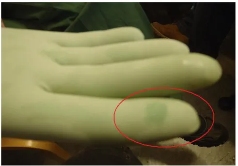
wearing the typical surgical gown, gloves, helmet and face mask………...… 29 Figure 2.10. The red circle points out a stronger color contrast which is the hole indicator……….………....… 30
Figure 2.11. Monopolar diathermy attached to local exhaust system…...… 33 Figure 2.12. Bipolar diathermy tweezers model……… 34 Figure 2.13. Ultrasonic dissector, forceps model…………...…………...… 34 Figure 2.14. Argon electrode handle………...………….. 35 Figure 2.15. Technical representation of the ventilation system divided in two
main parts, the HVAC system and the airflow diffusion system……… 36 Figure 2.16. Technical representation of the ventilation system, with the indication of the 3 AHUs and the local cooling coil signed with the letter “C”……….………. 38 Figure 2.17. Representation of the clean area (orange) and the periphery of the clean area (green)………..… 40 Figure 2.18. Representation of the different positions of the plenum (orange),
in grey are defined the 4 different positions of the HEPA filters in the peripheral zone. The figure indicates also in red the 4 different positions of the extraction grilles indicated by arrows……….... 42 Figure 2.19. Technical data related to the filter types. The installed filters in the Hybrid OT in the clean zone are described in the table….. 43 Figure 2.20. Touch screen control panel positioned inside the Hybrid OT of
Sahlgrenska University Hospital………... 46
Figure 3.1. P-TRAK™ 8525 TSI…………..………... 50
Figure 3.2. OPC Metone 3313……….…… 51
Figure 3.3. Innovation by Climet CI-500 (Certification and calibration
Service, 2014)………..………. 52
Figure 3.4. Ultrafine particle measurements in two operating rooms with two
different ventilation systems. On the left, the particle concentrations with conventional ventilation. On the right, the particle concentrations with unidirectional ventilation……... 54
Figure 3.5. Instrument positions during operation n°1, inside the room
Figure 3.6. Instrument positions during operation n°8, inside the room equipped with unidirectional airflow……….…... 59
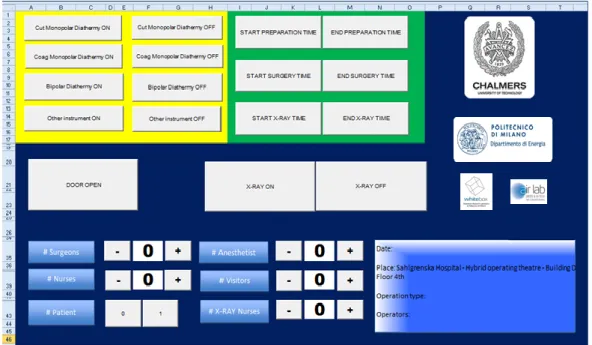
Figure 3.7. The Excel sheet designed by Daniele Meda and Leonardo
Claudio Amato for this measurement campaign for data collection………... 62 Figure 3.8. Excel sheet concerning the orthopedic surgery of the 28th of
October 2013 related to the number of surgeons inside the Hybrid OT……….… 63 Figure 3.9. Clean instruments. In order from left: P-trak, Tygon piping,
telescopic probe, telescopic instrument holder , probe holder.. 66 Figure 3.10. Office next to the Hybrid OT, position of the operator that used
to work with the Excel sheet……….……… 67 Figure 3.11. Sequence of pictures represents the rotational 3-D visualization
recorded with the X-ray scanner with a standard angiography during an EVAR surgery. The two wires in the pictures are the catheters used for the introduction of the stunts through the femoral arteries………..………... 70 Figure 3.12. Probes of UPC (P-TRAK™) and OPC (Climet) are placed in the
surgical zone. The sampling probes are positioned on the Mayo (trolley) at the height of 1.50 m and the distance between them is around 30 cm………. 71 Figure 3.13. Typical image that permits the surgeon to control the correct positioning of screws on the spinal column during orthopaedic surgeries………. 73 Figure 3.14. UPC (P-TRAK™) and OPC (Climet) placed in the surgical zone. The sampling probes are positioned on the Mayo (trolley) at the height of 1.50 m and the distance between them is around 30 cm……….. 74 Figure 3.15. UPC (P-TRAK™) positioned in the clean area with the red circle that indicates the position of the telescopic probe placed at the height of 2.80 m, which means at a distance of 20 cm under the H14 HEPA filters……….. 75 Figure 3.16. UPC (P-TRAK™) and OPC (Climet) near the surgical table; the
sampling probes were placed at the height of 1.50 m with a
Figure 3.17. The two sampling probes of UPC (P-TRAK™) and OPC (Climet) positioned near the surgeon’s nose during the simulated surgery (operation N°22)………..……….…… 79
Figure 4.1. Positioning of the sampling probe of the OPC for the 4 different
measuring points……….…………...… 89
Figure 4.2. Positioning of the sampling probe of the OPC for the 9 different
.measuring points………...…… 92
Figure 4.3. Surgery duration related to the different types of operations observed……….… 97
Figure 4.4. The mean number of people recorded during the various
operations inspected………...………... 99
Figure 4.5. Number of people recorded during the EVAR operation
performed on the 7-11-2013 (operation N°9)…………..…... 100
Figure 4.6. Number of people recorded during the orthopedic operation
performed on the 9-12-2013 (operation N° 21)……….. 101 Figure 4.7. Frequency of door openings per minute recorded during the
different types of operations………..…. 102 Figure 4.8. Position of the particle sampling probe during the different
ongoing surgeries, indicated by the red point………... 105 Figure 4.9. Mean concentration values related to particles ≥ 0.5 µm, recorded
near the surgical area during the different types of operations (18
surgeries for a total of 68 hours)………. 106 Figure 4.10. Mean concentration values related to particles ≥ 5 µm, recorded
near the surgical area during the different types of operations (18
surgeries for a total of 68 hours)………... 108 Figure 4.11. Mean concentration values related to particles of 0.02- 1 µm
(ultrafine particles), recorded in the surgical area during the different types of operations (18 surgeries for a total of 68 hours)... 110 Figure 5.1. Illustration of air pattern inside an operating room, produced by a
vertical unidirectional airflow from the ceiling………….….. 115
Figure 5.3. Pictures sequence about the smoke test performed on the 16th December 2013 (operation N° 23) in the Hybrid OT……….. 118 Figure 5.4. Mean e icienc (-) and ( 5 )(---) (calculated as the lower limit value for the 95 % confidence) as a function of the particle diameter d p ( 1822-3:2009) for H14 HEPA filters……. 119 Figure 5.5. Comparison of the dimensional ranges for particles spread by
electro surgical instruments and the MPPS of H14 HEPA filters…………...… 120
Figure 5.6. Positions of the sampling probes monitoring during open
surgeries………... 121
Figure 5.7. The two instrument positions defined for the measurements
performed during the simulated surgery (operation N°22). Position 1 indicates the sampling probe position of UPC that measures the UFP under H14 HEPA filters. In position 3 the
UPC and OPC were placed to detect the airborne particle
concentrations close to the surgeon’s nose……….…. 125 Figure 5.8. The disposition of the two measuring probes near the surgeon’s nose in position 3. UPC sampling probe on the left and the isokinetic probe of the OPC on the right ……… 126
Figure 5.9. Example of the procedure during the simulation with monopolar
diathermy (on the left) and the calf liver at the end of the simulation (on the right)………. 127 Figure 5.10. Data related to the simulated surgery on a calf liver performed on
the 9th of December 2013. Comparison of UFP measurements carried out in position 1 and position 3 in relation to the moments in which the different electrosurgical instruments were turned on……….………. 128 Figure 5.11. Data related to the first 30 minutes of the orthopedic surgery (operation N° 7) on the 4th of November 2013. In descending order: UFP measured in position 1, UFP in position 2 and
moments in which monopolar diathermy was turned
Figure 5.12. Data related to the liver resection (operation N° 13) on the 14th of November 2013. In order: UFP about position 1, UFP about position 2 and moments in which monopolar diathermy, bipolar diathermy, ultrasonic device and argon diathermy were turned on………..…... 133 Figure 5.13. Data related to the first 60 minutes of the orthopaedic surgery (operation N° 11) on the 11th of November 2013. In descending order: UFP about position 1, UFP detected in position 2 and
switching-on moments of monopolar and bipolar
diathermy………...….. 135 Figure 5.14. Data related to the first 1 hour and a half of the orthopaedic surgery on the 18th of November 2013. In descending order: UFP about position 1, UFP detected in position 2 and switching-on moments of monopolar and bipolar diathermy…………...… 136
Table index
Table 1.1. Risks of surgical smoke (Alp et al.[5])………. 6
Table 2.1. Technical data related to the filters. The red rectangle highlights the characteristics of the filters installed in the Hybrid OT in the clean zone……… 43
Table 2.2. Technical data related to the filters the red rectangle highlights the characteristics of the filters installed in the Hybrid OT in the periphery of the clean zone……….………. 44
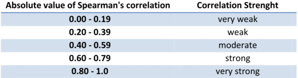
Table 3.1. Strength of the Spearman’s correlation taking into account the absolute value of the statistical test………..……… 57
Table 3.2. Spearman´s Coefficient related to measurements inside the room equipped with conventional airflow………... 58
Table 3.3. Spearman’s Coefficient of data related to measurements inside the room equipped with unidirectional airflow……… 59
Table 3.4. Summary of the measurements inside the Hybrid OT…………. 68
Table 3.5. Measurements days inside the Hybrid OT during EVAR surgery... 69
Table 3.6. Measurement days in the Hybrid OT during orthopaedic surgeries……….. 72
Table 3.7. Measurement day inside the Hybrid OT during liver resection surgery………. 76
Table 3.8. Measurement days inside the empty Hybrid OT……….. 78
Table 3.9. Measurements days inside the Hybrid OT during the simulated surgeries………. 79
Table 4.1. The different airborne particle threshold limits for the nine ISO cleanliness classes……… 84
Table 4.2. 95th percentile of the t-student distribution……... 86
Table 4.3. Data of the different measurements during the air cleanliness classification for the clean area……… 90
Table 4.4 Calculations for the different mean particle concentration measurements during the air cleanliness classification for the clean area. General average of the mean values, standard deviation of the mean values, 95th percentile of the t-student distribution, upper bounds of a 95% confidence interval of the general average.………..….. 90
Table 4.5. Data of the different measurements during the air cleanliness classification for the periphery of the clean area……….... 93
Table 4.6. Calculations for the different mean particle concentration measurements during the air cleanliness classification for the periphery of the clean area. General average of the mean values, standard deviation of the mean values, 95th percentile of the t-student distribution, upper bounds of a 95% confidence interval of the general average………...………... 94
Table 4.7. Different choices for the clothing system for EVAR surgeries and open surgeries……… 96
Table 5.1. Mean concentrations about ultrafine particles detected during a simulated operation on a calf liver. The sampling probe were positioned at 5-8 cm from the working instrument. The instruments used were monopolar, bipolar pincers, bipolar scissors, argon and ultrasonic device………..…… 123
Abstract
Air cleanliness in operating rooms is an important goal in order to preserve both the patient’s and the medical staff's health. Particle contamination inside an Operating Theatre (OT) is supposed to depend mainly on the surgery process, clothing system and personnel routines. However, during open surgeries, a significant airborne contaminant load is given by the release of surgical smoke from electrosurgical tools. This is a work environment problem because of the smell and the risk for health consequences. Ordinary surgical masks are inadequate for filtering these ultrafine particles, emitted when using electrosurgical tools, due to their small size. This study is based on the analysis of airborne particle concentrations, obtained during a measurement campaign carried out at Sahlgrenska University Hospital of Gothenburg, Sweden in the new built Hybrid OT. The measurements were performed continuously for 18 operations. The OT comprises X-ray scanner while the air is supplied with a unidirectional vertical air flow by a ceiling equipped with H14 HEPA filters. Due to the air diffusion principle, surgical smoke may represent an health hazard in every part of the room. A data analysis was conducted between different kinds of surgeries, such as endovascular surgeries or open surgeries, taking into account valuable info about number of people inside operating room, door openings and clothing system adopted. The measurements were carried out with two optical particle counters and two ultrafine particle counters. Results revealed a strict connection between the use of electrosurgical tools and the increase of particles concentration near surgical area due to the surgical smoke motion from the operating table to the other part of the Hybrid operating theatre. Furthermore, the data analysis highlighted that, during activities which require an extremely clean environment such as open surgeries because of the massive use of electrosurgical tools, a part of the ultrafine particles flow back in the clean zone via the recirculation air system. The reintroduction of ultrafine particles can be motivated by the low air filter efficiency at low size range.
Keywords: Contamination control, Ultrafine particles, Airborne Particles, Electrosurgical instruments, Surgical smoke, Hybrid Operating Theatre
Sommario
La pulizia dell’aria all’interno delle sale operatorie e uno dei requisiti fondamentali nella salvaguardia della salute sia dei pazienti sia del personale medico stesso. La contaminazione particellare dell’aria in questi locali dipende da diversi fattori, legati principalmente all’attività operatoria, agli indumenti utilizzati, ai movimenti del personale e al paziente stesso. Più nello specifico, le principali fonti di contaminazione presenti sono la flora batterica endogena del paziente e quella esogena della sala stessa, emessa per lo più dallo lo staff chirurgico. Inoltre, durante le operazioni di chirurgia invasiva, l’utilizzo di strumenti elettrochirurgici gioca un ruolo fondamentale in termini di generazione particellare.
L'elettrochirurgia, infatti, è una tecnica chirurgica che si basa sull’utilizzo della corrente elettrica alternata ad alta frequenza direttamente sul tessuto biologico con effetto termico, in modo da ottenere un taglio oppure un coagulo. Questa particolare tecnica genera in larga misura il cosiddetto “fumo chirurgico”, (surgical smoke in anglosassone) che, oltre a produrre un cattivo odore all’interno della sala operatoria, è stato dimostrato provocare problemi alla salute del personale chirurgico, così come aumentare il rischio di infezioni per il paziente stesso.
Questo studio si basa sull’analisi delle misure di contaminazione particellare non biologica condotte presso il Sahlgrenska University Hospital di Göteborg, in Svezia, all’interno di una sala operatoria ibrida di nuova concezione, dove gli stessi strumenti elettrochirurgici sono utilizzati durante operazioni di tipo invasivo. Le conte particellari sono state realizzate in maniera continuativa per l’intera durata di 18 operazioni chirurgiche di varia natura, a queste si aggiungono altre attività di rilevazione e analisi a sala vuota, per un totale di circa 90 ore di misura.
Nella prima parte di questo lavoro verranno introdotti gli aspetti fondamentali della ricerca. Sarà inoltre fornita una valutazione preliminare dei problemi di salute riscontrati in seguito alla prolungata respirazione del fumo chirurgico quali enfisemi, asma, bronchiti croniche, dermatiti, irritazioni agli occhi e mal di testa etc. Ulteriori ricerche risultano necessarie per confermare i potenziali danni alla salute per il personale stesso. A tal proposito, è di fondamentale importanza l’analisi delle dimensioni delle particelle generate dai vari strumenti elettrochirurgici. Questi strumenti si basano su diversi principi di funzionamento, come ad esempio la diatermia con generazione di particelle di
dimensioni che oscillano in media tra 0.007 µm e 0.42 µm, oppure apparecchi ultrasonici che producono particelle nell’intervallo di grandezza compreso tra 0.35 e 6.5 µm.
Il problema messo in luce da tali osservazioni ricorrente è l’inefficienza delle mascherine chirurgiche in termini di filtrazione di queste particelle ultra-fini. In altre parole, si è rilevato che le stesse mascherine sono ottimi sistemi filtranti solo ed esclusivamente per particelle con dimensioni maggiori a 5 µm. Da questo dettaglio tecnico deriva l’importanza di valutare le concentrazioni particellari rappresentative durante l’utilizzo di questi strumenti.
Una volta presentate queste prime valutazioni, si passa alla descrizione delle caratteristiche generali delle sale operatorie ibride nelle loro peculiarità costruttive e impiantistiche. Queste sale, infatti, possiedono una tecnologia diagnostica elevatissima grazie a sistemi angiografici (scanner X-ray) installati nella sala operatoria stessa che permettono l’integrazione delle funzioni diagnostiche e terapeutiche. Data l’avanguardia tecnologica delle attrezzature, la sala operatoria ibrida è utilizzata in compartecipazione da diversi reparti ed è caratterizzata dalla presenza di un team multidisciplinare durante gli interventi chirurgici.
In seguito, verrà descritta in dettaglio la sala operatoria ibrida del Sahlgrenska University Hospital, focalizzando l’attenzione sulle attrezzature e sulla routine operatoria a livello di comportamenti e vestizione dello staff chirurgico. A tal proposito, merita una particolare considerazione il riferimento e la descrizione dell’impianto HVAC (Heating, Ventilation and Air Conditioning) che, grazie all’utilizzo di filtri H14 HEPA (High Efficiency Particulate Air filters), garantisce alti livelli di qualità dell’aria in termini di filtrazione particellare. Inoltre tale impianto assicura un flusso unidirezionale dell’aria dal soffitto verso il basso, che permette la pulizia della zona chirurgica dalle particelle generate durante le operazioni. Risulta anche degno di nota l’utilizzo di un sistema di ricircolo dell’aria che entra in conflitto con le elevate concentrazioni di particelle emesse durante l’impiego degli strumenti elettrochirurgici.
Successivamente verranno presentati gli strumenti di misura particellare usati durante la campagna di misure: due contatori di particelle ultra-fini e due OPC (Optical Particle Counter). I primi strumenti misurano la concentrazione totale delle particelle le cui dimensioni oscillano tra 0.02 e 1 µm, range tipico delle particelle ultra-fini. Gli OPC, invece, registrano le concentrazioni cumulate di particelle con dimensioni maggiori di 0.5 µm e di 5 µm.
Si passa poi all’esposizione delle valutazioni statistiche effettuate sui dati di una precedente tesi sullo stesso argomento, da cui è stato possibile ricavare importanti risultati che permettono di motivare la scelta delle posizioni adottate
per le misurazioni realizzate all’interno di questo lavoro di tesi. Inoltre, verranno elencate le date delle misurazioni e le caratteristiche del foglio Excel progettato ad hoc per questa campagna di misure che ha consentito di rilevare i dati relativi al numero delle persone nonché all’apertura delle porte, nei periodi in cui gli strumenti elettrochirurgici erano in funzione. I valori così ottenuti hanno permesso di completare le misure particellari realizzate durante le operazioni con dati sulle attività della sala operatoria.
Le concentrazioni rilevate durante le misure nella sala operatoria ibrida in condizioni simili a quelle di riposo (at rest), caratterizzata dalla presenza di due operatori all’interno, hanno reso possibile la classificazione della pulizia dell’aria nella sala operatoria seguendo le procedure suggerite dalla norma ISO 14644-1:2001. Tali dati confermano che i valori di concentrazione particellare non superano i limiti dettati dalla norma per le sale operatorie, infatti almeno la classe di pulizia ISO 5 risulta soddisfatta.
Gli interventi chirurgici seguiti per tale studio sono di due tipi: endovascolare e a cielo aperto (invasive). Del primo gruppo fanno parte le EVAR (riparazioni endovascolari dell’aneurisma), operazioni caratterizzate da un livello minimo di invasività e realizzate grazie all’utilizzo dello scanner a raggi X che consente la visualizzazione delle parti interne del corpo del paziente per il corretto posizionamento dello stent. Invece, per quanto riguarda gli interventi a cielo aperto (invasivi), il campione di analisi comprende alcune operazioni di ortopedia, in particolare fusioni spinali, e una operazione di resezione del fegato. In questi casi, lo scanner a raggi X è stato usato solo per le procedure ortopediche relative alla diagnosi iniziale e per il controllo del risultato finale dell’operazione.
I dati raccolti con il foglio Excel hanno permesso di effettuare alcuni utili confronti tra le operazioni endovascolari e a cielo aperto. La durata dei due tipi di operazione non può essere utilizzata come indicatore di differenza. Tuttavia, tramite la valutazione del numero medio di persone durante il periodo operatorio, sono stati riscontrati dei valori maggiori nel caso delle operazioni endovascolari, dovuti al team multidisciplinare richiesto. E’ possibile osservare lo stesso trend prendendo in considerazione la frequenza di apertura della porta di ingresso della sala operatoria ibrida, che risulta maggiore per le operazioni endovascolari rispetto a quelle a cielo aperto. Inoltre, sono state rilevate delle differenze a livello di abbigliamento tecnico dello staff chirurgico. Tutto il personale, durante le operazioni endovascolari, indossa gli stessi indumenti che caratterizzano l’intero staff ospedaliero. Al contrario, durante le operazioni a cielo aperto in generale viene riposta maggiore attenzione all’abbigliamento adottato, i cui capi sono fatti di un materiale particolare, volto a limitare la
dispersione particellare, e si richiede a tutto il personale medico di indossare le mascherine chirurgiche.
Dall’analisi di questi dati risulta evidente che le operazioni a cielo aperto (invasive) sono maggiormente protette, a causa del minor numero di persone nella sala ibrida, e della minore frequenza di apertura delle porte, in modo da limitare la contaminazione interna e l’ingresso di particelle. Le suddette valutazioni confermano in altre parole una attenzione maggiore alla pulizia interna della sala dovuta al maggior rischio di infezione per il paziente. D’altro canto, la natura stessa delle operazioni endovascolari, che sono meno invasive, implica un’attenzione inferiore sia al contenimento del numero delle persone, e delle aperture della porta, sia alla qualità del vestiario tecnico.
Nonostante la maggiore cura riscontrata nel mantenere un elevato livello di pulizia all’interno della sala operatoria ibrida durante operazioni a cielo aperto le concentrazioni particellari rilevate tra la zona chirurgica e le griglie di ripresa dell’aria, all’altezza di 1.5 m dal pavimento, presentano valori più alti rispetto alle operazioni endovascolari. La causa di tali risultati imprevisti può essere associata unicamente all’uso degli strumenti elettrochirurgici e la conseguente generazione di fumo chirurgico. I dati medi relativi alle operazioni a cielo aperto risultano maggiori sia per particelle di dimensioni 0.5 µm, e 5 µm, che per particelle ultra-fini nell’intervallo 0.02-1 µm.
La ricerca di dati significativi in merito al fumo chirurgico è partita dall’analisi delle concentrazioni particellari generate dagli strumenti elettrochirurgici a 5-8 cm dalla punta dello strumento. Queste misure sono state possibili solo durante una operazione simulata su di un fegato di vitello, dove i vari strumenti sono stati utilizzati a rotazione, campionando le particelle generate durante tale pratica. I dati hanno dimostrato maggiori concentrazioni durante l’uso di strumenti chirurgici basati sul principio della diatermia.
Una seconda operazione simulata è stata realizzata al fine di indagare la reintroduzione di particelle ultra-fini dai filtri attraverso il sistema di ricircolo dell’aria. In tal caso si è nuovamente lavorato su di un fegato di vitello per simulare l’utilizzo degli strumenti elettrochirurgici. Ulteriore scopo della simulazione è stato quello di indagare le concentrazioni di particelle ultra-fini che raggiungono il chirurgo durante le fasi di un’operazione. La reintroduzione di particelle ultra-fini è stata confermata durante questa procedura e in aggiunta è stato osservato che le particelle che raggiungono la zona della testa del chirurgo arrivano direttamente dal soffitto filtrante grazie al flusso unidirezionale discendente dell’aria anziché dallo strumento. Questo testimonia la potenziale pericolosità dell’ingresso di queste particelle ultra-fini dai filtri HEPA posti sopra la zona chirurgica, grazie al sistema HVAC che ricircola aria proveniente dalla sala stessa durante l’uso degli strumenti elettrochirurgici che
generano fumo chirurgico. Le maggiori concentrazioni rilevate in questo caso confermano gli stessi dati della precedente simulazione.
In conseguenza a questi risultati si è voluto indagare approfonditamente quali fossero le concentrazioni delle particelle ultra-fini disperse durante le reali operazioni invasive. Durante tali interventi sono stati confrontati i dati relativi alle concentrazioni rilevate in prossimità della zona chirurgica e quelli raccolti a 20 cm sotto filtro nella zona chirurgica, al fine di verificare se tramite il sistema di ricircolo si verifica una reintroduzione di particelle nella zona pulita attraverso il soffitto filtrante posto sopra il letto operatorio. Inoltre, la rilevazione di particelle ultra-fini è stata messa a confronto con la generazione delle stesse tramite l’uso degli strumenti elettrochirurgici, mettendone in luce una stretta correlazione.
Il dato principale derivante da questo studio è la presenza di particelle ultra-fini nella zona chirurgica a valle dei filtri HEPA, i quali non riescono ad arrestare la totalità di questo tipo di particelle che rientrano dal sistema di ricircolo dell’aria. Tale problema può essere associato alle peculiarità intrinseche dei filtri, che hanno un MPPS (Most Penetrating Particle Size) - la dimensione delle particelle per le quali l’efficienza è minore – nell’intervallo 0.12-0.25 µm, pari proprio alle dimensioni caratteristiche del fumo chirurgico. Ciò implica un potenziale rischio per la salute del personale chirurgico che, come già anticipato, non è in grado di proteggersi dalla inalazione di queste particelle.
I dati raccolti in riferimento alle particelle ultra-fini sotto filtro hanno evidenziato, inoltre, dei picchi in corrispondenza degli stessi massimi rilevati vicino alla zona chirurgica parallelamente ad un uso massiccio degli strumenti elettrochirurgici, e ciò conferma ulteriormente la tesi che la presenza di queste particelle sia da attribuire direttamente all’uso di tali strumenti, e non ad altri fattori.
Studi più approfonditi volti all’indagine della natura microbiologica di queste particelle e della loro pericolosità risultano più che mai necessari al fine di confermare il rischio di problemi di salute per il personale medico, derivanti dall’inalazione del fumo chirurgico. Questione peraltro molto delicata e dibattuta negli ambienti ospedalieri. Inoltre, si palesa l’utilità di testare l’efficienza di filtri maggiormente performanti per verificarne la migliore azione filtrante nei confronti del fumo chirurgico.
Parole Chiave: Controllo della contaminazione particellare, Particelle Ultra-fini, Particolato, Strumenti elettrochirurgici, Fumo chirurgico, Sala operatoria ibrida.
1 Introduction
1.1 Purpose
Operating rooms are very clean environments and a lot of studies were performed to confirm that. Moreover, the cleaning operations and the use of clean clothes by the personnel play a primary function keeping a proper level of cleanliness. However the most important role to maintain a good cleanliness in operating rooms belongs to the ventilation system. The main aim of the ventilation system is to sweep away all the airborne contaminant particles spread by personnel movements and surgical procedures.
The purpose of this work is to measure and analyse particle concentrations inside the Hybrid OT (Operating Theatre) at the Sahlgrenska University Hospital of Gothenburg, Sweden in order to be able to improve the safeguarding of the surgical staff’s and patients’ health. The feature of such project is that particle measurements were performed continuously during ongoing operations. The Site Surgical Infection risk is a key concept during the design process of the ventilation system in an operating room. However, the principal focus of this work is on the occupational problems related to the presence of surgical smoke inside operating rooms and how to preserve surgical personnel’s health.
There is a general consensus in hospital environments, where surgeons and nurses are worried about the surgical smoke contamination, in relation to their unsecure exposure to these ultrafine particles spread during the use of electro surgical tools, commonly called diathermy tools. In addition, it is fundamental for the surgical staff to know whether the use of these instruments actually affects the air quality, thus increasing the risk of infections and health risk hazards.
The principle idea of this work is to follow the prescriptions of the Swedish technical specification SIS-TS 39:2012 [1] about prevention of airborne contamination in operating room environments which state that “The outdoor air flow must ensure a good air quality with acceptable contamination levels. Consideration must be given to the number of people, usage and management of non-microbiological contaminants (such as anaesthetic gases and diathermy smoke), the use of equipment and the maintenance of positive pressure with respect to adjacent rooms”.
Owing to these, this work is focused on the experimental measure of airborne particle concentrations generated within the Hybrid OT under different scenarios, in order to compare the production and the spread of contaminants during different types of operations.
The airborne particle concentrations were measured with two OPCs (Optical Particle Counter) and two Ultrafine Particle Counters (UPC). The measurements of particle concentrations were completed by the recording of the number of people, the door openings and the use of electrosurgical tools during the different operations involved, as suggested by the Swedish technical specification [1] as already mentioned.
The data were collected during different types of ongoing surgeries, for example orthopaedic surgery, endovascular aortic repair (EVAR) and liver resection performed inside the Hybrid OT. In addition, the analysis included the experimental measurements performed inside the empty operating room, which allowed to assess the concentrations of particles in absence of particle sources. Moreover, a more detailed evaluation on the concentration of ultrafine particles was undertaken in the case of open surgeries, which require a massive use of the electrosurgical instruments.
It follows, a detailed evaluation of the air movement inside the environment, that lead to the understanding of surgical smoke motion which is swept away by the supply air.
Afterwards, a measurement was performed about the local particle source during simulated surgery on a calf liver, that revealed higher ultrafine concentrations during the use of monopolar diathermy, argon diathermy and ultrasonic device than bipolar diathermy.
These values are confirmed during another simulated surgery on a calf liver, where the presence of ultrafine particles was revealed in the air coming from the H14 HEPA filters due to the use of the same surgical instruments. It was also shown that the ultrafine particles breathed by surgeons comes only from the recirculation system.
Moreover, part of the reintroduction of the potentially harmful surgical smoke, generated by such electrosurgical procedures, were detected and reported during real open surgeries. Results were in line with the outcomes of the simulations characterized by similar proportions among different particle concentrations detected.
Finally, it is important to highlight that the measurement results were not evaluated from a medical point of view.
1.2 Method
The Master Thesis work hereinafter proposed is organized in 5 chapters.
Chapter 1 gives a general description of the research work undertaken as well as of the problems related to the surgical smoke.
Chapter 2 presents a general overview of the characteristic of hybrid operating rooms, followed by a detailed description of the Hybrid OT at Sahlgrenska University Hospital – that is where the experimental measurements were carried out. The report mainly focuses on the technical equipment, the medical activities and the clothing system, and finally concentrates on the ventilation system that supplies air to the OT.
Chapter 3 deals with the description of the airborne particle counters adopted for measurements. Later, it is possible to examine the statistical analysis of rough data concerning the previous study, which was important to plan and optimise the new measurement campaign showed in this work. After that, the characteristics of the measurement campaign are outlined, including all details about the positioning of the instruments and the working strategy.
Chapter 4 initially introduces a sort of classification of the air cleanliness of the Hybrid OT, which followed the rules defined by the standard ISO 14644-1:2001. Then, the chapter offers an exhaustive discussion of the typical characteristics of the different operations in relation with the clothing system, the number of people inside the OT and the frequency of door openings. Moreover, it shows and analyses the particle concentration related to the three different kinds of operations attended, namely endovascular aortic repair (EVAR), liver resection and orthopaedic surgery. Particular attention is reserved to the use of electrosurgical instruments and their influence on the inert particles concentrations.
In Chapter 5 a special focus is provided on the presence of ultrafine particles during liver resection and orthopaedic surgery, in which the electrosurgical instruments are profusely used. Two simulated operations on a calf liver were performed, the first one was carried out in order to measure the different sources of particles spread during the use of these tools, the second to delineate the ultrafine particle concentrations of the supplied to the surgical area with the unidirectional air flow from the ceiling, which revealed the presence of ultrafine particles flowing back through the recirculation air system. These outcomes were confirmed by various measurements during real surgeries. The presence of these ultrafine particles can be considered a work environment problem due to the risk for personnel’s and patient’s health.
1.3
Previous work
This study starts from the previous evaluations of Nilsson and Lundblad, collected and explained in their Master of Science Thesis titled “Diathermy and airborne particles in operating rooms”[2]. Their data were useful to plan the new measuring campaign shown in this Master Thesis work.
Their research was carried out at the Sahlgrenska University Hospital of Gothenburg, Sweden and it was focused on the inspection of particle contamination in operating rooms. These authors collected data from several measuring points in the operating theatre and compared the different values obtained in order to determine which correlations existed between particle concentration and the operating instruments used by surgeons, like monopolar diathermy, bipolar diathermy, argon diathermy and ultrasonic devices. They carried out measurements in different operating theatres, equipped with two different types of ventilation: the conventional ventilation and the unidirectional air flow from the ceiling. The main conclusions of the abovementioned authors were:
With conventional ventilation the operating staff is exposed to higher particle concentration compared with unidirectional one.
The largest mean concentration was reached during the usage of argon diathermy
Ultrafine particles (0,02 µm – 1 µm) do not seem to be affected by door
openings and the number of people inside the operating room
In the surgical zone the mean concentration of particles is about twice as high as the other areas
This preliminary analysis was essential to understand the better cleaning action of unidirectional air flow compared to the conventional one. The observation of the measuring technique previously adopted was necessary to design a new efficient measurement system inside the operating room supplied by the unidirectional air flow. In effect, the rough data of the above-mentioned evaluations allowed to develop a preliminary statistical analysis related to particle concentration inside an Hybrid OT – here presented in Chapter 3. As a result, the collection of different statistical information was supposed to represent a valuable starting point for the preparation of the new measurement campaign performed inside the Hybrid OT.
Such research project can be consider unique due to its specific goal, which consists in detecting the airborne particle concentration during ongoing surgeries. Unfortunately, as a matter of fact, documentation on this topic is rather poor, since it cannot be assumed as a standard procedure.
1.4 Problem
Inside hospitals, one of the most sterile environments to find are operating rooms. Such rooms are expected to be clean, firstly, in order to prevent patients from being infected and, secondly, to preserve also the personnel health. Moreover, the ventilation system plays a principal role inside them, thus guaranteeing fresh and filtered air to keep the right environmental conditions in terms of indoor comfort and contamination control. In the normal practice the main causes of particle contamination in operating theatres are personnel and surgical procedures.
1.4.1 People as a contamination source
As state by Mangram et al [3], the microbial level in an operating room is proportional to the number of people inside. Moreover, the personnel movements are likely to increase the presence of skin debris, lint and respiratory droplets. As a result, during any kind of operation the staff movements should be minimized to avoid the spread of particles and the consequent rise in the risk of SSI (Surgical Site Infection). Furthermore, the authors state that the personnel technical clothing can limit the spread of microorganisms from hair, skin and mucous membranes. Their evaluations came from clinical studies which assessed the correlation between the personnel garments and the SSI risk. Therefore, it is important to underline the importance of using protective barriers, on the one hand, to reduce the patient’s exposure and, on the other hand, to safeguard the surgical staff from the contact with blood and blood borne pathogens carried together with airborne particles.
1.4.2 Surgical procedures
During an operation, the surgeon usually uses a lot of different tools. Some of these medical devices release surgical smoke, which is a gaseous by-product coming out of the burnt tissue. These instruments are, for example, electro cautery, laser and ultrasonic devices, which generate potentially dangerous contaminating airborne particles of different sizes – that are likely to create problems for the surgical staff. As mentioned by Ulmer [4], electro cauterization is responsible for the production of the smallest and most harmful particles, with the mean aerodynamic size around 0.07 µm, which may easily penetrate the standard surgical masks.
On the other hand, electrosurgical laser-based tools (0.31 µm) and ultrasonic devices (0.35-6.5 µm) generate particles with larger sizes, which can be filtered
and stopped without difficulty by surgical masks. Almost equal values in terms of particle sizes are analysed by Alp et al [5].
Additionally, the dimension ranges of particles already presented are confirmed by the work of Fan et al [6] with similar outcomes. According to these authors, particle sizes range between 0.007 µm and 0.42 µm in the case of electro cautery instruments, between 0.1 µm and 0.8 µm with laser devices and, finally, between 0.35 µm and 6.5 µm in the case of ultrasonic scalpels.
Furthermore, Ulmer [4] states that the surgical smoke is composed of 95% water and steam and 5% cellular debris. Obviously, water and steam are supposed to be harmless for human health. On the contrary, the cellular debris are potentially hazardous due to their main chemical composition of acetonitrile and benzene. On the same subject, Barrett and Garber [7] specify that the main chemicals in the electro cautery smoke are hydrocarbons, nitriles, fatty acids and phenols. These harmful substances and all the others contained in surgical smoke could bring about many different kinds of diseases. In particular, the most frequent long-term diseases are acute and chronic inflammatory respiratory changes, such as emphysema, asthma and chronic bronchitis, whereas the short-term ones are dermatitis, eye irritation and headache.
A more detailed description is available in the study carried out by Alp et al.[5], which provides a list of the main risks related to surgical smoke, that can be summarized in Table 1.1.
Table 1.1. Risks of surgical smoke (Alp et al.[5])
Acute and chronic inflammatory changes in respiratory tract (emphysema, asthma, chronic bronchitis) Hypoxia/dizziness Eye irritation Nausea/vomiting Headache Sneezing Weakness Light-headedness Carcinoma Dermatitis Cardiovascular dysfunction Throat irritation Lacrimation Colic Anxiety Anaemia Leukaemia Nasopharyngeal lesions Human immunodeficiency virus
The main protection from inhaling the surgical smoke is supposed to be offered by medical face masks. Several studies confirm that ordinary surgical masks are efficiently filter particles with a size of 5 µm or more, as shown in Figure 1.1. However, the particles spread by electro cautery are smaller, which allows them to pass; therefore the ordinary mask’s protective purpose is not achieved [7].
Figure 1.1. Characteristic sizes of particles generated by the use of electro cautery, laser and ultrasonic devices. These particles are too small to be filtered by ordinary surgical masks, which are able to block only particles 5 µm.
As a result, it is necessary to evacuate the smoke near the site of generation, due to the difficulty in filtering it once it is spread. To this end, the authors suggest the use of a suction device at a distance of 3-4 cm from the blade of the instrument. Nevertheless, the wearing of the mask is compulsory, and sometimes it is better to use a double mask, besides keeping oneself as far as possible from the smoke plume.
To sum up, surgical smoke is likely to be harmful especially for surgeons and nurses. Therefore, they are required to be aware of the risks, in order to take the proper precautions to preserve their health.
1.5 Limitations
The particle analysis has been conducted on total concentrations of airborne particles without any distinction between viable and non-viable contaminants. It means that the results are not univocally related to an actual risk of infection for the patients [8].
2 The Hybrid Operating Theatre
The main purpose of this chapter is to describe the main characteristics of hybrid operating rooms with a special focus on the one located within Sahlgrenska University Hospital of Gothenburg, Sweden where the measurement campaign was carried out.
The importance of this Chapter is to describe the technical features and procedures inside the operating room in order to be able to interpret better the results about inert particle contamination. The close connections and interferences between surgical procedures and the ventilation system which aims to maintain the supply air clean will be fundamental in this work
Chapter 2 is divided in two parts:
A general overview of the features of hybrid operating rooms in terms of planning process, typical size, equipment, ventilation system and building costs
An accurate description of the procedures and characteristics of the
Hybrid OT inside Sahlgrenska University Hospital of Gothenburg, Sweden
2.1 General features of hybrid operating theatres
2.1.1 Definition
In scientific literature related with new medical technologies, it is possible to find a great variety of definitions of the hybrid procedures, which include all the specific operations carried out inside the hybrid operating room. Firstly, it is necessary to provide a general definition of the term hybrid. A general meaning of the term can be: “Anything derived from heterogeneous sources, or composed of elements of different or incongruous kinds”. In this case, the heterogeneous sources are the different procedures carried out during surgery in the same operating room.
One of the definitions of the above mentioned combination of hybrid operations was given by Nollert et al [9], who has stated that it is a kind of “procedure that combines a conventional surgical part including a skin incision with an interventional part using some sort of catheter-based procedure guided by fluoroscopic or MRI (Magnetic Resonance Imaging) in a hybrid room without interruption”. Owing to this definition, it is understandable the extremely relevant role of the fusion of the diagnostic and therapeutic functions. Obviously, the diagnostic and therapeutic techniques are usually available, but they are often applied in different operating rooms. For this reason, the newness is to find both of them in the same OT (Operating Theatre) or OR (Operating Room).
In addition, the level of overall care provided to patients has considerably increased due to the less invasiveness of these operating procedures. Moreover, the recovery length decreases if the endovascular approach is successful [10].
2.1.2 Description
The hybrid operating theatre is a new and innovative concept in the field of surgery. The basic aim of hybrid OT is to match the diagnostic and the therapeutic functions. As a result, the combination of these two elements implies a great challenge in relation with the development of advanced constructive technologies connected with new operative methods. To this end, Schaadt and Landau [11] specify that “the major differentiators of a hybrid OR versus a
standard OR include the system configuration and its imaging system, additional monitors, and the OR bed”.
Moreover, the planning process to design a new hybrid operating theatre involves a multifaceted team from various disciplines. As a matter of fact, this kind of projects needs the expertise of a variety of professional qualified people working in different fields, such as administrators, engineers, architects, nurses, anaesthetists, surgeons in various disciplines, interventionists.
Furthermore, the fusion of different techniques leads to an increase in dimensions of the operating room, compared with the old and standard OTs. In effect, as Urbanowicz [10] states, the new hybrid OTs are characterized by a floor area between around 1000 ft2(~93 m2) and 1200 ft2(~111 m2), bigger than conventional OTs, which are around 400 ft2(~37 m2) and 700 ft2(~65 m2). One of the most relevant innovations of the hybrid OT is the introduction of an X-ray scanner inside the operating room, which allows surgeons to benefit from a real time visualization of the internal anatomic structure. Therefore, the application of this solution leads the operative technique to the next step, which entails the passage from open surgery to endovascular surgery. The type and the positioning of the X-ray scanner are defined in relation with the utilization of the OT and the needs of the operating staff.
The typical complexity and disposal of equipment is shown in Figure 2.1 about the hybrid operating room at Providence St. Vincent Medical Center in Portland.
Figure 2.1. This hybrid operating room, completed in 2004 at Providence St. Vincent Medical Center in Portland, is one of the first of its kind in the Northwest.
2.1.3 Planning
Urbanowicz [10] states that at the beginning of the planning process in order to design a functional hybrid OT, it is fundamental to define which will be its final utilization. All the features and factors of the various disciplines (radiology, trauma, orthopaedic, urology, vascular, neurology) connected with the use of the room need to be considered in the designing process.
In addition, the author highlights in his paper the importance of joining together different backgrounds and expertise in a multidisciplinary team. In other words, Urbanowicz suggests that this specialized decision maker team should include hospital administrators, engineers, architects, nurses, anaesthetists, surgeons of various disciplines, interventionists and the radiology technologists.
This polyhedral point of view is likely to lead the designing process towards the desired best outcome. Additionally, Nollert et al [9] stress the fundamental involvement of all technicians during the entire planning and implementation process, as well as a frequent sharing of ideas among them.
Another basic suggestion that Urbanowicz [10] makes is to visit other facilities which have already built hybrid OT. The visit can be helpful for having feedbacks from the OT users on their choices and on the problems they have met after the construction and utilization of the hybrid OT.
2.1.4 Size and workflow
As it was explained above, the hybrid OT cannot be considered a traditional operating room. In fact, the presence of all the instruments typical of the different disciplines involved is supposed to create a problem of space. For this reason, it is predictable that the size of the hybrid OT must be definitely larger. According to Urbanowicz [10], a common hybrid OT requires approximately 1000 ft2 (~93m2) – 1200 ft2 (~111 m2), which implies a significant increase in size compared to the traditional OT, whose dimensions are between 400 ft2 (~37 m2) and 700 ft2 (~65 m2). In other words, it means more or less a double size for a single OT. In the same paper, the importance of an efficient use of space is stressed. For instance, the storage of all equipment and materials for any kind of surgery, such as wires, stents, catheters and implantable material, needs to be inside the OT for simple access and availability. In addition, keeping the equipment in a clean environment, as the hybrid OT is, can permit to save time avoiding the cleaning procedures.
Further studies, carried out by Schaadt and Landau [11], have confirmed that the size of an efficiently functioning hybrid OT is between 1000 ft2 (~93 m2) and 1200 ft2 (~111 m2). Moreover, the latter authors state that “Adequate space means not only square footage, but the arrangement of equipment, supplies, and people, such that smooth transitions between and minimal protrusion into traffic areas are ensured. Drawings depict the overall room layout but do not convey the dynamic ability to ‘work’ in the room. According to AORN (Association of periOperative Registered Nurses), policies and procedures for traffic patterns for personnel, patients, supplies, and equipment in the operating room should be developed and reviewed periodically. The hybrid operating room is no exception”.
In addition, Rostenberg et al [12] state that the typical operating rooms size is between 500 ft2 (~47 m2) and 650 ft2 (~60 m2), but new hybrid operating rooms due to the increasing equipment quantities have a dimension between 800 ft2 (~74 m2) and 1000 ft2 (~93 m2).
Nollert et al [9] highlight in their paper the fundamental importance of the layout, which needs to be ergonomic and workflow driven. Also, the angiography system is a non-standard installation, which requires particular structural conditions to be installed as well as a certain space to be correctly positioned and used inside the OT.
2.1.5 Lighting
An efficient lighting system is fundamental in operating rooms. Usually, it is possible to find two different types of light sources:
Surgical lights, mainly used for open surgery, shown in the middle of Figure 2.2, left picture
Ambient lights illustrated in Figure 2.2, it is in the left picture characterized by a yellow color and in the right by green and orange color.
Figure 2.2. Typical surgical lights during open surgeries in the middle of the picture on the left , different ambient lights in both of the pictures.
The former light source need to cover the whole surgical area over the operating table. The most frequent position for the surgical lights is above the table, with a particular attention to the possible interferences with the X-ray scanner. In general, a double arm OR-light system is required, which is installed against the ceiling in the central part, as shown by Figure 2.1 in both of pictures . Moreover, there are several restrictions about the surgical lamps as state by [13] “The shape and position of the operating lamps are of great importance. Small cross-formed lamps should be used. The lamps must never be positioned directly over instrument tables or over the wound area but instead at approximately a 45-degree angle, leaving free space for the sterile LAF (Laminar Air Flow)” [9].
On the other hand, ambient lights pose less problems due to the normal kind of lights mounted in the suspended ceiling and the less importance of their function.
2.1.6 Imaging system configuration
2.1.6.1 Magnetic Resonance ImagingAs it has been mentioned before, the new concept expressed with hybrid theatres is the meeting of diagnostic and therapeutic functions. There are two different approaches for the design of an hybrid operating room in relation with the position and features of the X-ray machine inside it. In more detail, Rogosky et al [14] point out that the first approach presupposes the patient’s movement from the surgical area to the MRI (Magnetic Resonance Imaging), which has a fixed position in the room. The second manner, instead, allows the patient to lie in an immovable position, since it focuses on the possibility to move the X-ray scanner. As a result, all the team components are expected to collaborate effectively during the preliminary planning in order to decide which is the best solution for their requirements.
2.1.6.2 Ceiling vs floor system
The presence of the X-ray scanner is newness inside operating rooms and it is fundamental to understand which are the best position and the best configuration for it. The X-ray scanner is also called C-arm because of its shape that resembles the letter “C”. Schaadt and Landau [11] assert that there are two different configurations for the so-called C-arm:
Ceiling mounted, as illustrated in Figure 2.3 on the left Floor mounted, as shown in Figure 2.3 on the right.
Figure 2.3.On the left, a ceiling mounted C-arm (Schaadt and Landau, (2013)), on the right a floor mounted X-ray scanner (Nollert et al, (2012)).
The ceiling mounted system leaves more space available on the floor. In addition, this system can be placed in a corner of the OT if not in use, for example during traditional surgery when no images are needed. Nevertheless, the movements of the C-arm are performed by means of tracks. These tracks - installed below the ceiling from where the air usually comes - may become sites of dust deposition, which implies the risk of obstructing the unidirectional air flow coming from the filters. Furthermore, the ceiling mounted scanner can prevent surgeons and nurses from watching the monitors.
The floor mounting system permanently occupies a large portion of space on the floor and often impedes the anaesthetists’ movements due to its position near the patient’s head. However, unlike the other type of scanner, the floor mounted system makes the space near the ceiling free, thus removing any interference with vertical unidirectional airflow and less risk of dust deposition.
2.1.6.3 Biplane vs Multi-axis imaging
Another fundamental step in the definition of the X-ray system is to describe the different technical solutions for image acquisition. According to Schaadt and Landau [11], the two basic principles which make it possible to acquire x-ray images are:
Biplane imaging
Multi-axis imaging
Biplane imaging is based on the utilization of both the different models of C-arm mentioned above, one ceiling mounted and the other floor mounted. Moreover, the main concept of the biplane acquisition method is to collect images from two distinct points of view, in order to examine the same object from different perspectives. In addition, the images are visualized on two different screens. Thanks to the double image visualization, the physicians are able to obtain a 3-D picture of small vessels and arteries. Another important issue to take into account is that the visualization of small vessels is not possible with a single X-ray scanner that can only be used to detect larger vessels. On the other hand, thanks to its 3-D visualization, biplane imaging allows paediatric and neural-angiography procedures to be carried out without any difficulties. Nevertheless, the installation of two different C-arms inside the OT causes critical space problems to arise, and may also produce interference with personnel movements and other instruments.
Multi-axis imaging is the most recent technique. It consists of a single plane, floor-mounted X-ray scanner, which, however, has the peculiarity to rotate on eight different axes. This feature makes it possible to acquire different images