INTRODUCTION
CHAPTER 1
Viruses occupy a unique position in biology; unlike most bacteria, fungi, and parasites, they are obligate intracellular parasites that depend on the biochemical machinery of the host cell for replication, therefore viruses cannot capture and store free energy, and they are not functionally active outside their host cells. Viruses are nonliving infectious entities that can be said to lead a kind of borrow life, becoming part of living system only after they have infected a host cells and their genome become integrated with that of the cells. Looking back to the history, viruses were described as “filterable agents” for their small size allowing them to pass through filters that are designed to retain bacteria. By this principle, Martinus Beijerinck was the first to discover the tobacco mosaic virus in 1899, and since then more than 6,000 types of viruses have been classified in 1,950 species and in more than 391 different higher taxa edited by the International Committee on Taxonomy of viruses (ICTV), a body empowered by the International of Microbiological Societies to have authority on matters of virus classification and nomenclature containing succinct and accurate information about all virus species: their taxonomic position, morphology, genome organization and replication, antigenic properties, and biological properties (Van Regenmortel, 2003; Fauquet et al, 2005; Mayo and Ball, 2006). Further, it is available an accessible Genbank system (part of International Nucleotide Sequence Database Collaboration, comprising the DNA Databank of Japan, the European Molecular Biology Laboratory, and Genbank, which is located at the National Centre for biotechnology Information (NCBI) in the U.S. National Institutes of Health) that allows molecular biologist and virologists world-wide to search all viruses and to identify new sequences and new virus names. The Genbank system contains currently 3,142 “species” of viruses not present in the ICTV current master list of 2005, but their sequences are collected in this system (Fauquet and Fargette, 2005).
The past 33 years have seen a rapid acceleration in the impact of scientific and technological progress. This situation, which is unprecedented in the history of mankind, also goes hand in hand with the globalisation process affecting communications, international trade and the economies of different countries and
regions of the world (Annual reports of OIE reference laboratories and collaborating centres, 2006). The world organization for animal health (OIE) was established in 1924 and in 2008 comprised 172 Member Countries and Territories (Manual of diagnostic tests and vaccines for terrestrial animals, 2008), is the international standard-setting scientific organization concerned with matters relating to the health and welfare of terrestrial and aquatic animals, has fully embraced these changes. The aim of OIE is to collect, analyse and disseminate relevant information on the diagnosis, control and surveillance of animal disease through its four Specialist Commission, including Biological Standard Commission, Reference Laboratories and Collaborating Centres and international renowned expert by them reaches its decisions on solid scientific grounds, and incorporates them into its Codes and Manuals of standards. In order to reach out the target it needs increasing the accuracy, transparency and speed of animal disease, including zoonosis reporting (Annual reports of OIE reference laboratories and collaborating centres, 2006). Disease like Hepatitis E Virus (HEV) to be considered at international level and included in the OIE List need at least one ‘yes’ answer, meaning that the criterion has been met, to the following basic criteria Parameters (Fig.1):
• International Spread
• Significant Spread within Naïve Populations
• Zoonotic Potential
• Emerging Diseases
After considering these parameters it is clear that potentially infection by HEV satisfies most of all of these criteria. The other side, epidemiology aspects, pathobiology, real potential infection transmission route, as direct and indirect contact human to human, are not at all clear. It needs understand more deeply our knowledge already acquired in order to analyses risk in the animal and meat trade in front of animal and public health concern in zoonotic way.
INTRODUCTION
CHAPTER 1.1
HISTORY
Hepatitis E is an infectious viral disease with clinical and morphological features of acute hepatitis observed in young, middle-aged adults and among pregnant women, and also recently reported in kids (youngest ever reported case: 7 years old boy in India) (Thapa, 2009). Hepatitis E is typically a self limited, acute viral hepatitis lasting 1-4 weeks; it does not progress to chronic disease. In rare cases, some patients have severe disease, which progresses to fulminant liver failure; the overall case fatality rate for the general population in disease-endemic countries ranges from 0.1 to 4%. Case fatality rates are much higher (up to 25%) among pregnant women infected with HEV during the third trimester (Krawczynski, 2007). The substantial morbidity is associated with large epidemics of hepatitis E disease initially described from the Indian subcontinent where in the winter 1955-56 a large epidemic of acute viral hepatitis, more than 29 000 icteric cases, resulted from contamination of a major water treatment plant with raw sewage in New Delhi, India (Vishwanathan, 1957; Chuttani et al., 1966). The cause was originally considered to be an example of water-borne hepatitis A. Nevertheless, several other epidemics occurred in Europe and United States in the 18th and 19th centuries with epidemiological features similar to acute viral hepatitis (Cockayne, 1912; Blumer, 1923) as reported from Asia Indian subcontinent indicated an epidemiological pattern distinct from that observed for HAV (Khuroo et al., 1980; Khuroo et al., 1983). The disease was first recognized as a distinct clinical entity in the 1980s when retrospective serological tests performed on stored clinical samples collected during water-borne epidemics in New Delhi during 1955-56 and another epidemic in Kashmir, were found to lack serological markers for acute hepatitis A and B (Wong et al., 1980), suggesting that a new viral hepatitis agent was responsible for the epidemic. A the beginning the disease was classified in the group of enteric non-A non-B hepatitis (ET- NANBH) regarding all viral hepatitis
resulting from viruses other than HAV or HBV, other well-characterised viruses, or predisposing conditions referred to collectively as non-A non-B hepatitis.
The first identification of virions by Transmission Electronic Microscopy (TEM) was detect since middle of 70’ by Feinstone (Feinstone et al., 1975) like possible cause of ET-NANBH and temporary classified in Picornaviridae family like HAV type II (Panda et al., 2007). The first proof viral etiologic and faecal route of transmission of this form of hepatitis was obtained in 1983 (Balayan et al., 1983) when an investigator deliberately ingested pooled stool extracted from presumed case of ET-NANBH in Russia 1983 and virions were visualized by TEM (27-34 nm of diameter) in stool samples collected in preclinical and early post clinical phases. The disease was subsequently transmitted to cynomolgus macaques (cyno,) by intravenous inoculation of virus containing stool extract, which excreted similar viral particles in the faeces.
In 1990, Reyes cloned the genome, of 7.6Kb by infected cyno’s bile, after inoculation of human faecal material from Burma’s patient (Reyes et al., 1990). The genome Burma strain, first identified, sequenced the following year was antigenically and biophysically unrelated to the picornaviruses (Arankalle, 1988). It was classified in the new member of Caliciviridae family under the separate genus Hepevirus, this name deriving from the sigla of Hepatitis E virus (Tam et al., 1991), and currently it is the sole member of Hepeviridae family (Fauquet et al., 2005). After two distinct isolates first recognized and designated as Burma and Mexico strains, others from Sargodha, Pakistan, China etc. have subsequently been sequenced, although the majority of HEV isolates have only been sequenced partially.
The disease became known as hepatitis E and its agent as HEV. The syllable ‘E’ can describe the tree features of the epidemiology of HEV: ‘enteric’ (in the gut), ‘epidemic’ like in tropical or subtropical areas: Asia and Middle East, northern and western parts of Africa and North America (Mexico) (Krawczynski, 1993; Aggarwal and Krawczynski, 2000) transmitted primarily by the faecal-oral route associated with poor sanitation and weak public health infrastructures or ‘endemic’ in much of Asia and Africa and Latin America, where it causes substantial morbidity and mortality, (1% of general population). Hepatitis E predilection is for older men and women during pregnancy (up to 25%) (Hamid et al., 1996) in developing countries, but it represents a public health concern worldwide (Khuroo et al., 1981; Suzuki et al., 2002; Emerson and Purcell, 2003; Khuroo et al., 2003; Okamoto et al., 2003; Dalton
et al., 2008). Industrialised countries where the sanitation systems are well established are considered traditionally non-endemic for HEV, except possibly rare disease cases travel-associated to countries where HEV is hyperendemic. However, autochthonous cases of sporadic hepatitis E in people with no pre-illness history of recent travel abroad have been reported in many developed regions such as North America, Europe like England and Wales, Spain, France, Netherlands, Germany, Austria, Italy, Greece even in Japan, Taiwan, New Zealand and Australia (Chapman et al., 1993; Psichogiou et al., 1995; Heath et al., 1995; Worm et al., 1998; Hsieh et al., 1998; Zanetti et al., 1999; Tsang et al., 2000; Pina et al., 2000; Teich et al., 2003; Widdowson et al., 2003; Mansuy et al., 2004; Sainokami et al., 2004; Ijaz et al., 2005; Dalton et al., 2007a;). The source of HEV infection in industrialized countries is not known. Serological tests have detected anti-HEV antibodies in healthy individual of developed countries ranging from 0.5% and up to 25% in some areas depending on the geographic location. The higher rates suggested that subclinical or unrecognized infection may be common (Thomas et al., 1997; Zanetti et al., 1994; Aggarwall and Krawcznski, 2000; Dalton et al., 2007b).
Since the 90’, the prevalence of anti-HEV antibodies has been detected also in a wide range of domestic and feral mammals including: monkeys, swine, rodents, chickens, dogs, cats, cattle, sheep, goats, horses, donkeys and mice. Increasing evidence supports the hypothesis of a zoonotic infection, as long as the animal can be infected by virus like-HEV and be the source or reservoir of infection for human beings (Arankalle et al., 2001; Banks et al., 2004a; Meng et al., 2002; Hirano et al., 2003b).
In 1997 a novel virus was first identified in pigs, in the Midwestern United States, characterized and designated swine hepatitis E virus (swHEV) to distinguish it from human hepatitis E virus (hHEV) (Meng et al., 1997). However, two cases of acute clinical human hepatitis E - first case (US-1) involved a patient who had not been in endemic countries, second case (US-2) that had travelled to Mexico prior diagnosis of the disease were reported in the same area, caused by virus strains very closely genetically and phylogenetically related to the swine HEV recovered from pigs in the same country and differing extensively from other strains of HEV (Meng et al., 1998). Since then, in many parts of the world several other porcine strains have been identified and characterized, sharing high sequence identity at the nucleotide level and at the aminoacid level with human HEV strains belonging to the same
geographic location (Hsieh et al., 1999; Banks et al., 2004a; Buti et al., 2004; Caprioli et al., 2007; Goens and Perdue, 2004; Peron et al., 2006; Zheng et al., 2006).
Balayan’s group first demonstrated that not alone cynomolgus monkeys and rhesus monkeys resulted successfully infected with a hHEV isolate from adult patient, but also domestic pigs (Sus scrofa domestica) were reported to be susceptible to infection with a hHEV strain (Balayan et al., 1990). Experimental infections showed that swine HEV can cross species barriers and infect non-human primates and that US-2 strain of hHEV could infect specific-pathogen free (SPF) pigs (Meng et al., 1998; Halbur et al., 2001; Williams et al., 2001; Meng et al., 2002; Banks et al., 2004a). Following studies assessed the potential risk of infection in swine veterinarians, and pig handlers resulted highly positive for anti-HEV compared to control subjects (Drobeniuc et al., 2001; Meng et al., 2002; Withers et al., 2002; Siochu et al., 2004). The first compelling evidence for zoonotic food-borne transmission was obtained from clusters of cases in Japan related to ingestion of meat at shared meal of raw Sika deer meat (Tei et al., 2003; Takahashi et al., 2004) or undercooked pork liver (Matsuda et al., 2003; Tamada et al., 2004). Of interest, swHEV strain (swJL145) isolated from a packaged pig liver purchased from local grocery stores in Japan was 100% identical to the virus recovered from an 86-year-old patient who had contracted sporadic hepatitis E after ingestion of undercooked pig liver/intestine few weeks before onset of the symptoms of disease. This is a confirmation of potential risk factor for HEV infection (Yazaki et al., 2003). Recently, presence of HEV genome has been reported also in commercial pig livers in the United States (Feagins et al., 2007).
In 2001 another animal strain of HEV, designated as avian HEV (aHEV) to distinguish from mammalian HEV, was firstly discover from bile samples of chickens associated with Hepatitis-Splenomegaly (HS) syndrome in the USA (Hasquenas et al., 2001). Although the aHEV strain is related genetically and antigenically to hHEV and swHEV, it apparently does not infect humans and experimental infection in rhesus macaques and mice failed (Huang et al., 2004; Meng et al., 2009), whilst it was able via oronasal route inoculation to infect specific-pathogen-free (SPF) chickens and turkeys (Sun et al., 2004; Billam et al., 2005).
Nowadays HEV disease is considered to be an emerging zoonosis (Péron et al., 2006).
1.2 General Features
Hepatitis E virus consists of a 7.2 Kb positive stranded (+) RNA polyadenilated viral genome packaged within a non-enveloped capsid with icosahedron symmetry varying between 27-34 nm in diameter (Fauquet et al., 2005), and believed to be composed of a single capsid protein (Fig.1.1). The variable size of virions depends from the laboratory where it has been identified, possibly depending on proteolytic digestion in the passage through the gut and on its sensitivity to freeze-thaw cycles or storage of stool preparation. The buoyant density of HEV is 1.35 g/cm3 in CsCl and 1.29g/cm3 in potassium tartrate and glycerol gradient and sedimentation coefficient computed is found ~183S; sometimes the HEV particle was found to sediment at 165S (Fauquet et al., 2005; Panda et al., 2007).
Figure 1.1. Hepatitis E Virus Particle. The three-dimensional structure of a
self-assembled, recombinant HEV particle has been solved to 22A˚ resolution by cryo-electron microscopy and three-dimensional image reconstruction (adapted from
The viral genome encompasses three open reading frames (ORFs: ORF1, ORF2 and ORF3 flanked by short untranslated region) encoding respectively the non-structural polyprotein (186KDa), the major capsid protein (72KDa) and an immunogenic small protein with an unidentified role (~ 13.5KDa) (Zafrullah et al, 1997; Ansari et al., 2000). The ORF2 of HEV has been expressed using various expression systems including Escherichia coli, insect cells using baculoviruses, and animal’s cells using transfection. Baculovirus expression system revealed multiple forms of pORF2 ranging in size from 72 to 52KDa, of which the 50-53 KDa forms are secreted as virus-like particles (VLPs), which are labile, like Calicivirus, being degraded following high speed pelleting in sucrose.
HEV does not seem to tolerate exposure to high concentration of salts (including caesium chloride), but is almost certainly resistant to changes of pH because it is able to survive in the gastrointestinal environment (Zafrullah et al., 2004; Panda et al., 2007).
In literature, three studies have been reported related to the thermal stability of HEV (Emerson et al., 2005; Feagins et al., 2008), but before them there was another single report of HEV (isolated in Guangzhou, China) being inactivated by heating at 56°C for 30 min, but time course or range of temperature tested was not reported, and the results have not been confirmed (Huang et al., 1999). The first study compares the thermal stability of three HEV strains, belonging to three different genotypes of HEV, between them and toward a strain of HAV. Range of incubation’s temperatures used was 0°C to 70°C for 1h before being used for infecting the hepatoblastoma cell line Hep G2/C3a. The first strain tested was Akluj strain, belonging to genotype III and collected from an Indian patient infected with HEV; a 50% of inactivation resulted at a temperature between 45°C and 50°C, and almost all the virus was inactivated at 56°C. To determine the rate of inactivation, the Akluj strain was heated at 56°C for 0, 15, 30, 60 min and 60°C before inoculation onto the cells. The second strain, Mex14, belonged to genotype II and was collected from an experimentally infected rhesus macaque, and was not inactivated by incubation at 56°C being almost totally inactivated at 60°C (80%). The last strain, SAR55 collected from a Pakistan patient, belongs to genotype I, and resulted to be inactivated by approximately 50% at 56°C, and 96% at 60°C. The HAV HM175 strain (previously reported to be relatively stable at 60°C) was only 50% inactivated by incubation at 60°C and almost totally inactivated at 66°C.
In the second study (Feagins et al., 2008) Meng’s group tried to inactivate by traditional cooking methods a genotype III HEV that contaminated commercial pig livers sold in United States grocery stores. Since a reliably successful cell culture system for HEV propagation is not available, an experimental infection was conducted on five groups of pigs (SPS) involving negative and positive control, inoculated intravenously with pool of homogenates of two HEV-positive livers incubated in three different ways and times: 56°C in a water bath for 1h, stir-fried at 191°C (internal temperature of 71°C) for 5 min or boiled in water for 5 min, respectively. The results demonstrated that incubation of homogenates of the contaminated pig livers at 56°C for 1h did not inactivate the virus confirming in vitro results of Purcell’s group.
In the third study, HEV (JE03-1760F strain) in faecal specimens, obtained in the acute phase from a 67-year-old Japanese patient with chronic renal failure who contracted domestic infection of genotype III HEV in 2003, was inactivated by heat-treated at following temperatures: 56°C for 30 min, 70°C for 10min, 95°C for 1 min, 95°C for 10 min before infecting 21 different cell lines, including PLC/PRF/5 (human hepatocarcinoma cells) and A549 (human lung carcinoma cells). The results corroborated previous reports: HEV at 56°C HEV was still infectious and could be propagated in two of the 21 cell lines, but for the other temperatures considered HEV was not detectable in the culture medium throughout the observation period of 50 days after inoculation (Tanaka et al., 2007).
1.3 GENOME ORGANISATION
The HEV viral genome consists of a single sense positive stranded RNA genome of approximately 7.2 Kb in length (Tam et al., 1991; Fauquet et al., 2005). The entire molecular sequence shows: a 7-methilguanosine cap (m7G) at the 5’ end carrying a short non-coding region (NCR) of 27-35 nucleotides (nt) forms a hairpin structure possibly involved in virus replication; the following encoding region that consists of three open reading frames (ORFs: ORF1, ORF2, ORF3), and the end at 3’ with terminal 65-74 nucleotides comprising another NCR that terminates at a polyadenylated tail with approximately 150-200nt long implicated in the initiation of
virus replication (Okamoto et al., 2007). In experimentally infected cynomolgus macaques (Tam et al., 1991) and in cell culture (Xia et al., 2000), it has been shown the presence of one genomic RNA (∼7.5Kb) and two subgenomic (∼3.7Kb and ∼2Kb) HEVs RNA (Fig.1.2).
Fig.1.2. The genome of HEV consists of a single-stranded, positive sense RNA with a size of 7.2 kb. There are three open reading frames (ORFs) that encode the non-structural proteins, a small protein of unknown function and the capsid protein, respectively. The genome also encodes putative phosphorylation and glycosylation sites and contains a cis-reactive element (CRE). Two subgenomic RNAs were reported previously; the smaller of the two has been shown to express both ORF2 and ORF3 (Purcell and Emerson, 2008). Humoral immune response has been detected against all three ORFs (Aggarwall et al., 2007).
1.2 ORF1 and viral encoded protein
The open reading frame one (ORF1) is the largest (5079nt) of three ORFs, begins after 28nt down of the 5’NCR of the viral genome and terminates at nucleotide position 5109 and encodes a 1693 aminoacid polyprotein including viral non-structural proteins such as methyltransferase, a papain-like cysteine protease, a
helicase and an RNA-dependent RNA Polimerase (RdRp) (Koonin et al., 1992; Krawczynski, 1993; Aggarwall and Krawczynsky, 2000; Magden et al., 2001).
1. METHYLTRANSFERASE
The Methyltransferase domain has been suggested by computer– assisted assignments to encompass an amino terminal domain between 60 to 240 aminoacids. The viral enzyme presents properties similar to members of the large alpha-virus like superfamily of positive-strand RNA viruses such as alpha-virus nsP1, brome mosaic virus replicase protein 1a, bamboo mosaic virus etc. This suggested that these viruses could have evolved from a common ancestor virus (Koonin and Dolja, 1993; Panda et al., 2007). Downstream of methyltransferase domain there is: Y domain with 200 aminoacids showing similarity to rubella virus, but at the present no particular function is known (Panda et al., 2007).
2. PAPAIN-LIKE CYSTEINE PROTEASE
A Papain-like protease domain follows the Y domain encompassing 440-610 aminoacids, and has been identified in other virus-like alphavirus and rubella virus and others like hepatitis C virus (HCV). It is postulated that this viral protease is involved in either co- or post-translational viral polyprotein processing to yield discrete non-structural gene products (Panda et al., 2007). A conserved “X domain” of unknown function flanks the papaine-like protease domains, preceded by a proline-rich region “P” that might constitute a flexible hinge between the X domain and the upstream domains (Koonin et al., 1992).
3. HELICASE
The Helicase domain, encompassing 960-1204 aminoacids of the full-length polypeptide, belongs to the typical Helicase superfamily and shows the highest overall similarity with the helicase of beet necrotic yellow vein virus. It promotes unwinding of DNA, RNA or
DNA-duplexes required for genome replication, recombination, repair and transcription (Panda et al., 2007).
4. RNA-DIPENDENT RNA POLIMERASE (RdRP)
The RdRP domain, encompassing 1200-1700 aminoacids of the carboxy terminal part of ORF1, shows a conserved aminoacid motif recognised in all positive strand RNA viruses as the canonical Glicine-Aspartate-Aspartate (GDD). It has been observed that mutations in this motif (GDD to GAD) generate replication-deficient replicone unable to replicate or do so very inefficiently. RdRP spreads out a crucial role in replication binding to the 3’NCR of HEV directing the synthesis of the complementary strand RNA (Panda et al., 2007). Several linear B-cell epitopes have been identified in the ORF1 protein, and appear to be particularly concentrated in the region of the RdRP (Kaur et al., 1992).
1.3.2 ORF2 AND VIRAL ENCODED PROTEIN
The Open reading frame 2 consists of ca. 1980nt beginning downstream of ORF1 from 5147nt to 7124nt. Translation of this region produces the HEV structural polypeptide (pORF2) of 660/599 aminoacids (Okamoto, 2007), highly conserved. The 5’ end of ORF2 region presents a range of approximately 350-450nt most conserved among HEV isolates; recently it has been used for classifying different subtypes of genotypes of HEV (Lu et al., 2006). In animal cells, the major capsid protein is expressed in a ~74KDa form (pORF2) and a ~88KDa glycosylated form (gpORF2) that was immunoreactive with sera from chimpanzees infected with HEV (Jameel et al., 1996). pORF2 is synthesized as an 82KDa precursor (ppORF2) cotranslationally translocated via N-terminal signal sequence to the endoplasmic reticulum (ER) membrane. The putative signal peptides consist of three regions an amino terminal region of 22 aminoacids stretch positively charged residues (Arg), a central hydrophobic core with 14-residues and the third region contains a turn-inducing stretch of proline residues, followed by the signal peptidase cleavage site. ppORF2 is processed by cleavage in the endoplasmic reticulum into the mature
polypeptide (pORF2), and then glycosylated (gpORF2) at N-linked glycosylation sites “Asn-X-Ser/Thr” (X-S-T) at residues 137, 310 (appear to be the major site of N-Glican addition) and 561, attached to them as a core unit of oligosaccharides (Glc3Man9Glc-NAc2) while the polypeptide chains are being translocated across the
ER membrane (Zafrullah et al., 1999). This process occurs usually for the synthesis of envelope proteins but is rare for capsid proteins. The glycosylation sites are conserved in the ORF2 sequences of all HEV isolates sequenced so far (Tam et al., 1991; Huang et al., 1992; Tsarev et al., 1992) as well as in swHEV (Meng et al., 1997). Mutations in the pORF2 glycosylation sites prevented the formation of infectious virus particles and resulted into low infectivity in macaques (Graff et al., 2008). The 88KDa gpORF2 obtained is transported to the cell surface by a bulk flow mechanism in the absence of any signal of retention in the endoplasmic reticulum. Final assembly occurs at the cytoplasmic membrane with encapsidation of HEV positive-stranded genomic RNA. Expression of gpORF2 in mammalian cells (COS-1 and HepG2) showed that it is expressed intracellularly, as well as on the cell surface and has the potential to form noncovalent homodimers (Pelham and Munro 1993; Jameel et al., 1996; Zafrullah et al., 1999; Panda et al., 2007). Recently, it has been suggested that gpORF2 is an unstable form of protein (Torresi et al., 1999). Although pORF2 is proposed to take part in the capsid assembly, the role of gpORF2 is not clear being possibly involved in apoptotic signalling (Jameel et al., 1999). The ORF-2 has been expressed in vitro and characterized by heterologous expression systems including Escherichia coli (Panda et al., 1995), mammalian cells using plasmids (Jameel et al., 1996), alphavirus vectors (Torresi et al., 1997; Torresi et al., 1999), baculovirus expression systems (McAtee et al., 1996; Robinson et al., 1998), recombinant vaccinia virus (Carl et al., 1994) and yeast (Tyagi et al., 2001). However, the results from infection of the insect Spodoptera frugiperda (Sf-9 cell line) by recombinant baculoviruses appear to be impressive for the multiples forms obtained (of ~72KDa, ~59-62KDa, ~ 50-55 KDa) varying with respect to size and solubility of stable protein products. The full length ORF2 product from insect cells are insoluble, whereas the truncated products, mapping to aminoacids 112-660 assemble into virus-particles, indicating that cleavage and assembly of the capsid protein occur in the system (Tsarev et al., 1993; McAtee et al., 1996; Zhang et al, 1997). Moreover, expression of the truncated 112-660 pORF2 (belonging to a strain from Myamar) undergoes further processing at the carboxy- terminus in insect Trichopulsia ni (Tn-5 cell line),
generating with high efficiency (1.0 mg/107cells) a secretory 50 KDa pORF2, which was capable of self-assembling into empty VLP secreted into the culture medium (Li et al., 1997). However, the VLPs deleted of the N-Terminal region rich in basic residues resulting by this last intracellular process led by protease can be found only in insect cells, but not in vertebrate cells (where glycosylation also occurs). The size of empty VLPs (23.7nm) is smaller than that authentic native HEV virion (27nm), and similar virus particles have not been found in the bile or stools from patients infected with hepatitis E or from experimentally infected monkeys. Expressed VLPs were used as an antigen for enzyme-linked immunosorbent assay (ELISA) against antibodies to HEV, appearing to be specific and sensitive enough to detect anti-HEV IgG as well as IgM in human and experimentally infected monkey sera (Li et al., 1997; Li et al., 2000). Immunodominant epitopes in ORF2 and ORF3 have been included in commercially available diagnostic ELISA for HEV (Yarborough et al., 1991; Dawson et al., 1992; Courasaget et al., 1993; Khudyakov et al., 1993; Khudyakov et al., 1994). The ORF2 epitopes are located at the extreme 3’ end of that reading frame (Yarborough et al., 1991). Antibody response to pORF2, highly immunogenic, neutralizes the virus and is protective (Purcell and Emerson, 2008). Currently, a single serotype has been described, with extensive cross-reactivity among circulating human and swine and chickens strains (Fauquet et al., 2005; Okamoto, 2007).
1.3.3 ORF3 AND VIRAL ENCODED PROTEIN
The third and the last open reading frame 3 (ORF3) consists of 369/366 nt, partially overlaps with the first ORF1 by 4 nt, and shares most of the remaining nucleotides of ORF2 at the 3’ end (Panda et al., 2007). ORF3 encodes for a 123/122 aminoacid immunogenic phosphoprotein of 13.5 KDa (pORF3) with a not fully defined function (Tam et al., 1991). Recombinant ORF3 protein expressed in eukaryotic cells accumulates in the cytoplasm and is associated with the cytoskeleton in cell fractionation studies, appears non-glycosylated and it does not undergo post-translational modification that would significantly alter its size (Zafrullah et al., 1997). Recent study using a replicon with deleted ORF3 in cell culture showed a normal RNA replication, suggesting that ORF3 is neither required for HEV replication nor for virion assembly or infection of culture cells (Emerson et al., 2006).
Another study (Yamada et al., 2009) provides evidence that the pORF3 is required for virion egress from infected cells. In addition, pORF3 is present on the surface of HEV particles suggesting that the HEV particles released from infected cells are lipid-associated. In its primary sequence, pORF3 contains two large hydrophobic domains in N-terminus of ORF3 rich in polycysteine stretch. Domain 1 may serve as a cytoskeleton anchor at which pORF2 can assemble the viral nucleocapsid, although it was reported that recombinant ORF2 protein assembled into small but typical icosahedrons in the total absence of ORF3 (Zafrullah et al., 1997; Xing et al., 1999) and bound also mitogen-activated protein kinase phosphatase (MAPKP) Kar-Roy et al., 2004). Another smaller hydrophobic domain (Domain 2) follows in the primary sequence, that has been shown to homo-dimerize (43 aminoacids region) in yeast cellular environment (Fields et al., 1989; Chien et al., 1991), and in human hepatoma cells it was demonstrated to interact with another host protein endogenous hemopexin (Hpx), an acute-phase plasma glycoprotein that plays important roles in inflammation. The pORF3-Hpx interactions must have significant importance on viral pathogenesis (Ratra et al., 2008) (Figure 1.3).
Figure 1.3. Genome organization and proteins of HEV. (A) The ~7.2 kb positive strand RNA genome of HEV is capped at the 5′ end and polyadenylated at the 3′ end.
It contains short stretches of untranslated regions (UTR) at both ends (red box). Other structural features proposed to be important for replication are also indicated. (B) The three open reading frames (ORFs) are shown. ORF1 encodes the nonstructural polyprotein (nsp) that contains various functional units – methyltransferase (MeT), papain-like cysteine protease (PCP), RNA helicase (Hel) and RNA dependent RNA polymerase (RdRp). ORF2 encodes the viral capsid protein; the N-terminal signal sequence (blue box) and glycosylation sites are indicated. ORF3 encodes a small regulatory phosphoprotein. Details of the ORF3 proteins are shown, including two N-terminal hydrophobic domains (blue boxes) and two C-N-terminal proline-rich regions (red boxes). Functions discovered for these domains are indicated below the illustration (Chandra et al., 2008).
C-terminal end of ORF3 contains two stretches with homology to the polyproline helices (PXXPXXP motif encompassing aminoacids 75 to 86) that binds several proteins containing src hmology y 3 (SH3) binding domain, as protein tyrosine kinase (PTKs); it also contains a mitogen-activated protein kinase (MAPK) phosphorylation site (Ser-80) conserved in genotype I and III strains but not in genotype II and IV strains of HEV (Tyagi et al., 2001). The overlaps of the dimerisation domain with SH3 binding and phosphorylation site suggested that pORF3 might have a dimerisation-dependent regulatory role to play in signal transduction pathway (Panda et al, 2007). Further, it is possible that the pORF3 protein forms a dimer prior to interacting with full-length ORF2. After dimerization, pORF3 gets phosphorylated, which makes it capable of binding to non-gpORF2 (Tyagy et al., 2002). C-terminal part of the pORF3 contains also several antigenic epitopes located in the most variable positions in the Burmese strain (aminoacids positions 112 to 117) in contrast with the Mexico strain (aminoacids positions 95 to 101) where these are located in the N-terminal part (Kudyakov et al., 1994). The epitopes specifically react with human acute- and convalescent-phase sera of infected patients and were also recognized by sera from experimentally infected cynomolgus macaques (Yarborough et al., 1991; Khudyakov et al., 1993). Antibody response to pORF3 is short-lived and does not neutralize the virus (Purcell and Emerson, 2008).
1.4 GENOTYPES
Extensive genomic diversity has been observed among HEV isolates, but a single serotype is recognised (Emerson and Purcell, 2003; Okamoto, 2007). Current classification encompasses V genotypes. Genotypes I and II were isolate only in human beings except one case, i.e. a Cambodian swine strain belonging to genotype I identified by Caron et al., 2006. Genotypes III and IV have been isolated in both human and swine, and also wild boar, deer and mongooses (Meng et al., 1997; Goens and Perdue, 2004; Okamoto, 2007; Panda, 2007).
Genotype I was first identified and subjected to sequencing in 1991 (Tam et al., 1991) from a sample that came from Myanmar (Burma strain) showing more than 88% of nucleotide identity with other genotype I strains isolated in Asia (China, India, Nepal and Pakistan) and Africa (Chad and Morocco) (Okamoto, 2007).
In 1992, a new strain completely different than the Burma strain was sequenced from outbreaks in Mexico (1986) and classified as genotype II. Differently than genotype I, outspread in many geographic regions, genotype II occurs in fewer countries (Lu et al., 2006).
Genotypes III was identified in 1997 in the USA from an autochthonous patient without history of travel abroad sequenced and became the first strain belonging to genotype III (Kwo et al., 1997). Later on, genotype III HEV has been shown to be distributed in many countries worldwide including Asia, Europe, Oceania, North and South America (Pina et al., 2000; Takahashi et al., 2003; Banks et al., 2004a; Peron et al., 2006.)
Genotype IV was identified in 1999 in China, and was different from strains belonging to genotype I from the same areas (Wang et al., 1999). Other isolates were identified exclusively in Asia, particularly in Japan and Taiwan (Takahashi et al., 2002b; Takahashi et al., 2003).
Genotype V encompasses the avian HEV (Huang et al 2004; Purcell and Emerson 2008). Phylogenetic analyses based on the complete genomic sequence of HEV confirmed that avian HEV was segregated into a distinct branch separate from human and swine HEVs of the four known genotypes (Huang et al., 2004, Meng, 2009). Avian HEV was shown to share about 57-61% nucleotide sequence identity
over the entire region with mammalian HEVs, common antigenic epitope(s) in the capsid protein with hHEV and swHEV (Hasquenas et al., 2002) but approximately 80% nucleotide identity with the big liver and spleen disease (BLSV) virus identified in Australian chickens (Payne et al. 1999); Hasquenas et al, 2001). Additional studies are needed for classifying avian HEV definitively in the 5th genotype or in a separate genus.
Currently, the four genotypes I-IV are classified into different subtypes, based on approximately 300-450 nucleotides of sequence in the 5’ end of ORF2 region which are most conserved among all HEV isolates. The phylogenetic analysis demonstrated that HEV can be divided into total 24 subtypes. Genotype 1 was divided in 5 subtypes (1a, 1b, 1c, 1d, 1e), genotype 2 in two subtypes (2a, 2b), genotypes 3 segregate in 10 subtypes (3a, 3b, 3c, 3d, 3e, 3f, 3g, 3h, 3i, 3j) and genotype 4 in 7 subtypes (4a, 4b, 4c, 4d, 4e, 4f and 4g) (Lu et al., 2006) (Fig.1.4) (Meng, 2009).
Fig.1.4. A phylogenetic tree based on the complete genomic sequences of 30 human, swine, and avian HEV strains. A scale bar, indicating the number of characters that change is proportional to the genetic distance. Modified with permission by the Society for General Microbiology from Huang (Huang et al, 2004).
1.5 GENETIC VARIABILITY AND QUASISPECIES
Comparing nucleotide sequences of 75 strains (a total of whole or nearly entire genomic sequences of HEV are available for comparison in Gen Bank, as 11th of January 2007) belonging to different genotypes demonstrated that at aminoacid level the difference of ORF2 genomic region was about 6.5%-11.7% among all isolates (Okamoto, 2007). The high degree of conservation of the aminoacid sequences of the capsid structural protein observed among distinct genotypes from different regions in the world is correlated to a little antigenic diversity, which confirms the presence of just one serotype; also, these isolates showed significant degree of nucleic acid variability depending on the high error rate of the viral RNA-dependent RNA polymerase and the absence of proofreading mechanisms (Okamoto, 2007). The high genetic variability among different isolates is a feature of RNA viruses: the mutation frequency of different varieties of RNA virus’s ranges from 10-4-10-5 substitutions per base per round of copying (Domingo, 1996). In fact, quasispecies is designated as a level of genomic diversity that characterizes RNA viruses in which the non-proofreading polymerase leads to error-prone replication, resulting in better environment adaptability and capacity for rapid evolution during passage from host to host (Schneider and Roossinck, 2001). That implies that we can find diverse nucleotide sequence in an outbreak source represented by one strain, and not just the relation “one outbreak, one strain”. The quasispecies structure describes an equilibrium status between variants in a replicating virus population under selective pressure such as immunological response (Grandadam et al., 2004). This diversity confers an advantage for survival and evolution as documented for human, animal and plants (Domingo et al., 1998, Schneider and Roossinck, 2001) persistently infected by RNA viruses as HIV type I (Wolinsky et al., 1996), HCV (Farci et al., 2000), and less in acute and self-limited infection like dengue virus (Wang et al., 2002), HAV (Sanchez et al., 2003) and HEV. Large epidemics of HEV are mainly caused by faecal contamination of drinking water resources rather than by person-to-person transmission. Therefore, the diffusion of HEV among humans is assumed to be clonal according to “one outbreak, one strain” (Arankalle et al., 2001). The first proof of the quasispecies nature of epidemic HEV was based on retrospective analyses and inter
and intra patient sequence diversity studies, assessed by restriction fragment length polymorphism (RFLP) and by sequencing a 448bp sequence corresponding to ORF2, examining 23 serum samples collected during a water-borne outbreak of HEV genotype 1 in 1986-1987 in Tanefdour (Algeria) (Grandadam et al., 2004). This outbreak was caused by faecal contamination of the water in the rain season in the course of flooding, and was expected to follow the “one outbreak, one strain” scheme; however, the study revealed the contrary, showing an inter-patient heterogeneity by RFLP which divided 23 isolates into three separate profiles (A, B and C). A following molecular epidemiology study on HEV infection on Kathmandu valley (Nepal) (Okamoto, 2007) investigated the genetic changes in HEV strains in the community, analyzing nucleotide sequences of HEV isolates, collected from patients recovered every two years (1997, 1999, 2000, 2002). HEV-viremic samples were typed as genotype I and further as subgenotype Ia (further segregated into five clusters) and Ic (detected only in 1997), highlighting the chance of mixed viral infections. High genetic variability in the community was observed among HEV strains and even among HEV strains of the same subtype obtained each year throughout the observed period, but no significant aminoacid substitutions were recognized in the HEV strains isolates. This fact suggests that genomic mutation of HEV may occur naturally in infected individuals without immunological pressure from the host.
However, among the four genotypes of HEV, less genomic variability appears in HEV strains of genotype I and II than genotypes III and IV. Pairwise comparison of the 75 entire sequences of HEV isolates reveals (Okamoto, 2007) an inter-genotype difference of 23.6-27.7%. In genotype I, the intra-genotype diversity was up to 11.8%, while for genotype 3 and 4 it showed a wider range of up to 19.3% and 17%, respectively. For genotype II, only one entire sequence is available on line (Mexican strain, Mex-14) (Huang et al., 1992), and partial sequences for 16 African strains belong to the same sub-genotype. Phylogenetic analyses suggest that genotype II HEV strains segregate into at least two subgenotypes, i.e. Mexican and African. The 16 African isolates differ from each other by up to 10.3% and from the Mexican isolates by up to 16.8% (Okamoto, 2007). Then the inter-genotype difference is higher in genotype III than genotype IV, corroborating the classification of four genotypes of HEV in total 22 subgenotypes (Lu et al., 2006), with genotype III being divided in 10 subgenotypes (IIIa-IIIg) and genotype 4 in 7 subgenotypes (IVa-IVg).
Hence, genotype I and II appear to be more conserved that may be due to the finding that, except for the Cambodian swine strain case mentioned above (Caron et al.,2006), they have so far been identified only in human beings, during large epidemics in developing countries where they are endemic. The geographical distribution is distinct between these two genotypes (Fig.1.5). Genotype I is present in Asia (India, Pakistan, Nepal, China, Bangladesh, Uzbekistan, and Kyrgyzstan) and Africa (Central African Republic, Morocco, Algeria, Namibia, Sudan, Egypt, and Chad). Genotype II is present in Mexico and occasionally in Africa (Nigeria, Namibia, Egypt, Central African Republic, and Chad) (Pavio et al., 2008).
Fig.1.5. Geographical distribution of HEV isolates according to genotypes (Gt). HEV Gt I ad II: epidemic strains causing human infection. HEV Gt III and I zoonotic strains isolated from humans and a variety of animals, particularly pigs. In some countries, different genotypes co-circulate in distinct ecological niches: Gt I and IV in China, India, and Vietnam; Gt I and II in several African countries, including Namibia, Chad, and Sudan; Gt III and IV in Japan; Gt I and III in Cambodia; Gt II and III in Mexico (Pelosi and Clarke, 2008).
Genotypes III and IV were not associated with large epidemics but with sporadic cases and identified in human and swine, wild deer and boars that suggests much greater genetic diversity including different species and different geographic areas (Lu et al., 2006). Genotype III is present in Europe (France, Germany, Austria, Greece, Italy, Spain, Great Britain, Netherlands, and Sweden) and also in the United States, Canada, Argentina, Brazil, Chile, Mexico, Australia, New Zealand, Russia, Kurdistan, Korea, Kampuchea, Thailand, Taiwan and South Africa. Genotype IV, the geographical distribution is more limited: Japan, China, Indonesia, South Africa, Taiwan, Vietnam and India (Pavio et al., 2008). Geographic distribution is considered to be an important parameter (Clemente-Casares et al., 2003; Okamoto et al., 2007; Panda et al., 2007) as isolates circulating in the same area show higher sequence similarity with human and animals isolates derived from the same area than to distant area (Banks, 2004a; Clemente- Casares et al., 2003; Peron et al., 2006; Zheng et al., 2006).
A few studies conducted in India (Arankalle et al, 2002; Arankalle et al., 2003) demonstrated that human and swine strains evolved separately. These data are in contrast with to the reports indicated above and others from USA (Meng et al., 1997; Huang et al., 2002) and Taiwan (Wu et al., 2000), where in both humans and swine HEV belonged to the same genotype, i.e., III and IV, respectively. In India, hHEV isolates collected during 1976-2001 from different parts of India were all classified as genotype I, while the swine HEV recovered from Western India (2000) belonged to genotype IV. The swine isolates in 1999 and 1985 from Southern India clearly showed that HEV infection has been highly endemic in this species for a long time. The second study confirms the earlier observation in that none of the pigs resulted to belong at genotype I, except for three pigs imported from USA belonging to genotype III, and some others belonging predominantly to genotype IV, standing for a significantly distinct circulation of genotypes among HEV-infected humans (type I) and pigs (type IV) in the same country. However, a recent study suggests the possible occurrence of an event of recombination between human genotype III and swine genotype III strains (van Cuyck et al., 2005), that implies the simultaneous infection of a single host with both human and animal HEV. Combining HEV quasispecies variability with possible co-infection and recombination events, viral variant with increase pathogenicity for humans might be selected (Pavio et al., 2008).
1.6 TAXONOMY OF HEV
Since identification of HEV by electron microscopy in 1983, the agent of ET-NANBH was assumed to be an RNA virus and it was suggested to group this virus into the Picornaviridae family as HAV type 2 (Balayan et al., 1983). Further, HEV presented a certain number of homologies in the non-structural polyprotein with Rubella virus of the Togaviridae family or plant fluvovirus (Koonin et al., 1992). The first HEV strain cloned and sequenced came from a Burma patient (Burma strain), and was shown antigenically and biophysically unrelated to the picornaviruses. The genome organization of HEV and its morphology present similarities with members of the Caliciviridae family. Therefore it was provisionally classified in this family as a separate virus cluster in addition to the 4 mainly genera of Norovirus and Sapovirus, affecting humans and animals, and the animal Lagovirus and Vesivirus. Looking more deeply into the coding region for helicase and polymerase that is more conserved than the structural protein region only the latter four groups were confirmed members of the Caliciviridae family, while HEV strains were removed from this family (Berke and Matson, 2000).
Comparative analysis of the polymerase for the phylogenetic study of HEV, Pircornaviridae and Togaviridae suggested that HEV was more closely related to the Togaviridae family (Emerson and Purcell, 2003) (Fig. 1.5). Presently, HEV is now classified as the sole member of the genus Hepevirus in the family Hepeviridae (Fauquet, 2005).
Fig.1.6. Maximum likelihood phylogram of partial polymerase nucleotide sequences showing that HEV segregates away from the Caliciviridae and is more closely related to the Togaviridae (Berke and Matson, 2000).
1.7 ZOONOSIS
The possibility that HEV infection can be a zoonosis in industrialized countries where HEV is not endemic is based on the isolation and characterisation of virus in pigs, initially in the USA with high nucleotide homology with human strains from the same country (Meng et al., 1997) and subsequently in Taiwan (Hsieh et al., 1999), in Japan (Okamoto et al., 2001). In 2001 (van der Poel et al., 2001) HEV swine strains were identified in The Netherlands, showing close genetic similarity to European human strains. In 2002 field isolates of swine HEV were identified from different geographic area (Huang et al., 2002) demonstrating nucleotide identity between swine (88-100%) and against human strains (89-98%). In 2004 in United Kingdom HEV were identified two UK pig strains with 100% aminoacid sequence identity to one autochthonous human case of HEV in UK (Banks et al., 2004a) (Fig.1.6). In Spain, 2006, de Deus et al., (2007) identified in swine affected by various pathology, HEV strains with nucleotide identity (85.7%-100%) between swine and 85-97.6% nucleotide identity against to human strains. In Italy Caprioli et al., (2007) identified swine strains with close nucleotide homology against Spanish human strains. Finally, swine may act as a reservoir for HEV infection in humans, as suggested by the high genetic homology between HEV strains isolated from pigs and human HEV strains from the same geographic area “sympatric”, suggesting cross-species infection of HEV (Meng et al., 1998b; Nishizawa et al., 2003; Okamoto et al., 2004).
The first human strain was identified in 1990 (Balayan et al., 1990) and was subsequently used in the experimental infection of pigs, although the isolate from the infected pig was not sequenced, which would have not enabled differentiation of the inoculum strain from any potential pre-existing infection with swine HEV (Balayan et al, 1990). Two humans strains, genotype 1 (Sar-55) and genotype 2 (Mex-14), were unsuccessfully used in the experimental infection of SPF pigs (Meng et al. 1998a), but this may have been due to the genetic diversity between the inoculum strain and those that typically infect swine successfully genotype 3 (75% homology at the nucleotide level). However, SPF pigs were successfully infected with human strain genotype 3 (US-2), and infection was confirmed by sequencing to demonstrate identity with the inoculum strain (Meng et al. 1998b). Further, human strain genotypes 1, 2 and 3 have been used in the experimental infection of monkeys (Erker et al., 1999b). A swine
strain isolated from a raw sewage sample from a purification plant located close to an abattoir, in an area where hepatitis E is not endemic (Barcelona, Spain), was shown to infect monkeys (Pina et al., 1998).
Figure 1.6. Human United Kingdom isolate (AY362357) is shown in bold and compared with closely related swine and human hepatitis E virus isolates (GenBank accession no., country of origin, and host are indicated). Bootstrap values greater than 70% are considered significant and are indicated. (Banks et al., 2004a – used with permission from the authors)
However, HEV replication has been demonstrated to occur in the liver and in intestinal tract (Ha et al., 2004); further, in Hokkaido (Japan) 9 patients (90%) had a history of consuming grilled or undercooked pig liver 2-8 weeks before the disease onset. Packages of raw pig liver sold in grocery stores as food in Hokkaido were tested for the presence of hepatitis E virus (HEV) with 1.9% found positivite. One swine HEV isolate (swJL145) from a packaged pig liver had 100 % identity with the HE-JA18 isolate recovered from an 86-year-old patient in Hokkaido. Two swine HEV isolates (swJL234 and swJL325) had 98.5-100 % identity with the HE-JA4 isolate obtained from a 44-year-old patient in Hokkaido (Yazaki et al., 2003). Further in
USA, recently, (Feagins et al., 2007), found 11% of liver HEV positive in the retail store. Homogenates of these positive livers were used to successfully infect pigs SPF, demonstrating that the HEV in the liver was positive and also infectious (Feagins et al., 2007). Further, the same group (Feagins et al., 2008) determined if the infectious HEV in contaminated commercial pig livers could be inactivated by traditional cooking methods before inoculating the pigs. Liver incubated at 56°C for 1h was still infectious, stir-fried at 191°C (internal temperature of 71°C) for 5 min or boiled in water for 5 min, respectively, appeared inactivated because the pigs did not become infected. So, HEV in contaminated commercial pig livers can be effectively inactivated if cooked properly, although incubation at 56 degrees C for 1 h cannot inactivate the virus. Thus, to reduce the risk of food-borne HEV transmission, pig livers must be thoroughly cooked.
In the muscle there did not appear to be extra-hepatic sites replication of the virus, but probably virus was present during the viremia phase (Teo, 2006).
Recently, a hepatitis E outbreak on board a UK cruise ship in returning from an 80 night world cruise has been investigated. Health Protection Agency (HPA) was informed of four cases of jaundice on board a cruise ship departed from Southampton on 7 January and returned on 28 March 2008, and an epidemiological investigation was launched by HPA to identify any additional case of HEV and potential risk factor for infection. The investigation was a cohort study to include all 2850 UK passengers who were on the cruise at any point. A total of 851 of the 2850 eligible passengers took part in the investigation. Finally, 33 (4%) individuals were identified with recent acute HEV infection, although only 11 of these were symptomatic cases. A common source outbreak was shellfish eaten on board the cruise ship. The causative agent was identified as HEV genotype 3 which was closely related to the other genotype 3 strains isolated in Europe. (Hepatitis A &E Symposium, London, 25-3-09) This habit to eat raw shellfish corroborating the potential risk of HEV infection, as demonstrated by high seroprevalence in people (Cacopardo et al., 1997). Further, swine may be a reservoir of infection because seriepidemiological studies show higher seroprevalence found in people with occupational exposure to swine (farmer, veterinarians, butchers, merchant,) than control people (Drobeniuc et al., 2001; Withers et al., 2002; Meng et al., 2002).
1.8. PATHOGENESIS
Hepatitis study has been facilitated by laboratory determination of HEV replication, immune response, and liver pathologic features in patients and in infected primates with hepatitis E (Balayan et al., 1983; Krawczynski K., et al, 1999). It has been estimated that the infectivity titre of HEV for macaques is 10000-fold higher when inoculated intravenously compared with when it is ingested (Emerson and Purcell, 2003). After ingestion, probably, the virus replicates in the intestinal tract (the primary site of replication has not been identified yet) and reaches the liver, but it is presumably via the portal vein serving the liver (Panda et al., 2007). It replicates in the cytoplasm of hepatocytes (Hussaini et al., 1997) and is released into the bile and bloodstream, by mechanism that are still not understood, and excreted in the faeces. It is not known if the virus replicates in the intestinal tract or if the all virus in the faeces originates in the liver (Emerson and Purcell, 2003). The incubation period (the time from infection to clinical symptoms) based upon limited study of oral infection in human volunteers (Balayan et al., 1983; Chauhan et al., 1993), was found to be 4-5 weeks. Viral excretion in the faeces begins approximately 1 week prior to the onset of illness and persist for 2-4 weeks, in some cases RT-PCR has yielded positive results for as long as 52 days after onset (Nanda et al., 1995). The viremia can be detected in the first 2 weeks after the onset of illness (Clayson et al., 1995), during the late phase of the incubation period, but may also be present transiently and may disappear before the onset of clinical symptoms; prolonged periods of HEV-RNA positivity in serum ranging from 4 to 16 weeks have also been reported (Chauhan et al., 1993; (Nanda et al., 1995). Viral excretion and viremia has been detected by RT-PCR also prior to liver abnormalites which normally appear with a rise in aminotransferase level, by as long as 10 days and reaches a peak by the end of the first week. Simultaneously the humoral immune response appears that is measured as anti-HEV IgM or IgG level, detected by enzyme immunoassay on serum samples (Jameel et al., 1999; Aggarwall et al., 2000). Immunoglobulin M antibody to HEV (anti-HEV) appears first during clinical illness but disappears rapidly over a few months (4-5 months) and is closely followed by Immunoglobulin G anti-HEV that appears a few days later and persists for at least a few years; its titre increases throughout the acute phase into the convalescent phase, and may remain high from 1 to 4.5 years after the acute phase of illness (Dawson et al., 1992; Favorov et al., 1992). The exact duration of persistence
of anti-HEV is not known. In one study, anti-HEV was detected in 47% of persons 14years after acute HEV infection (Khuroo et al., 1993). Determination of IgM HEV is useful for the diagnosis of acute infection, whereas the presence of IgG anti-HEV indicates anti-HEV infection, not necessarily recent (Aggarwall et al., 2000).
The symptoms of hepatitis E are typical of acute icteric viral hepatitis; the commonest recognizable form of illness has an initial prodromal phase (preicteric phase) lasting a few days, with a variable combination of flu-like symptoms, fever, mild chills, abdominal pain, anorexia, nausea, aversion to smoking, vomiting, clay-coloured stools, dark or tea clay-coloured urine, diarrhoea, arthralgia, asthenia and a transient macular skin rash (Aggarwall et al., 2000). These symptoms are followed in a few days by lightening of the stool colour and the appearance of jaundice. Itching may also occur. With the onset of jaundice, fever and other prodromal symptoms tend to diminish rapidly and soon disappear entirely. Laboratory test abnormalities include bilirubinuria, variable degree of rise in serum bilirubin (predominantly conjugated), marked elevation in serum alanine aminotransferase (ALT), aspartate aminotransferase and gammaglutamyltransferase activities, and a mild rise in serum alkaline phosphatase activity. The magnitude of transaminase rise does not correlate well with the severity of liver injury. The illness is usually self-limiting and typically lasts 1–4 weeks (Aggarwall et al., 2000). No evidence of chronic hepatitis has been reported in general nor has cirrhosis been detected following acute hepatitis E. However, two recent reports present biochemical histological and genetic evidence of chronic HEV infection in transplant patients (Haagsma et al., 2008; Kamar rt al., 2008). A few patients, however, have a prolonged clinical illness with marked cholestasis (cholestatic hepatitis), including persistent jaundice and prominent itching. In these cases, laboratory tests show a rise in alkaline phosphatase and a persistent bilirubin rise even after transaminase levels have returned to normal (Aggarwall et al., 2000). The prognosis is good as jaundice finally resolves spontaneously after 2–6 months. Other infected individuals have a milder clinical course and develop only non-specific symptoms that resemble those of an acute viral febrile illness without jaundice (anicteric hepatitis) (Aggarwall et al., 2000). Histological features of hepatitis E may differ from those of other forms of acute viral hepatitis. Nearly half of hepatitis E patients have a cholestatic-type of hepatitis, which is characterized by canalicular bile stasis and gland-like transformation of parenchymal cells. In these patients, degenerative changes in hepatocytes are less marked (Gupta et al., 1957,
Aggarwal et al., 2000) In other patients, changes resemble those of other forms of acute hepatitis, such as the presence of ballooned hepatocytes and acidophilic bodies, and focal or confluent hepatocyte necrosis. No particular zonal distribution of hepatocyte damage is observed. In both forms, lobules contain an inflammatory infiltrate consisting predominantly of macrophages and lymphocytes and, in patients with a cholestatic type of hepatitis, of a few polymorphonuclear leucocytes. The Kupffer cells appear prominent. Portal tracts are enlarged and contain an inflammatory infiltrate consisting of lymphocytes and a few polymorphonuclear leucocytes and eosinophils; polymorphonuclear cells are particularly increased in the cholestatic type of lesion (Gupta et al., 1957, Aggarwal et al., 2000). In cases with severe liver injury, a large proportion of hepatocytes are affected, leading to submassive or massive necrosis with collapse of liver parenchyma (Aggarwal et al., 2000). In its most benign form, HEV infection is entirely unapparent and asymptomatic and passes unnoticed. A small proportion of patients have a more severe disease with fulminant or subacute (or late-onset) hepatic failure. The exact frequencies of asymptomatic infection and of anicteric hepatitis are not known but probably far exceed that of icteric disease as, in disease endemic areas, a large proportion of individuals who test positive for anti-HEV antibodies do not recall having had jaundice (Aggarwall et al., 2000). Hepatitis E has a mortality rate of 0.2-1% in general population (Chandra et al., 2008). In developing regions hepatitis E is most common in young adults (15-40 years of age), and in disease-endemic regions, this infection constitutes an important cause of acute and fulminant hepatic failure. Hepatitis E appears to cause more-severe disease among pregnant women, particularly those in the second and third trimesters; they are more frequently affected during hepatitis E outbreaks and have a worse outcome (Aggarwall et al., 200).It has been shown that HEV commonly causes intrauterine infection as well as substantial prenatal morbidity and mortality (Khuroo et al., 1995), suggesting that the placenta may be a preferred site of viral replication as Lassa fever (McCormick et al., 1986; Hamid et al., 1996). Death is usually due to encephalopathy, haemorrhagic diathesis or renal failure. In a preliminary report (Longer et al., 1993) cynomolgus monkeys infected intravenously with HEV developed acute tubular necrosis with focal haemorrhages suggesting that HEV may replicate in monkey kidneys. In pregnant monkeys, however, no increased mortality has been observed (Arankalle et al.,1993; Krawczynski et al.1989). In endemic countries such as India, the mortality rates of
women with acute genotype 1 hepatitis E in the third trimester of pregnancy are usually fairly high (26-64%) (Navaneethan et al., 2008). Why acute HEV infection in pregnant women causes severe liver disfunction is not known.
2. TECHNIQUES FOR INVESTIGATION OF HEV
Several methods have been available for investigate HEV, differing one another by sensitivity and specificity, as qualitative and quantitative PCR, cell culture, confocal, and transmission and scanning electron, microscopy).
2.1 QUALITATIVE PCR
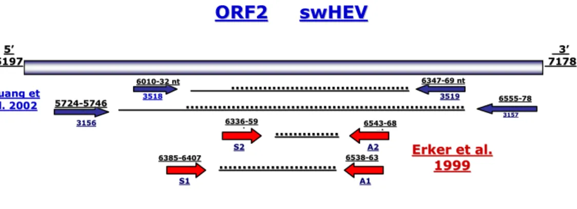
Qualitative PCR or conventional RT-PCR assays is mostly utilized in direct diagnosis of HEV. The samples collected may be faeces, serum, from animal or human, cultures of infected cells cultivated in 2D and 3D configurations, or necroscopic tissue highly positive as bile and liver (Panda et al., 2007; de Deus et al., 2007). HEV is an RNA virus and needs to be extracted before being subjected to retrotranscription reaction phase to cDNA. This is a limiting step, because cDNA it easily degradable, if in the samples the viral load is so low at initial state, may give rise at the end to false negativity. Various sets of sense and antisense synthetic oligonucleotide primers may be used for the detection of HEV genome, differing based on conservative region target in the genome against middle or terminal part of ORF1, C terminal of ORF2 (Panda et al., 2007). There are reports which indicate universal degenerate primers (Erker et al., 1999a; Inoue et al., 2006b), for identifying positives samples even though the strains belong to the different genotype. In this study e.g. has been used then following primers A1/S1 (Erker et al., 1999a) and 3156/7 primers (Huang et al, 2002) used to amplify the ORF2 region. Most of all the time the first product of PCR amplification it is not sufficient quantity to be visualized on electrophoresis gel. However, if the first product of PCR has been amplified by nested- PCR with internal primers: A2S2 (Erker et al., 1999a; Di Bartolo et al., 2008) and 3158/9 (Huang et al, 2002), respectively, the PCR product became clearly visible on the electrophoresis gel through ethidium bromide dye. This absorbs UV light intercalating into DNA and makes it fluoresce orange when visualized under UV