Molecular Neurobiology
11β-HSD1 inhibition rescues SAMP8 cognitive impairment induced by metabolic stress
--Manuscript
Draft--Manuscript Number: MOLN-D-19-00481R1 Article Type: Original Article
Keywords: Glucocorticoids, cognition, neurodegeneration, high-fat diet, neuroinflammation, oxidative stress, endoplasmic reticulum stress, ageing, Alzheimer's disease Corresponding Author: Merce Pallàs
University of Barcelona
BARCELONA, BARCELONA SPAIN First Author: Dolors Puigoriol-Illamola
Order of Authors: Dolors Puigoriol-Illamola Rosana Leiva
Manuel Vazquez-Carrera Santiago Vazquez Christian Griñan-Ferré Merce Pallàs
Abstract: Aging and obesity have been shown to increase the risk of cognitive decline and Alzheimer’s disease (AD). Besides, elevated glucocorticoids (GCs) levels cause metabolic stress and have been associated with the neurodegenerative process. Direct pieces of evidence link the reduction of GCs caused by the inhibition of 11β-HSD type 1 (11β-HSD1) with cognitive improvement.
In the present study, we investigated the beneficial effects of 11β-HSD1 inhibitor (i) RL-118 after high-fat diet (HFD) treatment in the senescence-accelerated mouse prone 8 (SAMP8). We found an improvement in glucose intolerance induced by HFD in mice treated with RL-118, a significant reduction in 11β-HSD1 and glucocorticoid receptor (GR) protein levels. Furthermore, specific modifications in the FGF21 activation after treatment with 11β-HSD1i, RL-118, which induced changes in SIRT1/PGC1α/AMPKα pathway, were found. Oxidative stress (OS) and reactive oxygen species (ROS), as well as inflammatory markers and microglial activation, were significantly diminished in HFD mice treated with 11β-HSD1i. Remarkably, treatment with 11β-HSD1i altered PERK pathway in both diet groups, increasing autophagy only in HFD mice group. After RL-118 treatment, a decrease in glycogen synthase kinase 3 (GSK3β) activation, Tau hyperphosphorylation, BACE1 protein levels and the product β-CTF were found. Increases in the non-amyloidogenic secretase ADAM10 protein levels and the product sAPPα were found in both treated mice, regardless of the diet. Consequently,
beneficial effects on social behaviour and cognitive performance were found in treated mice. Thus, our results support the therapeutic strategy of selective 11β-HSD1i for the treatment of age-related cognitive decline and AD.
Mercè Pallàs LLiberia
Departament de Farmacologia, Toxicologia i Quimica Terapèutica
Secció de Farmacologia
Facultat de Farmàcia i Ciències de l’Alimentació Av. Joan XXIII, 27-31 08028 Barcelona Tel. +34 934 024 531 [email protected]
http://www.ub.edu/farmaco/
30 June 2019
Chief Editor, Molecular Neurobiology
Title: “11β-HSD1 inhibition rescues SAMP8 cognitive impairment induced by
metabolic stress”
Authors:, Dolors Puigoriol-Illamola, Rosana Leiva, Manuel Vázquez-Carrera, Santiago Vázquez, Christian Griñán-Ferré and Mercè Pallàs
Dear Editor,
Please find attached our revised manuscript that we wish to be considered for publication as an Article in Molecular Neurobiology.
We would like to thank the reviewers for their valuable comments and helpful suggestions for the improvement of our research paper #MOLN-D-19-00481, "11β-HSD1 inhibition rescues SAMP8 cognitive impairment induced by metabolic stress".
We have carefully incorporated most of the reviewers’ suggestions. We hope that we have addressed all the concerns and questions of the reviewers and that this manuscript may now be suitable for publication.
Sincerely,
Dr Mercè Pallàs
Pharmacology professor at the University of Barcelona
Cover Letter Click here to access/download;Cover Letter;Revision_letter.docx
Mercè Pallàs LLiberia
Departament de Farmacologia, Toxicologia i Quimica Terapèutica
Secció de Farmacologia
Facultat de Farmàcia i Ciències de l’Alimentació Av. Joan XXIII, 27-31 08028 Barcelona Tel. +34 934 024 531 [email protected]
http://www.ub.edu/farmaco/
30 June 2019
Chief Editor, Molecular Neurobiology
Title: “11β-HSD1 inhibition rescues SAMP8 cognitive impairment induced by
metabolic stress
”
Authors:, Dolors Puigoriol-Illamola, Rosana Leiva, Manuel Vázquez-Carrera, Santiago Vázquez, Christian Griñán-Ferré and Mercè Pallàs
Dear Editor,
Please find attached our revised manuscript that we wish to be considered for publication as an Article in Molecular Neurobiology.
We would like to thank the reviewers for their valuable comments and helpful suggestions for the improvement of our research paper #MOLN-D-19-00481, "11β-HSD1 inhibition rescues SAMP8 cognitive impairment induced by metabolic stress".
We have carefully incorporated most of the reviewers’ suggestions, and by answered point by point in the text at the end of this document. We hope that we have addressed all the concerns and questions of the reviewers and that this manuscript may now be suitable for publication.
Sincerely,
Dr Mercè Pallàs
Pharmacology professor at the University of Barcelona
Reviewer #2: The manuscript entitled "11β-HSD1 inhibition rescues SAMP8 cognitive impairment induced by metabolic stress" by
Illamola et al is well-planned, nicely written, novel with its results showing the inhibition of 11β-HSD type 1 reduces neuro-inflammation and improve cognition in senescence mice.
Please address following questions:
To the reviewer #2:
We thank the reviewer for the relevant suggestions, which helped us to improve our manuscript.
Question 1. Please describe how the dose of RL-118 was selected and reason behind this time frame, provide references.
Answer: We thank the reviewer for the relevant question. The dose was selected according to another paper published in 2017 in which RL-118 at 21mg/kg produced changes in different oxidative stress and inflammation markers. There it is described as: concentration of 105 mg l-1 (average body weight for 48-week-old mice is 25g; fluid consumption is 5mL, therefore the dose was 0.105 mg/ml x 5 ml/0.025 kg= 21 mg/kg (Leiva et al., 2017).
Question 2. In TCT behavioural paradigm, please define the 'interaction with intruder'.
Answer: We thank the reviewer for this comment. The Interaction with the intruder is the measure of the time spent in social investigation by pairs (File and Hyde 1978). The pairs of mice are placed in the TCT, and the time that they spend in active social interaction (e.g., sniffing, grooming) is measured. When the cognitive impairment or aggressive behaviour appears in mice, the overall level of social interaction is suppressed, as you can see in SAMP8 Control mice group. On the other hand, when the SAMP8 was treated with RL-118, the social interaction was increased.
Question 3. Was any mouse excluded from NORT test? If yes, what was the exclusion criteria? Does any of the mouse spent more time on either of the identical objects in first trial.
Answer: We thank the reviewer for this question. In this study, no mouse was excluded from the NORT. However, in order to clarify our experimental procedure, we have two exclusion criteria. The first one is that the mouse explores both objects and the second one, is related to statistical analysis. We use Grubb's test to eliminate outliers.
Regarding the time spent in each identical object during the familiarization phase (first trial), there were no differences in the time they explore each of them. Otherwise, we should have shown the results of the familiarization phase (first trial) and explain the results in a different way. Importantly, to avoid object preference biases, objects were counterbalanced, not only in the test phase but also in the familiarization phase.
Question 4. Fig. 7G, negative discrimination index of ND Ct mice seems unrealistic. Is this common in this mice line? Please clarify!
Answer: We thank the reviewer for noticing this. However, this is a common result, due to the discrimination index is a ratio between time spent in the novel object minus time spent in the old object, divided between the sum of both. This implies that the range of the DI is -1 to 1. This implies that the range of the discrimination index is -1 to 1, and whenever the mouse scans the old object longer than the new the ID will be negative. Furthermore, our group has previously described SAMP8 mice and compared to its control (SAMR1), as well as, we have performed NORT in several studies. So, we have a broad knowledge of NORT test in these mice under different conditions (Griñán-Ferré et al., 2016; Palomera et al., 2016; Leiva et al., 2017). Also, the same is described in other groups (Fujiwara et al., 2018; Dobarro et al., 2013).
Question 5. In Fig. 4C, the images are not presented uniformly, this is not publication quality! Please select images from same brain region in each
group. Even the orientation of images are not uniform. Also, outline the area, where cells were counted.
Answer: We thank the reviewer for noticing this. We agree with the reviewer that there were mistakes in the images. Accordingly, we have replaced five of them and put in the same orientation. Furthermore, we have revised the intensity and quantification of all of them, and we have corrected. On the other hand, regarding the quantification, we do not count cells; we quantify the average intensity of the IHQ through ImageJ/Fiji software, with different corrections such as average darkness, threshold colour and the areas that display the tissue. Therefore, is a semi-automatic analysis that allows us to replicate the evaluation, really robust and widely used.
Reviewer #3: The authors present very valuable work concerning role of RL- 118 inhibitor in Alzheimer's disease. Notably, the results were obtained from in vivo - animal model. Manuscript is very well written. The results are discussed extensively. However, the quality of images should be increased. My additional comment is to place the values of densitometric analysis above western blots.
To the reviewer #3:
Answer: We thank the reviewer for highlighting this. According to the reviewer comment, we have revised exhaustively all western blot figures and we believe that images are with good quality and representative of the results, even so, in order to demonstrate our analysis. We annex here additional information about densitometric analysis of the representative western blots presented in the manuscript to suppor the answer.We hope this point is clearer now.
11β-HSD1 inhibition rescues SAMP8 cognitive impairment induced by metabolic stress
Dolors Puigoriol-Illamola1,3, Rosana Leiva2,4, Manel Vázquez-Carrera1,4,5,6, Santiago
Vázquez2,4, Christian Griñán-Ferré1,3* and Mercè Pallàs1,3*.
1Pharmacology Section, Department of Pharmacology, Toxicology and Therapeutic Chemistry. Faculty of
Pharmacy and Food Sciences. University of Barcelona, Av Joan XXIII 27-31, 08028 Barcelona, Spain.
2Laboratori de Química Farmacèutica (Unitat Associada al CSIC), Department de Farmacologia, Toxicologia
i Química Terapèutica, Facultat de Farmàcia i Ciències de l’Alimentació, and Institute of Biomedicine (IBUB), Universitat de Barcelona, Av. Joan XXIII, 27-31, E-08028 Barcelona, Spain.
3Institute of Neuroscience, University of Barcelona (NeuroUB), Barcelona, Spain. 4Institute of Biomedicine, University of Barcelona (IBUB), Barcelona, Spain.
5Spanish Biomedical Research Center in Diabetes and Associated Metabolic Diseases
(CIBERDEM)-Instituto de Salud Carlos III.
6Pediatric Research Institute-Hospital Sant Joan de Déu, Esplugues de Llobregat, Spain
*These authors contributed equally
Corresponding author: Mercè Pallàs, PhD.
Pharmacology Section in Pharmacology, Toxicology, and Therapeutic Chemistry Department, Faculty of Pharmacy and Food Sciences, University of Barcelona, Av Joan XXIII 27-31, 08028 Barcelona, Spain.
e-mail: [email protected]
Keywords: Glucocorticoids, cognition, neurodegeneration, high-fat diet, neuroinflammation, oxidative stress, endoplasmic reticulum stress, ageing, Alzheimer’s disease
Manuscript Click here to access/download;Manuscript;Puigoriol-illamola.V3.docx
Abstract
Aging and obesity have been shown to increase the risk of cognitive decline and Alzheimer’s disease (AD). Besides, elevated glucocorticoids (GCs) levels cause metabolic stress and have been associated with the neurodegenerative process. Direct pieces of evidence link the reduction of GCs caused by the inhibition of 11β-HSD type 1 (11β-HSD1) with cognitive improvement.
In the present study, we investigated the beneficial effects of 11β-HSD1 inhibitor (i) RL-118 after high-fat diet (HFD) treatment in the senescence-accelerated mouse prone 8 (SAMP8). We found an improvement in glucose intolerance induced by HFD in mice treated with RL-118, a significant reduction in 11β-HSD1 and glucocorticoid receptor (GR) protein levels. Furthermore, specific modifications in the FGF21 activation after treatment with 11β-HSD1i, RL-118, which induced changes in SIRT1/PGC1α/AMPKα pathway, were found. Oxidative stress (OS) and reactive oxygen species (ROS), as well as inflammatory markers and microglial activation, were significantly diminished in HFD mice treated with 11β-HSD1i. Remarkably, treatment with 11β-HSD1i altered PERK pathway in both diet groups, increasing autophagy only in HFD mice group.
After RL-118 treatment, a decrease in glycogen synthase kinase 3 (GSK3β) activation, Tau hyperphosphorylation, BACE1 protein levels and the product β-CTF were found. Increases in the non-amyloidogenic secretase ADAM10 protein levels and the product sAPPα were found in both treated mice, regardless of the diet. Consequently, beneficial effects on social behaviour and cognitive performance were found in treated mice. Thus, our results support the therapeutic strategy of selective 11β-HSD1i for the treatment of age-related cognitive decline and AD.
Aging is the most significant risk factor for a majority of chronic diseases, such as age-related cognitive decline and Alzheimer’s disease (AD) [1]. Obesity has recently been considered to play an essential role in mild cognitive impairment (MCI) and dementia [2,3]. Abundant pieces of evidence suggest that pathological glucocorticoids (GCs) excess not only is associated with age-related cognitive decline but also with metabolic stress, obesity, and diabetes [4-7]. Thus, it has been described that prolonged exposure to increased levels of GCs exerts deleterious effects on hippocampal electrophysiology, structure and function, both in rodents [8] and humans [9], being part of altered intercellular communication, considered one of the nine hallmarks of aging [10,11]. The availability of natural GCs in tissues is regulated by corticosteroid-binding globulin in serum and by locally expressed 11β-hydroxysteroid dehydrogenase (11β-HSD) enzyme, a microsomal enzyme that catalyses the interconversion of active GCs (corticosterone in rodents but cortisol in humans) and inert 11-keto-forms [11-dehydrocorticosterone (11-DHC), cortisone] [12]. Two isoenzymes have been identified: 11β-HSD type 1 (11β-HSD1) and type 2 (11β-HSD2) [13]. 11β-HSD1 acts as a reductase, thus locally potentiating GCs activity, and is the predominant form in the brain, both in rodents and humans. 11β-HSD1 expression in mouse hippocampus and parietal cortex increases with aging, correlating with impaired spatial memory [14] and its overexpression accelerates related cognitive decline [6]. Conversely, 11β-HSD1 knockout mice resist age-dependent cognitive loss [15, 16]. In line with these findings, some preclinical studies demonstrate that 11β-HSD1 inhibition improved cognition and AD hallmarks suggesting a neuroprotective effect [17-18]. On the other hand, preclinical studies have demonstrated that sub-maximal inhibition of central 11β-HSD1 is able to prevent cognitive impairments in AD and ageing [19,20] and early preclinical studies demonstrated that administration of brain penetrant 11β-HSD1 (UE2343) is well tolerated [21]. Besides, many selective 11β-HSD1 inhibitors have reached clinical stages for metabolic diseases like type 2 diabetes mellitus (e.g., AZD8329, ABT-384, and BVT.2733).
In addition, several hypotheses argument a link between altered glucose metabolism and dementia, considering an altered metabolic pathway as a potential contributor to
persistent oxidative stress (OS) that culminates into neuronal dysfunction and dementia [22]. Increased OS, neuroinflammation, endoplasmic reticulum (ER) stress, misfolded proteins removal pathways, and autophagy, have been identified as components of neuronal metabolic stress, thus developing and aggravating neurological disorders and cognitive impairment, pointing to their implication in the dysregulated energy metabolism characteristic of ageing [23]. Thus, recent studies have focused on the pathology of cognitive decline and neurodegeneration through high-fat diet (HFD) intervention that leads to metabolic stress [24,25]. For instance, HFD-fed aged animals showed insulin resistance and increased weight gain among others, both signs of pre-diabetes, obesity, cardiovascular disease, depressive-like behaviour and mental health problems [26-28]. However, the current knowledge about the molecular mechanism responsible for these affections is controversial, even though most of the information is related to reduced insulin sensitivity mediated by HFD. Hence, HFD-induced metabolic stress could be linked with the development of physiopathological conditions, such as AD and other neurodegenerative diseases [29].
Recently, we have developed a brain penetrant 11β-HSD1 inhibitor (RL-118) that was characterized chemically and pharmacologically in vivo [30]. RL-118 attenuates neuroinflammation, increases the antioxidant defence, promotes autophagy, improves mitochondrial function and reverts memory deficits in senescent mice model [17,30]. Therefore, the targeted inhibition of 11β-HSD1 may become a potential therapeutic strategy for age-related cognitive decline and AD. Here, we assessed the neuroprotective effects of 11β-HSD1 inhibition on metabolic stress through behaviour, cognitive and molecular changes induced by HFD in a mice model of age-related cognitive decline and late-onset AD (LOAD), the senescence-accelerated mouse prone 8 (SAMP8).
2. Materials and methods 2.1. Animals
Females SAMP8 mice (n=48) were used to carry out behavioural, cognitive and molecular analyses. We divided these animals into four groups: normal diet chow (ND Ct, n=12), ND treated with the RL-118 11β-HSD1 inhibitor (ND+11β-HSD1i, n=12), HFD
(HFD Ct, n=12) and HFD treated with RL-118 (HFD+11β-HSD1i, n=12). Animals had free access to food and water and were kept under standard temperature conditions (22±2°C) and 12h: 12h light-dark cycles (300lux/0 lux). Animals were fed with both diets since the weaning (1-month-old) up to sacrifice. RL-118 was administered at 21 (mg/kg/day) by oral gavage from 4 months old to end of behavioural test (Figure 1A). ND provided 3,8 Kcal/g meanwhile HFD 4,7 Kcal/g – 45% fatty acids (D12451 Research Diets, Inc.). The weight of the animals and the ingested food were monitored weekly. Before the performance of the cognitive tests, the glucose tolerance test was conducted.
Studies and procedures involving mice brain dissection and subcellular fractionation were performed following the institutional guidelines for the care and use of laboratory animals established by the Ethical Committee for Animal Experimentation at the University of Barcelona.
2.2. Glucocorticoid, glucose tolerance test and triglyceride determination
Plasma and brain corticosterone concentrations were measured using a commercially available RIA (MP Biomedicals, Irvine, CA). Blood extraction and brain dissection were done among 4 pm and 5 pm in ND+11β-HSD1i and ND Ct mice.
Intraperitoneal (i.p.) glucose tolerance test was performed following 12 weeks of HFD feeding and 4 weeks of 11β-HSD1i/vehicle treatments, as described previously. In brief, mice fasted overnight for 12 hours. The test was performed in a quiet room and 2g/kg i.p. glucose injection was administered (diluted in H2O) and blood glucose levels were
measured at 0, 5, 10, 15, 30, 60 and 120 minutes after the injection with the Accu-Chek® Aviva blood glucose meter (Accu-Chek® Aviva, Roche, Barcelona, Spain). The determination of triglyceride concentration was performed by using a triglyceride meter device (Accutrend® Plus, Cobas, Roche).
2.3. Behavioural and cognitive test 2.3.1. Three-Chamber test (TCT)
The Three-Chamber test assesses cognition through sociability and interest in social novelty [31]. Testing occurs in a box with three equally dimensioned rooms. Each test consists of 20 minutes and is recorded with a camera. The animal is placed in the center of the box and allowed to explore the three chambers for 10 minutes (Habituation
phase). The time spent in each chamber was evaluated. Afterwards, an intruder (same sex and age) was added to one of the rooms in a metal cage and behaviour is recorded for 10 minutes. In this phase, the time spent in each room is assessed as well as the time interacting with the intruder (e.g., sniffing, grooming).
2.3.2. Morris Water Maze (MWM)
This test evaluates both learning and spatial memory [32]. An open circular pool (100 cm in diameter, 50 cm in height) filled with water was used. Water was painted white with latex in order to make it opaque and its temperature was 22± 1°C. Two main perpendicular axes were established (North-South and East-West), thus configuring four equal quadrants (NE, NW, SE, and SW). Four visual clues (N, S, E, W) were placed on the walls of the tank so that the animal could orientate and could fulfil the objective. The test consists of training a mouse to find a submerged platform (Learning phase) and assess whether the animal has learned and remembered where was the platform the day that it is removed (Test). The training lasts five consecutive days and every day five trials are performed, which have a different starting point (NE, E, SE, S, and SW), with the aim that the animal recognizes the visual clues and learns how to locate the platform, avoiding learning the same path. At each trial, the mouse was placed gently into the water, facing the wall of the pool, allowed to swim for 60 seconds and there was not a resting time between trials. If the animal was not able to locate the platform, the investigator guided it to the platform and was allowed to rest and orientate for 30 seconds. The platform was placed approximately in the middle of one of the quadrants, 1.5 cm below the water level. Above the pool there was a camera that recorded the animals’ swimming paths and the data was analysed with the statistical program SMART® ver.3.0. During the learning phase, a learning curve was drawn, in which is represented the latency to find the platform every training day. On the day test, more parameters were measured, such as the target crossings and the swum distance in the platform zone.
2.3.3. Novel Object Recognition Test (NORT)
The Novel Object Recognition Test (NORT) protocol employed was a modification of [33]. In brief, mice were placed in a 90°, two-arms, 25-cm-long, 20-cm-high, 5-cm-wide black maze. Before performing the test, the mice were individually habituated to the
apparatus for 10 min for 3 days. On day 4, the animals were submitted to a 10-min acquisition trial (first trial), during which they were placed in the maze in the presence of two identical, novel objects at the end of each arm. After a delay (2h and 24h), the animal was exposed to two objects one old object and one novel object. The Time that mice explored the Novel object (TN) and Time that mice explored the Old object (TO) were measured. A Discrimination Index (DI) was defined as (TN−TO)/(TN+TO). To avoid object preference biases, objects were counterbalanced. The maze, the surface, and the objects were cleaned with 70% ethanol between the animals’ trials to eliminate olfactory cues.
2.4. Immunodetection experiments 2.4.1. Brain processing
Three days after the behavioural and cognitive tests, 8 animals per group were euthanized for protein extraction, RNA and DNA isolation, and 4 animals per group were euthanized for immunohistochemistry (IHQ).
When the animals were for protein extraction, RNA and DNA isolation, brains were immediately removed and the hippocampus was isolated, frozen on powdered dry ice and maintained at -80°C until procedures. When the animals were for IHQ, mice were intracardially perfused with 4 % paraformaldehyde (PFA) diluted in 0.1M phosphate buffer solution after being anesthetized by intraperitoneal injection of ketamine 100 mg/Kg and xylazine 10 mg/Kg. Afterwards, brains were removed and post-fixed in 4% PFA overnight 4oC then the solution was changed into PFA + 15% sucrose. Finally, brains
were frozen burying directly on powdered dry ice (around 5 minutes) and store at -80°C until sectioned.
2.4.2. Western blotting
Tissue samples were homogenized in lysis buffer (Tris HCl pH 7.4 50mM, NaCl 150mM, EDTA 5mM and 1X-Triton X-100) containing phosphatase and protease inhibitors (Cocktail II, Sigma-Aldrich) to obtain total protein homogenates.
For Western Blotting (WB), aliquots of 15 μg of hippocampal protein extraction per sample were used. Protein samples were separated by Sodium dodecyl sulphate-polyacrylamide gel electrophoresis (SDS-PAGE) (8-14%) and transferred onto Polyvinylidene difluoride (PVDF) membranes (Millipore). Afterwards, membranes were blocked in 5% non-fat milk in Tris-buffered saline (TBS) solution containing 0.1% Tween
20 TBS (TBS-T) for 1 hour at room temperature, followed by overnight incubation at a 4°C with the primary antibodies listed in (Supplementary Table 1). Then, the membranes were washed and incubated with secondary antibodies listed in (Table1) for 1 hour at room temperature. Immunoreactive proteins were viewed with the chemiluminescence-based ChemiLucentTM detection kit, following the manufacturer’s
protocol (ECL Kit, Millipore), and digital images were acquired using ChemiDoc XRS+System (BioRad). Semi-quantitative analyses were done using ImageLab software (BioRad) and results were expressed in Arbitrary Units (AU), considering control protein levels as 100%. Protein loading was routinely monitored by immunodetection of Glyceraldehyde-3-phosphate dehydrogenase (GAPDH) or β-tubulin.
2.4.3. Immunofluorescence
Coronal section of 30 μm was obtained by a cryostat (Leica Microsystems CM 3050S, Wetzlar, Germany) and kept in a cryoprotectant solution at -20°C.
First, free-floating slices were selected and placed on a 24-wells plaque. After that, were washed five times with PBS 0.01M + 1% Triton X-100. Then, free-floating sections were blocked with a solution containing 5% fetal bovine serum (FBS), 1% Triton X-100, PBS 0.01M + gelatine 0.2% for 2h at room temperature. Afterwards, slices were washed with PBST (PBS 0.1M, 1% Triton X-100) five times for 5 min each and were incubated with the primary antibodies over-night at 4°C (Supplementary Table 2). On the following day, coronal slices were washed with PBST 6 times for 5 min and then incubated with the secondary antibodies (Supplementary Table 2) at room temperature for 2h. Later, sections were co-incubated with,1mg/ml DAPI staining solution (Sigma-Aldrich, St. Louis, MI) for 5 min in the dark at room temperature and washed with PBS 0.01M. Finally, the slices were mounted using Fluoromount G (EMS, USA) and image acquisition was performed with a fluorescence laser microscope (Olympus BX41, Germany). At least four images from 4 different individuals by the group were analysed with ImageJ/Fiji software available online from the National Institutes of Health.
2.5. RNA extraction and gene expression determination by q-PCR
Total RNA isolation was carried out using TRIsureTM reagent according to the
manufacturer’s instructions (Bioline Reagent, UK). The yield, purity, and quality of RNA were determined spectrophotometrically with a NanoDrop™ ND-1000 (Thermo
with 260/280 ratios and RIN higher than 1.9 and 7.5, respectively, were selected. Reverse Transcription-Polymerase Chain Reaction (RT-PCR) was performed as follows: 2 μg of messenger RNA (mRNA) was reverse-transcribed using the High Capacity cDNA Reverse Transcription Kit (Applied Biosystems). Real-time quantitative PCR (qPCR) was used to quantify mRNA expression of oxidative stress and inflammatory genes listed in (Supplementary Table 3). SYBR® Green real-time PCR was performed in a Step One Plus
Detection System (Applied-Biosystems) employing SYBR® Green PCR Master Mix
(Applied-Biosystems). Each reaction mixture contained 6.75 μL of complementary DNA (cDNA) (which concentration was 2 μg), 0.75 μL of each primer (which concentration was 100 nM), and 6.75 μL of SYBR® Green PCR Master Mix (2X).
Data were analyzed utilizing the comparative Cycle threshold (Ct) method (ΔΔCt), where the housekeeping gene level was used to normalize differences in sample loading and preparation [31]. Normalization of expression levels was performed with β-actin for SYBR® Green-based real-time PCR results. Each sample was analyzed in duplicate, and the results represent the n-fold difference of the transcript levels among different groups.
2.6. Oxidative stress determination
Hydrogen peroxide was measured in hippocampus protein homogenates as an indicator of oxidative stress, and it was quantified using the Hydrogen Peroxide Assay Kit (Sigma-Aldrich, St. Louis, MI) according to the manufacturer’s instructions.
2.7. Data analysis
Data analysis was conducted using GraphPad Prism ver. 7 statistical software. Data are expressed as the mean ± standard error of the mean (SEM) of at least 6 samples per group. Diet and treatment effects were assessed by the Two-Way ANOVA analysis of variance, followed by Tukey post-hoc analysis or two-tail Student’s t-test when it was necessary. Statistical significance was considered when p-values were <0.05. The statistical outliers were determined with Grubbs’ test and subsequently removed from the analysis.
3. Results
3.1. Treatment with 11β-HSD1i decreases GC levels and Improves Glucose Intolerance Induced by HFD
As expected, corticosterone levels were significantly reduced in blood and brain tissue after 11β-HSD1i treatment (Figure 1B-C). Body weight was measured weekly during the intervention. All animal groups significantly increased body weight over time (Figure 1D). Furthermore, both HFD-fed mice groups exhibited increased in body weight from 12 weeks to the end time point (Figure 1D), correlating with the higher caloric intake (Figure 1E). However, 11β-HSD1i treatment did not significantly modify those parameters. Likewise, significant higher glucose levels between 15 and 90 min were found in HFD Ct (Figure 1F). Noteworthy, 11β-HSD1i treatment reduced significantly glucose levels in HFD fed mice (Figure 1F). On the other hand, triglyceride plasma concentration was higher in HFD mice groups but did not differ from 11β-HSD1i treated groups (Figure 1G). In conjunction, 11β-HSD1i ameliorated glucose metabolism only after metabolic stress induced by HDF feeding.
Finally, WB analysis revealed a significant reduction in 11β-HSD1 protein levels in treated mice, both in ND and HFD fed mice (Figure 1H). Additionally, the inhibition of 11β-HSD1 diminished GR protein levels significantly regardless of the diet (Figure 1I).
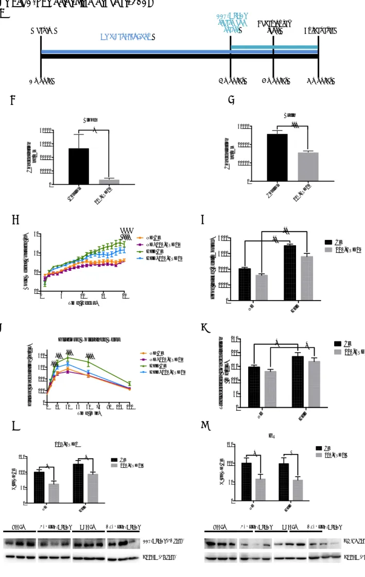
3.2. Restoration of FGF21 levels after treatment with 11β-HSD1i is accompanied by changes in the nutrient Sensor SIRT1/PGC1α/AMPKα pathway
Fibroblast growth factor 21 (FGF21) and SIRT1/PGC1α/AMPKα axis were evaluated by immunoblot. A significant reduction in FGF21 in HFD Ct group compared to the ND Ct group (Figure 2A) was observed, demonstrating that HFD reduced FGF21 protein levels. Of note, a significant increase in FGF21 was found in the HFD+11β-HSD1i group in comparison with the HFD Ct group (Figure 2A). No significant changes in FGF21 protein levels were found in ND+11β-HSD1i compared to ND Ct group, although there was a slight increase. Likewise, analysis of the nutrient sensor axis showed a significant reduction in SIRT1 protein levels in the HFD group, but RL-118 was unable to prevent it. Conversely, ND+11β-HSD1i showed a significant increase in SIRT1 compared to ND Ct group (Figure 2B). Importantly, liver kinase B1 (LKB1) was activated in 11β-HSD1i treated groups compared to the control mice regardless of the diet (Figure 2C). A significant increase in the phosphorylated activated protein kinase (p-AMPKα) ratio levels in the HFD+11β-HSD1i group was observed compared to the HFD Ct mice (Figure 2D). Finally, peroxisome proliferator-activated receptor-gamma coactivator 1-α (PGC1α) protein
levels were increased in a significant way only in the ND+11β-HSD1i compared to the ND Ct group, but not to HFD groups (Figure 2E).
By contrast, 11β-HSD1 pharmacological inhibition through RL-118 was only able to increase SIRT1, LKB1, and PGC1α in ND-fed animals. Albeit RL-118 did not cause apparent changes in SIRT1 and PGC1α in HFD-fed mice, the increase in AMPKα phosphorylation indicated that improvements in nutrient sensing and mitochondrial function after 11β-HSD1 inhibition also occurs in HFD. In whole results, our findings pinpoint the beneficial effect of reducing GC signalling by 11β-HSD1 inhibition in SAMP8 under metabolic stress.
3.3. Treatment with 11β-HSD1i Reduced OS Markers
HFD induced a significant increase in GPX1 and a moderate increase in SOD1 protein levels that were prevented by RL-118 treatment (Figure 3A-B). In addition, iNOS gene expression was reduced in 11β-HSD1i treated animals in comparison with the control groups that reached significance in HFD Ct mice (Figure 3C). Likewise, analysis of hydrogen peroxide levels showed that 11β-HSD1i can reduce although in a not significant way (Figure 3D).
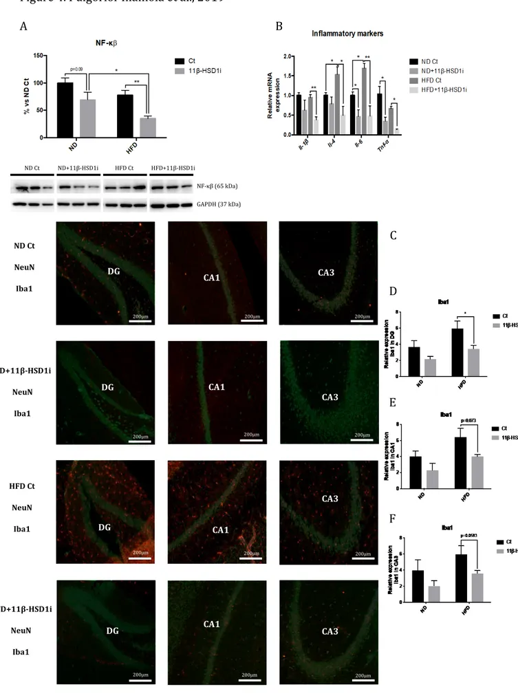
3.4. Reduction of Inflammatory Markers and Microglial Activation after Treatment with 11β-HSD1i
HFD did not modify NF-κβ protein levels. However, 11β-HSD1i treatment induced a significant diminution in NF-κβ protein levels both in ND and HFD mice (Figure 4A).
Il-1β, Il-4, Il-6, and Tnf-α gene expression was reduced after 11β-HSD1i treatment, being
significant in Il-6, and Tnf-α, regardless of the diet (Figure 4B). By last, immunostaining quantification of Iba1 fluorescence intensity demonstrated that 11β-HSD1i treatment reduced Iba1 staining, especially in the dentate gyrus (DG) and CA1 regions (Figures 4C-F).
3.5. Treatment with 11β-HSD1i Increased Autophagy through PERK pathway
Next, we evaluated the ER stress response. PERK pathway revealed changes in phosphorylated PKR-like endoplasmic reticulum kinase (p-PERK) and phosphorylated
eukaryotic translation-initiation factor 2 (p-eIF2α) activating transcription factor 4 (ATF4), but not in and C/EBP homologous protein (CHOP). All these protein levels were higher in 11β-HSD1i treated groups compared to its Ct group, regardless of the diet (Figure 5A-D). Moreover, the HFD Ct group showed less phosphorylated PERK and eIF2α protein levels compared to ND Ct (Figure 5A-B). Regarding BCL-2 protein levels, no changes were found among groups (Figure 5E). However, Beclin 1 protein levels were slightly increased in HFD+11β-HSD1i compared to the HFD Ct group (Figure 5F).
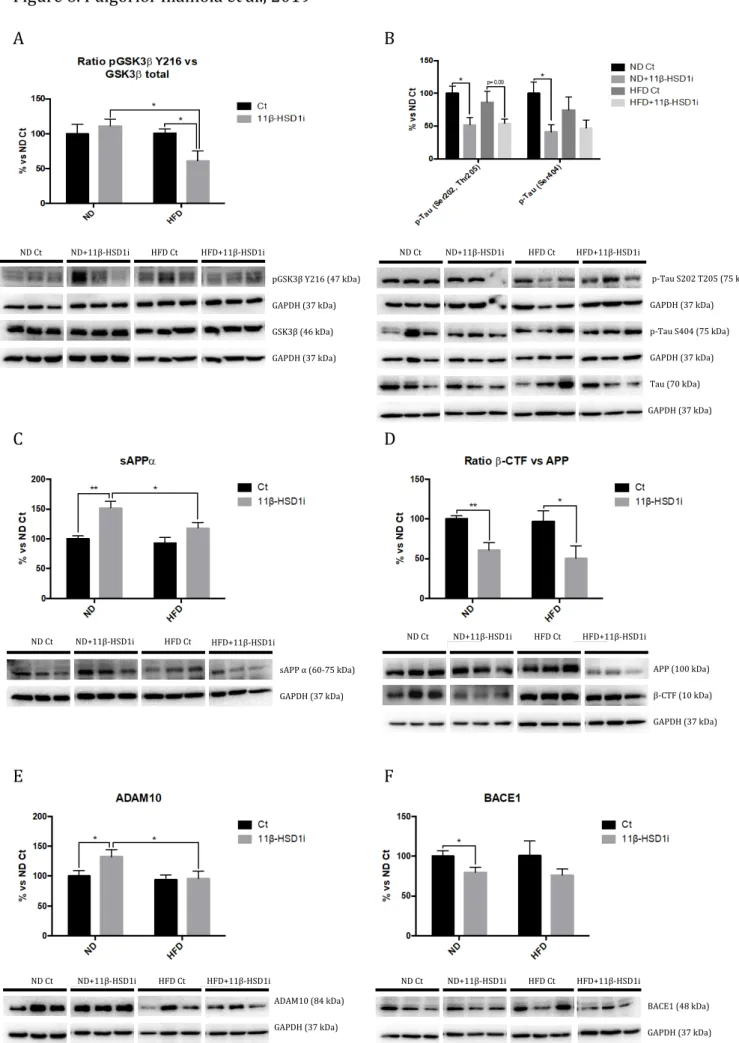
3.6. Reduction of AD Hallmarks after Treatment with 11β-HSD1i are Associated with Glycogen Synthase Kinase 3 Signalling Pathway Induced by HFD
HFD did not alter phosphorylation in Tyr217 of glycogen synthase kinase 3 beta (p-GSK3β) and Tau hyperphosphorylation. However, p-GSK3β (Tyr217) was significantly diminished in the HFD+11β-HSD1i group compared to HFD Ct (Figure 6A). In parallel, a reduction in p-Tau (Ser202, Thr205), as well as p-Tau (Ser404) protein levels after the 11β-HSD1i treatment were found in HFD Ct mice (Figure 6B).
HFD was unable to alter APP processing in SAMP8, neither soluble APP fragment alpha (sAPPα) nor the fragment delivered by β-secretase (β-CTF) protein levels were modified. 11β-HSD1i treatment caused a significant increase in sAPPα in the ND-fed group but not in the HFD-fed mice (Figure 6C). β-CTF protein levels were decreased after 11β-HSD1i regardless of the diet (Figure 6D). Finally, a significant increase in ADAM10 protein levels was only found in ND+11β-HSD1i group compared to the ND Ct group (Figure 6E), whereas a significant reduction in BACE1 protein levels was found in 11β-HSD1i treated animals, reaching significance in ND-fed mice (Figure 6F).
3.7. Beneficial Effects on Social Behaviour and Cognitive Performance after Treatment with 11β-HSD1i
Social behaviour was investigated by TCT. HFD did not alter the preference for a specific chamber during the habituation, neither the treatment with 11β-HSD1i (Figure 7A). Moreover, in all the experimental groups the presence of an intruder increase significantly the time spent in this chamber, regardless of diet or treatment (Figure 7B). Higher interaction in HFD+11β-HSD1i group compared to the HFD Ct group was found,
although no changes in the interaction between the resident and the intruder between ND groups occurred (Figure 7C).
Furthermore, cognitive performance was measured by the MWM and NORT tests. Regarding MWM, all mice groups were able to learn through the training period; no differences were found among groups (Figure 7D). On the test day, 11β-HSD1i treated mice increased the swim length in the platform zone compared to Ct animals indicating higher cognitive abilities (Figure 7F). Besides, the number of target crossings was significantly increased in HFD+11β-HSD1i mice in comparison with the ND Ct (Figure 7F).
Regarding NORT, there were no differences in the exploration time between identical
objects during acquisition trial (first trial). In test trial analysis 11β-HSD1i treated mice
exhibited a significant improvement in cognitive performance both in short-(2h) and long-(24h) term recognition memory in comparison with Ct groups, obtaining higher DI values (Figure 7G).
4. Discussion
In this study, we provide new evidence that inhibition of 11β-HSD1 improves cognitive impairment after metabolic stress induced by HFD intervention in a model of cognitive decline. To this end, various molecular pathways influenced by HFD and GCs, as well as social and cognitive impairment were evaluated to elucidate new mechanisms by which 11β-HSD1 inhibition exerts neuroprotection.
Our group has previously described that HFD induced metabolic stress in a murine model of accelerated senescence, SAMP8 [34] and in C57BL/6J aged mice [35]. Those metabolic disturbances were prevented by resveratrol [34, 35]. Moreover, in previous reports, the neuroprotective effect of 11β-HSD1 inhibitor (RL-118) was demonstrated in old female SAMP8 [17,30].
Animals fed with HFD increased body weight in comparison with ND fed mice showing an alteration in glucose metabolism. The hugest increase in animal weight is not only associated with impaired glucose tolerance but also with an alteration in lipid metabolism. Barroso and co-workers described that the excessive consumption of hypercaloric and high saturated fat food causes an increase in serum triglyceride levels,
which is the first step for the development of the atherogenic dyslipidaemia found in obese and diabetic patients [36]. Accordingly, our results showed higher triglyceride concentration in blood in HFD-fed groups.
As aforementioned, there is extensive evidence that HFD impairs glucose metabolism, altering the insulin signalling pathway [37-39]. Recently, a close relationship between insulin insensitivity and neurodegenerative disorders, such as AD, has been extensively reported [40,41] and it is still being discussed whether to consider AD as a type 3
Diabetes mellitus (DM3) [42]. Additionally, high GC levels can mediate insulin resistance
response, increasing glucose levels and favouring insulin resistance and obesity [43, 44]. Thus, selective inhibitors of 11β-HSD1 have been postulated as a neuroprotective strategy in several pathological scenarios [45]. RL-118 treatment reduced the 11β-HSD1 enzyme and GC receptor protein levels both in ND- and HFD-fed SAMP8. In concordance, GC levels were reduced both in blood and in plasma of RL-118 treated animals. Results are in line with reports describing a reduction in gene expression of 11β-HSD1 and GR in diet-induced obese mice after treatment with carbenoxolone, a 11β-HSD1 inhibitor [46].
Recently, FGF21 has been demonstrated to modulate energy homeostasis of glucose and lipid through activation of SIRT1/PGC1α/AMPKα axis, mainly through LKB1 activation [47]. Consistent with this hypothesis, treatment with 11β-HSD1 inhibitor significantly increased protein levels of FGF21 and LKB1 under both dietary conditions. Albeit RL-118 did not induce direct changes in SIRT1 and PGC1α protein levels in HFD fed SAMP8, the increase in AMPKα phosphorylation suggests an improvement in nutrient sensing and mitochondrial function. In sum, these results demonstrate the beneficial effects of reducing GC signalling by 11β-HSD1 inhibition in SAMP8 under HFD induced metabolic stress.
It has been reported that HFD increases OS and inflammation [48]. Accordingly, OS markers, such as GPX1, SOD1 and iNOS were increased in HFD treated groups, and 11β-HSD1i treatment was only able to reduce them when animals were fed with HFD. Whereas a clear tendency to reduce hippocampal ROS levels in all 11β-HSD1i treated
animals was found. Regarding neuroinflammation, Il-6 and Il-4 gene expression and microglial activation evaluated through Iba-1, increased under HFD confirming the cellular dysfunction induced by impaired energy metabolism. Importantly, 11β-HSD1 inhibition diminished gene expression of both Tnf-α and Il-6, as well as p65 protein levels, inhibiting microglial reactivity. These findings agree with already published evidence describing that 11β-HSD1 inhibition modulates OS and inflammatory processes [17,30,49].
11β-HSD1 inhibition by RL-118 reduced autophagy and apoptosis markers in old SAMP8 [17], but in the present work, we did not observe significant changes in Beclin 1 or BCL2, probably because of the young age of mice. Because ER stress response activates proteostatic mechanisms [50,51], ER stress markers were studied. While HFD did not modify ATF4 protein levels in a significant way, PERK and eIF2α activation were significantly reduced in those animals. Of interest, inhibition of 11β-HSD1 by RL-118 recovered the ratio of phosphorylation of PERK and eIF2α, and in addition, increased ATF4 protein levels. By contrast, in our hands, CHOP showed a narrow but not significant increase after RL-118 treatment [52]. Regarding the accumulation of misfolded proteins, a characteristic of ageing and AD, 11β-HSD1 inhibition reduced GSK3β activation and tau phosphorylation after HFD feeding. Regarding the β-amyloid pathway, RL-118 promoted APP processing by the non-amyloidogenic pathway, decreasing pro-amyloidogenic APP fragments, as well as reducing BACE1 and increasing ADAM10 protein levels. Results suggest that 11β-HSD1 inhibition reduced the negative impact of HFD on cellular and tissue hallmarks of cognitive decline and AD [34,35,53].
Of paramount importance, RL-118 promoted changes in sociability behaviour, recognition memory, in both short- and long-term, and spatial memory in both dietary conditions. These results point out that the GC levels have a key role in memory and learning processes, influencing mood-like behaviours in mice, both physiological and pathological conditions, supported by the molecular results.
In conclusion, our results demonstrate that HFD induced systemic metabolic dysfunctions and exacerbated cognitive impairment in adult SAMP8 mediated by
alterations in insulin signalling, OS, neuroinflammation, and aberrant protein processing. Decreasing GC levels with RL-118 reduced global stress and led to beneficial effects in most of the markers evaluated in HFD and ND fed mice. Finally, modulation of GC activity by 11β-HSD1 inhibition contribute to enhance cognitive performance in senescent mice regardless of the dietary influence. Because new approaches are needed to fight against cognitive decline and dementia, such as AD, the control of GC levels may open new avenues to prevent these devastating conditions.
Disclosure statement
The authors have no actual or potential conflicts of interest.
Aknowledgments
This study was supported by Ministerio de Economía y Competitividad of Spain and FEDER (SAF2016-77703 to MP and SAF2017-82771 to SV, and SAF2015-64146-R to MVC) and 2017SGR106 (AGAUR, Catalonia). Financial support was provided for D.P.I. and R.L. (FPU program). We thank ACCIÓ (Generalitat de Catalunya) for financial support (Programa Nuclis, RD14-1-0057, SAFNAD).
5. References
1. Guerreiro R, Bras J (2015) The age factor in Alzheimer's disease. Genome medicine 7: 106 doi:10.1186/s13073-015-0232-5
2. Bischof GN, Park DC (2015) Obesity and Aging: Consequences for Cognition, Brain Structure, and Brain Function. Psychosomatic medicine 77(6):697–709.fe doi:10.1097/PSY.0000000000000212
3. Feinkohl I, Lachmann G, Brockhaus WR, Borchers F, Piper SK, Ottens TH, Nathoe HM, Sauer AM et al (2018) Association of obesity, diabetes and hypertension with cognitive impairment in older age. Clinical epidemiology 10:853–862. doi:10.2147/CLEP.S164793
4. De Quervain DJ, Poirier R, Wollmer MA, Grimaldi LM, Tsolaki M, Streffer JR, Hock C, Nitsch RM et al (2004). Glucocorticoid-related genetic susceptibility for Alzheimer’s disease. Hum Mol Genet 13(1):47-52
5. Kumar A, Datusalia AK (2018) Metabolic Stress and Inflammation: Implication in Treatment for Neurological Disorders. CNS Neurol Disord Drug Targets 17(9):642-643. doi: 10.2174/187152731709180926121555
6. Holmes MC, Carter RN, Noble J, Chitnis S, Dutia A, Paterson JM, Mullins JJ, Seckl JR et al (2010) 11beta-hydroxysteroid dehydrogenase type 1 expression is increased in the aged mouse hippocampus and parietal cortex and causes memory impairments. J Neurosci 30(20):6916-6920. doi: 10.1523/JNEUROSCI.0731-10.2010
7. Bujalska IJ, Quinkler M, Tomlinson JW, Montague CT, Smith DM, Stewart PM (2006) Expression profiling of 11β-hydroxysteroid dehydrogenase type-1 and glucocorticoid-target genes in subcutaneous and omental human preadipocytes. J Mol Endocrinol 37(2):327-340
8. Canet G, Chevallier N, Zussy C, Desrumaux C, Givalois L (2018) Central Role of Glucocorticoid Receptors in Alzheimer's Disease and Depression. Frontiers in neuroscience 12:739. doi:10.3389/fnins.2018.00739
9. Tatomir A, Micu C, Crivii C (2014) The impact of stress and glucocorticoids on memory. Clujul medical 87(1): 3–6. doi:10.15386/cjm.2014.8872.871.at1cm2 10. López-Otín C, Blasco MA, Partridge L, Serrano M, Kroemer G (2013) The hallmarks of
aging. Cell 153(6):1194–1217. doi:10.1016/j.cell.2013.05.039
11. Mattson MP, Arumugam TV (2018) Hallmarks of Brain Aging: Adaptive and Pathological Modification by Metabolic States. Cell metabolism 27(6):1176–1199. doi:10.1016/j.cmet.2018.05.011
12. Yau JL, Seckl JR (2012) Local amplification of glucocorticoids in the aging brain and impaired spatial memory. Front Aging Neurosci 4:24. doi: 10.3389/fnagi.2012.00024 13. Kadmiel M, Cidlowski JA (2013) Glucocorticoid receptor signalling in health and
disease. Trends Pharmacol Sci 34(9):518-530. doi: 10.1016/j.tips.2013.07.003 14. Yau JL, Wheelan N, Noble J, Walker BR, Webster SP, Kenyon CJ, Ludwig M, Seckl JR
(2015) Intrahippocampal glucocorticoids generated by 11β-HSD1 affect memory in aged mice. Neurobiol Aging 36(1):334-343 doi: 10.1016/j.neurobiolaging.2014.07.007
15. Yau JL, Noble J, Kenyon CJ, Hibberd C, Kotelevstev Y, Mullins JJ, Seckl JR (2001) Lack of tissue glucocorticoid reactivation in 11beta-hydroxysteroid dehydrogenase type 1 knockout mice ameliorates age-related learning impairments. Proc Natl Acad Sci U S A 98(8):4716-4721
16. Yau JL, McNair KM, Noble J, Brownstein D, Hibberd C, Morton N, Mullins JJ, Morris RG et al (2007) Enhanced hippocampal long-term potentiation and spatial learning in aged 11beta-hydroxysteroid dehydrogenase type 1 knock-out mice. J Neurosci 27(39):10487-10496
17. Puigoriol-Illamola D, Griñán-Ferré C, Vasilopoulou F, Leiva R, Vázquez S, Pallàs M, (2018) 11β-HSD1 inhibition by RL-118 promotes autophagy and correlates with reduced oxidative stress and inflammation, enhancing cognitive performance in SAMP8 mouse model. Mol Neurobiol 55(12):8904-8915. doi: 10.1007/s12035-018-1026-8
18. Mohler EG, Browman KE, Roderwald VA, Cronin EA, Markosyan S, Scott Bitner R, Strakhova MI, Drescher KU et al (2011) Acute inhibition of 11beta-hydroxysteroid dehydrogenase type-1 improves memory in rodent models of cognition. J Neurosci 31(14):5406-5413. doi: 10.1523/JNEUROSCI.4046-10.2011
19. Sooy K, Webster SP, Noble J, Binnie M, Walker BR, Seckl JR, Yau JL (2010) Partial deficiency or short term inhibition of 11β-hydroxysteroid dehydrogenase type 1 improves cognitive function in aging mice. J Neurosci 30:13867–13872
20. Sooy K, Noble J, McBride A, Binnie M, Yau JL, Seckl JR, Walker BR, Webster SP (2015) Cognitive and disease-modifying effects of 11ß-hydroxysteroid dehydrogenase type 1 inhibition in male Tg2576 mice, a model of Alzheimer’s disease. Endocrinology 156:4592–4603
21. Webster SP, McBride A, Binnie M, Sooy K, Seckl JR, Andrew R, Pallin TD, Hunt HJ et al (2017) Selection and early clinical evaluation of the brain‐penetrant 11 β -hydroxysteroid dehydrogenase type 1 (11β‐HSD1) inhibitor UE2343 (Xanamem™) Br J Pharmacol 174(5):396–408
22. Mule NK, Singh JN (2018) Diabetes Mellitus to Neurodegenerative Disorders: Is Oxidative Stress Fueling the Flame? CNS Neurol Disord Drug Targets 17(9):644-653. doi: 10.2174/1871527317666180809092359
23. Kumar A, Datusalia AK (2018) Metabolic Stress and Inflammation: Implication in Treatment for Neurological Disorders. CNS Neurol Disord Drug Targets 17(9):642-643. doi: 10.2174/187152731709180926121555
24. Lee S, Kim JY, Kim E, Seo K, Kang YJ, Kim JY, Kim CH, Song HT et al (2018) Assessment of Cognitive Impairment in a Mouse Model of High-Fat Diet-Induced Metabolic Stress with Touchscreen-Based Automated Battery System. Experimental neurobiology 27(4):277–286. doi:10.5607/en.2018.27.4.277
25. Wei L, Yao M, Zhao Z, Jiang H, Ge S (2018) High-fat diet aggravates postoperative cognitive dysfunction in aged mice. BMC anesthesiology 18(1):20 doi:10.1186/s12871-018-0482-z
26. Karatsoreos IN, Bhagat SM, Bowles NP, Weil ZM, Pfaff DW, McEwen BS (2010) Endocrine and physiological changes in response to chronic corticosterone: a potential model of the metabolic syndrome in mouse. Endocrinology 151(5):2117-2127. doi: 10.1210/en.2009-1436
27. McEwen BS, Karatsoreos IN (2015) Sleep Deprivation and Circadian Disruption: Stress, Allostasis and Allostatic Load. Sleep Med Clin 10(1):1-10. doi: 10.1016/j.jsmc.2014.11.007
28. Bowles NP, McEwen BS, Boutin-Foster C (2017) Trouble in transit: organizational barriers to worker’s health. Am J Ind Med 60:350-367
29. Cai D (2012) Neuroinflammation and neurodegeneration in overnutrition-induced diseases. Trends in endocrinology and metabolism: TEM 24(1):40–47. doi:10.1016/j.tem.2012.11.003
30. Leiva R, Griñán-Ferré C, Seira C, Valverde E, McBride A, Binnie M, Pérez B, Luque FJ et al (2017) Design, synthesis and in vivo study of novel pyrrolidine-based 11β–HSD1 inhibitors for age-related cognitive dysfunction. Eur J Med Chem 139:412-428. doi: 10.1016/j.ejmech.2017.08.003
31. Griñan-Ferré C, Pérez-Cáceres D, Gutiérrez-Zetina SM, Camins A, Palomera-Ávalos V, Ortuño-Sahagún D, Rodrigo MT, Pallàs M (2016) Environmental Enrichment Improves Behavior, Cognition, and Brain Functional Markers in Young Senescence-Accelerated Prone Mice (SAMP8). Mol Neurobiol 53(4):2435-2450. doi: 10.1007/s12035-015-9210-6
32. Vorhees CV, Williams MT (2006) Morris water maze: procedures for assessing spatial and related forms of learning and memory. Nature protocols 1(2):848–858. doi:10.1038/nprot.2006.116
33. Ennaceur A, Delacour J (1988) A new one-trial test for neurobiological studies of memory in rats. 1: Behavioral data. Behav Brain Res 31:47–59.
34. Palomera-Ávalos V, Griñán-Ferré C, Puigoriol-Ilamola D, Camins A, Sanfeliu C, Canudas AM, Pallàs M (2017a) Resveratrol Protects SAMP8 Brain Under Metabolic Stress: Focus on Mitochondrial Function and Wnt Pathway. Mol Neurobiol 54(3):1661-1676
35. Palomera-Ávalos V, Griñán-Ferré C, Izquierdo V, Camins A, Sanfeliu C, Pallàs M (2017b) Metabolic Stress Induces Cognitive Disturbances and Inflammation in Aged Mice: Protective Role of Resveratrol. Rejuvenation Res 20(3):202-217. doi: 10.1089/rej.2016.1885
36. Barroso E, Astudillo AM, Balsinde J, Vázquez-Carrera M (2013) PPARβ/δ Activation prevents hypertriglyceridemia caused by a high fat diet. Involvement of AMPK and PGC-1α-Lipin1-PPARα pathway. Clínica e Investigación en Arteriosclerosis 25(2):63-73. doi: 10.1016/j.arteri. 2013.01.001
37. Shoelson SE, Herrero L, Naaz A (2007) Obesity, Inflammation, and insulin resistance. Gastroenterology 132(6):2169-2180
38. von Frankenberg AD, Marina A, Song X, Callahan HS, Kratz M, Utzschneider KM (2015) A high-fat, high-saturated fat diet decreases insulin sensitivity without changing intra-abdominal fat in weight-stable overweight and obese adults. European journal of nutrition 56(1):431–443. doi:10.1007/s00394-015-1108-6
39. Kesby JP, Kim JJ, Scadeng M, Woods G, Kado DM, Olefsky JM, Jeste DV, Achim CL et al (2015) Spatial Cognition in Adult and Aged Mice Exposed to High-Fat Diet. PLoS One 10(10):e0140034. doi: 10.1371/journal.pone.0140034
40. Tumminia A, Vinciguerra F, Parisi M, Frittitta L (2018) Type 2 Diabetes Mellitus and Alzheimer's Disease: Role of Insulin Signalling and Therapeutic Implications. International journal of molecular sciences 19(11):3306. doi:10.3390/ijms19113306
41. Griffith CM, Eid T, Rose GM, Patrylo PR (2018) Evidence for altered insulin receptor signalling in Alzheimer’s disease. Neuropharmacology 136:202-215. doi:10.1016/j.neuropharm.2018.01.008
42. De la Monte SM, Wands JR (2008) Alzheimer's disease is type 3 diabetes-evidence reviewed. Journal of diabetes science and technology 2(6):1101–1113. doi:10.1177/193229680800200619
43. Ye J (2013) Mechanisms of insulin resistance in obesity. Frontiers of medicine 7(1):14–24. doi:10.1007/s11684-013-0262-6
44. Geer EB, Islam J, Buettner C (2014) Mechanisms of glucocorticoid-induced insulin resistance: focus on adipose tissue function and lipid metabolism. Metab Clin North Am 43(1):75-102. doi.10.1016/j.ecl.2013.10.005
45. Joharapurkar A, Dhanesha N, Shah G, Kharul R, Jain M (2012) 11β-Hydroxysteroid dehydrogenase type 1: potential therapeutic target for metabolic syndrome. Pharmacol Rep 64(5):1055-65
46. Liu Y, Nakagawa Y, Wang Y, Liu L, Du H, Wang W, Ren X, Lutfy K et al (2008) Reduction of hepatic glucocorticoid receptor and hexose-6-phosphate dehydrogenase expression ameliorates diet-induced obesity and insulin resistance in mice. Journal of molecular endocrinology 41(2):53–64. doi:10.1677/JME-08-0004
47. Chau MD, Gao J, Yang Q, Wu Z, Gromada J (2010) Fibroblast growth factor 21 regulates energy metabolism by activating the AMPK-SIRT1-PGC-1alpha pathway. Proceedings of the National Academy of Sciences of the United States of America 107(28):12553–12558. doi:10.1073/pnas.1006962107
48. Newsholme P, Cruzat VF, Keane KN, Carlessi R, De Bittencourt PI Jr (2016) Molecular mechanisms of ROS production and oxidative stress in diabetes. Biochem J 473(24):4527-4550
49. Gathercole LL, Lavery GG, Morgan SA, Cooper MS, Sinclair AJ, Tomlinson JW, Stewart PM (2013) 11β-Hydroxysteroid dehydrogenase 1: translational and therapeutic aspects. Endocr Rev 34(4):525-555. doi: 10.1210/er.2012-1050
50. Cuanalo-Contreras K, Mukherjee A, Soto C (2013) Role of protein misfolding and proteostasis deficiency in protein misfolding diseases and aging. International journal of cell biology. doi:10.1155/2013/638083
51. Hetz C, Chevet E, Oakes SA (2015) Proteostasis control by the unfolded protein response. Nature cell biology 17(7):829–838. doi:10.1038/ncb3184
52. Mahdi AA, Rizvi SH, Parveen A (2015) Role of Endoplasmic Reticulum Stress and Unfolded Protein Responses in Health and Diseases. Indian journal of clinical biochemistry : IJCB 31(2):127–137. doi:10.1007/s12291-015-0502-4
53. Petrov D, Pedrós I, Artiach G, Sureda FX, Barroso E, Pallàs M, Casadesús G, Beas-Zarate C et al (2015) High-fat diet-induced deregulation of hippocampal insulin signalling and mitochondrial homeostasis deficiencies contribute to Alzheimer disease pathology in rodents. Biochim Biophys Acta 1852(9):1687-1699. doi: 10.1016/j.bbadis.2015.05.004
Figure 1. Puigoriol-Illamola et al., 2019 A B C D E F G H I ND HFD 0 50 100 150 % v s N D C t 11b-HSD1 Ct 11β-HSD1i * * ND HFD 0 50 100 150 GR % v s N D C t Ct 11β-HSD1i * * 0 5 10 15 20 10 20 30 40 Time (weeks) B o d y W e ig h t A v a ra g e ( g ) ND Ct ND+11β-HSD1i HFD Ct HFD+11β-HSD1i #### **** Weaning 1 month
High-Fat Diet Chow
11β-HSD1i treatment starts 4 months Behavioural tests 5 months Euthanasia 6 months 0 15 30 45 60 75 90 105 120 0 100 200 300 400 Time (min) G lu c o s e c o n c e n tr a ti o n ( n g /d L )
Glucose Tolerance Test
ND Ct ND+11β-HSD1i HFD Ct HFD+11β-HSD1i *** *** *** **** $$ ND HFD 0 50 100 150 200 250 T r ig ly c e r id e s c o n c e n tr a ti o n ( m g /d L ) Ct 11β-HSD1i * * ND HFD 0 1000 2000 3000 4000 F o o d i n ta k e ( k c a l/ m o u s e ) Ct 11β-HSD1i ** ** ND Ct ND+11β-HSD1i HFD Ct HFD+11β-HSD1i 11β-HSD1i (30 kDa) GAPDH (37 kDa) GR (86 kDa) GAPDH (37 kDa) ND Ct ND+11β-HSD1i HFD Ct HFD+11β-HSD1i Con trol 11 -HS D1i 0 10000 20000 30000 40000 C o n c e n tr a ti o n n g /m l * Blood Con trol 11 -HS D1i 0 10000 20000 30000 C o n c e n tr a ti o n n g /m l *** Brain
Figure 2. Puigoriol-Illamola et al., 2019 A B ND HFD 0 50 100 150 LKB1 % v s N D C t Ct 11β-HSD1i ** * ND Ct ND+11β-HSD1i HFD Ct HFD+11β-HSD1i LKB1 (55 kDa) GAPDH (37 kDa) ND HFD 0 50 100 150 200 PGC1a % v s N D C t Ct 11β-HSD1i * ** ND Ct ND+11β-HSD1i HFD Ct HFD+11β-HSD1i PGC1α (90 kDa) GAPDH (37 kDa) ND HFD 0 50 100 150 % v s S P 8 C t N D FGF-21 Ct 11β-HSD1i *** ** FGF-21 (21 kDa) GAPDH (37 kDa) ND Ct ND+11β-HSD1i HFD Ct HFD+11β-HSD1i C D E ND HFD 0 50 100 150 200
Ratio p-AMPKa vs AMPKa total
% v s N D C t Ct 11β-HSD1i ** p-AMPKα (62 kDa) GAPDH (37 kDa) AMPKα (62 kDa) GAPDH (37 kDa) ND Ct ND+11β-HSD1i HFD Ct HFD+11β-HSD1i ND Ct ND+11β-HSD1i HFD Ct HFD+11β-HSD1i SIRT1 (120 kDa) GAPDH (37 kDa) ND HFD 0 50 100 150 SIRT1 % v s N D C t Ct 11β-HSD1i * **
Figure 3. Puigoriol-Illamola et al., 2019 A B C D GPX1 (22 kDa) GAPDH (37 kDa) ND Ct ND+11β-HSD1i HFD Ct HFD+11β-HSD1i SOD1 (17 kDa) GAPDH (37 kDa) ND Ct ND+11β-HSD1i HFD Ct HFD+11β-HSD1i
Figure 4. Puigoriol-Illamola et al., 2019 A B C ND Ct ND+11β-HSD1i HFD Ct HFD+11β-HSD1i NF-κβ (65 kDa) GAPDH (37 kDa) DG ND Ct NeuN Iba1 ND+11β-HSD1i NeuN Iba1 HFD+11β-HSD1i NeuN Iba1 HFD Ct NeuN Iba1 DG 200μm 200μm 200μm CA3 CA1 200μm CA3 DG CA1 CA3 200μm 200μm 200μm DG CA1 CA3 200μm 200μm E D F CA1 CA1 DG DG 200μm 200μm 200μm DG 200μm
Figure 5. Puigoriol-Illamola et al., 2019 A B C D E F ND Ct ND+11β-HSD1i HFD Ct HFD+11β-HSD1i p-eIF2α (38 kDa) β-Tubulin (55 kDa) eIF2α (38 kDa) β-Tubulin (55 kDa) ND Ct ND+11β-HSD1i HFD Ct HFD+11β-HSD1i ATF4 (38 kDa) β-Tubulin (55 kDa) ND Ct ND+11β-HSD1i HFD Ct HFD+11β-HSD1i CHOP (30 kDa) β-Tubulin (55 kDa) ND Ct ND+11β-HSD1i HFD Ct HFD+11β-HSD1i Beclin 1 (60 kDa) GAPDH (37 kDa) ND Ct ND+11β-HSD1i HFD Ct HFD+11β-HSD1i BCL-2 (26 kDa) GAPDH (37 kDa) p-PERK (140 kDa) β-Tubulin (55 kDa) PERK (140 kDa) β-Tubulin (55 kDa) ND Ct ND+11β-HSD1i HFD Ct HFD+11β-HSD1i
Figure 6. Puigoriol-Illamola et al., 2019 A B C D E F ND Ct ND+11β-HSD1i HFD Ct HFD+11β-HSD1i p-Tau S202 T205 (75 kDa) GAPDH (37 kDa) p-Tau S404 (75 kDa) GAPDH (37 kDa) Tau (70 kDa) GAPDH (37 kDa) ND Ct ND+11β-HSD1i HFD Ct HFD+11β-HSD1i sAPP α (60-75 kDa) GAPDH (37 kDa) APP (100 kDa) β-CTF (10 kDa) GAPDH (37 kDa) ND Ct ND+11β-HSD1i HFD Ct HFD+11β-HSD1i ND Ct ND+11β-HSD1i HFD Ct HFD+11β-HSD1i ADAM10 (84 kDa) GAPDH (37 kDa) ND Ct ND+11β-HSD1i HFD Ct HFD+11β-HSD1i BACE1 (48 kDa) GAPDH (37 kDa) ND Ct ND+11β-HSD1i HFD Ct HFD+11β-HSD1i pGSK3β Y216 (47 kDa) GAPDH (37 kDa) GSK3β (46 kDa) GAPDH (37 kDa)
Figure 7. Puigoriol-Illamola et al., 2019 A B C D E F G
Click here to access/download
Supplementary Material
Supplementary Table 1.docx
Click here to access/download
Supplementary Material
Supplementary Table 2.docx