S H O R T R E P O R T
Open Access
A primate-specific short GluN2A-NMDA
receptor isoform is expressed in the human
brain
Hannah Warming
1, Chrysia-Maria Pegasiou
1, Aleksandra P. Pitera
1, Hanna Kariis
1, Steven D. Houghton
1,
Ksenia Kurbatskaya
1, Aminul Ahmed
2, Paul Grundy
2, Girish Vajramani
1,2, Diederik Bulters
1,2, Xavier Altafaj
3,
Katrin Deinhardt
1and Mariana Vargas-Caballero
1*Abstract
Glutamate receptors of the N-methyl-D-aspartate (NMDA) family are coincident detectors of pre- and postsynaptic
activity, allowing Ca
2+influx into neurons. These properties are central to neurological disease mechanisms and are
proposed to be the basis of associative learning and memory. In addition to the well-characterised canonical GluN2A
NMDAR isoform, large-scale open reading frames in human tissues had suggested the expression of a primate-specific
short GluN2A isoform referred to as GluN2A-S. Here, we confirm the expression of both GluN2A transcripts in human and
primate but not rodent brain tissue, and show that they are translated to two corresponding GluN2A proteins present in
human brain. Furthermore, we demonstrate that recombinant GluN2A-S co-assembles with the obligatory NMDAR
subunit GluN1 to form functional NMDA receptors. These findings suggest a more complex NMDAR repertoire in human
brain than previously thought.
Keywords: NMDA receptor, Synapses, Human, Primate, Resected, Neurosurgery, PSD-95, Glutamatergic
Introduction
NMDA receptors are activated by coincident glutamate
binding and intracellular depolarisation. Ca
2+entry via
NMDARs can gate long-term biochemical and gene
ex-pression changes that alter synaptic strength, which are
proposed as central to mechanisms of memory storage
[
17
] and neurodegenerative processes [
9
]. Our current
knowledge of NMDAR function is largely derived from
the study of rodent receptors and heterologous expression
of cloned rodent genes. Tetrameric NMDARs comprise
two obligatory GluN1 subunits and two GluN2 or GluN3
subunits, and in the adult forebrain GluN1/GluN2A,
GluN1/GluN2B
diheteromers,
and
GluN1/GluN2A/
GluN2B triheteromers are the most common [
18
,
19
].
The subunit combination confers the distinct biophysical
and pharmacological properties to the receptor channel.
The developmentally and anatomically regulated gene
ex-pression of NMDAR subunits, together with diverse
post-translational modification mechanisms and protein
inter-actions, determines the assembly, trafficking, synaptic or
extrasynaptic localisation and internalisation of NMDARs
(Reviewed in [
16
]) and their correct functioning is
neces-sary for human brain functions [
5
,
6
,
21
].
Homologous rodent and human NMDARs do share
highly conserved subunit sequences and exhibit almost
identical pharmacological properties [
10
]. However, large
scale open reading frame studies performed with mRNA
from a mix of human tissues [
20
,
28
] have suggested that
in addition to the conserved NMDAR canonical isoform
of GluN2A in chordates, a shorter isoform is produced in
humans (GluN2A-S) generated by alternative splicing of
human
GRIN2A (Fig.
1
a). Here, we show that this
alterna-tive NMDAR isoform is expressed in the human and
pri-mate brain, and that it forms functional receptors together
with the obligatory subunit GluN1 [
15
]. The presence of
alternative NMDAR subunits not expressed in rodent
model systems indicates the existence of unexplored
neural mechanisms in human synapses with relevance to
normal function, ageing and neurological disease.
© The Author(s). 2019 Open Access This article is distributed under the terms of the Creative Commons Attribution 4.0 International License (http://creativecommons.org/licenses/by/4.0/), which permits unrestricted use, distribution, and reproduction in any medium, provided you give appropriate credit to the original author(s) and the source, provide a link to the Creative Commons license, and indicate if changes were made. The Creative Commons Public Domain Dedication waiver (http://creativecommons.org/publicdomain/zero/1.0/) applies to the data made available in this article, unless otherwise stated.
* Correspondence:[email protected]
1School of Biological Sciences, University of Southampton, University Road,
Southampton SO17 1BJ, UK
Full list of author information is available at the end of the article
Warminget al. Molecular Brain (2019) 12:64 https://doi.org/10.1186/s13041-019-0485-9
Results and discussion
To test whether the GluN2A-S mRNA (GRIN2A) is
expressed in human brain, we designed primers (Fw1/
Rv1) flanking the region of exon 13 containing the 343
base pairs (bp) present only in
GRIN2A (Fig.
1
a). We
predicted two distinct amplicons (474 bp and 131 bp)
that would distinguish the
GRIN2A and GRIN2A-S
tran-scripts, respectively (Fig.
1
a). Following PCR using
cDNA from human brain (Table
1
) as template, we
ob-served the presence of a ~ 131 bp amplicon. In contrast,
in mouse we observed one product of 474 bp,
corre-sponding to the canonical isoform, using Fw1/Rv1
primers and a pair of primers modified to exactly match
mouse
Grin2A at the same location (mFw1/mRv1, Fig.
1
b). If both short and long
GRIN2A cDNAs are present in
the human sample, the synthesis of shorter cDNA could
overwhelm the early PCR cycles [
27
] and only generate the
short amplicon. Using an additional
GRIN2A specific
re-verse primer (Rv2), we confirmed the presence of canonical
GRIN2A in this human cDNA sample (Fig.
1
c). We
ob-served the 131 bp band in further adult brain cDNAs tested
with Fw1/Rv1 primer pair (Fig.
1
d, two further samples not
shown). Although its expression levels increase
develop-mentally, GluN2A is expressed throughout the life course
[
2
]. We tested foetal human brain cDNA and confirmed
the expression of both
GRIN2A and GRIN2A-S (Fig.
1
e).
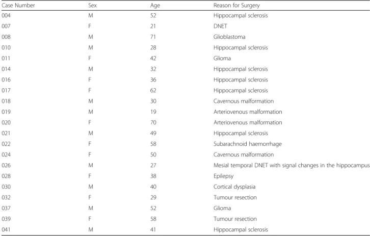
Table 1 Details of resected human brain tissue samples
Case Number Sex Age Reason for Surgery 004 M 52 Hippocampal sclerosis 007 F 21 DNET 008 M 71 Glioblastoma 010 M 28 Hippocampal sclerosis 011 F 42 Glioma 014 M 32 Hippocampal sclerosis 016 F 36 Hippocampal sclerosis 017 F 62 Hippocampal sclerosis 018 M 30 Cavernous malformation 019 M 19 Arteriovenous malformation 020 F 70 Arteriovenous malformation 021 M 49 Hippocampal sclerosis 022 F 58 Subarachnoid haemorrhage 024 F 50 Cavernous malformation
026 M 27 Mesial temporal DNET with signal changes in the hippocampus
028 F 38 Epilepsy 030 M 40 Cortical dysplasia 032 F 29 Tumour resection 037 M 52 Glioma 039 F 58 Tumour resection 041 M 41 Hippocampal sclerosis
M Male, F Female, DNET Dysembryoplastic neuroepithelial tumour
(See figure on previous page.)
Fig. 1 TheGRIN2A gene has two transcript variants in human and primate but not mouse brain. a A short isoform of GRIN2A transcript was predicted by open reading frame studies. The published sequence ofGRIN2A-S suggests that the final exon (exon 13) is missing two nucleotide regions compared to the canonical transcript: firstly, the lack of 343 nucleotides generates a putative exon 14 inGRIN2A-S (splice site shown in Ai)
and the final 206 nucleotides of the canonical form are lost altogether. We designed primers to amplify the region of variance between the two isoforms (Fig.1, Fw1/Rv1) generating an amplicon of 474 bp inGRIN2A and 131 bp in GRIN2A-S. A second reverse primer was designed to amplify canonicalGRIN2A selectively (Fig.1a, Rv2) generating an amplicon of 380 bp. b-f RT-PCR amplification end products. Control conditions indicate no cDNA template was used in PCR b In human cDNA, only the short form ofGRIN2A was observed likely due to preferential amplification in PCR, whereas only a long product of 474 bp was seen in mouse using either human or mouse specific primers. c The Fw1/Rv2 primer pair was used to confirm expression of canonicalGRIN2A in the same human sample as shown in (B). d 3 other human cortical samples with GRIN2A-S amplified. e Both the short and long amplicons were observed in human foetal cDNA. fGRIN2A-S was observed in primate (Rhesus) brain cDNA. g Sequencing of human and primate RT-PCR short amplicons confirmed the presence of the putative splice site shown in Ai
Thus, our data confirm the presence of both canonical
GRIN2A and the novel GRIN2A-S transcripts in human
brain tissue samples.
A BLAST search of the 131 bp sequence amplified by
primers Fw1/Rv1 provided primate-specific predictive
hits. To confirm whether
GRIN2A-S transcript is present
in primate brain, we tested Rhesus macaque brain cDNA
with Fw1/Rv1 primers and confirmed the presence of
GRIN2A-S (Fig.
1
f ). We sequenced the shorter PCR
products for both the adult human and primate samples
and confirmed the presence of the exact splice site
re-ported in the European Nucleotide Archive (Coding:
AAI17132.1; [
20
] (Fig.
1
a,g).
Furthermore, we aimed to evaluate whether the
GRIN2A and GRIN2A-S transcripts were translated into
the corresponding proteins. To this end, we
hypothe-sised that if both transcripts are translated into proteins,
two protein bands corresponding to GluN2A and
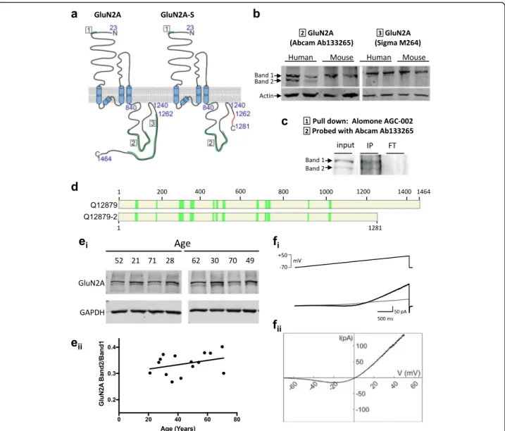
Fig. 2 Two GluN2A protein bands are observed in human but not mouse brain. a Topology of the GluN2A subunit of the NMDA receptor and of GluN2A-S predicted from human mRNA studies. A spliced region is retained in canonical GluN2A leading to an alteration of the reading frame to generate a diverging C-terminal sequence (red) with early truncation. Epitopes for the antibodies used are shown in green and are numbered. b Immunoblots with specific antibodies against the canonical GluN2A and putative GluN2A-S protein in human and mouse cortical lysates. c GluN2A proteins were pulled down with an N-terminal antibody. Band 2 was cut from Coomassie-stained polyacrylamide gel and analysed by mass spectrometry. IP, immunoprecipitate. FT, flowthrough d A set of 14 peptides were confirmed to be present in this band. Figure shows tryptic peptides from band 2 coverage to either canonical GluN2A amino acid sequence (Uniprot: Q12879) or GluN2AS (Q12879–2, predicted) confirming band 2 contains GluN2A protein. e Homogenate from freshly frozen cortical human tissue probed with the GluN2A antibody Abcam 133,265 (select blots shown on top) and quantification of GluN2A-S / GluN2A immunoreactivity (bottom). See Table1for human tissue sample details. f Recombinant GluN2A-S co-expressed with GluN1 in HEK293 cells produces functional NMDARs as demonstrated by a typical J-shaped curve in response to 40 mM NMDA in response to a slow ramp of voltage (− 70 to + 50 mV, 3 s)
GluN2A-S in human homogenate would be
immunode-tected by a GluN2A antibody targeting an epitope
con-served between the canonical and short GluN2A
isoforms (Fig.
2
a,b). Importantly, Western blot analysis
from human brain homogenates confirmed the presence
of two immunoreactive bands with a molecular weight
corresponding to the predicted GluN2A and GluN2A-S
isoforms. On the contrary, a single immunoreactive
band with the high molecular weight (corresponding to
the canonical GluN2A) was detected in mouse brain
ly-sates (Fig.
2
b). Furthermore, using an antibody
specific-ally detecting a carboxy terminal epitope (exclusively
present in the canonical GluN2A isoform), we detected
the presence of a single band, with a molecular weight
corresponding to the canonical GluN2A subunit (Fig.
2
a,
b). To confirm the identity of the low molecular weight
band, we immunoprecipitated GluN2A from human
tis-sue homogenates with an antibody against the conserved
N-terminal domain and analysed the primate-specific
band (Band 2, Fig.
2
c) by mass spectrometry. This
un-biased method allowed the identification of 14 unique
peptides located within GluN2A residues 81–1022,
con-firming that this low molecular band contains the
prox-imal part of GluN2A, and thus discarding the potential
cross-reactivity of anti-GluN2A N-terminal antibody
(Fig.
2
d). To assess the relative levels of GluN2A-S vs.
total GluN2A, human cortical homogenates from
fresh-frozen tissue resected from individuals 27–61 years of
age were analysed by Western blot and the
quantifica-tion showed that GluN2A-S immunoreactivity accounts
for 34 ± 4% of canonical GluN2A protein in cortical
hu-man brain homogenate (Table
1
) and this fraction
re-mains constant across age (Fig.
2
ei).
Finally, to test whether GluN2A-S can be incorporated
into functional NMDARs, we co-transfected HEK293 cells
with plasmids expressing GluN2A-S and GluN1 subunits
[
15
]. A slow voltage ramp delivered during local perfusion
with 40
μM NMDA and 10 μM glycine elicited a typical
J-shaped curve (Fig.
2
fi), and subtraction of responses
with-out agonist (leak subtraction) revealed a typical NMDA
current with reversal potential near 0 mV (Fig.
2
eii). This
confirms that GluN2A-S subunits are able to assemble
with GluN1 subunits and become inserted into the plasma
membrane to form a functional NMDAR that likely plays
a role in human neural function.
Here we describe for the first time the brain
expres-sion of an uncharacterised, primate-specific NMDAR
subunit. The splice site for GluN2A-S suggests that it
will contain a diverging 19 aa sequence in its extreme
C-terminal domain (Fig.
2
a), in addition to lacking the
dis-tal carboxy terminal domain (183 amino acids) that
con-tains PKC/SFK phosphorylation sites, two PDZ binding
motifs that allow synaptic localisation [
4
,
12
,
14
], and a
dileucin clathrin adaptor motif involved in receptor
internalisation [
13
]. Following many lines of published
evidence, these differences suggest that the dynamic
regu-lation of GluN2A-S in response to stimuli could diverge
from GluN2A subunit-containing NMDARs. This could
impact the number of receptors present synaptically or
extrasynaptically, the insertion of new receptors into the
membrane, their lateral diffusion and clustering into
synapses and their active removal. The potential changes
in human synapses compared to mouse neurons void of
GluN2A-S could result in distinct mechanisms involved in
activity-dependent plasticity of synapses, which will highly
depend upon its triheteromeric partners [
1
,
8
,
19
].
Together, our data suggest that GluN2A-S is a
primate-specific NMDAR subunit and a substantial component of
functional NMDARs in the adult human brain. Many
neur-onal mechanisms discovered in mice have been
success-fully
recapitulated
in
humans.
However,
mounting
evidence suggests that there are important differences
be-tween rodent and human neurons that result in distinct
signal integration properties [
22
,
23
,
26
] and proteomic
composition of synapses [
3
]. Species differences may
ultim-ately impact the way in which human neural circuits can
be computationally modelled [
7
], and the translation of
pre-clinical findings into approved therapies [
24
]. Further
analyses of GluN2A-S spatio-temporal gene expression and
synaptic/ extrasynaptic localisation will enhance our
know-ledge of their functional role and may uncover NMDAR
trafficking mechanisms present only in primates and
diver-ging sequences may uncover novel therapeutic targets.
Materials and methods
Human brain tissue samples
All samples consisted of cortical tissue resected for access
to deeper brain lesions such as sclerotic hippocampus in
epileptic patients (the pathological tissue for these lesions
was not used). Informed consent was obtained from all
pa-tients to use surgically-resected putatively non-pathological
tissue not required for diagnostic purposes (see ethical
ap-proval declaration). Briefly, resected tissue was obtained
from temporal cortex of patients undergoing surgery for
the removal of deeper structures. Tissue was collected in
ice cold artificial cerebrospinal fluid [
26
] then taken to the
laboratory, frozen and kept at
− 80 °C. Transfer time was of
the order of 10–15 min.
Mouse brain tissue
Mice were decapitated following isoflurane anaesthesia
(see ethical approval declaration). Brains were extracted
in ice-cold ACSF and sliced or snap-frozen. All brain
tis-sue samples were stored in the -80C freezer until lysed.
RNA extraction and cDNA synthesis
Total RNA was isolated from human and mouse tissue
using Trizol and then purified using the RNeasy Mini kit
(QIAGEN) following the manufacturer’s instructions.
cDNA was synthesised immediately from 200 ng of total
RNA per reaction using the SuperScript IV reverse
tran-scriptase and cDNA synthesis kit (INVITROGEN)
ac-cording to the manufacturer’s instructions. The cDNA
obtained was stored at
− 80 °C. Human foetal cDNA
was obtained from Takara (Normal brain tissue
cDNA, pooled from 59 spontaneously aborted male/
female Caucasian fetuses, ages: 20–33 weeks). Rhesus
macaque cDNA was obtained from Amsbio (Normal
brain tissue, Female, 4.5 years).
PCR conditions and primers
PCR was performed on 1
μg of cDNA using primers and
REDTaq® Readymix
™ PCR Reaction Mix (Sigma-Aldrich)
for 40 cycles. DNA was denatured at 95 degree C and
extended at 72 degree C for 45 s each cycle. Products
were separated on a 1.5% agarose gel.
The following primers were used: Fw1:
ATTCAGGC-CACTTCACCATGAG,
Rv1:
ATCTCCCAATAAC-CAAGCGTTG, Rv2: CTTGCTGTCCTCCAGACCTTGG
mFw1: ACTCAGGCCACTTTACCATGAG, and mRv1:
ATCTCCCAATAACTAAGCGTTG.
Plasmids
pEYFP-NR1a was a gift from Stefano Vicini (Addgene
plasmid # 17928; [
15
]).
GRIN2A-S plasmid from Broad
Institute, was acquired from Source Bioscience
(Tran-script NM_001134408.2).
Western blotting
Equal amounts of protein (28
μg) were separated in 7.5%
acrylamide gels by SDS-PAGE and transferred onto
nitro-cellulose membranes. Membranes were blocked in 5% (w/
v) non-fat milk for 1 h at room temperature and incubated
overnight at 4 C in 5% (w/v) bovine serum albumin (BSA)
containing 0.1% (v/v) Tween-20 and one of the following
primary antibodies: anti-NMDAR2A (ab133265; 1:1000;
Abcam); and GAPDH (D16H11; 1:1000; CST) actin.
Membranes were washed 3 times with Tris-buffered saline
(TBS) containing 0.1% Tween-20 (TBS-T) and probed
with fluorophore-conjugated goat anti-mouse/
−rabbit
sec-ondary antibody (1:10000; LI-COR). Proteins were
visua-lised using the Odyssey infrared scanner (LI-COR) using
Image Studio Light Software.
GluN2A pulldown
GluN2A was pulled down from 1 mg of protein
homogenate (in RIPA buffer) using 2
μg of Alomone
antibody AGG-002 beads. Eluate was run in SDS
page and stained with Coomassie dye. The lighter
band corresponding to putative GluN2A-S was cut
and analysed by LC-MS/MS [
11
].
HEK293T cell recordings
HEK293T cells were cultured at 37 C with 5% CO
2in
Dulbecco’s Modified Eagle Medium with glucose,
L-glutamine and pyruvate, 10% FBS and 1% Pen-Strep and
seeded at low density onto poly-L-lysine coated glass
coverslips for electrophysiology. Adherent cells were
transfected
using
JetPEI
reagent
with
NR1a
and
GluN2A-S plasmids at a 1:1 ratio and recorded after 48
h. Borosilicate glass micropipettes were pulled to
pro-duce a resistance of 4–6 mOhm and filled with
intracel-lular recording solution containing in mM: Gluconic
acid 70, Caesium chloride 10, sodium chloride 5, BAPTA
10, HEPES 10, GTP 0.3 ATP 4 and pH balanced to 7.3
with caesium hydroxide. Cells were perfused with aCSF
throughout recording containing, in mM: sodium
chlor-ide 126, calcium dichlorchlor-ide 2, glucose 10, magnesium
sulfate 2, potassium chloride 3, NaH
2PO
41.25 and
NaHCO
326.4, and glycine 10
μM and pH regulated by
continuous bubbling of 95% oxygen, 5% CO
2.
Record-ings with or without addition of NMDA 40
μM were
made in whole-cell voltage clamp and Matlab software
and amplified using an Axopatch 200B as previously
de-scribed [
28
].
Abbreviations
bp:Base pairs; cDNA: Copy deoxyribose nucleic acid; GAPDH: Glyceraldehyde 3-phosphate dehydrogenase; LC-MS: Liquid chromatography– mass spectrometry; mRNA: Messenger ribonucleic acid; NMDA: N-methyl-D-aspartate; NMDAR: N-methyl-D-aspartate receptor; PCR: Polymerase chain reaction; SDS-PAGE: Sodium dodecyl sulfate polyacrylamide gel electrophoresis
Acknowledgements
We thank Dr. Kate J Heesom from the Proteomics Facility, University of Bristol for advising on and performing the mass spectrometry analysis. We thank Dr. Ian Galea as chief investigator of the NOII study.
Authors’ contributions
MVC, KD, XA, HW designed research; HW, CMP, AP, HK, SDH, KK and MVC performed research; HW, CMP, AP, KK, MVC analyzed data; DB, AA, PG, GV performed neurosurgery to resect human brain tissue used in this work, MVC, KD, HW wrote the paper. All authors revised and approved the manuscript.
Funding
MVC was funded by the Institute for Life Sciences (IfLS) and Wessex Innovation Fund. HW was funded by an Alzheimer’s Research UK Southcoast Network Summer Studentship, HW and CMP PhD studentships were funded by the Gerald Kerkut Trust, CMP was co-funded by IfLS studentship, KK and AP were funded by Alzheimer’s Society, KD was funded by BBSRC [BB/ L007576/1].
Availability of data and materials
The datasets used and/or analysed during the current study are available from the corresponding author on reasonable request.
Ethics approval and consent to participate
Anonymised human brain tissue samples: All patients provided informed consent for participation. Ethical approval: REC Reference: 12/NW/0 794, HTA LN: 12009 under the Southampton Biorepository Research. Study reference number: SRB002/14 or the NOII study: REC reference: 11/SC/0204 Sponsor number: RHM NEU0169.
Animal tissue: Animal care and experimental procedures were conducted in accordance with UK Home Office regulations under the Animals (Scientific Procedures) Act of 1986.
Consent for publication Not applicable.
Competing interests
The authors declare that the research was conducted in the absence of any commercial or financial relationships that could be construed as a potential conflict of interest.
Author details
1
School of Biological Sciences, University of Southampton, University Road, Southampton SO17 1BJ, UK.2Wessex Neurological Centre, University Hospital
Southampton, University of Southampton, Southampton, SO16 6YD, UK.
3Neuropharmacology Unit, Bellvitge Biomedical Research Institute (IDIBELL),
L’Hospitalet de Llobregat, Barcelona, Spain.
Received: 4 June 2019 Accepted: 21 June 2019
References
1. Al-Hallaq RA, Conrads TP, Veenstra TD, Wenthold RJ. NMDA Di-Heteromeric receptor populations and associated proteins in rat Hippocampus. J Neurosci. 2007;27:8334–43.
2. Bar-Shira O, Maor R, Chechik G. Gene expression switching of receptor subunits in human brain development. PLoS Comput Biol. 2015;11: e1004559.
3. Bayés A, Collins MO, Croning MDR, van de Lagemaat LN, Choudhary JS, Grant SGN. Comparative study of human and mouse postsynaptic proteomes finds high compositional conservation and abundance differences for key synaptic proteins. PLoS One. 2012;7:e46683.
4. Cousins SL, Kenny AV, Stephenson FA. Delineation of additional PSD-95 binding domains within NMDA receptor NR2 subunits reveals differences between NR2A/PSD-95 and NR2B/PSD-95 association. Neuroscience. 2009;158:89–95.
5. de Ligt J, Willemsen MH, van Bon BWM, Kleefstra T, Yntema HG, Kroes T, Vulto-van Silfhout AT, Koolen DA, de Vries P, Gilissen C, del Rosario M, Hoischen A, Scheffer H, de Vries BBA, Brunner HG, Veltman JA, Vissers LELM. Diagnostic exome sequencing in persons with severe intellectual disability. N Engl J Med. 2012;367:1921–9.
6. Endele S, Rosenberger G, Geider K, Popp B, Tamer C, Stefanova I, Milh M, Kortüm F, Fritsch A, Pientka FK, Hellenbroich Y, Kalscheuer VM, Kohlhase J, Moog U, Rappold G, Rauch A, Ropers H-H, von Spiczak S, Tönnies H, Villeneuve N, Villard L, Zabel B, Zenker M, Laube B, Reis A, Wieczorek D, Van Maldergem L, Kutsche K. Mutations in GRIN2A and GRIN2B encoding regulatory subunits of NMDA receptors cause variable neurodevelopmental phenotypes. Nat Genet. 2010;42:1021–6.
7. Eyal G, Verhoog MB, Testa-Silva G, Deitcher Y, Lodder JC, Benavides-Piccione R, Morales J, DeFelipe J, de Kock CP, Mansvelder HD, Segev I. Unique membrane properties and enhanced signal processing in human neocortical neurons. Elife. 2016;5:1–18.
8. Hansen KB, Ogden KK, Yuan H, Traynelis SF. Distinct functional and pharmacological properties of Triheteromeric GluN1/GluN2A/GluN2B NMDA receptors. Neuron. 2014;81:1084–96.
9. Hardingham GE, Bading H. Synaptic versus extrasynaptic NMDA receptor signalling: implications for neurodegenerative disorders. Nat Rev Neurosci. 2010;11:682–96.
10. Hedegaard MK, Hansena KB, Andersena KT, Bräuner-Osborne H, Traynelis SF. Molecular pharmacology of human NMDA receptors. Neurochemistry International. 2012;61(4):601–9.
11. Jiménez-Castellanos J-C, Wan Nur Ismah WAK, Takebayashi Y, Findlay J, Schneiders T, Heesom KJ, Avison MB. Envelope proteome changes driven by RamA overproduction in Klebsiella pneumoniae that enhance acquired β-lactam resistance. J Antimicrob Chemother. 2018;73:88–94.
12. Köhr G, Jensen V, Koester HJ, AL a M, Utvik JK, Kvello A, Ottersen OP, Seeburg PH, Sprengel R, Hvalby Ø. Intracellular domains of NMDA receptor subtypes are determinants for long-term potentiation induction. J Neurosci. 2003;23:10791–9.
13. Lavezzari G. Subunit-specific regulation of NMDA receptor endocytosis. J Neurosci. 2004;24:6383–91.
14. Lim IA, Merrill MA, Chen Y, Hell JW. Disruption of the NMDA receptor –PSD-95 interaction in hippocampal neurons with no obvious physiological short-term effect. Neuropharmacology. 2003;45:738–54.
15. Luo J-H, Fu Z-Y, Losi G, Kim BG, Prybylowski K, Vissel B, Vicini S. Functional expression of distinct NMDA channel subunits tagged with green fluorescent protein in hippocampal neurons in culture. Neuropharmacology. 2002;42:306–18.
16. Lussier MP, Sanz-Clemente A, Roche KW. Dynamic regulation of NMDA and AMPA receptors by posttranslational modifications. J Biol Chem. 2015;290: jbc.R115.652750.
17. Martin SJ, Grimwood PD, Morris RGM. Synaptic plasticity and memory: an evaluation of the hypothesis. Annu Rev Neurosci. 2000;23:649–711.https:// doi.org/10.1146/annurev.neuro.23.1.649.
18. Paoletti P, Bellone C, Zhou Q. NMDA receptor subunit diversity: impact on receptor properties, synaptic plasticity and disease. Nat Rev Neurosci. 2013;14:383–400.
19. Rauner C, Köhr G. Triheteromeric NR1/NR2A/NR2B receptors constitute the major N-methyl-D-aspartate receptor population in adult hippocampal synapses. J Biol Chem. 2011;286(9):7558–66.
20. Strausberg RL, Feingold EA, Grouse LH, Derge JG, Klausner RD, Collins FS, Wagner L, Shenmen CM, Schuler GD, Altschul SF, Zeeberg B, Buetow KH, Schaefer CF, Bhat NK, Hopkins RF, Jordan H, Moore T, Max SI, Wang J, Hsieh F, Diatchenko L, Marusina K, Farmer AA, Rubin GM, Hong L, Stapleton M, Soares MB, Bonaldo MF, Casavant TL, Scheetz TE, Brownstein MJ, Usdin TB, Toshiyuki S, Carninci P, Prange C, Raha SS, Loquellano NA, Peters GJ, Abramson RD, Mullahy SJ, Bosak SA, McEwan PJ, McKernan KJ, Malek JA, Gunaratne PH, Richards S, Worley KC, Hale S, Garcia AM, Gay LJ, Hulyk SW, Villalon DK, Muzny DM, Sodergren EJ, Lu X, Gibbs RA, Fahey J, Helton E, Ketteman M, Madan A, Rodrigues S, Sanchez A, Whiting M, Madan A, Young AC, Shevchenko Y, Bouffard GG, Blakesley RW, Touchman JW, Green ED, Dickson MC, Rodriguez AC, Grimwood J, Schmutz J, Myers RM, Butterfield YSN, Krzywinski MI, Skalska U, Smailus DE, Schnerch A, Schein JE, Jones SJM, Marra MA. Generation and initial analysis of more than 15,000 full-length human and mouse cDNA sequences. Proc Natl Acad Sci U S A. 2002;99:16899–903.
21. Strehlow V, Heyne HO, Vlaskamp DRM, Marwick KFM, Rudolf G, de Bellescize J, Biskup S, Brilstra EH, Brouwer OF, Callenbach PMC, Hentschel J, Hirsch E, Kind PC, Mignot C, Platzer K, Rump P, Skehel PA, Wyllie DJA, Hardingham GE, van Ravenswaaij-Arts CMA, Lesca G, Lemke JR, Arzimanoglou A, Augustijn PB, Van Bogaert P, Bourry H, Burfeind P, Chu Y, Chung B, Doummar D, Edery P, Fattal-Valevski A, Fradin M, Gerard M, de Geus C, Gunning B, Hasaerts D, Helbig I, Helbig KL, Jamra R, Lyver MJ, Wassink-Ruiter JSK, Koolen DA, Lederer D, Lunsing RJ, Mathot M, Maurey H, Menascu S, Michel A, Mirzaa G, Mitter D, Muhle H, Møller RS, Nava C, O’Brien M, van Pinxteren-Nagler E, van Riesen A, Rougeot C, Sanlaville D, Schieving JH, Syrbe S, Veenstra-Knol HE, Verbeek N, Ville D, Vos YJ, Vrielynck P, Wagner S, Weckhuysen S, Willemsen MH. GRIN2A -related disorders: genotype and functional consequence predict phenotype. Brain. 2019;142:80–92. 22. Testa-Silva G, Verhoog MB, Goriounova NA, Loebel A, Hjorth J, Baayen
JC, de Kock CPJ, Mansvelder HD. Human synapses show a wide temporal window for spike-timing-dependent plasticity. Front Synaptic Neurosci. 2010;2:12.
23. Testa-Silva G, Verhoog MB, Linaro D, de Kock CPJ, Baayen JC, Meredith RM, De Zeeuw CI, Giugliano M, Mansvelder HD. High bandwidth synaptic communication and frequency tracking in human neocortex. PLoS Biol. 2014;12:e1002007.
24. Vargas-Caballero M, Willaime-Morawek S, Gomez-Nicola D, Perry VH, Bulters D, Mudher A. The use of human neurons for novel drug discovery in dementia research. Expert Opin Drug Discov. 2016;11(4).https://doi.org/10. 1517/17460441.2016.1154528.
25. Vargas-Caballero M, Robinson HPC. 2003. A slow fraction of Mg2+ unblock of NMDA receptors limits their contribution to spike generation in cortical pyramidal neurons. J Neurophysiol 89:2778–2783.
26. Verhoog MB, Goriounova NA, Obermayer J, Stroeder J, Hjorth JJJ, Testa-Silva G, Baayen JC, de Kock CPJ, Meredith RM, Mansvelder HD. Mechanisms underlying the rules for associative plasticity at adult human neocortical synapses. J Neurosci. 2013a;33:17197–208.
27. Walsh PS, Erlich HA, Higuchi R. Preferential PCR amplification of alleles: mechanisms and solutions. PCR Methods Appl. 1992;1:241–50.
28. Yang X, Boehm JS, Yang X, Salehi-Ashtiani K, Hao T, Shen Y, Lubonja R, Thomas SR, Alkan O, Bhimdi T, Green TM, Johannessen CM, Silver SJ, Nguyen C, Murray RR, Hieronymus H, Balcha D, Fan C, Lin C, Ghamsari L, Vidal M, Hahn WC, Hill DE, Root DE. A public genome-scale lentiviral expression library of human ORFs. Nat Methods. 2011;8:659–61.
Publisher
’s Note
Springer Nature remains neutral with regard to jurisdictional claims in published maps and institutional affiliations.