SCUOLA DOTTORALE IN BIOLOGIA
BIOLOGIA APPLICATA ALLA SALUTE
DELL’UOMO
CICLO XXIV
Approach in Aptamer Based Biosensors For
Human Health Applications
Studio di Biosensori Basati su Aptameri
Applicati alla Salute dell’Uomo
Dr. Ilaria Lamberti
Tutor: prof. Antonio Antoccia
Coordinator: prof. Paolo Visca
DOCTORAL SCHOOL IN BIOLOGY:
BIOLOGY APPLIED TO HUMAN HEALTH
COURSE XXIV
Approach in Aptamer Based Biosensors For
Human Health Applications
Studio di Biosensori basati su aptameri
Applicati alla Salute dell’Uomo
Dr. Ilaria Lamberti
CONTENTS Abstract ………....page 1 1 General background………....3 1.1 What is a biosensor………..…....5 1.2 Bioreceptors………...6 1.2.1 Immunosensors………..6 1.2.2 Aptasensors………....7 1.3 Types of Trasducers:………..10 1.3.1 Densitometric………..11 1.3.2 Mass sensitive……….13 1.3.3 Optical………...…..15 References………..…..17 Aims Aim 1: Protein Microarray Applications………...21
Aim 2: Quartz Crystal Sensor...36
Aim 3: Sensing Surface for SERS Application...48
Publications...51
1
Abstract
In recent decades the combination of different technologies and scientific disciplines like bioelectronics, genomics, materials science, biochemistry and computer science, has allowed the development and implementation of innovative analytical devices - biosensors - which allow to find new solutions in the field of diagnostic. In such devices the measurement of the target analytes is achieved by selective transduction of a parameter of the biomolecule-analyte reaction into a quantifiable signal, providing a selective identification of toxic chemical compounds at ultra trace levels in industrial products, chemical substances, environmental samples (e.g., air, soil, and water) or biological systems (e.g., bacteria, virus, or tissue components) for biomedical diagnosis. In life sciences, biosensors have offered new options for clinical diagnostic procedures. In the development of biosensors, nanotechnology is playing an increasingly important role, improving sensitivity and performance of their construction and allowing the capability of unambiguous identification and accurate quantification of big chemical constituents in complex systems.
Recently, biosensors based on the use of monoclonal or polyclonal antibodies have seen a great development in the field of small molecules analytical determination and specifically in the mycotoxins analyses for food safety application and in biomarkers detection for human health (i.e. caspases detection of apoptosis, playing a critical role in the development of therapeutics in many different fields, including cancer).
Affinity to specific molecular targets (such as nucleic acids, proteins, small compounds, or cells) of antibodies is overcome by aptamers, single stranded of DNA or RNA oligonucleotides of 15 to 60 base. In contrast to antibodies, aptamers are prepared by in vitro selection procedure called SELEX. The purpose of this PhD thesis is to provide a study of a range of technologies and innovations in the field of the bio-sensible component of a biosensor, comparing different experimental approaches.
At the beginning, using as biological component monoclonal e polyclonal antibody, we performed an antibodies- based microarray biosensor for applications in proteome analysis, disease diagnostics and quantitative small molecules analysis. We discussed different microarray surfaces, immobilization techniques, detection systems and advantages and disadvantages of antibody microarrays.
During the second year, we approached a different bioreceptor: aptamers, that exhibit many advantages as recognition elements in biosensing, compared to traditional antibodies. The unique properties of aptamers and
the possibility of developing aptamers against different binding sites on the target analyte allowed high variability on the assay and a good use in biosensor field.
We used an acoustic sensor in a TSM format (Transverse Shear Mode methods) for studying protein/aptamer interaction. We detected subtle structural effects at the sensor surface connected with conformation changes of thrombin aptamers and ochratoxin induced by protein or low molecular mass ligand.
In the last year, an aptamer terminated sensing surface, was exploited for designing a nano-aptasensor for microfluidic device, coupled with a Surface Enhanced Raman Spectroscopy (SERS). In order to validate this strategy, the already experienced aptamer for thrombin is used.
During the PhD we moved in this direction, studying and defining different approaches to develop new biosensors for the most popular toxic and carcinogenic food contaminant class: micotoxins. We also focused our interest in thrombin and caspases, due to their importance as biomarker in several human pathologies, and in the development of the analytical devices.
3
1. General Background
The purpose of this PhD thesis is to provide a study of a range of technologies and innovations in the field of the bio-sensible component of a biosensor, comparing different experimental approaches. The availability of innovative tools for the detection of food contaminants and for the analysis of biomarkers of pathological conditions is closely related to the protection of human health. During my PhD, we moved in this direction, studying and defining different approaches to develop new biosensors.
1.1 What is a Biosensor?
A biosensor is a device which converts biological activity into a quantifiable signal, providing rapid analysis and real time detection [Schmid, 1987; Turner et al., 1987]. It consists of two main components: a bio-receptor and a transducer. The bioreceptor (or biological sensing
element or biological component) is a biomolecule, such as an antibodies, enzymes, cells or aptamers, in close contact with the transducer, which binds specifically to the analyte of interest. The transducer translates the binding event into detectable and measurable signal (fig.1). Interactions, via coupling of a biological recognition element to a transducer, are capable of providing qualitative or quantitative information [Malhotra et al. 2005]. The two most important properties of any proposed biosensor are the specificity and the sensitivity toward the target analyte. The specificity of a biosensor is entirely governed by the properties of biological component, because this is where the analyte interacts with the sensor. The sensitivity of the
integrated device is dependent on both the biological component and the transducer, because there must be a significant biomolecule-analyte interaction and high efficiency of subsequence detection of this reaction by the transducer. The range of potential biological components and the range of transducer technologies available for use in biosensors require a truly multi and interdisciplinary approach to research and development in this area. Much of the basic transducer technologies already exist, though it requires further development and optimization. A number of different techniques for measuring the response, as well as various types of transducer which are currently being evaluated for their suitability for a range of sensing applications, are listed in table 1. Biosensors based on DNA or RNA aptamers (aptasensors) represent new type of sensors that utilize unique properties of artificial receptor – aptamers. Aptasensors are of considerable interest, due to their application in detection of practically unlimited kinds of compound [Hianik and Wang, 2009].
Table 1
Biological applications Tranducer system Cofactors Antibodies Receptors Enzymes Membranes Organelles Cells Aptamers Optical Electrochemical Piezo-electric Calorimetric Acoustic Mechanical Densitometric
The main advantages of biosensor technology, in comparison with traditional analytical methods, are fast detection (minutes) and response (seconds), high sensitivity (typically nM, improved sensitivity with nanoparticles pM and better), high selectivity, easy preparation and operation assay method. In addition, most of these devices are reusable and show low cost assay.
The methodology of surface chemistry is the basic know-how for obtaining reproducible results with biosensors and various strategies can be used
5
The key points to consider when selecting an appropriate surface and coating procedure are the low degree of unspecific binding sites and the uniform distribution of functional groups on the substrate surface.
For this reason, during biosensor development and testing, particular attention has to be focused on:
• Surface (on which sensing layer will be coated) characterisation
• Biological reagent (immunoglobulin, nucleic acid, ecc.)
characterisation
• Uniformity of biological element • Standard solution preparation
1.2 Bioreceptor components
The variety of bioreceptors used in vast majority of biosensors, reported to date, have been based on antibodies, oligonucleotides, enzymes, whole cells, membrane and, more recently, aptamers. A brief overview of different types will be described next.
1.2.1 Immunosensors
Immunosensors use antibodies (monoclonal or polyclonal) as biological element and are defined affinity sensors. They are based on the immunological technique that can be applied to investigate and manipulate minute concentrations of complex molecules. Current immunological assays capable of detecting many biological agents utilize the sensitivity and specificity of polyclonal and monoclonal antibodies [Emanuel et al., 2000]. Polyclonal antibodies are obtained from animal serum and are produced by two type of blood cell, B lymphocytes and plasma cells, in response to a foreign substance. Monoclonal antibodies are produced in vitro by hibridoma technology. From the analytical point of view, the main difference between polyclonal and monoclonal antibodies is the fact that monoclonal recognizes on the molecule of antigen a single epitope only [Pohanka, et al., 2007]. Antibodies consist of four polypeptide sub-units comprising two identical large or heavy chains and two identical small and light chains which are held together by non covalent force and covalent inter chain disulphide bonds. The carbohydrate residues in antibodies are covalently bonded to the C-terminal half (Fc) of the molecule. The key portions of the antibody molecule that contain the antigen binding sites are calledthe Fab fragments. Each Fab fragment comprises an entire light chain and a segment of the heavy chain [Byfield and Abuknesha, 1994].
In biosensor based on antibodies the analyte is either the corresponding antigen of the antibody used. An antibody-antigen interaction is characterized by two major properties that may be exploited for sensing or detection purposes: a very high affinity constant (10-18 M of antigens can be detected) and a low cross-reactivity. The main important property of antibodies is their ability to bind to an extremely wide range of natural and man-made compounds [Vo-Dinh and Cullm, 2000].
7
1.2.2 Aptasensors
Aptamers are ligands with a high affinity for considerably differing molecules ranging from large targets as proteins over peptides, complex molecules to drugs and organic small molecules or even metal ions. They are widely used, including medical and pharmaceutical basic research, drug development, diagnosis, and therapy. Analytical and separation tools bearing aptamers as molecular recognition and binding elements are another big field of application.
Aptamers have become important tools for molecular diagnostics and therapeutics. In particular, the aptamers-based biosensors possess unprecedented benefits for their high specificity and affinity. In principle, they can be selected in vitro for each target, ranging from small molecules to large proteins, also cells. They can be synthesized with high
reproducibility and purity, DNA aptamers are usually highly chemically stable. The application of aptamers as biocomponents in biosensors offers a
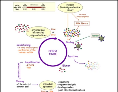
Fig. 2: Systematic Evolution of Ligands by Exponential enrichment - SELEX
multitude of advantages over the state of the art in affinity sensing [Song et al, 2008]. Aptamers are single-stranded RNA or DNA oligonucleotides 15 to 60 base in length that bind with high affinity to specific molecular targets such as nucleic acids, proteins, small compounds or cells. This oligonucleotides bind to their target with high selectivity and sensitivity due to their three-dimensional shape. Their specificity is comparable and in certain cases even higher than those of antibodies. In contrast to antibodies, aptamers are prepared by in vitro selection procedure: Systematic Evolution of Ligands by Exponential enrichment, also called SELEX, developed simultaneously in early 1990s by L. Gold and A. Ellington laboratories [Ellington and Szostak, 1990; Tuerk and Gold, 1990]; SELEX, aimed at the development of aptamers [Stoltenburg et al., 2000], involves three processes, namely: selection of ligand sequences that bind to a target;
partitioning of aptamers from non-aptamers via affinity methods; amplification of bound aptamers (Fig.2) [Gopinath, 2007]. Aptamers have been selected to hundreds of small molecules and protein targets including, for example, ATP, GTP, B12, malachite green and caffeine, HIV-1 Rev peptide, MS2 coat protein, and thrombin. Several aptamer structures have been shown to be readily evolvable in terms of specificity. For example, three mutations within the binding loop of an in vitro selected l-citrulline aptamer were enough to change its specificity to l-arginine because the pattern of hydrogen bond donors and acceptors could be flipped in a simple way [Yang et al., 1996; Klussmann 2006].
Among aptamers, the thrombin aptamer was most extensively studied. The composition of the thrombin aptamer is as follows: d(T15GGTTGGTGTGGTTGG). A pair of thymines on the G- quandruplex of the aptamer characterizes thrombin Fig. 3: Structures of DNA
aptamers that selectively bind thrombind in its fibrionogen binding site [Hianik, 2009].
9
oligonucleotides that bind a target protein with unknown specificity for nucleic acids and it is largely use in biosensor field [Bock et al., 1992]. Thrombin is a multifunctional serine protease, which plays an important role in pro-coagulant and anti-coagulant function. This protease converts soluble fibrinogen into insoluble strands of fibrin, which is responsible either for a physiological plug or pathological thrombus. Thrombin has two binding sites that are spatially separated and localized at opposite poles of thrombin molecules. These binding sites are sensitive to fibrinogen and heparin, respectively. Aptamers can be used as potential inhibitors of thrombin, therefore study of the mechanisms of interaction of thrombin with aptamers is of great importance for medicine [Coughlin, 2000].
Recently, also a DNA aptamer sensitive to ochratoxin A (OTA) has been developed [Cruz-Aguado and Penner, 2008]. OTA A is a fungal toxin, discovered as a metabolite of aspergillus ochraceus [Turner et al., 2009]. This aptamer is able to recognize OTA A with sensitivity in a ppb level and with high selectivity.
Mycotoxins, such are aflatoxins, fumonisine and ochratoxin, are toxical fungal metabolites that can occur in primary food products. This mycotoxin generally appears during storage of cereals, coffee, cocoa, dried fruit, pork etc. and occasionally in the field of grapes. It may also be present in blood and kidneys of animals that have been fed on contaminated feeds. Animal studies indicated that this toxin is carcinogenic [Turner et al., 2009]. Therefore, the European Commission has fixed maximum concentration of OTA in foodstuffs: 3 µg/kg (7.4 nM) for cereal products and 5 µg/kg (12.4 nM) for roasted coffee, respectively (Commission Regulation No. 1881/2006, 19 December 2006).
The establishment of an efficient method of this analyte detection is therefore of high importance. In addition to traditional, but expensive and time-consuming methods such as liquid chromatography, new trends consist in development of portable and easy to use biosensors [Tsai and Hsieh., 2007].
1.3 Types of Biosensor Trasducers
The availability of rapid and reliable methods for rapid determination of small molecules is an increasing need for human health. In order to monitoring small molecules proteins/marker related on Human Health, Gas Chromatographic (GC) High Pressure Liquid Chromatography (HPLC) methods and immunoassay are generally utilized, due to their high detection sensitivity and selectivity. However, GC and HPLC analyses are time consuming and needs sample pre-treatment or pre-concentration procedures [Sethi, 1994].
Immunoassays and biosensors are becoming a recognized alternative or complementary to conventional analytical techniques for the detection of different analytes. Recently, biosensors based on the use of monoclonal or polyclonal antibodies have seen a great development in the field of small molecules analytical determination. The absence of cross-reactivity obtained with most of these biosensor, the possibility of on-line measurement, the absence of sample pre-treatment, can really put it in competition with other conventional systems such as HPLC and ELISA [Pacheco, 2011].
In particular, we focused our attention on biosensors that utilize immunoglobulins or aptamer showing affinity for a correspondent analyte, associated to various transduction elements. Several biosensing platforms have been introduced for the Surface Enhanced Raman Spectroscopy (SERS) and for the Quartz Microbalance Crystals (QCM). Examples of microarray densitometric platform is been also presented.
Analytical methods used for small molecules determination for human health are mainly based on immunoassay as ELISA. Actually, biosensor and microsystem technologies are used for different applications including studies of human and veterinary diseases, drug discovery, genetic screening, clinical and food diagnostics. According to these approaches, the goal of many authors has been to transfer the methods of immunoassay from microtiter plates into a biosensor format to develop a fast, sensitive and inexpensive detection of protein markers for various diseases.
Microarray and biosensor technology enables the fast and parallel analysis of a multitude of biologically relevant parameters. Not only nucleic acid-based tests, but also peptide, enzyme antibody and aptamer assays using different formats of biosensor evolved within the last decade. Microarrays and biosensors are a powerful assay technology that can be used to generate
11
1.3.1 Densitometric
Densitometry is the quantitative measurement of optical density in light-sensitive materials. Densitometric analysis is the quantitative and qualitative study of color levels of the image obtained. It seems expecially suitable, since it allows rapid quantification. Densitometric evaluation is based on measurement of light reflected by a spot or of a native fluorescence. Today, modern computer-controlled densitometers allow quantitative determination of almost all chemicals, both colorless and colored which have UV-Vis absorbance or a capable of measuring fluorescence. The method is rapid and it offers possibility of analyzing simultaneously a large number of sample along with consecutive qualitative or semi-quantitative analysis. Several colorimetric and fluorometric methods are currently available for the measurement of proteins in biological extracts [Ghosh, 1988].
Enzyme linked immunosorbent assay (ELISA) and fluorescence immunoassay (FIA) are excellent survey tools for many analytical purposes because of their high-throughput, user friendliness, and field portability. Immunoassay is traditionally performed as individual test, however in many cases it is necessary to perform a panel of tests on each sample (detection of drug residues). To address this requirement, microarray-based immunoassay technologies have been developing utilizing microarray platform (multianalyte analysis) and classic immunoassay (multi-samples analysis). In recent years, the antibody microarray technology has made significant progress, going from proof-of-concept designs to established high-performing technology platforms capable of targeting non-fractionated complex samples, as proteoma [Blohm and Guiseppi-Elie, 2001]. Microarrays provide a powerful analytical tool for the simultaneous detection of multiple analytes in a single experiment and consist of a biosensor micro o nano arrays.
Microarrays consist of immobilized biomolecules spatially addressed on planar surfaces, microchannels or microwells, or an array of beads immobilized with different biomolecules. Biomolecules commonly immobilized on microarrays include oligonucleotides, polymerase chain reaction (PCR) products, proteins, lipids, peptides and carbohydrates. Ideally, the immobilized biomolecules must retain activity, remain stable, and not desorb during reaction and washing steps. The immobilization procedure must ensure that the biomolecules are immobilized at optimal density to the microarray surface for efficient binding [Venkatasubbarao et al., 2004].
Some microarray applications are focused on current trends in the movement of this technology from being a purely research method to
becoming an analytical instrument applicable in the clinic and as well as in human health [Koppal, 2004].
Research on microarrays as multianalyte biosystems has generated increased interest in the last decade. The main feature of the microarray technology is the ability to simultaneously detect multiple analytes in one sample by an affinity-binding event at a surface interface. In some cases immunoanalytical microarrays have the potential to replace conventional chromatographic techniques. They are applied if the number of samples is high or analysis by current methods is difficult and/or expensive. Therefore, microarray platforms have a great potential as monitoring systems for the rapid assessment of water or food samples. Antibody-based microarrays are a powerful tool for analytical purposes, also for aflatoxins and caspases detection application. Immunoanalytical microarrays are a quantitative analytical technique using antibodies as highly specific biological recognition elements.
In our two applications we developed a competitive immunoassay in a microarray format and observed, using the method described, different microarray patterns in samples containing aflatoxin-B1 or fumonisine at a ppb concentration range and either analytes in order to detect different caspases. The quality of the microarray data was comparable to data generated by a microplate-based immunoassay, but further investigations are needed in order to better characterize these methods.
13
1.3.2 Mass sensitive
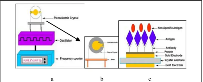
Another form of transduction that has been used for biosensors is the measurement of small changes in mass. This has already been shown to capable of very sensitive measurements. The principle means of mass
analysis relies on the use of piezoelectric crystals. These crystals can be made to vibrate at a specific frequency with the application of an electrical signal of a specific frequency. The frequency of oscillation is therefore dependent on the electrical frequency applied to the crystal as well as the crystal’s mass. Therefore, when the mass increases due to binding of chemicals, the oscillation frequency of the crystal changes and the resulting change can be measured electrically and be used to determine the additional mass of the crystal [Vo-Dinh and Cullum, 2000].
Quartz crystal microbalances (QCM) are particularly suitable for the nucleic acids hybridization detection because no label is required to reveal the interaction between a strand immobilized on the sensor surface and the corresponding interacting molecule in solution. The limitation of this approach is non-specific adsorption of molecules present in real matrices. However, previous experiments with piezoelectric and DNA probes showed that non-specific binding can be avoided using an appropriate immobilisation chemistry [Tombelli et al., 2000]. Mass sensitive piezoelectric transducers are usually based on AT-cut quartz crystal covered by gold electrodes. The external alternating voltage induces oscillation of the quartz. The frequency of this oscillation depends on the transducer thickness (Fig. 4a and 4b).
a b c
Fig. 4 - a) and b): Mass piezoelectric transducer; c): A bioreceptor
In these biosensors the frequency value of the oscillation of the quartz is proportional to the mass of the crystal following the Sauerbrey law and decreases with increasing of the mass (Equation 1) [Sauerbrey, 1959]. However, exact relation between the changes of the resonant frequency and
mass is valid, according to Sauerbrey, only for dry crystal. In a solution, the contribution of viscoelasticity should be considered due to the possible friction between the biolayer at crystal surface and surrounding liquid. The analysis of viscoelastic contribution can be made by Thikness Shear Mode (TSM) method [Hianik et al., 2009]. This method is based on analysis of complex impedance spectra of quartz transducer. In addition to the resonant frequency also so called motional resistance (Rm), can be determined in this method. The Rm value is measure of viscoelastic contribution to the crystal oscillation [O’Sullivan and Guilbault 1990].
TSM is certain analogy of QCM, however, in addition to mass, the TSM determines also the viscosity contribution arising from the friction between biolayer and the surrounding buffer (Fig. 5). This is important for detection of small molecules, such are mycotoxins for which the QCM detection is difficult due to small molecular weight of the analyte [Lamberti et al., 2011].
∆f= -2.26x10⁻⁶ f
02(∆m/A)
Equation 1: Sauerbrey equation
Fig. 5: Propagation of acoustic wave from the sensor surface [Pacheco, 2011].
15
1.3.3 Optical
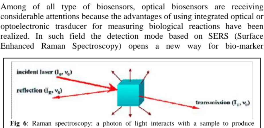
Among of all type of biosensors, optical biosensors are receiving considerable attentions because the advantages of using integrated optical or optoelectronic trasducer for measuring biological reactions have been realized. In such field the detection mode based on SERS (Surface Enhanced Raman Spectroscopy) opens a new way for bio-marker
recognition [Wang et al., 2007].
In laser-based optical spectroscopy, Raman scattering generates a fingerprint-like vibrational spectrum for individual molecular species with features that are much narrower than fluorescence. Raman scattering can be generated using monochromatic far-red or near-IR light, photon energies too low to excite the inherent background fluorescence in biological samples (fig. 6). In addition, water is a very poor Raman scatterer, and combined with all the features described, makes Raman a useful tool in detecting molecular species in biological samples.
SERS takes advantage of strongly increased Raman scattering signals generated by local field enhancements near metallic nanostructure and can be applied for label free analyte detection, revealing molecular fingerprints. A Raman dye can be either fluorescent or non fluorescent, and a minor chemical modification of a dye molecule can lead to a new dye with a different Raman spectrum, even if the two dyes exhibit virtually indistinguishable fluorescence spectra [Sassolas et al., 2006]. A variety of metal structure (Au Ag and Cu) are used to induce SERS effect. These metal are used in different format as metal plates, colloids rods coating [Knauer et al., 2010].
The spectral specificity of a SERS scattering probe is excellent in comparison to that of the fluorescence method. For example, the spectral bandwidths of cresyl fast violet in UV adsorption and fluorescence are broad whereas the bandwidth of the SERS spectrum of the same dye is
Fig 6: Raman spectroscopy: a photon of light interacts with a sample to produce
narrower. The dye-labeled SERS active substrates can provide richer spectral information than fluorescence based signatures, which are often limited by spectral overlap of the chromophores and by background signals due to other components in the sample [Kneipp et al., 1997; Wang et al. 2007].
The SERS technique was reported as a tool for detecting specific nucleic acid sequences. A SERS aptasensor was described to detect thrombin. Thiolated thrombin-binding aptamers were immobilized onto a gold substrate. A sandwich structure was formed between the immobilized aptamer, the protein target and a secondary aptamer bound to AuNPs, which were labeled by a Raman reporter (R6G). Then, silver NPs aggregated on AuNPs yielding electromagnetic hot spots. Thus, the Raman signal of the R6G was greatly enhanced due to the large electromagnetic coupling effect produced by the hot spots between AgNPs and AuNPs. The detection limit of this SERS aptasensor was 5×10⁻¹⁰M. A reagentless aptameric biosensor based on SERS spectroscopy was also developed to detect cocaine. Tetramethylrhodamine (TMR)-labeled aptamer was immobilized on a SERS substrate. In the absence of the target, the aptamer was partially unfolded. So the TMR moiety remained away from the sub-strate and yielded a weak SERS signal. In the presence of cocaine, the aptamer folded into stable three-way junction, in which the TMR moiety came in close proximity to the SERS substrate, generating an enhanced SERS signal. Detection limit was 10−6M . SERS “aptatags” were used to develop a biosensor for thrombin detection. “Aptatags” were composed by AgNPs linked together using a small organic molecule bearing two thiol functionalities: biphenyl-4,4’-dithiol (DBDT). DBDT served as the linker and as the SERS reporters. Then, “aptatags” were functionalized with thiolated thrombin-binding aptamers. First, aptamers were immobilized onto a silver layer deposited over a silicon wafer. After interactions between thrombin and specific aptamers, the surface was treated with the SERS “aptatags”. A sandwich complex was formed between immobilized aptamers, the target and aptamers on the SERS “aptatags”. The SERS signature of the linker holding the nanoparticles together indicated the presence of thrombin. Detection limit was 10−10M [Sassolas et al., 2006]. An optical aptamer-based detection system label free appears as highly efficient device with enormous potential. Unfortunately, such systems are still immature compared to immunoassays, reflecting the limited availability
17
Reference
Byfield, M.P. and Abuknesha, R.A. (1994). Biochemical aspects of biosensors Biosensors and Bioelectronics 9 (4-5): 373-400.
Blohm, D.H., and Guiseppi-Elie, A. (2001). New developments in microarray technology. Current Opinion in Biotechnology, 12 (1): 41-47.
Bock, L.C., Griffin, L.C., Latham, J.A. Vermaas, E.H. and Toole J.J. (1992). Selection of single-stranded DNA molecules that bind and inhibit human thrombin Nature 355: 564-566.
Coughlin, S.R. (2000). Thrombin signalling and protease-activated receptors. Nature 407: 258–264.
Cruz-Aguado, J.A., and Penner, G. (2008). Determination of ochratoxin A with a DNA aptamer. Journal of Agricultural and Food Chemistry, 56 (22): 10456-10461.
Ellington, A.D., Szostak, J.W. (1990). In vitro selection of RNA molecules that bind specific ligands. Nature 346: 818–822.
Emanuel, P.A., Dang, J., Gebhardt, J.S., Aldrich, J., Garber, E.A.E., Henrieta, K., Stopa, P., Valdes, J.J., Schultz, A.D. (2000). Recombinant antibodies: a new reagent for biological agent detection. Biosens. Bioelectron., 14: 761-770.
Gagliardi, S., Rapone, B., Mosiello, L., Luciani, D., Gerardino, A., Morales, P. (2007). Laser- assisted fabrication of biomolecular sensing microarrays, IEEE Transactions on Nanobioscience, 6 (3): 242-248.
Ghosh, S, Gepsten, S., Heikkila, J.J., Dumbroff, E.B. (1988).Use of a Scanning Densitometer or an ELISA Plate Reader for Measurement of Nanogram Amounts of Protein in Crude Extracts from Biological Tissues Analytical Biochemistry 169. 377-233
Gopinath, S.C.B. (2007). Methods developed for SELEX Anal. Bioanal. Chem., 387:171–182.
Hianik, T., and Wang, J. (2009) Electrochemical Aptasensors – Recent Achievements and Perspectives. Electroanalysis, 21: (11) 1223-1235.
Hianik, T., Grman, I. and Karpisova, I. (2009). The effect of DNA aptamer configuration on the sensitivity of detection thrombin at surface by acoustic method. Chem. Commun., 7 (41): 6303–6305.
Klussmann, S. (2006). The Aptamers Handbook, Wiley-VCH.
Knauer, M., Ivleva, N.P., Liu, X., Niessner, R. and Haisch, C. (2010). Surface-Enhanced Raman Scattering-Based Label-Free Microarray Readout for the Detection of Microorganisms. Anal. Chem., 82 (7): 2766–2772.
Kneipp, K., Wang, Y., Kneipp, H, Perelman, L.T., Itzkan, I, Dasari, R.R, and Feld, M.S. (1997). Single Molecule Detection Using Surface-Enhanced Raman Scattering (SERS). Phys. Rev. Lett., 78: 1667–1670.
Koppal, T. (2004). Microarrays: migrating from discovery to diagnostics. Drug Discovery Development, 7: 30–34.
Kumar, A. (2000). Biosensors Based on Piezoelectric Crystal Detectors: Theory and Application JOM-e, 52 (10) on line.
Lamberti, I., Mosiello L., and Hianik, T. (2011). Development of thickness shear mode biosensor based on DNA aptamers for detection ochratoxin. Chemical Sensors, 1: 11-15.
Malhotra, B.D., Singhal, R., Chaubey, A., Sharma, S.K. and Kumar A. (2005). Recent Trends in Biosensors. Current Applied Physics, 5: 92-97.
O’Sullivan, C.K. and Guilbault, G.G. (1999). Commercial quartz crystal microbalances – theory and applications. Biosensor and Bioelectronics, 14:
19
Pacheco I.T. (2011). Aflatoxin - Detection, Measurement and Control (from chapter Biosensors for Aflatoxin Detection by Mosiello L. and Lamberti I.). InTech.
Pohanka, M., Pavliš, O., and Skládal, P. (2007). Rapid Characterization of Monoclonal Antibodies using the Piezoelectric. Immunosensor Sensors, 7: 341-353.
Sassolas A, Blum L. J., Leca-Bouvier B. D. (2011). Optical detection systems using immobilized aptamers. Biosensors and Bioelectronics, 26: 3725–3736
Sauerbrey, G. (1959). The use of oscillator for weighing thin layers and for microweighing. Z. Phys., 155: 206–210.
Schmid, R.D. (1987). Biosensors, Nachr. Chem. Tech. Lab., 35: 910-914.
Sethi, R.S. (1994). Transducer aspects of biosensors Biosensors and Bioelectronics, 9 (3):243-264
Song, S.; Wang, L.; Li, J.; Fan, C.; Zhao, J. (2008) Aptamer-based biosensors. Trends in Analytical Chemistry, 27: 108-117.
Stoltenburg, R., Reinemann, C., Strehlitz, B. (2007). SELEX—A (r)evolutionary method to generate high-affinity nucleic acid ligands Biomolecular Engineering, 24: 381–403.
Tombelli, S., Mascini, M., Sacco, C., Turner, A.P.F. (2000). A DNA piezoelectric biosensor assay coupled with a polymerase chain reaction for bacterial toxicity determination in environmental samples. Analytica Chimica Acta, 418: 1–9.
Tsai, W.-C., Hsieh, C.-K. (2007). QCM-based immunosensor for the determination of ochratoxin A. Analytical Letters, 40:(10) 1979-1991.
Tuerk, C., Gold, L. (1990). Systematic evolution of ligands by exponential enrichment: RNA ligands to bacteriophage T4 DNA polymerase. Science, 249: 505–510.
Turner, A.P.F., Karube, I. and Wilson, G.S. (1987). Biosensors - Fundamentals and Applications., Oxford University Press.
Turner, N.W., Subrahmanyam, S., Piletsky, S.A. (2009). Analytical methods for determination of mycotoxins: A review. Analytica Chimica Acta, 632 (2): 168-180.
Venkatasubbarao, S. (2004). Microarrays – status and prospects. Trends in Biotechnology, 22 (12): 630-637.
Vo-Dinh, T. and Cullum, B. (2000). Biosensors and biochips: advances in biological s in biological and medical diagnostics. Fresenius J Anal Chem, 366: 540–551.
Wang, Y., Wei, H., Li, B., Ren, W., Guo, S., Dong, S., Wang, E. (2007). SERS opens a new way in aptasensor for protein recognition with high sensitivity and selectivity. Chem.Commun., 28 (48):5220-5222.
Yang, Y., Kochoyan, M., Burgstaller, P., Westhof, E., Famulok, M. (1996). Structural basis of ligand discrimination by two related RNA aptamers resolved by NMR spectroscopy. Science, 272 (5266): 1343-1347
21
Aims
Aim 1: Protein Microarray Applications
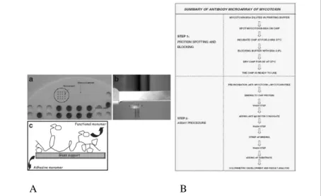
Research on microarrays as multi-analyte biosystems has generated increased interest in the last decade. According to this trend, we have tried to transfer the immunoassay method from microtiter plates into a microarray format in order to develop a multiparametric, rapid, sensitive and inexpensive method for the detection of mycotoxins for food safety application. To perform our test and check the feasibility of this format, we focused our studies on the most popular mycotoxins Aflatoxin B1 and Fumonisin B1 and developed a competitive immunoassay in a microarray format, using the Dr.Chip platform provided by Life Line Lab Co. (Pomezia, Italy) and used also for other applications.
Microarray platform is equipment to create microarrays and to read the final results, via densitometric detection, based on the enzymatic and colorimetric assay. In Fig. 7 A is reported a detail of the plastic probe tray for protein spotting and pins. In the same picture is also shown the scheme of the glass treated with functional protein linker.
As in other conventional competitive immunoassay, the color intensity and
A B
Fig. 7-A: Microarray spotting platform used for Aflatoxin B1 detection; B: Scheme of
corrispondent grey values obtained from antigen microarrays BSA-Afla B1, prepared as described in this paper and used in our immunological tests, are in inverse proportion to antigen concentration in standard solutions. Assay method for Aflatoxin is described in Fig. 7 B.
The aim of our second application was to develop a new and rapid method for studying H460 cells that have been induced to apoptosis as a possible new tool for oncology studies. As main focus of this aim, we study the feasibility to transfer the methods of the immunological assay for caspases from classical western blot into a microarray format in order to develop a multiparametric, rapid immunoassay, sensitive and inexpensive assay. Caspases are specific cytosolic proteases that are activated during apoptosis. Programmed Cell Death – PCD, or apoptosis, is a highly regulated process characterized by morphological and biochemical cellular changes. The selective induction of apoptosis in malignant cells may be an attractive mechanism to control neoplastic cell proliferation. Thus, factors that affect caspase activation and apoptosis might be important determinants for drug sensitivity. In addition, forms of cell death that are caspase dependent may also have a crucial role in the treatment response.
To test the feasibility of this format, we focused our studies on the three caspases that are critically involved in apoptosis: caspase-3 (both cleaved and uncleaved forms), caspase-8 and caspase-9. We developed different microarray formats using the commercial platform DR.Chip. Our results demonstrate that this platform is suitable for carrying out rapid immunological tests to identify cleaved and uncleaved caspase-3 as well as caspase-9. Simultaneous analyses for more of these proteases are mainly limited by the densitometric detection system associated with the microarray platform. In our opinion, the future use of fluorometric detection would offer great improvement for the microarray-based identification of caspases described in our work.
Our results demonstrate that this platform is suitable for carrying out rapid immunological tests to identify cleaved and uncleaved caspase-3 as well as caspase-9. Simultaneous analyses for more of these proteases are mainly limited by the densitometric detection system associated with the microarray platform. In our opinion, the future use of fluorometric detection would offer great improvement for the microarray-based identification of caspases described in our work.
23
A Novel Based Protein Microarray for the Simultaneous Analysis of Activated Caspases
(2010) Lamberti I, Mosiello L, Cenciarelli C, Antoccia A. and Tanzarella C. SENSORS AND MICROSYSTEMS, Lecture Notes in Electrical Engineering, 54 (4), pp. 323-326, DOI: 10.1007/978-90-481-3606-3_64
25
Aim 2: Quartz Crystal Sensor
The aim of this work was to analyse the binding of human thrombin to the DNA aptamers that differ in structure of binding site. As a basic we used aptamer that selectively bind thrombin in its fibrinogen-binding site. A pair of thymines on the G- quandruplex of the aptamer characterize thrombin binding site. We analysed the aptamers in which AA replaced TT pair, (AA, AT or TA aptamers, respectively). The aptamers were purchased from Thermo Scientific (Ulm, Germany). We used acoustic method based on quartz crystal microbalance (QCM) and thickness shear mode (TSM) for monitoring the aptamer-thrombin interaction. We showed that substitution thymines by adenines in the thrombin-binding site of G-quadruplex substantially affect both thermodynamic and binding properties of the DNA aptamers immobilised on TSM transducer.
In a second application we used a DNA aptamer sensitive to ochratoxin A. The aptamers were immobilised at the gold layer of quartz crystal transducer.. We showed that TSM allowing detecting this mycotoxin with LOD 30 nM and with good selectivity. He also studied the stability of DNA aptamers depending on concentration of calcium ions, that are important for binding OTA to DNA aptamer.
A Piezoelectric Quartz Crystal Sensor Applied for Thrombin-Binding Aptamers (2011)
Rakitka J., Hianik t. Lamberti I., Mosiello L., SENSORS AND MICROSYSTEMS 91 (5): 449-453, ISBN: 9789400713239, DOI: 10.1007/978-94-007-1324-6_73
Development of a Thickness Shear Mode Biosensor based on DNA Aptamers for detection Ochratoxin A (2011) Lamberti I Mosiello L Hianik T Chemical Sensors 1, 2011:11
Aim 3: Sensing Surface for SERS Application
We present the preparation of an aptamer terminated sensing surface, allowing a fast and cheap system for assay of analytes unlimited by size and tossicity. The designed platform is suitable for an aptamer-based microfluidic device, coupled with a Surface Enhanced Raman Spectroscopy (SERS). To accomplish this, a multi-step deposition sequence was performed: as a start, a mixed self assembled monolayer (SAM) containing a binary mixture of biotinylated alkylthiol (BAT) [1]. with the capacity to graft neutravidin proteins and diluent methyl-terminated alkylthiol, was prepared on a gold thin film. The chemical and electronic structure of the mixed SAMs was investigated by X-ray Photoelectron Spectroscopy and IRRAS (Infrared Spectroscopy in Reflection Mode) [2].
In a second step, the bioconjugation with an avidin-gold sol is performed. Monodispersed 5 nm gold particles were prepared in aqueous medium and covered with neutravidin, overcoming possible non-specific bindings, taking usually place at the isolectric point (~ 6) of neutravidin [3, 4].
The sample morphologies were observed by atom force microscopy (AFM), the size was determined by Dynamic Light Scattering and the concentration of gold species in the colloid was monitored by UV-vis spectra [5, 6]. In the ending step, a sensing aptamer (i. e. thrombin-binding aptamer) is bond to the surface through the avidin-biotin linkage. The resulting SERS changes involved was observed when the target molecule (i.e. thrombin) interacts with its own aptamer [7].
This aptamer terminated sensing surface is fitted for affinity based microfluidic devices, providing controlled fluid transport, rapid affinity assay and cost saving advantages over conventional methods for biological and medical applications. Through such design, the sensing surface overcomes the sandwich structure, formed between the immobilized aptamer, the protein target and a secondary aptamer bound to the Raman probe (i.e. gold nanoparticles ), usually realized in SERS aptasensors, limited by consuming and inconvenient handling step [8].
References
[1] Ptrats-Alfonso E., Garcia-Martin F.,Bayo N., Cruz L.J., Pla-Roca M., Samitier J., Errachid A., Albericio F., (2006) Tetrahedron, 62, 6876-6881.
49
Surface Characterization of Mixed Self-Assembled Monolayers Designed for Streptavidin Immobilization Langmuir. 17: 2807–2816.
[3] Grabar, K.C., Freeman R.G., Hommer M.B., and Natan M.J. (1995) Preparation and Characterization of Au colloid Monolayers, Anal. Chem., 67: 735-743.
[4] Morris R.E. and Saelinger C.B., (1984) Visualization of intracellular trafficking: use of biotinylated ligands in conjunction with avidin-gold colloids. J.Histochemistry and Cytochemistry, 32, 124-128.
[5] Kimling J, Maier M, Okenve B, Kotaidis V, Ballot H, Plech A. (2006) Turkevich method for gold nanoparticle synthesis revisited. J Phys Chem B., 110 (32):15700-15707.
[6] Ji X, Song X, Li J, Bai Y, Yang W, Peng X.,. (2007) Size control of gold nanocrystals in citrate reduction: the third role of citrate.
J.Am.Chem.Soc. 129: 13939-13948.
[7] Nie S.; and Emory S. R., (1997) Probing Single Molecules and Single Nanoparticles by Surface-Enhanced Raman Scattering Science 275: 1102,.
[8]. Sassolas A, Blum L. J., Leca-Bouvier B. D., (2011) Optical detection systems using immobilized aptamers Biosensors and Bioelectronics 26: 3725–3736
51
Publications
Impedimetric DNA Aptasensor for Sensitive Detection of Ochratoxin A in a Food. Castillo G., Lamberti I., Mosiello L., Hianik T.
ELECTROANALYSIS (in press)
Biosensors for Aflatoxin Detection Mosiello L., and Lamberti I., Chapter
Title from Aflatoxin - Detection, Measurement and Control by Irineo Torres Pacheco. ISBN:978 953 307 711 6
High Sensitive Impedimetric Aptasensor for Detection Ochratoxin A In Food Castillo G., Lamberti I., Mosiello L., Hianik T. SENSORS AND
MICROSYSTEMS World Scientific Publishing co. ISBN:978114609342 in press
*A Piezoelectric Quartz Crystal Sensor Applied for Thrombin-Binding Aptamers (2011) Rakitka J., Hianik T., Lamberti I., Mosiello L.,
SENSORS AND MICROSYSTEMS 91 (5): 449-453, ISBN:
9789400713239, DOI: 10.1007/978-94-007-1324-6_73
*An antibody-based microarray assay for the simultaneous detection of aflatoxin B1 and fumonisin B1 (2009) Lamberti I., Tanzarella C.,
Solinas I., Padula C., Mosiello L. Mycotoxin Research, 25 (4): 193-200. MYCOTOXIN RESEARCH ISSN: 01787888 DOI: 10.1007/s12550-009-0028-9
*Development of a thickness shear mode biosensor based on DNA aptamers for detection ochratoxin A (2011) Lamberti I., Mosiello L.,
Hianik T., Chemical Sensors 1: 11-15 ISSN 2231-6035.
*A Novel Based Protein Microarray for the Simultaneous Analysis of Activated Caspases (2010) Lamberti I, Mosiello L, Cenciarelli C,
Antoccia A. and Tanzarella C. SENSORS AND MICROSYSTEMS, Lecture Notes in Electrical Engineering, 54 (4): 323-326, DOI: 10.1007/978-90-481-3606-3_64.
Development of a Protein Microarray System Antibody based for Mycotoxins Determination Mosiello L, Lamberti I, Vitali F, Misiti S And
Di Giorgio G, SENSORS AND MICROSYSTEMS World Scientific Publishing co. ISBN: 10 981-283-597-0.
Toxicity of toner nanoparticles on RT112 cell cultures Mosiello, L.,
Zappa, G., Zoani, C., Lamberti, I., Gatti, R., Pilloni, L.(2009) Proceedings Of The 9th IEEE Conference on Nanotechnology, IEEE NANO 2009, art. no. 5394752, pp. 616-618.
Conference
Integrated approach to a sensitive platform for SERS aptamer biosensors, suitable for a microfluidic device. I. Lamberti, A. Antoccia,
F. Duconge, G. Iucci, M. Papi, L.G. Quagliano and S. Foglia CONVEGNO NAZIONALE SENSORI (2012) (submitted)
Overcoming of gold nanoparticles sandwich for sensitive platforms in microfluidic devices, based on surface-enhanced Raman scattering Foglia S., Lamberti I.,Quagliano L.G., Papi M., Antonini G., Polzonetti G., and Tanzarella C. NANOTECHITALY 2011
NOVEMBER 23rd, 24th November
Aptamer based SERS sensing surface for direct detection of proteins.
Lamberti I., Antoccia A., Battocchio C. Quagliano L, Tanzarella C, Iucci G., and Foglia S. Proceedings Of The 6th Mondial Conference on Biomimetics, Artificial Muscles and Nano-Bio Cergy-Pontoise, France 25-26-27 October 2011.
Development of thickness shear mode biosensor based on DNA aptamers for detection ochratoxin A Lamberti I., Mosiello L., Hianik T.
Sensors And Microsystems, Proceedings Of The 16th Italian Conference, Rome, Italy 7- 9 February 2011 ENEA –C.R. Casaccia - Via Anguillarese, 301 - 00123 Roma; Edited By C Di Natale, A D'amico (University Of Rome "Tor Vergata" and Cnr-Imm, Italy)
53
(Università degli Studi di Messina, Italy) A D'amico (University Of Rome "Tor Vergata" and Cnr-Imm, Italy)
A Novel Based Protein Microarray for the Simultaneous Analysis of Activated Caspases Lamberti I., Mosiello L., Cenciarelli C., Antoccia A.,
Tanzarella C. Sensors And Microsystems, Proceedings Of The 14th Italian Conference, Pavia, Italy 24-26 February 2009 Edited By Andrea Baschirotto, Piero Malcovati e Arnaldo D'Amico (University Of Rome "Tor Vergata" and Cnr-Imm, Italy)
Development of a Protein Microarray System Antibody based for Mycotoxins Determination Mosiello L, Lamberti I, Vitali F, Misiti S
And Di Giorgio G, Sensors And Microsystems, Proceedings Of The 13th Italian Conference, Rome, Italy 19- 21 February 2008 Edited By C Di Natale, A D'amico (University Of Rome "Tor Vergata" and Cnr-Imm, Italy)
Toxicity of toner nanoparticles on RT112 Cell Cultures Lucia Mosiello,
Giovanna Zappa, Claudia Zoani, Ilaria Lamberti, Rosanna Gatti, Luciano Pilloni Proceedings Of The 9th Nanotechnology Conference IEEE NANO 2009 Genoa July 26-30 2009
Materiali di Riferimento per la determinazione di micotossine e la valutazione di contaminazioni fungine in prodotti alimentari Rosanna
Gatti, Ilaria Lamberti, Lucia Mosiello, Giovanna Zappa, Claudia Zoani ENEA – Dipartimento Biotecnologie Agroindustria e protezione della Salute C.R. Casaccia - Via Anguillarese, 301 - 00123 Roma; [email protected] III Congresso Nazionale: Le Micotossine nella Filiera Agro-Alimentare e Zootecnica 28-29-30 settembre 2009 Roma By Istituto superiore di Sanità Dipartimento Sanità Pubblica Veterinaria e Sicurezza Alimentare Reparto OGM e Xenobiotici di Origine Fungina
Conclusions
The main results obtained during the PhD work consist in the characterization of the best approach for the development of biosensors in order to detect concentrations and ligand affinity parameters of free unlabeled molecules in real time.
In recent years, the antibody microarray technology has made significant progress, going from proof-of-concept design to established high-performing technology platforms capable of targeting non-fractionated complex samples, as proteoma.
We have studied microarray detection in standard solutions with a low detection limits and developed a competitive immunoassay in microarray format for simultaneous analysis. The quality of the microarray data is comparable to data generated by microplate based immunoassay, and for this reason it represents just a useful insight in order to arrange further experiments to develop a rapid test or kits. Our method could be used as a semi quantitative tool for rapid pre-screening and further investigations are needed in order to improve the reported microarray performance mainly in term of sensitivity.
We moved from antibodies to aptamers because the use of functional aptamers as bioreceptors has become a new interdisciplinary field that aims at providing new hybrid sensing systems for specific and high sensitive molecular recognitions. This novel integration has yield various types of sensor for selective and sensitive detection of a wide range of analytes. Then, we focused on QCM aptasensor. We used, as model systems, two different analytes, thrombin and ochratoxin, in association with specific their aptamers as immobilized ligands. Our experiments prove that aptamers are equivalent to antibodies in terms of specificity and sensitivity and are one of the best approach for affinity biosensors construction.
We showed that acoustic methods (such as QCM) are perspective tool for label-free detection of low molecular toxins and protein markers. The detection of aptamers requires the present of ions that are probably responsible for their stability and formation of 3-D structure of the specific binding site.
We tried to develop a biosensor, based on direct method that do not require additional modification of receptor or complicated multi stage assay, with the aim to overcome the indirect detection methods in published literature.
55
In comparison to antibodies as capture molecules in biosensors, small aptamer receptors have a number of advantages. Because of their small size, denser receptor layers can be generated, meaning for a given receptor affinity, the sensitivity of these layers can be increased. They are produced in vitro; indeed no animals are needed.
The improvement of sensitivity, produced by changing the selective element (from antibodies to aptamers), has been assisted by a sensitility enhancement, applying a different trasducer (from acustic to optical methods).
We have approached the development of an affinity based detection system for analytes unlimited by size and tossicity, designing a nanoaptasensor sensing surface for microfluidic device, coupled with Surface Enhanced Raman Spectroscopy (SERS). Among the optical detection methods, SERS distinguishes itself with several advantages: the spectrum change brought by a single molecule, is induced by the specific interaction between aptamers and their own proteins and the spectral probe specificity is excellent in comparison to the fluorescence methods.
A thrombin SERS aptasensor is usually realized by a sandwich structure, formed between the immobilized aptamer, the protein target and a secondary aptamer bound to a Raman probe (i.e. gold nanoparticles), which requires a consuming and inconvenient handling step.
In attempt to overcome this drawback we designed an aptamer terminated sensing surface, allowing a faster and cheaper system and suitable for an analytical label free measurements, in order to perform a direct analysis of biological samples.
We also developed a multi-step depositional sequence where the last stage is the assay itself of the target molecules (i.e. thrombin), instead of the SERS probe deposition (i.e. gold nanoparticle), as planned to a classic sandwich structure. Such advantage allows to make improvements to costly and inconvenient handling steps.
In conclusion, we have investigated different experimental approaches of biosensible components of biosensors. Firstly, we developed a multi-parametric, rapid, sensitive and inexpensive method for the detection of mycotoxins for food safety and caspases for human health applications (immunoassay in a microarray format). Then, we moved to a different bioreceptor, choosing aptamers as selective element, improving the method sensitivity (QCM). Finally we designed a novel sensitive surface for a different detection mode (i.e. optical method, SERS) with higher sensitivity compared to the acoustic methods. This final improvement allows the possibility of submitting a patent application.
Acknowledgement
I would like to express my gratitude to Prof.ssa Caterina Tanzarella and Prof. Antonio Antoccia, my super advisors at the biologist department, University of RomaTre, my tutors Dr. Lucia Mosiello, (ENEA) and Dr Sabrina Foglia (CNR) for their great support.
I also would like to thank for their helpful collaboration:
Prof. Tibor Hianik, head of the Laboratory of Biophysics at Department of Nuclear Physics and Biophysics, Faculty of Mathematics, Physics and Computer Sciences, Comenius University.
Prof Giovanna Iucci, Dr Chiara Battocchio and Prof G. Polzonetti, CISDiC University of RomaTre and M. Papi, Physics departement, Catholic University S.C. of Rome.
![Fig. 1: Scheme of a Biosensor [Pacheco, 2011].](https://thumb-eu.123doks.com/thumbv2/123dokorg/2843261.5355/6.629.188.441.392.571/fig-scheme-of-biosensor-pacheco.webp)
![Fig. 5: Propagation of acoustic wave from the sensor surface [Pacheco, 2011].](https://thumb-eu.123doks.com/thumbv2/123dokorg/2843261.5355/17.629.168.454.435.586/fig-propagation-acoustic-wave-sensor-surface-pacheco.webp)