INDEX
INTRODUCTION
pag. I - VICHAPTER 1: Endocrine Disruptor Pesticides
1.1. Effects of Endocrine Disruptor Pesticides » 2
1.2. Fungicides » 4
1.2.1. Use » 4
1.2.2 Classification » 5
1.2.3. Environmental risk and toxicology » 6
CHAPTER 2: Pyrimethanil and Tebuconazole
2.1. Pyrimethanil » 10
2.1.1. Toxicity of pyrimethanil » 13
2.2. Tebuconazole » 16
2.1.1. Toxicity of tebuconazole » 20
CHAPTER 3: Studied species (Hyla intermedia)
3.1. Taxonomy » 23
3.2. General distribution » 23
3.3. Morphology » 25
3.4. Habitat and ecology » 26
3.5. Reproduction and development » 27
3.6. Population and conservation actions » 28
CHAPTER 4: Target organs
4.1. Urogenital system » 30 4.1.1. Kidneys » 30 4.1.2. Gonads » 32 4.1.3. Urogenital ducts » 35 4.1.4 Fat bodies » 36 4.2. Liver » 36
CHAPTER 5: Materials and Methods
5.1. Collection and animal husbandry » 39
5.2. Experimental design and exposure conditions » 39
5.3. Endpoints » 42
5.4. Statistical analyses » 43
5.5. Morphofunctional analysis » 44
5.1.1. Light microscopy analysis » 45
5.1.2. Immunofluorescence » 46
5.1.3. Sex ratio, LSI, GMCI. » 46
CHAPTER 6: Results
6.1. Effects of long-term exposure to pyrimethanil and tebuconazole, on survival and life history traits
» 48
6.1.1. Survival » 48
6.1.2. Morphological abnormalities » 52
6.1.3. GS 25-42: growth and general development » 55
6.1.4. GS 46: length, mass and time to metamorphosis » 57 6.2. Effects of long-term exposure to pyrimethanil on gonads, liver
and kidney
» 59
6.2.1. Control group » 59
6.2.1.1. Gonadal differentiation and histology » 59
6.2.1.2. Kidney morphology » 61
6.2.1.3. Liver morphology » 62
6.2.2. Pyrimethanil exposed groups » 62
6.2.2.1. Sex ratio and gonadal morphology » 62
6.2.2.2. Kidney histopathology » 64
6.2.2.3. Liver histopathology » 65
6.2.3. Immunofluorescence » 66
PLATES
» 69CHAPTER 7: Discussion
7.1. Effects of long-term exposure to pyrimethanil and tebuconazole, on survival and life history traits
» 78
7.1.1. Survival » 78
7.1.2. Morphological abnormalities » 80
7.1.3. Metamorphic traits » 80
7.1.3.1. Success at metamorphosis » 80
7.1.3.2. Mass and time to metamorphosis » 81
Conclusion » 83
7.2. Effects of long-term exposure to pyrimethanil on gonads, liver and kidney. » 84 7.2.1. Gonads » 84 7.2.2. Liver » 88 7.2.3. Kidneys » 90 Conclusion » 92
REFERENCES
» 93I
Introduction
A consequence of human population growth is the increase of global agricultural production and therefore the increased use, abuse, or misuse of pesticides all over the world (Köhler and Triebskorn, 2013). Aquatic biota inhabiting agricultural areas are often exposed to a wide variety of pesticides that reach freshwater ecosystems through un-intended direct application, spray drift and runoff, thus posing a potential risk for non-target species (Wagner et al., 2014).
Amphibians are currently the most globally threatened group of vertebrates and have experienced a drastic population decline. Among other established causes for this decline (e.g. habitat loss, increased disease susceptibility, climate changes), pesticide contamination is considered a primary factor (Bernabò et al., 2011a; Brunelli et al., 2009; Smalling et al., 2013, 2015). Amphibians are particularly sensitive to pollution due to their complex life cycle and permeable skin; in fact, in amphibians skin permeability is higher than in any other order of vertebrates and therefore the percutaneous absorption of xenobiotics is greater (Quaranta et al., 2009). Aquatic habitats, in which amphibians live, breed and develop, are easily contaminated by a range of pollutants, and in agricultural landscapes the likely exposure scenario is represented by pesticides (Aldrich et al., 2016; Brühl et al., 2011, 2013; Fryday and Thompson, 2012; Mann et al., 2009). Many of these substances have been shown to exert their adverse effects through modulation and/or disruption of endocrine functions, and are known as endocrine disrupting chemicals (EDCs) (Hayes et al., 2006; McKinlay, 2008; Orton and Tayler 2015, Orton et al., 2011,). Given the crucial role of the endocrine system in the
II
maintenance of numerous biological, physiological and behavioural functions, damage in any part of this complex system can lead to serious disease or death. A number of laboratory-based studies demonstrated that in amphibians, a perturbation of the endocrine system has the potential to dramatically affect all biological processes, including growth, development, gonadal differentiation, hormone levels and liver function (Bernabò et al., 2011b; Hayes et al., 2006, 2010; Higley et al., 2013; Kloas, 2002; Navarro-Martín et al., 2014; Orton and Tayler, 2015). There is also good evidence that amphibian populations, living in agricultural areas, have been affected by many endocrine-related disorders that can be linked to endocrine disrupting potential of pesticides (e.g. skewed sex ratio, greater incidences of gonadal anomalies, male and female reproductive dysgenesis, altered secondary sex characteristics, sex steroid and thyroid hormone disruption) (McCoy et al., 2008; McDaniel et al., 2008; Orton and Routledge, 2011; Papoulias et
al., 2013).
In recent years pesticide use is changing and fungicides have become the most important component of pest and disease management programs in modern et al., 2014; Reilly et al et al., 2014). As a result of repeated applications, chronic exposure scenario becomes most likely, and the concentrations often exceed the chronic toxicity values of concern (Belden et al., 2010; Deb et al., 2010; Reilly et al., 2012). Furthermore, most fungicides do not have specific modes of action and they may be toxic to a wide range of organisms (Maltby et al., 2009). Despite the widespread occurrence of fungicides in aquatic
III
environments, ecotoxicological data are surprisingly sparse, compared with other types of pesticides (Reilly et al., 2012; Smalling et al., 2013; Wightwick et al., 2012). Fungicides are generally or entirely overlooked by amphibian conservation biologists (Ghose et al., 2014).
Available data indicate that fungicides, at environmentally relevant concentrations, can induce several harmful effects on amphibians, such as increased mortality and deformity (Brühl et al., 2013; Méndez et al., 2016; Yu et al., 2013a), decreased and/or increased growth rate and development (Brande-Lavridsen et al., 2010; Hartman et al., 2014; Higley et al., 2013; Yu et al., 2013b), alteration of behaviour (Teplitsky et al., 2005), immunosuppression and lipid peroxidation (McMahon et
al., 2011; Strong et al., 2016a). Most if not all of those harmful effects could be
related to endocrine disturbance and actually a variety of fungicides are known or suspected to act as EDCs (Matthiessen and Weltje, 2015; McKinlay, 2008; Orton et
al., 2011; Poulsen et al., 2015).
In this context, we simulated a chronic contamination using two different common-used compounds, tebuconazole and pyrimethanil. These two fungicides are extensively applied on cereals, vineyards, fruits, vegetables and ornamentals with protective, curative, and eradicative purposes (EFSA, 2006, 2014) and are among the most frequently detected and relatively persistent pesticides in both surface waters and sediments (De Gerónimo et al., 2014; Herrero-Hernández et al., 2013; Robles-Molina et al., 2014; Smalling et al., 2012, 2015; Thomatou et al., 2012; Wightwick et al., 2012).
Information on pyrimethanil acute and chronic toxicity is scarce especially on vertebrates. Seeland and colleagues (2012) reported that pyrimethanil LC50/EC50
IV
aquatic organism range from 1.18 to 46.1 mg/L. Available study on fish reported that lethal concentration for the rainbow trout (Oncorhynchus mykiss) ranges between 14 and 35 mg/L (van Leeuwen and Vonk, 2008), whereas Araújo and collaborators (2014a) found that pyrimethanil, even at non-lethal concentrations, could be environmentally disruptive by triggering spatial avoidance in juveniles of
Danio rerioexposed to a pyrimethanil gradient. The same authors demonstrated in two amphibian species exposed to pyrimethanil-contaminated water that spatial distribution was influenced by the presence of fungicide (Araújo et al., 2014b). Tebuconazole is moderately toxic to freshwater fishes and LC50-96h values range
from 2.37 to 19.6 mg/L (EFSA, 2014; Kreutz et al., 2008; Sancho et al., 2010; Toni
et al., 2011a). The exposure to sublethal concentrations of tebuconazole may affect
fish growth and survival, and induce oxidative stress, physiological impairment, changes in metabolism, chronic inhibition of the stress response and severe hepatic cell injuries (Ferreira et al., 2010; Koakoski et al., 2014; Sancho et al., 2010; Toni et al., 2011a,b). Moreover, it has been shown that this fungicide has bioaccumulation potential in fish (Andreu-Sánchez et al., 2012; Konwick et al., 2006). Concerning amphibians, previous studies have reported the occurrence of tebuconazole in frog tissue (Hansen et al., 2014; Poulsen et al., 2015; Smalling et
al., 2013).
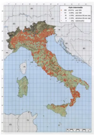
In spite of their widespread occurrence in environmental compartments, no previous study has examined sublethal effects of tebuconazole and pyrimethanil on amphibians. On this basis, we performed a long-term exposure to environmentally relevant concentrations (5 and 50 µg/L) of tebuconazole and pyrimethanil, on a native species to Italy Hyla intermedia. This species was chosen
V
because typically lives in inland water bodies and it is frequently found in agricultural areas.
In order to have a more comprehensive overview of the fungicides induced effects we evaluated survival, growth, developmental traits and incidence of deformities, and we also explored indicators of post-metamorphic fitness such as mass at and time to metamorphosis. This analysis is very informative to determine the factors that may affect amphibian population in nature.
In evaluating the effects of a toxicant in amphibians it is important to consider also lagged effects that may not become evident until metamorphosis (Bernabò et al., 2011b; Hayes et al., 2006; Tamschick, 2016b). On this basis, here we focused on the effects induced by an exposure to pyrimethanil during the whole developmental period, in H. intermedia juveniles.
Several studies in mammalian and fish models, both in vitro and in vivo, suggested that pyrimethanil may influence the biosynthesis of sexual hormones and/or interact with sexual hormone receptor thus acting as EDC (Ankley et al., 2005; Medjakovic et al., 2014; Orton et al., 2011; 2014; Prutner et al., 2013;). Given the fact that EDCs are responsible of many adverse reproductive outcomes in developing amphibians we first evaluated gonads histology, in order to identify putative effects on sex ratio and gonadal differentiation. EDCs may also act through broader mechanisms/pathways than firstly recognized, exerting different effects in a tissue specific manner (Bernabò et al., 2014; Haselman et al., 2016), therefore we also analysed morphological alterations in two organs highly susceptible to xenobiotic toxicity.
The kidney is an important site of injury after chemical exposure, due to their involvement in a number of interrelated functions (i.e. maintenance of internal
VI
water, ion, and acid-base balance, selective reabsorption and secretion of ions and organic molecules, and excretion of nitrogenous and other waste products of metabolism) (Cakici, 2015; Fenoglio et al., 2011; Strong et al., 2016b). The liver has long been considered the major target organ for most chemicals, including EDCs, in consequence of its essential functional features (i.e. maintaining of the metabolic homeostasis of the body including protein synthesis, storage metabolites, detoxification and inactivation of harmful substances) (Bernabò et al., 2014; de Oliveira et al., 2016; Melvin et al., 2013).
To our knowledge no previous studies have investigated the effects of pyrimethanil on the selected organs in amphibians.
1
Chapter 1
Endocrine Disruptor Pesticides
Endocrine disrupting chemicals (EDCs) are compounds that are able to modify the normal functioning of the endocrine system of both wildlife and humans. A large number of chemicals have been identified as endocrine disruptors, among them several pesticides (Mnif et al., 2011).In industrialized countries, with the Green Revolution of the 1960s the agricultural productivity greatly increased and numerous pesticides have been developed and used extensively worldwide, in order to eradicate the threatening pathogens, with few guidelines or restrictions (Mellanby, 1992; Briggs, 2009). This fight, therefore, requires the massive use of pesticides, which are hazardous chemicals designed to repel or kill rodents, fungi, insects, and weeds that undermine intensive farming. Pesticides represent a great benefit for increased food production (Cooper and Dobson, 2007). However, many pesticides for agricultural use, or agrochemicals, are harmful to the environment, in fact, they can persist in soils and aquatic sediments, bioconcentrate in the tissues of invertebrates and vertebrates, move up trophic chains, and affect top predators including humans (Mnif et al., 2011).
Worldwide consumption of agrochemicals is steadily rising and consequently humans and wildlife are now continuously exposed to various of agrochemicals though the
2 environment (surface water, groundwater, soil), food and drinking water (Kolpin et al., 2000)
The World Health Organization has reported that roughly three million pesticide poisonings occur annually, resulting in 220,000 deaths worldwide (WHO, 1992, 2007) In some cases, it has been suggested that diseases such as cancer, allergies, neurological and reproductive disorders may be connected to pesticide exposure.
1.1. Effects of Endocrine Disruptor Pesticides
More than 100 substances of the several identified EDCs are pesticides (Andersen et al., 2002; Kojima et al.,2004; Lemaire et al., 2006a,b; Vinggaard et al., 2000). Of these, 46% are insecticides, 21% herbicides and 31% fungicides (Mnif, et al., 2011). In general, EDCs interfere with the hormonal homeostasis binding to estrogen or androgen receptors. More in detail, EDCs can act as receptor agonist miming the natural hormone’s action, thus binding to and activating various hormone receptors, including androgen, estrogen and aryl-hydrocarbon receptors. EDCs can also act as an antagonist by binding to these receptors without activating them and consequently inhibiting their action. Finally, EDCs can interfere through different feedback mechanisms with the synthesis, transport, metabolism and elimination of hormones, thus decreasing the concentration of natural hormones (Tabb and Blumberg, 2006). At the environmental level, wildlife is particularly vulnerable to the endocrine disrupting effects of pesticides (Mnif et al., 2011). Endocrine-related effects have been amply observed in invertebrates (Ellis and Pattisina, 1990; Gooding et al., 2003; Heidrich et al., 2001), reptiles (Bishop et al., 1991,1995; Guillette et al., 1995, 1996,
3 1999), fish (Munkittrick et al.,1991; Purdom et al., 19994), birds(Crisp et al., 1998; Fry, 1981; Fry et al., 1987; Tyler et al., 1998 ) and mammals (Facemire et al., 1995; Oskam et al., 2003; Reijnders, 1986) as reviewed by Mnif and colleagues (2007). Most of this effects are related to exposure to organochlorine pesticides (OC) and disturb the reproductive function. For example, endosulfan sulphate affects embryonic development and juvenile hormone activity of Daphnia magna (Palma et al., 2009a,b). Another OC, linuron, influences the production of reproductive hormones in rats after an in utero exposure; testosterone levels was strongly reduced, whereas progesterone production was not affected (Wilson et al., 2009).
Endocrine disrupting pesticides are also able to interfere with the reproductive and sexual development in humans, and many of these detrimental effects occur during gametogenesis and the early development of the fetus (Anderson, 2000; Hardell et al., 2006; Sharpe, 2006; Skakkebaek, 2001; Sultan et al., 2001; Waliszewski et al., 2000). Nevertheless, the effects may not become apparent until adulthood. Also, fetuses and newborns receive larger doses of EDCs because of the mobilization of maternal fat stores during pregnancy and breastfeeding (Anderson, 2000; Hardell et al., 2006; Przyrembel et al., 2000; Skakkebaek, 2001; Waliszewski et al., 2000). Infants are tremendously susceptible to pre and postnatal exposure to these compounds, resulting in a large variety of negative health effects, including the likely long-term impact on intellectual function (Eskenazi et al, 2006 ;. Jacobson and Jacobson, 1996) and the lagged effects on the central nervous system functioning (Beard, 2006; Ribas-Fito et al., 2003).
4 1.2. Fungicides
Nowadays many fungicides are known or suspected to act as EDCs (Orton et al., 2011; Poulsen et al., 2015; Matthiessen and Weltje, 2015; McKinlay, 2008). Fungicides are chemical or biological agents that specifically inhibit or kill fungi underlying diseases important to man. Fungal infections may cause severe damage in agriculture resulting in critical loss of yield, quality and profit (Oruc, 2010; Rouabhi, 2010).
1.2.1. Use
Fungicides are used both in agriculture and in the struggle against fungal infections in humans and animals. In agriculture, these pesticides are applied to protect the fruits, tubers and vegetables during storage or are applied directly to grapes, ornamental plants, trees, field crops, cereals and turf grasses (Gupta and Aggarwal, 2007).
In veterinary medicine, fungicides are commonly used in the treatment of foot rot disease but are also utilized to repel and kill slugs and snails (Ortolani et al., 2004). Another example of dual use of fungicides is the treatment of intestinal parasites in both human and veterinary medicine with the widely used agricultural fungicide thiabendazole (Lorgue et al., 1996).
In addition, many fungicides are used to protect industrial products during shipment, remove molds from painted surfaces, wood preserving, controlling fungal growth in paper production and protect the carpets integrity (Osweiler et al., 1985).
5 1.2.2. Classification
The classification systems proposed by different authors for fungicides are numerous. Most of these are based on the chemical structure but this kind of classification somewhat lead to confusion rather than providing a streamlined list. In addition to classification for chemical structural group, fungicides can be classified according to their utilization (agricultural, clinical).
According to their origin, fungicides can be classed into two main groups (Rouabhi, 2010):
Biologically based fungicides (biofungicides) that contain living microorganisms, bacteria or fungi, that are antagonistic to the pathogens that cause the disease.
Chemically based fungicides that are synthesized from organic and inorganic chemicals. most of the fungicides that are sold throughout the world are chemically-based.
A further classification of fungicides can be made according to the topical activity, we distinguish four groups (Rouabhi, 2010):
Contact fungicides. They are able to act only on the surface of the plants without being absorbed from leaves, stems or roots, so they cannot inhibit the fungal growth from the inside of the plants (dithiocarbamates, nitriles, aromatic hydrocarbons, peroxides, phenylpyrolles, cyanoimidazoles).
Localized penetrants. These fungicides inhibit fungi on treated plant surfaces and inside treated leaves. They are absorbed only by the treated leaves and
6 cannot move from one leaf to another. They are not absorbed by the roots (dicarboximides, most of the strobilurins).
Acropetal penetrants. These fungicides can penetrate inside the plants through roots, shoots and leaves. They inhibit fungi on and in treated plant surfaces and inside plant parts that lie above the treated surface. (benzimidazoles, triazoles, pyrimidines, carboximides, acylalanines, plus the strobilurins azoxystrobin and fluoxastrobin).
Systemic fungicides. They are the only fungicides able to be absorbed into xylem and phloem and moves up and down in plants. These fungicides inhibit fungi on and in treated plant surfaces and inside plant parts that lie above or below the treated surfaces (phosphonates).
Fungicides can be also divided into two groups based on mode of action in fungal cells (Rouabhi, 2010):
Site-specific inhibitors, that target individual sites within the fungal cell. Multi-site inhibitors, that target many different sites in each fungal cell.
1.2.3. Environmental risk and toxicology
Given the crucial role in the agricultural sector, in recent years the use of fungicides has increased significantly (Battaglin et al., 2010). In fact, most of the diseases caused by fungi is difficult to eradicate and requires a massive and repeated use of fungicides. Furthermore, modern farming practices also include the adoption of preventive strategies to control the spread of fungal diseases and this involves a regular
7 application of these substances for the whole plant growing season, even when infection is not present (Wightwick et al., 2012).
As a result, the frequent use of fungicides may pose a risk to the environment, particularly if the residues persist in soil or migrate out of the application site, with negative consequences for the health of both terrestrial and aquatic ecosystems (Komarek et al., 2010; Wightwick and Allinson, 2007; Wightwick et al., 2010). A limited number of studies have taken into account the presence of these compounds in surface waters and sediments. The few available data reveals that residues of fungicides are the most frequent and can reach high percentages in the samples analyzed (Gregoire et al. 2010; Rabiet et al. 2010; Smalling et al., 2012; Wightwick et al. 2010; 2012). These studies also indicate that the risk of contamination for aquatic ecosystems is more likely ascribable to long-term rather than short-term exposure (Wightwick et al., 2012). Aquatic organisms, therefore, may be exposed to fungicides through: atmospheric transport, runoff, leaching and runoff, the direct application of spray, the spray drift and the movement of animals through the fields during application (Junges et al., 2012; Belden et al., 2010). Moreover, for the majority of fungicides on the market there are no guide values on the concentrations which may cause adverse effects on non-target organisms (e.g. LC50, EC50) (Frampton et al., 2006;
Maltby et al., 2009; Wightwick et al. 2010; 2012).
The understanding of the specific mechanisms of action of fungicide and their toxicity is important because even humans, cattle and pets meet these substances through a wide range of applications. In fact, every year, livestock are poisoned accidentally by these pesticides. The available toxicological data examined the detrimental effects of
8 fungicides on model laboratory animals (rats, mice, rabbits) offering limited data on livestock, wild animals and pets. (Gupta and Aggarwal, 2007). In general, fungicides have been defined as low/moderate toxic for mammalian, although they are believed to have a higher overall incidence than other pesticides to cause developmental toxicology and oncogenesis (Costa, 1997).
It was estimated that over 80 per cent of oncogenic risk related to pesticides use, comes from a few fungicides (NAS, 1987).
Theoretically, since morphology and physiology of fungi greatly differ from those of other forms of life, fungi can be efficaciously fought by low toxicity compounds to other organisms, particularly mammals. (Edwards et al., 1991). However, since the mechanism of injury to pathogenic fungi may be different from that affecting mammals, it is possible that the two properties may coexist in a fungicide molecule (Marrs and Ballantyne, 2004). Since the mechanisms of action and metabolic clearance differ among the various existing fungicides, the specific effects (reproductive, teratogenic, mutagenic, carcinogenic) that may occur will be very different according to the poison ingested.(Hayes and Laws, 1990; USEPA, 1999). In addition, some animals may be more vulnerable to fungicide contamination than others due to their physiology and/or behaviour. For example, some fungicides (e.g., copper sulphate, thiram, chlorothalonil and captan) have especially toxic effects on fish (Pimentel, 1971; Lorgue et al., 1996; Tomlin, 2000), and bees (Hartley and Kidd, 1983). The kinds of fungicides used in both agricultural and industrial practices range from those of relatively low toxicity to those that may be lethal to animals.
9 Fungicides that are frequently used around the home pose a serious risk to pets and livestock consequently to lack of attention and misapplication (Osweiler et al., 1985; Gupta and Aggarwal, 2007; Oruc et al., 2009). For example, fungicides have caused systemic poisoning in animals such as sheep (Ortolani et al., 2004; Oruc et al., 2009), poultry (Guitart et al., 1999), and humans (Israeli et al., 1983; Kintz et al., 1997; Chodorowski, 2003; Kayacan et al., 2007; Calvert et al., 2008; Mortazavi and Jafari-Javid, 2009). incorrect application while using fungicides are probably responsible for a disproportionately large number of injuries irritating to skin and mucous membranes, as well as skin sensitization. Fungicides are often utilized in combination with other pesticides and adjuvants or solvents, which, together, may be more toxic (Osweiler et al., 1985). In France, Lorgue et al. (1996) reported that pesticides are the most common cause of animal poisoning (45.5%), with fungicides accounting for 6.1% of all pesticides. The two most commonly involved species are dogs and cattle. In 2003, 992 cases involving dogs and cats were confirmed as poisoning in France, and fungicides caused 2.8% of all poisonings (Barbier, 2005). Acute fungicide poisonings was 4.4% in 129 poisoning cases in Greece (Berny et al., 2009). In Italy, poisoning related with fungicides account was 8.1% of pesticides in pet poisonings (Caloni et al., 2004).
10
Chapter 2
Pyrimethanil and Tebuconazole
2.1. PyrimethanilThe fungicide pyrimethanil [N-(4, 6-dimethylpyrimidin-2-yl)-aniline; CAS number 53112-28-0] is an anilinopyrimidine fungicide that inhibits the secretion of fungal enzymes produced in the infection process (FAO/WHO, 2007; EFSA, 2011). It was developed to act on resistant fungi strains, mainly to control Botrytis cinerea in grapes, Venturia inaequalis in apples and Botrytis spp. in protein peas (EFSA, 2006), consequently its use has increased greatly in the lasted years (Smilanick et al., 2006; Sugar and Basile, 2008). Pyrimethanil rapidly penetrates the cuticle and inhibits the secretion of fungal enzymes required for the infection process, blocking the ability of fungi to degrade and digest the plant tissues, thus stopping the disease (Araújo et al., 2015; EFSA, 2006).
The commercial products that contain pyrimethanil as active ingredient are Clarinet®, Mythos®, Rubin®, Scala®, Siganex®, Vision®, and Walabi®, which are
11 currently used both pre- and post-harvest to protect various crops such as apple, banana, carrot, citrus, grape, melon, onion, potato, strawberry, and tomato (Smilanick et al., 2006; EFSA, 2011; Sirtori et al., 2012)
According to the EFSA report (2006), pyrimethanil does not bioaccumulate, is quickly eliminate once orally absorbed, is not teratogenic and has a low acute toxicity. However, several authors have demonstrated that pyrimethanil has the potential to induce negative effects on non-target organism after both acute and chronic exposure (Verdisson et al., 2012; Shinn et al., 2015; Seeland et al., 2012, 2013; Araújo et al., 2014a,b). Unluckily, despite the intensive agricultural use of this fungicide, there is a lack of information regarding the effects on adjacent aquatic ecosystems.
This is perhaps in relation to the assumption that pyrimethanil has a short half-life and, consequently, the possible toxic effects may occur in the short term, but are reduced to a minimum at long-term (EFSA, 2006; PPDB, 2009). Chemical and (eco)toxicological characteristics of pyrimethanil, published by EFSA, are summarized in Table 2.1 (Araújo et al., 2015; EFSA, 2006).
The detected concentrations in surface water and sediments for pyrimethanil range between 0.06–90 µg/L for pyrimethanil (EFSA, 2006; Gregoire et al., 2010; Herrero Hernández et al., 2013; Kreuger et al., 2010; Seeland et al., 2013; Thomatou et al., 2012; Wightwick et al., 2012).
12
Chemical name (IUPAC) N-(4, 6-dimethylpyrimidin-2-yl)
aniline
Chemical name (CA) 4,
6-dimethyl-N-phenyl-2-pyrimidinamine
Molecular formula C12H13N3
Molecular mass 199.28 g mol-1
Temperature of decomposition 189.54 to 344.74 °C
Flammability Not flammable
Explosive properties Not explosive
Skin irritation Not irritating
Eye irritation Not irritating
Genotoxicity No evidence Degradation time in water and sediment DT50 water 8.9 to 24 days DT90 water 70 to 99 days
DT50 whole system 40 to 121 days
DT90 whole system Not stated and 134 days Toxicity for
aquatic organisms
Rainbow trout LC50 (96 h):10.56 mg/L
Daphnia sp. EC50 (96 h): 2.9 mg/L
Green alga EbC50/ ErC50 (96 h):
1.2/5.84 mg/L
Daphnia magna NOEC (reproduction, 21 d):0.94 mg/L
Chironomus riparius NOEC (emergence, 28 d) 4.0 mg/L
Ecotoxicological data Harmful
CA: Chemical Abstract; DT50 and DT90: period required for 50% and 90% dissipation; EC50: median effective concentration;EbC50: the concentration at which
50% reduction of biomass is observed, ErC50: the concentration at which 50%
reduction of growth rate is observed, IUPAC: International Union of Pure and Applied Chemistry; LC50: median lethal concentration; NOEC: no observed effect
concentration.
Table 2.1. Chemical and (eco)toxicological characteristics of pyrimethanil (from Araújo et al., 2015).
13 2.1.1. Toxicity of pyrimethanil
The toxic effects induced by pyrimethanil exposure on different aquatic animals have been investigated in the last years. For Daphnia magna the 96 h LC50 (lethal
concentration to 50% of exposed organisms) of pure pyrimethanil ranged from 1.2 to 2.9 mg/L , while the NOEC (no observed effect concentration) on reproduction after 21 days of exposure range from 0.5 to 0.9 mg/L (EFSA, 2006; Seeland et al., 2012). In addition, D. magna exposed to 1.0 mg/L pyrimethanil did not produce a F1-generation (Seeland et al., 2012). The EC50 (Half maximal effective concentration) for the
reproduction of the D. pulex was 0.69 mg/L and the NOEC was 0.015 mg/L (Scherer et al., 2013). The 96 h LC50 for a rainbow trout population (Oncorhynchus mykiss) was 14
mg/L- pyrimethanil, whereas the NOEC for the parameter dry weight was 0.07 mg/L pyrimethanil (van Leeuwen and Vonk ,2008).
In a recent study conducted by Mosleh and collegous (2014) the aquatic worm Tubifex tubifex has been used as model species to assess the toxicity of pyrimethanil. In particular, the endpoints of the study were survival rate and oxidative stress index. Despite the LC50 values after 7days was 39 mg/L, and after 1 day was 49 mg/L, the
authors observed after exposure to a sub-lethal concentration (25 mg/L ) an increased activity of catalase and a decreased activity of glutathione-S-transferase (Mosleh et al., 2014).
A novel approach to evaluate the toxicity of pyrimethanil in a probable global change scenario has been developed by a group of German researchers (Müller et al., 2012; Seeland et al., 2012; Scherer et al., 2013). Scientists have based these studies on the assumption that the warm and humid climate expected in the coming years, will
14 probably lead to the appropriate conditions for the fungus growth, and consequently an increase in the use of fungicides (Müller et al., 2012). Thus, they assessed if the harmful effects of pyrimethanil on the studied species (C. riparius, D. magna, D. pulex, P. acuta) change with increasing temperature. Lethal pyrimethanil toxicity to C. riparius increased when combined with increasing temperature (Seeland et al., 2012). They observed that when exposed to 2 mg/L of pyrimethanil the genetic diversity in C. riparius cohorts decreased for multiple generations in dependence of thermal variation; genetic diversity was reduced by about 20% under thermal simulation of a typical cold or hot year and by 42% in a temperature regime for a warm hypothetical year (Müller et al., 2012). Even the thermophilic snail P. acuta showed higher susceptibility to toxic effects of pyrimethanil at warmer temperatures (Seeland et al., 2013).
Recently, in an in vitro study it was demonstrated that pyrimethanil is able to modulate the pathway of estrogen (ERα) and androgen (AR) receptors and the activity of the aryl-hydrocarbon receptor (AhR). It has been observed that an AhR-agonist effect may involve alteration in several physiological pathways such as: cellular differentiation and division, hormonal and growth factors metabolism. Moreover, the activation of the AhR may also have antiestrogenic effects (Medjakovic et al., 2013).
As regards mammals, short term toxicity of pyrimethanil was studied in dietary 90-days studies in rats and mice, and in 90- 90-days and 1-year studies in dogs. In the rat study the main detrimental observed effect were increase of weight and hypertrophy of the liver and follicular epithelial hypertrophy and colloid depletion of thyroid. In
15 mice, the relevant findings concerned the liver were changes in clinical parameters and increased organ weight; in the thyroid necrosis of follicular epithelial cells were frequently detected along with tubular dilatation of the kidneys and urinary bladder hyperplasia. In dogs relevant findings comprised clinical signs, retardation of body weight gain and some minor effects in hematological and biochemical parameters. In addition, in both dog studies, a dose-related marked decrease in water intake was observed, which was considered an adverse effect. (EFSA, 2006).
For assessing long term toxicity of pyrimethanil a combined chronic toxicity/carcinogenicity 2-year study was conducted in rats and an 18-months carcinogenicity study in mice (EFSA, 2006). In rats liver and thyroid have been identified as the target organs. Liver pathology comprised changes in biochemical parameters, increased organ weight and histological alterations at 5000 ppm. In the thyroid, microscopic examination revealed higher incidences of colloid depletion, hypertrophy of the follicular epithelium, deposition of intra-cytoplasmic brown pigment and focal hyperplasia of the follicular epithelium also at 5000 ppm. In addition, increased incidences of benign follicular cell tumors of the thyroid gland were evident in males and females at this high dose level. However, statistical significance was not reached. In mice, there were no treatment-related increases in the incidence of tumors following long-term treatment with pyrimethanil up to 1600 ppm suggestive of a carcinogenic effect. Additionally, there were no treatment-related differences in mortality, clinical signs, body weight or hematological parameters at any dose level. There was an increased incidence in morbidity and mortality in males of all groups, particularly during the first 52 weeks of the study, which was associated
16 with lesions in the urogenital tract. These findings (with no evidence of a clear dose-response relationship) were mostly considered to be caused by male aggression. However, the slightly increased incidence of urinary bladder distension evident in decedent males at 1600 ppm was suggested to be a possible effect of treatment (EFSA, 2006).
2.2. Tebuconazole
Tebuconazole (TBZ) (α-[2 etile (4-chlorophenyl)] -1 dimethylethyl di a (1,)-1H-1,2, 4 triazole1-ethanol) is a broad-spectrum azole fungicide that inhibits the biosynthesis of ergosterol, a component of yeast and fungal cell membranes (EFSA, 2014). Tebuconazole is used in agriculture and viticulture to control a range of fungal diseases. Tebuconazole is currently registered for use on peanuts, and recently on turf (golf courses and sod farms), ornamentals (residential and commercial uses), almonds, asparagus, barley, beans, corn (foliar and seed treatment), cotton, cucurbits, hops, lychee, okra, pecan, pistachio, pome fruit, soybean, stone fruit (except cherries), sunflower, turnip, and wheat (USEPA, 2007).
17 The chemical structure of TBZ is of synthetic nature and some of its chemical and toxicological characteristics are summarized in the Table2.2.
Field monitoring studies reported for tebuconazole a large variation in surface water concentrations depending on the pesticide application calendar and entry routes (e.g. direct overspray, runoff and rainfall events). The detected concentrations range between 0.02–200 µg/L (Berenzen et al., 2005; Deb et al., 2010; Elsaesser and Schulz, 2008; Herrero-Hernández et al., 2013; Knäbel et al., 2014; Rabiet et al., 2010; Robles-Molina et al., 2014; Wightwick et al., 2012)
Citing the assessment report of the Standing Committee on Biocidal Products: “Tebuconazole is not readily biodegradable and the biodegradation half-life in surface water is estimated to about 198 days. However, tebuconazole will be adsorbed to the sediment and therefore a dissipation half-life in surface water is estimated to be 43 days based on a water/sediment study. Tebuconazole is not metabolised rapidly in soil in laboratory experiments, the half-life for primary degradation is greater than one year. In field studies the dissipation half lives are 77 days. An accumulation of Tebuconazole in soil is not anticipated when tebuconazole is used as a wood preservative” (SCBP, 2013).
The action mechanism of tebuconazole is similar to that of the other azole fungicides. Tebuconazole is able to penetrate into the tissues of plants, showing an excellent antifungal activity (Zarn et al., 2003).
The molecular mechanism behind the antifungal activity is due to the inhibition of the ergosterol synthesis. Ergosterol is a sterol precursor of vitamin D2, an essential
18 function in fungi that cholesterol does in animal cells. More in detail, the main effect is to inhibit 14α-demethylation of lanosterol in the ergosterol biosynthetic pathway (Vanden Bossche et al., 1995). In consequence of the ergosterol depletion, the normal permeability and fluidity of the fungal membrane is altered, with secondary consequences for membrane-bound enzymes, such as those involved in cell wall synthesis (Marichal et al., 1985). The principal molecular target of azole antifungals is a cytochrome P450–Erg11p or Cyp51p, which catalyses the oxidative removal of the 14α-methyl group of lanosterol and/or eburicol in fungi by a typical P450 mono-oxygenase activity (Frank et al.,2003).
Furthermore, tebuconazole belongs to the group of triazole fungicides which are suspected to have endocrine disrupting properties (EFSA, 2008).
19
Chemical name (IUPAC)
(RS)-1-p-chlorophenyl-4,4-dimethyl-3-(1H-1,2,4- triazol-1-ylmethyl)- pentan-3-ol
Chemical name (CA) (±)-α-[2-(4-chlorophenyl)ethyl]- α
-(1,1- dimethylethyl)-1H-1,2,4-triazole-1-ethanol
Molecular formula C16H22ClN3O
Molecular mass 307.8 g/mol
Temperature of decomposition DTA-measurement: Exothermal reaction above 350 °C. TGA-measurement: A weight loss was observed above 165 °C. (99.5%)
Flammability Not highly flammable (purity 98.1%)
Explosive properties No explosive properties (purity 97.6%)
Skin irritation Not irritating
Eye irritation Not irritating
Genotoxicity No evidence for genotoxic potential
Absorption, distribution, excretion and metabolism (toxicokinetics) Rate and extent of oral absorption > 98% (based on urinary (7.4%) and
biliary (90.9%) excretion within 48 hours
Distribution Widely distributed, highest
concentrations in kidney and liver
Potential for accumulation No potential
Rate and extent of excretion Rapid and extensively. 65-80% via faeces and 16-35% via urine
Metabolism in animals Extensively metabolised by phase-1
oxidation and phase-2 conjugation
Toxicologically relevant compounds
(animals and plants) Tebuconazole and triazole metabolites
Toxicologically relevant compounds
(environment) Tebuconazole and triazole metabolites
20 2.2.1. Toxicity of tebuconazole
Single-dose toxicity testing with tebuconazole indicates that it is low in toxicity to mammals, bees, and worms but moderately toxic to birds, fish and other aquatic organisms In detail, the LD50 corresponded to: 3,252 mg/kg for mammalian, 1,988
mg/kg for avian species, > 83 ug/bee for honey bee or other insect, 1,381 mg/kg for anellida (USEPA, 2011; IUPAC, 2012); the LC50 calculated for fish and crustacean were
respectively 4.4 mg/L and 2.8 mg/L, whereas no data is available for amphibians and mollusk (IUPAC, 2012). Unrefined risk assessments show that the level of concern is exceeded for freshwater fish, marine fish, and other aquatic organisms from runoff following three applications to turf at the rate of 1.4 pounds per acre (USEPA, 2011). Potential long-term exposures to birds eating contaminated vegetation and insects from large scale applications or exposures to eggs may cause toxicity (USEPA, 2011). The LC50-96h for tebuconazole on zebrafish was found to be 26.8 mg/L
(Andreu-Sánchez, et al., 2012), while for Rainbow trout was 4.4 mg/L and for Bluegill sunfish 5.7 mg/L (Tomlin, 200).
In a recent study, adult males of Danio rerio, were exposed to a sublethal tebuconazole concentration of 230 mg/L for 7 or 14 days and allowed to recover for 7 or 14 more days, respectively. After the first hours of contact with the toxic substance, all the fish showed clear signs of poisoning, with pale skin, a reduction in swimming performance followed by periods of inactivity. These clinical signs tend to disappear slowly during the recovery period in pesticide-free water. In addition, the same authors showed that tebuconazole induces increased production of vitellogenin (Vtg) during treatment and during the 14 days following treatment. Even the levels of glucose, cholesterol,
21 triglycerides and lactate increased after 7 days of treatment and 14 days after exposure (Sancho et al., 2010). Further studies on zebrafish have highlighted the many negative effects of such fungicide on homeostasis of thyroid hormones. In fact, after treatment with tebuconazole T4 levels are very low compared to control animals, while T3 levels are increased, indicating that the tissue of the thyroid gland may suffer significant damage (Yu et al., 2013a).
Sub-lethal concentration of tebuconazole are also able to inducechanges in oxidative stress parameters as well as hepatic cell injuries in Silver catfish (Ferreira et al., 2010) Studies conducted on adult male Xenopus laevis showed that the accumulation of tebuconazole is tissue-specific. High levels of this fungicide were found in adipose tissue, kidney, liver and brain; the presence of tebuconazole in the brain, indicates that it is able to pass through the blood-brain barrier and to determine neurological and neuroendocrine disorders (Poulsen et al., 2015).
To assess the toxicity of tebuconazole on mammalian species the EFSA committed some short- term and long-term studies (EFSA, 2008).
Two separated studies are conducted on rats: a 90-day oral and a 21-day inhalation study. With dogs a 90-day and two 1-year oral studies were performed. With rabbits a 21-day dermal study was presented. The NOAEL in the rat 90-day oral study was set at 9 mg/kg bw/d based on liver enzyme induction, growth retardation and histopathology in the adrenals. In the inhalation study with rats a NOAEL of 0.0106 mg/L was obtained based on observations of induction of liver enzymes and slight clinical symptoms.
22 From the 90-day dog study a NOAEL of 8.3 mg/kg bw/d was derived based on body weight effects and clinical changes at the next higher dose while an overall NOAEL of 3 mg/kg bw/d was derived from the two 1-year studies based on findings of hypertrophy in zona fasciculata cells of the adrenals. No adverse effects were seen in the dermal study in rabbits up to the highest dose of 1000 mg/kg bw/d (EFSA, 2008). Long term toxicity test were carried out on rats and mouse: a 2-year rat study and two 21-month mouse studies. In the chronic rat study a systemic NOAEL of 55.0 mg/kg bw/d was derived based on liver effects (pigment deposits in Kupffer cells). No tumours were observed up to the top dose.
From the two mouse carcinogenicity studies (employing the same strain) an overall systemic NOAEL of 5.9 mg/kg bw/d was derived from liver effects (changes in clinical chemistry and vacuolisation).
The experts concluded that the liver tumours occurring in the second study should be considered as not relevant for human risk assessment since the strain used was highly susceptible and the tumours occurred only at a dose exceeding the maximum tolerated dose (i.e. at the highest dose of 280 mg/kg bw/d). (EFSA, 2008).
23
Chapter
3
Studied species (Hyla intermedia)
Phylum Chordata Subphylum Vertebrata Class Amphibia Order Anura Family Hylidae Genus Hyla
Species Hyla intermedia
3.1. Taxonomy
The Italian tree frog (Hyla intermedia) is an endemic species of the Italian peninsula; based on genetic studies, it was distinct from the common tree frog (Hyla arborea), nevertheless, the two tree frogs share some morphological, ethological and ecological characteristics (Dubois, 1995; Nascetti et al., 1995; Sindaco et al., 2006)
3.2. General distribution
This species is largely restricted to mainland Italy and the island of Sicily (Italy); smaller populations are present on the edge of its range in southern Switzerland and western Slovenia (a single site on the Italian border) (Andreone et al., 2009; Lanza et al., 2006; Sindaco et al., 2006).
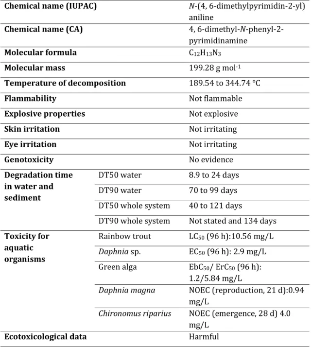
24 The Italian tree frog appears common and relatively well distributed across much of the Italian territory, except for Sardinia, the Tuscan Archipelago, the Alpine regions and the Apennine ridge, where is rare and usually limited to the valley floors. In Valle d'Aosta may be extinct as the last known observation dates back to 1983. In addition, it is uncommon in Liguria, where it is replaced by Hyla meridionalis (Sindaco et al., 2006). It appears also scarce in the southern regions of the peninsula, with the exception of Calabria where it is fairly widespread (Tripepi et al., 1999) (Fig. 3.1). The species has an altitudinal range ranging from sea level to at least 1,855m asl (Andreone et al., 2009). However, the Italian tree frog appears to be linked mainly to lowland areas and hill (with a marked preference for altitudes of less than 400 m), although it may exceed 1000 m s.l.m. in some regions and reach 1800 m of altitude in Nebrodi Mountains, in the province of Messina (Lanza et al., 2006; Sindaco et al., 2006).
Fig. 3.1.Distribution map of H. intermedia (from Sindaco et al., 2006)
25 3.3. Morphology
The Italian tree frog is a small arboreal anuran that rarely exceeds 5 cm in length from the apex of the snout to the cloaca. The eyes, on the sides, have a horizontal pupil; the golden iris is more or less colored with brown lines. The eardrum has a diameter at most equal to half of that of the eye. The long and slender legs are characterized by the presence of adhesive disc-shaped expansion at the ends of the fingers; the disks of the fingers are as big as the eardrum. The fingers of the forelimbs have a webbing barely visible, while the rear toes are webbed by half to 2/3 of their length (Lanza et al., 2006).
The skin is perfectly smooth dorsally but grainy on the abdomen, on the lower face of the thighs and, in females, even under the throat. In the dorsal area, the Italian tree frog shows a typical bright green color rather uniform, but the color variations are not uncommon; the color variations range from very dark shades to very clear in function of the fluctuations of some environmental parameters such as brightness, temperature and substrate. The throat and the ventral area are usually whitish, while the fingers have a pink or yellow pigmentation. Both males and females have a gray or black marginal strip (rimmed of white, cream or pale yellow) which runs from the nostrils, through the eye and the eardrum, along the sides of the body to the groin (Tripepi et al., 1999; Lanza et al., 2006).
As regards the sexual dimorphism, the males are slightly smaller than the females and are provided with a great vocal sac of spherical shape with a yellowish-brownish coloration; when inflated this sac is spherical and larger than the head, at rest does
26 not form longitudinal folds under the throat. In the breeding period the males develop nuptial pads under the first finger of the hand (Lanza et al., 2006).
3.4. Habitat and ecology
The Italian tree frog, as all species of the genus Hyla, has mainly arboreal habits but can also live in a wide range of environmental conditions. It inhabits open and well sunny environments, characterized by the presence of shrub or arboreal vegetation. It is a skilled swimmer and jumpers, it also has great qualities of climber (thanks to the presence of adhesive disks at the apex of the fingers that enable it to move easily even on smooth and vertical surfaces), therefore, it prefers the vegetation at a certain height from the ground, such as trees, shrubs and reed beds. On the contrary, juveniles stay close to the land becoming more distinctly arboreal over time (Lanza et al., 2006; Sindaco et al., 2006; Gentilli and Scali, 2007).
It is often observed in clearings, heathlands, scrub areas and is quite common even in cultivated areas, especially on the edge rice fields and orchards.
The Italian tree frog has mainly crepuscular and nocturnal habits. It is a eurythermal species and both hibernation and aestivation (that in particular conditions may not occur) occur not far from the breeding sites and usually take place in the ground or in burrows previously occupied by other animals, under rocks, in crevices between rocks, under or inside rotting logs, and in the hollows of tree roots (Lanza, 1983). This species feeds mainly on insects and other small invertebrates, which also captures in flight. Its predators are mammals, water birds, water snakes, carnivorous fish; when leading arboreal life it can be preyed upon by various diurnal and nocturnal raptors.
27 The main means of defense of the species is mimicry. It spends much of its life on land and its presence in water is limited to the breeding season; at the end of March, chorusing males concentrate near well-vegetated reproductive sites (Sindaco et al., 2006).
3.5. Reproduction and development
H. intermedia has annual cyclic reproductive activity regulated by endogenous hormonal factors, the seasonal cycles as well as various environmental factors such as environmental and water temperature. The breeding season begins between March (in more temperate locations and at low altitude) and May, and can last up to July-August; under optimal conditions they may also have autumn depositions. The breeding sites of the Italian tree frog are represented by standing water bodies (of natural or artificial origin), often only temporary, characterized by shrub or arboreal vegetation; its presence in these environments, such as pools, ponds, lakes, marshes, rice fields, marshes, reservoirs, channels and troughs, is limited to the reproductive period (Tripepi et al., 1999; Bologna et al., 2000; Ebisuno and Gentilli, 2002).
During night and evening hours the males in spawning emit their songs call audible even at great distances. The embrace is of type axillary and occurs mostly at dusk and during the night. For each breeding season, each female lays up to 1000 eggs with a diameter between 1.5 and 2 mm; the eggs are divided into small spherical gelatinous masses of 3-4 cm in diameter and attached to aquatic vegetation (Lanza et al., 2006). Hatching occurs after about 15 days of laying. The tadpoles show an olive-brown or yellowish-brown colour with golden hueshe in the dorsal part; in the sides are present
28 golden spots, while the belly is whitish with golden or nacreous spots. The larvae of tree frogs are distinguished from those from the other Anurans for the great distance between the two eyes, which are located in lateral position, and for the high and convex caudal ridges with the dorsal portion that extends forward up to eye level. The tail is relatively large and with more or less sharp apex. Under normal temperature conditions, the development of the larvae to the metamorphosis takes 2-3 months. The newly metamorphosed, already very similar to the adults, are approximately long 1.5 cm. They remain tied to the land and shrub vegetation for some time, then take arboreal habits; sexual maturity is probably reached at the second to third year of age (Lanza et al., 2006; Bologna et al., 2000; Andreone, 1995; Gentilli and Scali, 2007).
3.6. Population and conservation actions
The status and distribution of H. intermedia have not been studied in detail (Sindaco et al., 2006). Overall, by an analysis at regional and provincial levels of the distribution and populational consistency of the Italian tree frog, some authors state that this widespread species is subject to a low risk of extinction, thanks to its wide ecological value and its ability to colonize also altered environments (Andreone, 1995; Tripepi et al., 1999; Andreone and Luiselli, 2000). This opinion is reinforced by the inclusion in the category “least concern - LC” of the IUCN Red List (International Union for the Conservation of Nature), given to species with a stable trend of populations because widespread and abundant (Andreone et al., 2009; Temple and Cox, 2009). However, although the Italian tree frog is not, at the time, one of the most threatened species, the authors seem to agree in identifying numerous factors of human pressure
29 affecting the species, which can locally determine a regression (Giacoma and Balletto, 1993; Scoccianti, 2001; Lanza et al., 2006; Sindaco et al., 2006). The researchers identified as risk factors at the local level the disappearance and alteration of wetland breeding grounds (also intended as a simplification of the agricultural landscape and the changing modes of cultivation techniques management that can cause the modification of breeding sites or ecotone environments often used by this species), e.g. intensive agricultural exploitation of the plain areas, overuse of pesticides, removing marginal / riparian vegetation belts, remediation and draining, filling of ditches, canals, ponds, lakes and marshes. Furthermore, the placing of allochthonous fish in the aquatic environment is a serious problem that affects in particular eggs and larvae, but which can also be harmful to adults individuals (Scoccianti, 2004). Similar problems may arise from the placing of non-native freshwater crayfish, such as Procambarus clarkii (Cruz and Rebelo, 2005).
and is protected by national legislation in Italy and Switzerland. It is present in a number of protected areas. With regard to conservation measures, the species is protected by numerous international, national and regional laws: it is listed on Appendix III of the Bern Convention and and Annex IV of the EU Habitats Directive 92/43/CEE. Therefore, detention, capture, or sale of the Italian tree frogs are prohibited as it is forbidden to make any modification, likely to cause excessive disturbance, destruction or deterioration of the environment in which it lives and reproduces (Gentilli and Scali, 2007).
30
Chapters 4
Target organs
4.1. Urogenital SystemThe excretory and reproductive system are strictly associated in amphibians, as they are in all vertebrate, although these structures originate from different embryonic tissue (Duellmann and Trueb, 1986). Francis (1934) furnished a description of the urogenital system of salamanders, while Bhaduri (1932) and Bhaduri and Basu (1957) provided a detailed comparative explanation of this system in anurans. The morphology of the urogenital system in caecilians was described by Wake (1968, 1970a, 1970b,1972).
4.1.1. Kidneys
The kidneys in adult amphibians are bilateral structures lying on either side of the dorsal aorta. They develop from larval nephrostomes. In caecilians, anterior and posterior nephrostomes persist in the formation of the kidney; they have an opisthonephric kidney in which evident segmentation are visible in adult. Adult anurans and salamanders have a mesonephric kidney, in fact the larval pronephros is lost and the middle and posterior nephrostomes were they maintained.
By observing the gross morphology of the kidney in the three order of amphibians, great differences could be detected. In caecilians the kidneys are slender and long and extend from the heart’s region to the cloaca. In some salamanders the kidneys are long
31 with no differences from males and females, while in other salamanders, the kidneys are shorter and sexual dimorphism in shape is evident. The kidneys of most anurans species vary from long and slender three to four times as long as broad. (Duellman and Trueb, 1986).
The kidneys are highly vascularized organs; they are supplied with blood via both renal arteries and hepatic portal veins. The renal arteries are branches from the dorsal aorta, while the hepatic portal veins carry blood from the posterior part of the animal. The renal arteries enter the kidneys medially and branch into arterioles which branch further into capillary tufts known as glomeruli. Therefore, there is an afferent arteriole going to the glomerulus and an efferent arteriole carrying blood from the glomerulus, which then connect to peritubular capillaries. The peritubular capillaries also contain branches of the renal portal veins which enter the kidney from a dorsolateral direction. These anastomoses between venous and arterial supplies means the peritubular capillary carry both arterial and venous blood to the interstitium of the kidney. This peritubular capillary blood drains via efferent renal veins to the postcava where it enters the sinus venous.
The functional unit of the kidney is the nephron. The kidney is comprised of thousands of nephrons. Each nephron consist of a glomerulus, Bowman’s capsule surrounding the glomerulus, a slender ciliated neck region of the proximal convoluted tubule, a large-diameter proximal convoluted tubule, a thin ciliated intermediate segment, a slender distal convoluted tubule and finally a collecting tubule which drains into the collecting duct. The combination of a glomerulus and its surrounding Bowman’s capsule is known as the renal corpuscle. Bowman’s capsule is drained by
32 the ciliated neck segment. The hydrostatic pressure difference moves urine from Bowman’s space capsule to the proximal convoluted tubule. The cilia of the neck region contribute in maintaining this pressure difference. The distal end of the proximal convoluted tubule is connected to a second ciliated tubular segment, the intermediate segment, which is also partially responsible for contributing to the pressure difference that move fluid from the proximal to the distal tubule. The collecting ducts from all nephrons drain into the ureter. (Hillman et al., 2009)
4.1.2. Gonads
In anurans, the gonads of male and female larvae are paired structures, located near the kidneys, that develop from sexually undifferentiated primordial gonads. Gonadal differentiation starting at different time in a specie specific manner: in Xenopus at Nieuwkoop/Faber stage 49, in R. ridibunda at Gosner 26 (Ogielska and Wagner, 1990) and in Rana nigromaculata at Gosner stage 29 (Tanimura and Iwasawa, 1988) The differentiation is completed in young frog.
The primordial gonads appear as longitudinal bilateral thickenings covering the ventromedial region of the opisthonephros and are attached by their central region, the mesogonium, to the inner body wall. The central region leads to the gonad, while the anterior region, progonium, will develop in the fat bodies.
The primordial gonad is made up by medullary and cortical regions covered with a basal lamina, and its differentiation start from anterior to posterior region (Iwasawa and Yamaguchi, 1984; Lopez, 1989; Merchant-Larios and Villalpando 1981; Ogielska and Wagner, 1990; Tanimura and Iwasawa, 1986, 1988, 1989, 1991).
33 Medullary and cortical regions of primordial gonads have no ultra-structural differences indicating a cortical origin of the medullary tissue. In later larval stages, the central region is flooded by blood vessels, nerves and opisthonephric interstitial tissue. A primitive germinal epithelium, constituted by the primordial germ cells (PGCs) together with surrounding prefollicular cells, is located in the cortical region of the undifferentiated gonad. The primordial germ cells, in connotation with proliferation and digestion of yolk platelets, usually become smaller after they enter the gonadal ridge (Lopez, 1989; Ogielska andWagner, 1990; Züst and Dixon, 1975, 1977).
Gonadal sex differentiation differs temporally among species but usually coincides with metamorphic climax (McDiarmid and Altig, 1999). Female and males gonads are distinguishable by the number and size of germ cells and the amount of medullary tissue. (Ijiri and Egami; 1975; Tanimura and Iwasawa, 1987; Zaccanti et al., 1977). Male gonads can develop in a differentiated, semidifferentiated, or undifferentiated mode. The semidifferentiated and undifferentiated types often expressed spontaneous early female sexuality (Tanimura and Iwasawa, 1989; Takahashi, 1971; Zaccanti et al., 1977). In most anuran species, differentiation of testes starts prior to metamorphosis (Iwasawa and Yamaguchi, 1984; Lopez,1989; Ogielska and Wagner, 1990; Tanimura and Iwasawa,1989,1991). The primitive germinal epithelium, due to disintegrative modification, becomes a simple surface epithelium. The male gonial cells or spermatogonia, together with their enveloping follicular cells, are scattered throughout the compact medullary tissue. In the center of the medulla, a network of tubules becomes increasingly distinct and finally differentiates into seminiferous
34 tubules. Medullary cords, near the hilus of the primordium testis, form the rete testis. Spermatogenesis is practically identical among species (Deuchar, 1975; Kalt, 1973; Kerr and Dixon, 1974). In premetamorphic larvae, are detectable two types of spermatogonia: primary and secondary spermatogonia. Primary single spermatgonia are characterized by a lobed nucleus with diffuse chromatin, while secondary spermatogonia, which derive from primary spermatogonia, occur in cluster of cells of similar size (Fawcett et al., 1959; Rastogi et al., 1983). Meiosis typically begins in late metamorphosis when secondary spermatogonia will transform into primary spermatocytes.
Larval ovaries are bigger than testes and have a lightly uneven outline. Ovaries typically comprise both medullary and cortical tissue in which are present group of proliferating oogonia and oocytes (Iwasawa and Yamaguchi, 1984; Lopez, 1989; Ogielska and Wagner, 1990; Tanimura and Iwasawa, 1986, 1988, 1989, 1991). The cortex is distinctly separated from medulla by an acellular collagenous layer, and the medullary tissue has degenerated and lost its sex cord pattern. In this way small lumens are formed, and with the proceeding of development, they finally fuse to the ovarian cavity. The medullary connective tissue, at this point, becomes a simple epithelial layer covering the ovarian cavity. Oogonia, similar to the primordial germ cells despite devoid of pigmentation granules, undergo periodic proliferation to replenish oogonial nests (Coggins, 1973; Lopez, 1979; Redshaw, 1972). The passage from primary to secondary oogonia (primary oocyte) is associated with change in darkening cytoplasm (Eddy and Ito, 1971) and modification of nuclei from a lobed to a roundish shape.
35 Prior to metamorphosis, the primary oocytes enter into prophase (Coggins, 1973; Lopez, 1989; Ogielska and Wagner, 1990). In the larval ovaries, at metamorphic climax, coexist oogonia and leptotene, zygotene, pachytene and diplotene oocyte (McDiarmid and Altig, 1999). When an oocyte of a cell nest isolates from the others, it is enclosed by follicular cells which differentiate into steroid-producing follicular epithelium (Redshaw and Nicholls, 1971; Saidapur and Nadkarni, 1974). During folliculogenesis oocyte begin to develop asynchronously and the cytoplasm and the nucleus of diplotene oocytes greatly increase (Coggins, 1973).
4.1.3. Urogenital ducts
The Wolffian and Müllerian ducts of anurans transport gametes and, especially in males, nitrogenous wastes and water. Therefore, the anatomical structure of the genital ducts reflects their functions (Wake, 1979). The Wolffian ducts originate from the anterior region of each kidneys and continue to the cloaca; they are present in both sexes and will develop into primary nephric ducts.
In female they retain their excretory functions, while in males they also serve as urogenital ducts. In fact, in male they present several modification especially near the cloaca where a seminal vesicle develops in some species (Amer, 1972; Bhaduri and Basu, 1957). In young female anuran the Müllerian ducts or oviducts arise at metamorphosis (Hillman et al., 2009). The oviducts lie parallel and lateral to the kidneys and connect to the cloaca. In larvae the Müllerian ducts are thin walled tubules, in adult are greatly convoluted especially during the breeding season. Each oviduct comprises a short ostium and a long, coiled pars convoluta. The terminal
36 portion of the oviduct is enlarged to form the ovisacs (Amer, 1972; Bhaduri and Basu, 1957). In males, Müllerian ducts either degenerate or persist with no functions.
4.1.4 Fat bodies
The fat body, derived from the progonium, consists of proliferating somatic cells that form fingerlike projections aggregated at the anterior end of the gonads. No data are available on the functions of fat body in larvae (McDiarmid and Altig, 1999). In adults, fat bodies are a source of nutrients for the gonads, in fact, they are largest just before hibernation an smallest after breeding (Duellmann and Trueb, 1986).
4.2. Liver
In anurans, the liver occupies the cranioventral quadrant of the coelom. The parietal face lies directly under the ventral abdominal wall. The liver consists of two completely separated lobes and each of which may be subdivided into smaller lobes. A third, median lobe is present in many species. A large gallbladder lies on the midline in the interlobar connective tissue. The lungs lie dorsal to each lobe of the liver. The left lobe may also make contact with the stomach, whereas the caudal edge of the right lobe borders the intestine as it crosses the coelom and turns caudally. In mature females, the ova extend cranially and cover the parietal surface. The ventral abdominal vein runs forward inside the ventral coelomic wall, travels cranially between the lobes of the liver, and divides with a branch entering each lobe. As it approaches the liver, the vein is joined by the hepatic portal vein and vessels from the gallbladder (Crawshaw, 2000).