Verbale n.2/2015 del 27.10.2015 UNIVERSITA’ DELLA CALABRIA
Dottorato di Ricerca in Biochimica Cellulare ed Attività dei Farmaci in Oncologia.
Il giorno 27 Ottobre 2014 alle ore 11.30 si è riunito, per via telematica, il Collegio dei Docenti del Dottorato di Ricerca in Biochimica Cellulare ed Attività dei Farmaci in Oncologia per discutere il seguente ordine del giorno.
1. Valutazione relazioni fine anno dottorandi XXVIII ciclo. OMISSIS
Punto 1 all’ordine del giorno: Valutazione relazioni fine anno dottorandi XXVIII ciclo.
Sono pervenute le relazioni di fine anno dei Dottorandi del XXVIII ciclo. Tra queste anche le relazioni dei dottorandi con borsa FSE nell’ambito del Polo di Innovazione Regionale delle “Tecnologie della Salute”. Come ogni anno il Collegio dei Docenti è chiamato ad esprimere il proprio parere sul lavoro svolto dai dottorandi.
Il presidente chiede al Collegio dei Docenti di esprimere un giudizio sulle relazioni presentate dai Dottorandi con borsa FSE.
OMISSIS Con borsa d’Ateneo
Dott. Fiorillo Marco.
Durante il dottorato di Ricerca in “Biochimica Cellulare ed attività dei Farmaci in Oncologia” (XXVIII ciclo) l’attività scientifica del Dr. Marco Fiorillo è stata rivolta allo studio di due flavonoidi, statino-simili, (brutieridina e melitidina) estratte dal bergamotto (Citrus bergamia), ma presenti anche in altre specie del genere Citrus. Come molecola di riferimento è stata considerata la statina commerciale più prescritta nell’uomo, la simvastatina.
Durante i primi due anni il Dr. Fiorillo ha effettuato la valutazione, “in vivo”, dell’attività ipocolesterolemica e ipolipidemica delle frazioni oggetto di studio. In primo luogo è stato ottenuto un modello animale ipercolesterolemico, in seguito al trattamento degli animali (ratti) con una dieta opportuna. Successivamente, sul modello ottenuto è stato monitorato il metabolismo del colesterolo, mediante la valutazione del livello di espressione dei geni codificanti l’enzima HMGR ed il recettore delle LDL, sia a livello di trascritto che a livello proteico. Attraverso lo studio condotto dal Dr. Fiorillo è stato dimostrato che le due molecole estratte dal bergamotto, brutieridina e melitidina, sono dotate di attività ipocolesterolemica, dovuta all’azione inibitoria esercitata nei confronti dell’enzima HMGR. L’interesse scientifico, nel terzo anno di dottorato, è stato quello di valutare, “in vitro”, l’aspetto anti-proliferativo ed antinfiammatorio dei due flavonoidi studiati. Il contributo scientifico del Dr. Marco Fiorillo è dimostrato dai lavori pubblicati su riviste internazionali la cui rilevanza scientifica della collocazione editoriale è di ottimo livello e dalla partecipazione a diversi congressi Nazionali e Internazionali. Durante i tre anni di dottorato, ha dimostrato entusiasmo, notevole attitudine alla ricerca e spirito critico nella elaborazione e nella interpretazione dei risultati sperimentali; ha acquisito buona padronanza delle metodologie utilizzate che si evince dalla capacità di svolgere attività di ricerca in grande autonomia e con
Verbale n.2/2015 del 27.10.2015
Il Dr. Fiorillo ha svolto regolarmente le attività di studio e di ricerca previste per il terzo anno di corso; inoltre, l’esperienza maturata durante i tre anni di dottorato è stata accresciuta da un periodo di formazione svolto presso il laboratorio diretto dal Dr. Michael Lisanti presso il Breakthrough Breast Cancer Research Unit, University of Manchester (da Febbraio 2014 ad oggi). Pertanto si esprime parere estremamente positivo sull’attività scientifica svolta dal Dr. Marco Fiorillo.
Il Collegio dei docenti valutato il contenuto della relazione di fine anno del Dott. Marco Fiorillo esprime parere favorevole alla partecipazione all’esame finale del corso di Dottorato.
OMISSIS Rende, 27.10.2015
Il Coordinatore Prof. Diego Sisci
Abstract
Il rischio di malattia coronaria è aumentato negli individui che mostrano elevata concentrazione di
colesterolo nelle lipoproteine plasmatiche a bassa densità (LDL). E’ stato dimostrato che l’inibizione del 3-idrossi-3-metilglutaril-CoA reduttasi (HMGR), enzima che catalizza la
conversione di HMG-CoA in mevalonato (MVA), tappa limitante la velocità di biosintesi del
colesterolo, è l’approccio più efficace per la diminuzione plasmatica di LDL e la riduzione del tasso
di eventi cardiovascolari. Come parte di un meccanismo compensatorio, alla deplezione di colesterolo nel fegato, dovuto all’inibizione dell’enzima HMGR, segue l’aumento della produzione di recettori per le LDL e il successivo smaltimento di LDL dalla circolazione sistemica. Gli inibitori di HMGR rappresentano la classe di farmaci più efficaci e maneggevoli per la riduzione della concentrazione di LDL. Sebbene le statine siano gli inibitori di HMGR più largamente prescritti, sono associate a spiacevoli effetti collaterali quali severa miopatia e perdita della memoria statino-associata. In questo contesto l’identificazione di nuovi composti statino-simili, che agiscono come inibitori di HMGR, risulta utile per superare le limitazioni già descritte. E’ stato dimostrato che alcuni composti naturali, che si ritrovano nella nostra dieta, hanno proprietà terapeutiche e farmacologiche. In particolare studi condotti in seguito a somministrazione cronica di succo di alcune specie di Citrus hanno confermato che questa strategia influenza positivamente i livelli plasmatici dei lipidi e può essere associata alla riduzione del rischio di malattia coronarica. Durante questo lavoro di tesi , l’attenzione è stata rivolta allo studio di due flavonoidi, statino-simili, (brutieridina e melitidina) estratte dal bergamotto (Citrus bergamia), ma presenti anche in altre specie del genere Citrus. Come molecola di riferimento è stata considerata la statina commerciale
più prescritta nell’uomo, la simvastatina. Gli esperimenti sono stati condotti utilizzando una
frazione arricchita delle due molecole (EF) ed una purificata (BMF), al 99%. I composti testati, sono stati isolati e caratterizzati in maniera esaustiva, mediante spettrometria di massa e risonanza magnetica, dal gruppo di ricerca del Prof. G. Sindona (Dip.to di Chimica, Università della Calabria). In particolare, si è valutata “in vivo”, l’attività ipocolesterolemica e ipolipidemica delle frazioni oggetto di studio. In primo luogo è stato ottenuto un modello animale ipercolesterolemico, in seguito al trattamento degli animali (ratti) con una dieta opportuna. Successivamente, sul modello ottenuto è stato monitorato il metabolismo del colesterolo, mediante la valutazione del livello di
espressione dei geni codificanti l’enzima HMGR ed il recettore delle LDL, sia a livello di trascritto
che a livello proteico. Il metabolismo dei trigliceridi, invece, è stato monitorato valutando il livello di trascritto e di proteina del gene che codifica per il principale enzima della sintesi degli acidi grassi e quindi dei trigliceridi, il gene FASN. Inoltre sono stati valutati i livelli di colesterolo e di
enzimi coinvolti nella lipogenesi, in quanto responsabili della produzione di NADPH, utilizzato per la sintesi degli acidi grassi e del colesterolo, enzima malico e isocitrato deidrogenasi. I risultati ottenuti hanno evidenziato, nei ratti trattati con BMF, una riduzione dei livelli di colesterolo e dei trigliceridi, sia a livello epatico che sierico, tale decremento è risultato essere ancora più evidente nei ratti trattati con EF, rispetto ai controlli ipercolesterolemici. Dalla valutazione dei livelli di trascrizione di due principali proteine coinvolte nel metabolismo del colesterolo, HMGR ed LDLR e dell'enzima principale della biosintesi degli acidi grassi, FASN, è emerso chiaramente che il comportamento di BMF è simile a quello della simvastatina, uno dei farmaci ipocolesterolemici più utilizzati. Infine, dalla valutazione dell’attività degli enzimi HMGR, isocitrato deidrogenasi citoplasmatica ed enzima malico, negli epatociti degli animali trattati con le due frazioni rispetto a quella evidenziata negli epatociti dei ratti controllo, è stata riscontrata un’inibizione, anche in questo caso, paragonabile a quella osservata per i ratti trattati con simvastatina. Attraverso lo studio condotto è stato dimostrato che le due molecole estratte dal bergamotto, brutieridina e melitidina, sono dotate di attività ipocolesterolemica, dovuta all’azione inibitoria esercitata nei confronti
dell’enzima HMGR. L’interesse scientifico inoltre, è stato quello di valutare “in vitro”, l’aspetto
anti-proliferativo ed antinfiammatorio dei due flavonoidi studiati. Infatti, in letteratura è riportato
che le statine sintetiche riducono la proliferazione di un’ampia varietà di tipi cellulari, “in vitro”, inducendo l’arresto del ciclo cellulare nella fase G1. Questa sperimentazione è stata condotta su
cellule di carcinoma mammario umano, MCF7, utilizzando la frazione purificata (BMF) e comparando i risultati con quelli ottenuti dopo trattamento di cellule della stessa linea con simvastatina (profarmaco precedentemente utilizzato per gli studi “in vivo”) e pravastina. Il lavoro svolto ha previsto, in primo luogo, la creazione di una linea tumorale, stabile, che fosse in grado di over-esprimere il gene HMGCR (MCF-7-HMGCR), utilizzando un metodo di trasfezione virale; successivamente è stata valutata l’attività proliferativa delle cellule tumorali (MCF-7); delle cellule tumorali trasfettate con il gene HMGCR (MCF-7-HMGCR) e di quelle epiteliali non tumorali (fibroblasti hTERT-BJ1), dopo trattamento con BMF, pravastatina e simvastatina. I risultati ottenuti hanno dimostrato che alte concentrazioni di BMF svolgono un’azione antiproliferativa meno elevata di quella riscontrata con concentrazioni più basse di simvastatina. Tuttavia è doveroso sottolineare che mentre BMF e pravastatina non inibiscono la proliferazione cellulare, nelle cellule non tumorali, la simvstatina è risultata essere tossica anche a basse concentrazioni. Mediante analisi con XFe96 Seahorse Analyzersi è effettuata la valutazione metabolica delle linee cellulari MCF-7, MCF-7-HMGCR, hTERT-BJ1. I risultati ottenuti hanno evidenziato un’aumentata respirazione
la respirazione mitocondriale, in cellule MCF-7-HMGCR, dopo trattamento con BMF, ha evidenziato una riduzione della produzione di ATP e una diminuzione della respirazione massimale e della capacità respiratoria cellulare. Infine, le analisi OCR su cellule hTERT-BJ1 dopo trattamento con BMF, pravastatina e simvastatina hanno sottolineato una riduzione della respirazione mitocondriale minima, nelle cellule trattate con pravastatina; tale riduzione è risultata essere più marcata nelle cellule trattate con simvastatina. Nessuna differenza è stata, invece, riscontrata nelle cellule trattate con BMF. Questi risultati hanno confermato una leggera tossicità nelle cellule hTERT-BJ1 trattate con pravastatina ed una marcata tossicità in quelle trattate con simvastatina, a differenza delle cellule trattate con BMF. Inoltre, l’indagine effettuata su diversi pathways, implicati nella proliferazione cellulare e nell’infiammazione, ha evidenziato un potenziale effetto antinfiammatorio e antiossidante di BMF, come sottolineato da un aumento della risposta antiossidante e di quella immunitaria regolate dall’interferone I, da una diminuzione della
risposta infiammatoria mediata dall’interferone-gamma e da una down-regolazione della via
infiammatoria regolata dal gene STAT3, in cellule MCF-7, trattate con BMF. Il trattamento con BMF ha determinato, inoltre, la down-regolazione di due pathways coinvolti nella proliferazione tumorale e nella formazione di CSCs (cancer stem cells), regolati da Notch e Wnt. Questi risultati hanno portato ad indagare su un’eventuale coinvolgimento di BMF, nella formazione di
mammospheres e a dimostrare, dopo trattamento con BMF, un decremento nella efficienza di
formazione di mammospheres, dose dipendente, più marcato nelle cellule che over-esprimono
l’enzima HMGCR. Successivamente, è stata riscontrata, per BMF, la capacità di ridurre lo stress
ossidativo, la formazione di radicali liberi e la successiva risposta pro-infiammatoria, come evidenziato dalla diminuizione dell’espressione delle citochine pro-infiammatorie, regolata dal complesso proteico NF-kB e dalla diminuizione dell’espressione dei fattori inducibili l’ipossia, regolata dal gene HIF, riscontrate in cellule hTERT-BJ1, trasfettare con i gene reporter Nf-KB e HIF e trattate con BMF. Infine è stato valutato l’effetto antinfiammatorio ed antiossidante della frazione purificata di brutieridina e melitidina; dai risultati ottenuti è stato possibile evincere una riduzione dei fattori di stimolazione, coinvolti nella formazione dei granulociti e dei macrofagi, in cellule MCF-7 trattate con pravastatina ed, in egual misura, in quelle trattate con BMF. Anche la produzione di IL-8, in cellule trattate con BMF, ha mostrato un decremento, di poco inferiore a quello riscontrato in cellule trattate con pravastatina. Infine la riduzione di cancer stem cells (CSC) è stata valutata tramite l’impiego di un marker specifico (ALDH) che ha permesso di isolare la popolazione ALDEFLUOR-positiva relativa alla popolazione staminale, in cellule MCF7. Dopo trattamento con BMF e pravastatina, è emersa una netta diminuzione della popolazione
della biosintesi del mevalonato) al mezzo cellulare, in presenza di BMF e/o pravastatina , non ha cambiato la percentuale di riduzione della popolazione di CSCs. Al contrario l’aggiunta di mevalonato al mezzo di coltura, ha riportato sia il numero di mammospheres che la percentuale della popolazione di CSCs ai valori del controllo. Questi risultati hanno permesso di indicare la BMF come un composto con scarsa tossicità e capace di prevenire la crescita tumorale, l’espansione tumorale mediata da fattori pro-infiammatori e la fomazione di CSCs. L’impiego della BMF in concomitanza ai canonici chemioterapici, potrebbe migliorare l’effetto degli stessi e diventare un nuovo target drug nella terapia add-on.
Index
Chapter 1
Introduction
pag. 1
Materials and Methods
pag. 7
Results
pag. 16
Discussion
pag. 38
References
pag. 42
Chapter 2
Introduction
pag. 49
Materials and Methods
pag. 53
Results
pag. 65
Discussion
References
pag. 94
pag. 97
Introduction
CHAPTER 1
INTRODUCTION
Cholesterol is a molecule of primary importance in animals, including humans, but is not required in the diet because hepatocytes can synthesize it starting from acetyl-CoA. The 3-hydroxy-3-methylglutaryl-CoA reductase (HMGR) is the rate-limiting enzyme in endogenous cholesterol biosynthesis and catalyzes the conversion of HMG-CoA into mevalonate [1]. Inhibition of HMGR has proven to be one of the most effective approaches for lowering plasma LDL and reducing cardiovascular event rates [2]. As part of a compensatory mechanism due to cholesterol depletion in the liver, inhibition of HMGR leads to an increased synthesis of itself and low-density lipoprotein receptors (LDLR); this latter process allows a subsequent clearance of LDL from systemic circulation [3], lowering the risk of atherosclerosis and coronary heart diseases [4 ,5]. The risk for this disease is increased in patients with elevated serum concentrations of low-density lipoproteins cholesterol (LDL), total cholesterol (TC) and triglicerides (TG) [6-9]. Several meta-analysis studies showed that statin therapy can reduce the 5-year incidence of cardiovascular diseases, by about one fifth per mmol/L reduction in LDL cholesterol [10-12]. It is well-known that statins are able to inhibit HMGR activity. Statin administration is one of the most widely used approaches to lower serum LDL level and to reduce cardiovascular event rates [13-15], they are pretty safe, well tolerated and highly efficient, nevertheless cardiovascular disease remains the main cause of mortality in westernized countries. Moreover, many patients, especially those
Introduction
with the dyslipidemia associated with metabolic syndrome, are unable to reach their lipid treatment goals on statins alone [6]. Furthermore, patients might be statin-intolerant and experience significant side-effects [7], hence the importance of finding new drugs acting as statins. Some natural compounds found in the human diet have been shown to possess therapeutic and pharmacologic properties, in particular, the daily consumption of citrus fruit juice has been shown to positively influence plasma lipid levels and reduce the risk of coronary heart disease [16, 17]. Hypolipidaemic effects can be correlated to several components of citrus juice, such as flavonoids (naringin and hesperidin), pectins, and ascorbic acid, which have a high antioxidant potential and interfere with cholesterol metabolism [18-20]. Plant flavonoids are a large group of very different compounds sharing the common feature of phenolic moieties [21]. The presence of a relatively large number of flavonoids is the result of many different possible combinations among polyhydroxylated aglycones and a limited number of mono- and disaccharides. The most commonly found sugars are hexoses, such as glucose, galactose and rhamnose or pentoses such as arabinose and xylose. They are, with a few notable exceptions, plant metabolites deriving from the shikimate pathway and the phenylpropanoid metabolism [22]. In recent years, flavonoids have attracted tremendous attention due to the protection that they provide against some types of cardiovascular diseases [23]. As a consequence, many studies have been directed to the characterization of the flavonoid fractions and to the isolation of the most representative flavonoids present in the most common Citrus species, as well as of flavonoids present
Introduction
in many local species such as C. bergamia Risso [24, 25]. The classes of flavonoids present in C. bergamia Risso fractions are flavanones and flavones. Flavanones are present as flavanone-O-glycosides, recently, flavanones diglycosides carrying the 3-hydroxy-3-methylglutaric acid (HMG) moiety have also been detected [26, 27].
Flavones are present as flavone-O-glycosides, flavone-C-glycosides or
polymethoxyflavones Bergamot, the common name of the fruit Citrus bergamia Risso, belongs to the family Rutaceae, subfamily Esperidea, and is widespread in the Mediterranean area for centuries. The botanical and geographical origins are still uncertain; probably bergamot is native of Calabria, deriving by mutations of other citrus species, or even arrived in loco from Berga (hence the name of bergamot) otherwise from Antilles, Greece and Canary Islands. Afterwards, the terpene-rich essential oil, extracted from the peel of the pear-shaped fruit, and its volatile fraction have been largely employed in the cosmetic and perfumery products. Bergamot and its derivatives have also been used in calabrian folk medicine as fever palliative, antiseptic, anthelminthic, wound healing, anti-inflammatory and hypocholesterolaemic agent [28, 29]. To date, bergamot essences are used in the food industry for the preparation of teas, jams, sherbet and other commodities, but also for the domestic preparation of liquors,
ice creams and pastries by the calabrian confectionery industry
(www.rc.camcom.gov.it). Bergamot juice has been considered, for a long time, a secondary and waste by-product of the essential oil extraction and, because of its organoleptic properties and its bitter taste, did not reach the popularity of other citrus
Introduction
juices but was used to fortify fruit juice instead of synthetic [30]. Later on, the discovery of the abundance and variety of bioactive compounds in the juice (e.g. naringin, neoeriocitrin, and neohesperidin), led to the development of several nutraceuticals (capsules, pills or soluble granular powders) in the global market [31]. Furthermore, other flavonoids, such as rhoifolin, neodiosmin, and some chryosoeriol derivatives, have been found in smaller amounts [32-34] and different tissues of the fruit produce the flavonoids diosmin and poncirin [35]. In particular, naringin seems to be active on atherosclerosis, as demonstrated by animal studies [36], neoeriocitrin is believed to strongly inhibit LDL oxidation [37] whereas, HMG-flavonoids could be able to inhibit HMGR [26]. These observations have provided the rationale to investigate the protective hypolipidemic effect of bergamot extracts in animal models and in human patients. Miceli et al. [38] demonstrated that daily administration of bergamot juice to hypercholesterolemic rats caused a significant reduction in TC, TG and LDL levels, an increase in serum HDL levels and a protective effect on hepatic parenchyma. In addition, fecal output of total bile acids and neutral sterols was enhanced in the bergamot juice treated group in comparison with the hyperlipidemic group. These results are in agree with previous studies, which hypothesized that pectins and flavonoids were able to lower serum cholesterol levels by modulating hepatic HMG-CoA concentration. It could be noted that in this study, the potential side-effect due to bergamottin presence in bergamot juice was not investigated. Bergamot juice is rich in bergamottin (ranging from 18 to 61 mg/L) [39, 40], a furanocoumarin compound that
Introduction
inhibits cytochrome P450 34A enzyme activity, significantly increasing the oral bioavailability of several drugs metabolized primarily by this cytochrome [37, 41]. This problem was overcome by Mollace et al. [28], that analyzed the hypolipidemic effect of a defurocoumarinizated bergamot-derived polyphenolic fraction supplemented with ascorbic acid on animal models of diet-induced hyperlipidemia and in patients suffering from metabolic syndrome [28]. They found that oral administration of this fraction both in animal and in patients, caused a significant reduction of TC, TG and glycemia with a concomitant increase of HDL levels. Recent prospective studies, led on patients with hyperlipidemia demonstrated that administration of a defurocoumarinizated bergamot-derived polyphenolic fraction was able to reduce TC level. In the same conditions, rosuvastatin administration (10 mg/die) caused a similar reduction of TC content. Their association produced a considerable enhancement of rosuvastatin hypolipidemic effect, normalizing the serum lipid profile [42]. The authors suggested that the observed hypolipidemic effect could be mainly due to the presence in bergamot-derived polyphenolic fraction of melitidin, brutieridin and HMG-neoeriocitrin. This hypothesis was investigated by our research group [27], in a hypercholesterolemic rat model, by measuring the effects on lipid profile of administration of HMG-flavanones enriched fraction (62% of brutieridin, 14% of melitidin and 15% of HMG-neoeriocitrin), extracted from bergamot fruit, in comparison with simvastatin. HMGR, LDLR and FASN transcription levels and their correlated protein amounts were evaluated. In this study, simvastatin and HMG-flavanones enriched fraction singularly administrated
Introduction
reduced levels of TC, TG, VLDL and LDL, whereas an increase in HDL content was observed exclusively in rats treated by HMG-flavanones enriched fraction [27]. Furthermore, according to previously published data, HMGR, LDLR and FAS transcription levels were found up-regulated. An increased amount of their corresponding proteins was detected [43]. Genotoxicity and toxicity were not observed by testing HMG-flavanones enriched fraction in vitro. Our hypothesis was that HMGR inhibition leads to a reduction of endogenous cholesterol level which, in turn, is responsible of HMGR and LDLR transcriptional up-regulation, as well of the higher LDLR exposure within the hepatocytes membrane, through a compensatory mechanism based on SREBPs pathway. We undertook a detailed analysis of dry extract from bergamot peel fruit. The main aim of the present work was to investigate the effects of HMG-flavanones pure fraction (70% of brutieridin and 30% of melitidin), extracted from bergamot fruit, on the total lipid profile of hypercholesterolemic rats, in comparison with simvastatin. HMGR, LDLR and FASN transcription levels and their correlated protein amounts and activities were evaluated.
Materials and methods
MATERIALS AND METHODS
Preparation of Brutieridin and Melitidin Fraction (BMF).
Bergamot fruit was collected in December 2012 and then stored at -20° C. 7 kg of fruits were squeezed to obtain the juice (2000 mL) which was filtered and passed through a 10 g C18 cartridge (Supelco, USA) in 50 mL aliquots. The loaded stationary phase was initially washed with water (2 x 50 mL) to remove the sugars and water soluble fraction, and then eluted with 50 mL of methanol to collect the flavonoid fraction. Each aliquot passed through the resin provided ca. 80 mg raw flavonoid fraction, for a total amount of 3.2 g. The polyphenolic fraction coming from the SPE step was loaded onto a glass
column (46 x 2.6 cm) from Buchi (USA) packed with 100 g of C1880-60 mesh
(Sigma-Aldrich, USA) and connected to a Perkin Elmer 200 LC binary pump. H2O (solvent A)
and CH3OH (solvent B) at the flow rate of 1.5 mL/min were used as elution solvents at the following gradient steps: isocratic at 100% A for 40 min.; linear gradient from 100% A to 70% A in 60 min.; isocratic at 70% A for 60 min.; linear gradient from 70% A to 40% A in 60 min.; isocratic at 40% A for 60 min.; linear gradient from 40% A to 0% A in 10 min.; washing of the column at 0% A for 60 min. The initial water elution was discarded and the collected fractions starting from min 40 (20 mL each) were monitored by HPLC/UV-MS using a Fractionlynx semi-preparative HPLC system (Waters Corp., Milford, MA, USA) The system was composed by an autosampler/collector Waters 2767 Sample Manager, a 600E pump working in analytical mode, a 486 UV detector and a ZMD mass spectrometer equipped with an ESI source working in negative
Materials and methods
ionization mode. The HPLC separation was achieved using a 250 × 4.6 mm, 5 µm
reversed phase C18Luna-Phenomenex column at a flow rate of 1 mL/min. The run time
was 70 min and the mobile phase was composed by 0.1% formic acid in water (solvent A) and methanol (solvent B). The chromatographic run (70 min) consisted of the following steps: isocratic at 80% A for 7 min; linear gradient from 80% A to 40% A in 33 min; isocratic at 40% A for 5 min; linear gradient from 40% A to 20% A in 5 min; isocratic at 20% A for 7 min; linear gradient from 20% A to 80% A in 5 min; equilibration of the column for 8 min. The UV detector was set at 280 nm. The MS condition were the following: capillary voltage -3.15 kV, cone voltage -3 V, extractor -2 V, RF lens -0.34 V, source block and desolvation temperature 120, 250 °C respectively, ion energy 0.5 V, LM resolution 14.5, HM resolution 15.0 and multiplier 650 V. The nebuliser gas was set to 650 L/h. The fractions coming from the MPLC separation and containing respectively compound brutieridin and melitidin were evaporated under reduced pressure, lyophilized and submitted to the purification step using the Fractionlynx system working in semipreparative mode at the same experimental
condition reported above except for the use of the column that was a 250 × 10 mm C18
Luna from Phenomenex (Torrance, CA) and for the chromatographic run (30 min) that consisted in a isocratic at 55% A. The flow rate was set to 4.7 mL/min, and the fractions were collected every 30 seconds, while the injected sample volume was 1 mL. The purity of HMG flavonoid was verified by HPLC/UV.
Materials and methods
Hydrolyzed flavonoid fraction from bergamot (FF).
The polyphenolic fraction coming from the SPE step was submitted to hydrolysis experiments using saturated Na2CO3. The hydrolysis of the HMG moiety was monitored by HPLC/UV-MS (see above for the experimental conditions). When all the HMG signal disappeared the mixture was neutralized to pH 7 and lyophilized.
Animals and diet.
Male Wistar strain rats, weighing about 100 g each, were purchased from Charles River (Lecco, Italy). All of the animal experiment protocols followed the institutional guidelines of the Italian Ministry of Health for Animal Care (D.M. 116/1992). The animals were housed in rooms maintained at 23±1°C and 55±5% relative humidity and were allowed free access to water, a standard rodent chow diet during acclimatization. Then, they were divided, randomly, into five groups of twelve animals each:
• Group N (controls) received the regular diet for 4 weeks.
• Group H ( hypercholesterolemic ) received the hypercholesterolemic diet (regular diet
+ 2% cholesterol + 0.2% cholic acid) for 4 weeks.
• Group ( H+S ) received the hypercholesterolemic diet (regular diet + 2% cholesterol +
0.2% cholic acid) for 4 weeks; from the 3nd to the 4th week each rat was administered by gavage with simvastatin (20 mg/kg bw/day).
Materials and methods
• Group ( H+BMF ) received the hypercholesterolemic diet (regular diet + 2%
cholesterol + 0.2% cholic acid) for 4 weeks; from the 3nd to the 4th week each rat was administered by gavage with the BMF (60 mg/kg bw/day).
• Group ( H+EF ) received the hypercholesterolemic diet (regular diet + 2% cholesterol
+ 0.2% cholic acid) for 4 weeks; from the 3nd to the 4th week each rat was administered by gavage with EF, at the same amount employed with BMF.
• Group ( H+FF ) received the hypercholesterolemic diet (regular diet + 2% cholesterol
+ 0.2% cholic acid) for 4 weeks; from the 3nd to the 4th week each rat was administered by gavage with Flavonoids alone, at the same amount employed with BMF.
Regular and hypercholesterolemic diets were supplied by Charles River (Lecco, Italy). During the experiment, rats were weighed daily and the 24 h food consumption was recorded, then fasted for 24 h before experiments. At the end of the treatment, blood samples were collected respectively in EDTA-treated tubes and Silica-treated tubes for plasma and serum detention, and centrifuged at 2500 x g for 15 min; the serum was separated and stored at 20°C until analyzed. The animals were anesthetized with chloral hydrate (400 mg/kg, i.p.) and killed by perfusion through the left ventricle of the heart with 100 mL of heparinized saline (pH 7.4). The liver was excised and immediately frozen in liquid nitrogen, then stored at -80°C until required.
Materials and methods
Biochemical valuations.
The livers were cut into small pieces, washed several times in a cold buffer (0.25 M sucrose, 3 mM EDTA, 20 mM Tris–HCl, pH 7.0), next homogenized in the same buffer 9and treated as described in Muci et al. [44] for TC extraction. Briefly, an aliquot of 10 mg protein sample was saponified with alcoholic KOH for 90 min at 85–90 °C. Nonsaponificable constituents were extracted three times (5 mL each) with light petroleum (b.p. 40-60°C). The pooled extracts were then evaporated to dryness under N2 and the residue, dissolved in 2-propanol. Total lipids were extracted from liver homogenate (10 mg protein) with chloroform/methanol (1:1 v/v) according to [45]. The levels of hepatic TG and TC were measured respectively by Triglyceride Quantification Kit ab65336 (Abcam, Toronto Canada), Total Cholesterol Assay Kits (Cell Biolabs, Inc. San Diego, USA), and HPLC analysis as described in Didonna et al. [45]. The TG, TC, HDL, LDL and VLDL levels were evaluated by direct enzymatic assays using Total Cholesterol Assay Kits Cell Biolabs, Inc. San Diego, USA), Triglyceride Quantification Kit ab65336 (Abcam, Toronto Canada) HDL and LDL/VLDL Cholesterol Assay Kit
ab65390 (Abcam, Toronto Canada),, respectively according to the manufacture’s
instruction. Alanine transaminase (ALT), aspartate transaminase (AST), bilirubin and creatinine were measured using reagent kits from Alanine Transaminase Colorimetric Assay kit (CaymanChem, Michigan USA), Aspartate Aminotransferase Activity Assay kit (CaymanChem, Michigan USA), Bilirubin Assay kit (Abnova, Taiwan) and Creatinine Colorimetric Assay kit (CaymanChem, Michigan USA).
Materials and methods
RNA extraction, reverse transcription and quantitative real-time polymerase chain reaction.
RNAs were extracted from liver as described by Zara et al. [46] and aliquots of 1 μg were reverse-transcribed as reported in Iacopetta et al. [47]. Quantitative real-time polymerase chain reaction (RT-PCR) was performed with the obtained complementary
DNAs (cDNAs) using the Applied Biosystems StepOne™ Real-Time PCR System
(Applied Biosystems, Monza, Italy). Primers based on the cDNA sequences of the genes of interest were designed using Primer Express (Applied Biosystems, Monza,
Italy). In each sample of 20 μl real-time PCR reaction 10 ng of cDNA, 10 μl of the
Power SYBR ® PCR Master Mix (Applied Biosystems, Monza, Italy) and 0.3 μL (150
nM) of the specific primers for each genes analyzed were used. Each experiment was repeated at least 3 times. The comparative threshold cycle method was used in relative gene quantification as previously described [48], using 18S gene as the endogenous control.
Microsomal fraction extraction and Western blot analysis.
The liver homogenate, obtained as previously described, was centrifuged at 800 g for 8 min, the pellet was discarded and supernatant centrifuged for 10 min at 12,000 g. The pellet containing the mitochondrial fraction was discarded and the supernatant was first
Materials and methods
cytosolic fraction and pelleted microsomes. The latter were re-suspended in the homogenizing medium and centrifuged again under the same conditions. Contamination of microsomal preparation by other subcellular fractions, ranging from 5 to 9%, was determined by the assay of marker enzymes as described in [49] and protein concentration was determined as in [50]. Western blot analysis was conducted as described previously [51]. Antibodies anti-HMGR (H-300), fatty acid synthase (FASN, H-300), LDL receptor (C-20), anti-calnexin (C-20), anti-β-tubulin (D-10), anti-GAPDH (FL-335) were supplied by Santa Cruz Biotechnology Inc. (Santa Cruz, CA, USA) and used following the manufacturer’s instructions. Anti-calnexin, β-tubulin or anti-GAPDH antibodies were used to confirm the equal loading of proteins.
HPLC analysis of total cholesterol in liver samples.
The analyses of samples were performed following a method from literature [52].
Determination of HMGR activitiy.
The assay was carried out via the radioisotopic method, following the production of [14C] mevalonate from 3-[14C]- hydroxymethylglutaryl coenzyme A (3-[14 C]-HMG-CoA; specific activity 57.0 mCi/mmol;Amersham-Pharmacia, Little Chalfont, UK); as described in Pallottini et al. [53]. Briefly, five hundred milligrams of liver were
Materials and methods
homogenised in a phosphate buffer containing 0.1 M sucrose, 0.05 M KCl, 0.04 M
KH2PO4, 30 mM EDTA, pH 7.4.. The activation of the enzyme by lysosomal
phosphatases during the preparative procedure can be avoided by the addition of the phosphatase inhibitor NaF. Microsomes were prepared by centrifugation of the homogenates as already reported [54], microsomes (100 µg) were incubate in presence of cofactors (20 mM phosphate, 20 mM NADP sodium salt, 1 IU glucose-6-phosphate dehydrogenase and 5 mM dithiotreitol). The assay, in a final volume of 200 µl, was started by the addition of 10 µl (0.088 µ Ci/11.7 nmol) of 3-[14C]-HMG-CoA. The radioactivity of the produced [14C]-mevalonate, isolated by chromatography on AG1-X8 ion exchange resin (Bio-Rad Laboratories), was counted. An internal standard
(3-[3H]-mevalonate, specific activity 24.0 mCi/mmol; 24.0 mCi/mmol;
Amersham-Pharmacia, Little Chalfont, UK) was added to calculate the recovery.
Determination of Isocitrate dehydrogenase and Malic enzyme activities.
For lipogenic enzyme analysis, samples of liver (0.5 g) were homogenized in 5 mL of buffer (pH 7.4) containing 0.25 M sucrose, 3 mM EDTA, 20 mM Tris–HCl, pH 7.0.
The cytosolic fraction was obtained as described in par. “Microsomal fraction
extraction and Western blot analysis” and used for quantification of enzyme activities. Isocitrate dehydrogenase (IDH) activity was assayed by the method of Ochoa [55] and malic enzyme activity as described by Ochoa [56]. Enzyme activities were measured
Materials and methods
spectrophotometrically and were expressed either as nmol NADPH produced, per minute, per mg of protein. Protein concentration was determined by the Bradford method [57].
Statistical analysis.
All data is presented as means ± SD for the number of experiments indicated in each
case. Data was analyzed by Student’s t-test using the GraphPAD Prism5 software
(GraphPad Software, USA). Differences were considered statistically significant at P < 0.05.
Results
RESULTS
Food intake and body weight gain.
The data on food intake and body weight evidenced no significant alteration, for each group (i.e. simvastatin (H+S), or BMF (H+BMF), or EF (H+EF), or flavonoids alone (H+FF) treated rats versus control), during the experimental phase (data not shown). These observations indicate that the diet and treatments used in these studies were well tolerated in rats, given that no physical alteration, body weight loss or food intake reduction occurred over the period of study. Two groups of rats were fed a regular and
hypercholesterolemic diet respectively, in order to verify the induced
hypercholesterolemia.
Effect of simvastatin, BMF, EF and flavonoids on serum and hepatic lipid content.
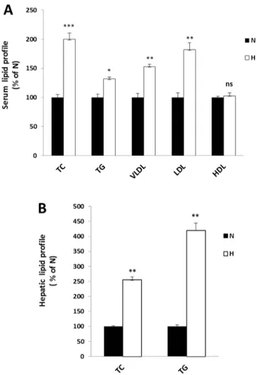
Firstly, in order to establish whether the hypercholesterolemic rat model has been achieved, we compared the serum and hepatic lipid profile of the regular diet fed rats (group N) and the hypercholesterolemic ones (group H). The TC, TG, VLDL, LDL and HDL levels, in rat serum of H group (Fig. 2, panel A), showed a significant increase (100, 32, 53 and 82% respectively) respect to those of N group, whose values were 105.85±5.6 mg/dl for TC, 204.7±12.3 mg/dl for TG, 39.02±1.5 mg/dl for VLDL and 49,03±3.1 mg/dl for LDL. Whereas no significant variation has been found for HDL levels (in N group, the HDL value was 45.52±3.3 mg/dl). Furthermore, a strong
Results
increase of the hepatic TC and TG levels (Fig. 2, panel B) has been observed in H group (150 and 300% respectively) compared to group N, whose values were 1.98±0.9 mg/g liver for TC and 4.87±0.39 mg/g liver for TG. These results confirmed that H group can be used as a hypercholesterolemic and hyperlipidemic model in the following experiments. Then, we investigated the effects triggered by BMF and simvastatin treatments on serum and hepatic TC of hypercholesterolemic rats, compared to group H. We observed a significant decrease of the serum TC that was more evident after BMF treatments (of about 40 and 55%, respectively). In rats treated with flavonoids fraction without brutieridin and melitidin (FF) was detected a less noticeable decrease (23%), compared to the hypercholesterolemic ones (Fig. 3, panel A). Moreover, the liver TC content showed a lower reduction, but with similar trends in H+S and H+BMF groups, of 25 and 45% respectively (Fig. 3, panel B). The direct correlation between an increase in the concentration of LDL in serum and the risk that the incidence of atherosclerosis and cardiovascular disease/cerebrovascular becomes higher is well known [58]. Besides, the HDL concentration has been correlated inversely with the onset of atherosclerosis associated diseases [59], whereas the data concerning the VLDL role in atherosclerotic plaque formation are still controversial [60]. For the above reason, we evaluated serum levels of LDL, HDL and VLDL. A significant reduction of VLDL and LDL was measured in H+S (25 and 30% lower, respectively), H+BMF groups (45 and 60% lower, respectively) and H+FF (55 and 25% lower, respectively) groups (Fig. 4, panels A and B, respectively). Conversely, any significant difference in the HDL
Results
cholesterol was found in the H+S group, whereas, surprisingly, an increase of 30% was found in the H+BMF group respect to H group (Fig. 4, panel C). The increase exceeded of 15% in rats of H+FF group. Since HMGR inhibitors have been shown to contribute at reducing serum triacylglycerols, in experimental animals [61], we measured serum and hepatic TG content, in our model. The obtained results showed that, in both cases, there was a decrease in H+S, H+BMF and H+FF groups respect to H group of about 30, 40 and 60%, respectively, for serum TG (Fig. 5, panel A) and of about 25, 63 and 60%, respectively, for hepatic TG (Fig. 5, panel B).
Effect of simvastatin and HMGF administration on hepatic HMGR, LDLR and FASN mRNAs and proteins levels.
In order to further investigate the HMGF effects in H groups, we evaluated the transcriptional levels of two main proteins involved in cholesterol metabolism, i.e. HMGR and LDLR, and the expression of the main enzyme of fatty acid biosynthesis, i.e. FASN [62]. Simvastatin and BMF treatments produced a similar modulation on the gene expression of the above mentioned proteins (Fig. 6; panel A). Particularly, simvastatin administration, respect to H group, brought to an increased transcription of HMGR, LDLR and FASN genes in an extent of 1.45, 1.88 and 1.50 fold respectively. As well BMF treatment increased the expression of HMGR, LDLR and FASN genes of 1.70, 1.80 and 1.90 fold respectively. In both cases, the mRNAs increase is appreciable
Results
respect to the untreated H group. Conversely, the FF treatment determine a significant increased transcription of LDLR and FASN genes. These data were confirned by Western blot analyses performed on the microsomal or cytoplasmic fractions or on liver homogenate extract, considering the different sub-localization of proteins analyzed. As shown in figure 6, (panels B-D), according to RT-PCR results, both simvastatin and BMF treatments led to a clear increase of HMGR, respect to H group, whereas the FF treatment determine a significant increase of LDLR protein that was more evident for FASN protein (1.45 and 2.8 fold respectively).
HMGR activity.
As HMGR is the rate limiting enzyme in cholesterol biosynthesis, we investigated whether reduction in cholesterol content in the liver and serum in hypercholesterolemic rats, upon treatment with BMF and FF, was due to the inhibition of HMGR. The HMGR activity was analysed in the liver microsomial fraction, following the production of [14C]-mevalonate from 3-[14C]-HMG-CoA. A significant decrease in the HMGR activity was detected when hypercholesterolemic rats were treated with simvastatin and BMF (Fig.7). Administration of two compounds decreased the HMGR activity by about 50% and 55%, respectively as compared to control hypercholesterolemic rats. No differences were found in HMGR activity, in hypercholesterolemic rats treated with FF. These results clearly demonstrated that
Results
brutieridin and melitidin (the two flavonoids in BMF) function as statin-like compounds, against HMGR.
Isocitrate dehydrogenase and malic enzyme activities.
It is know that the effects of statins on fat accumulation are controversial. Therefore, to establish whether brutieridin and melitidin, were able to influence de novo lipogenesis, we determine the activities of isocitrate dehydrogenase (Fig. 8, panel A) and malic enzyme (Fig. 8, panel B). These enzymes are related to this pathway as responsible for the formation of reduction potential, in the form of NADPH, required for de novo fatty acids synthesis. The results here reported show, in our animal model, a decrease of two tested enzyme activities, in statin and BMF treated rats. With regard to isocitrate dehydrogenase enzyme, the reduction was found about 50%, both after treatment with simvastatin that BMF. The decrease in the malic enzyme activity, instead, is more marked after treatment with simvastatin compared to that observed with BMF (lower 60 and 50%, respectively). Smaller decrease was detected in activities of both enzymes, in FF treated rats (30% isocitrate dehydrogenase and 40% malic enzyme, respectively)
Results
Influence of simvastatin, BMF and FF on rat ALT, AST, bilirubin and creatinine levels.
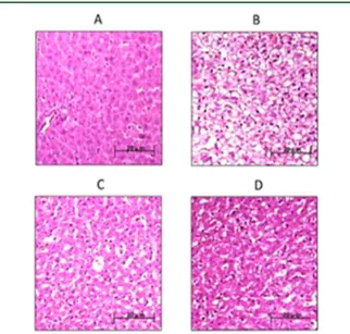
At last, we investigated, in our animal model, the effects triggered by simvastatin, BMF and FF, on hepatic and renal functional parameters, (e.g. ALT, AST, bilirubin and creatinine). The creatinine levels decreased in simvastatin and FF treated rats by 14% compared to those of hypercholesterolemic rats. This reduction is even more marked, reaching 25%, in BMF treated rats; this latter treatment brings the creatinine level to normal values (group N values). Conversely, no significant difference was found for serum total bilirubin in treated rats, compared to those of groups N and H (Table 1). Compared with N group, the serum ALT and AST levels of the H group rats have had a greater increase, of 83% and 54% respectively. Serum ALT and AST levels were lowered significantly in rats belonging to H+S and H+FF groups (28% and 21% respectively), when comparing them to the hypercholesterolemic rats. This reduction was greater, by 40% and 31% respectively, in rats belonging to H+BMF group (Table 1). The results obtained in the present study are in good accordance with those reported by Iseri et al. [63] who observed that daily simvastatin treatment of rats, markedly improves cisplatin-induced kidney and liver dysfunction, as confirmed by biochemical assays.
Results
FIGURE 1.
Fig. 1: HPLC/UV Chromatogram and structures of pure HMG flavonoid fraction (BMF). Panel A: structures of brutieridin and melitidin. Panel B: structures of brutieridin and melitidin. the revealed molecules were 75% w/w brutieridin (r.t. 41.64) and 24% w/w melitidin (r.t. 40.66).
Results
FIGURE 2.
Fig. 2: Serum and hepatic lipid profile variations between normal and hypercholesterolemic rats. Normal (white bars, N) and hypercholesterolemic (grey bars, H) rats groups were fed as described in par 2.3. The total cholesterol (TC), triglycerides (TG), very low density lipoproteins (VLDL), low density lipoproteins (LDL) and high density lipoproteins (HDL) levels were measured in N and H serum (panel A, N as control). TC and TG were also measured in N and H liver (panel B, N as control). The obtained results were plotted as percentage and columns are mean ± SD of three independent experiments performed in duplicate. *, P = 0.01 vs. control; **, P < 0.01 vs. control; ***, P < 0.005 vs. control; ns, nonsignificant.
Results
FIGURE 3.
Fig. 3: Serum and hepatic total cholesterol variations between hypercholesterolemic and treated rats. Hypercholesterolemic (grey bars, H), simvastatin treated (white bars, H+S), BMF treated (checked bars, H+BMF) FF treated (striped bars, H+FF) rats groups were fed as described in par 2.3. The total cholesterol (TC) levels were measured in H, H+S, H+BMF and H+FF serum (panel A, H as control). TC was also measured in H, H+S, H+BMF and H+FF liver (panel B, H as control). The obtained results were plotted as percentage and columns are mean ± SD of three independent experiments performed in duplicate. *, P = 0.01 vs. control; **, P < 0.01 vs. control; ***, P < 0.005 vs. control; ns, non significant.
Results
FIGURE 4.
Fig. 4: Serum lipid profile variations between hypercholesterolemic and treated rats. Hypercholesterolemic (grey bars, H), simvastatin treated (white bars, H+S), BMF treated (checked bars, H+BMF) FF treated (striped bars, H+FF) rats groups were fed as described in par 2.3. The very low density lipoproteins (VLDL), low density lipoproteins (LDL) and high density lipoproteins (HDL) levels were measured in H, H+S, H+BMF and H+FF serum (panels A, B and C, respectively, H as control). The obtained results were plotted as percentage and columns are mean ± SD of three independent experiments performed in duplicate. *, P = 0.01 vs. control; **, P < 0.01 vs. control; ***, P < 0.005 vs. control; ns, non significant.
Results
FIGURE 5.
Fig. 5 :Serum and hepatic triacylglycerols variations between hypercholesterolemic and treated rats. Hypercholesterolemic (grey bars, H), simvastatin treated (white bars, H+S), BMF treated (checked bars, H+BMF) and FF treated (striped bars, H+FF) rats groups were fed as described in par 2.3. The triacylglycerols (TG) levels were measured in H, H+S, H+BMF and H+FF serum (panel A, H as control). TG were also measured in H, H+S, H+BMF and H+FF liver (panel B, H as control). The obtained results were plotted as percentage and columns are mean ± SD of three independent experiments performed in duplicate. *, P = 0.01 vs. control; **, P < 0.01 vs. control; ***, P < 0.005 vs. control; ns, non significant.
Results
FIGURE 6.
Fig. 6: Effect of simvastatin, BMF and FF on HMGR, LDLR and FASN mRNAs and proteins expression in experimental rats. Panel A: livers were isolated from hypercholesterolemic untreated rats (grey bars, H), hypercholesterolaemic treated with
Results
simvastatin (white bars, H+S) or with BMF (checked bars, H+HMGF) or with FFF (striped bars, H+FF). The HMGR, LDLR and FASN mRNAs levels were analyzed by RT-PCR and normalized to that of 18S. The values are plotted as fold of H group and are representative of three independent experiments. *, P < 0.01 vs. H; **, P < 0.01 vs. control; ***, P < 0.005 vs. control. Panels B-D: protein levels of HMGR (97 kDa, panel B), LDLR (160 kDa, panel C) and FASN (270 kDa, panel D) are shown. A total of 100 μg of microsomes or cellular liver extracts were used for Western blot analysis; Calnexin (90 kDa), β-tubulin (55 kDa) or GAPDH (37 kDa) were used as a control for equal loading and transfer. Densitometric analyses of the blots are also shown. The immunoblots are representative of three separate experiments. *, P < 0.01 vs. H; **, P < 0.01 vs. control; ***, P < 0.005 vs. control; ns, non significant.
Results
FIGURE 7.
Fig. 7: Effect of simvastatin, BMF, and FF on HMGR activity, in experimental rats. Hypercholesterolemic (grey bars, H), simvastatin treated (white bars, H+S), BMF treated (checked bars, H+BMF) FF treated (striped bars, H+FF); rats groups were fed as described in par 2.3. The enzyme activity was measured spectrophotometrically in liver from rats of different experimental groups, as described in Materials and Methods, (H as control). It was expressed as pmol mevalonate produced, per minute, per mg of protein. The obtained results were plotted as percentage vs. control (control value was 265 pmol MV/min/mg protein). The columns are mean ± SD of three independent experiments performed in duplicate. ***, P < 0.005 vs. control; ns, non significant.
Results
FIGURE 8.
Fig. 8 Effect of simvastatin, BMF or FF and isocitrate dehydrogenase and malic enzyme activities, in experimental rats. Hypercholesterolemic (grey bars, H), simvastatin treated (white bars, H+S), BMF treated (checked bars, H+BMF) FF treated (striped bars, H+FF) rats groups were fed as described in par 2.3. The enzyme activities were measured spectrophotometrically in liver from rats of different experimental groups, as described in Materials and Methods, (H as control). They were expressed as nmol NADPH produced, per minute, per mg of protein. The obtained results were plotted as percentage vs. control (malic enzyme activity of control was 73 nmol NADPH/min/mg protein; isocitrate dehydrogenase of control was 186 nmol NADPH/min/mg protein). The columns are mean ± SD of three independent experiments performed in duplicate. **, P < 0.01 vs. control; ***, P < 0.005 vs. control.
Results
TABLE 1
Tab. 1: Bilirubin, creatinine ALT and AST serum levels in in experimental rats.
GROUP Total Bilirubin
(mg/dl)
Creatinin (mg/dl)
ALT (U/L) AST (U/L)
N 0.29 ± 0.05 0.52 ± 0.06 41.391 ± 2.730 102.996 ± 5.124
H 0.30 ± 0.04 0.69 ± 0.02 75.832 ± 3.654 157.019 ± 4.673
H+S 0.31 ± 0.05 0.60 ± 0.04 54.269 ± 4.002 123.998 ± 4.329
H+BMF 0.29 ± 0.06 0.52 ± 0.02 45.678 ± 3.423 109.375 ± 4.997
H+FF 0.29 ± 0.04 0.59 ± 0.03 54.269 ± 4.002 123.987 ± 5.689 ALT: alanine transferase; AST: aspartate transferase. Values are means ± SD.
Discussion
DISCUSSION
The cholesterol homeostasis is subtly regulated at several levels as intestinal absorption, hepatic uptake of LDL, de novo synthesis and excretion. When its blood concentration
raise over certain levels, the incidence of atherosclerosis and
cardiovascular/cerebrovascular diseases becomes higher [64], hence it followed the need to develop many statin classes, as advanced pharmacological treatment, during the twentieth century [65]. Besides, it became pretty clear that a more correct daily diet could prevent the hypercholesterolemia, lowering the risk of the associated-diseases onset, as already reported from traditional cures employed in folk medicine [38]. Concerning this, the right habit of eating foods containing many bioactive compounds, as flavonoids, pectins, ascorbic acid [66, 20], has been shown to positively influence serum lipid levels and, most importantly, to reduce atherogenic lipoproteins. Recent studies have shown the presence of some statin-like compounds in the Citrus bergamia Risso [26-28], to which can be ascribed the beneficial effects exerted on human health. Starting from the latter data, in the present study we demonstrated that the two statin-like flavanones, extracted from bergamot and contained in BMF, exert a similar behaviour respect to commercial simvastatin on a model of hypercholesterolemic rats. The achievement of this model has been validated by the serum and liver lipid profile comparison of N versus H group, which has been used in the subsequent experiments in order to test BMF effects. The daily diet supplementation of H group with BMF, over the period of the study, led to a decrease of serum TC and TG, as in H+S group, but
Discussion
exhibited a higher ability in decreasing LDL levels, accompanied by a significant increase in serum HDL content. The latter peculiarity represents a favourable event, given that HDL are able to picking up cholesterol from peripheral tissues or cells and carries it back to liver [67], where they are readily catabolized contributing to prevent atherosclerosis. It has been reported that elevated LDL levels and decreased HDL levels in serum represent independent risk factors for the onset of atherosclerosis associated diseases [59], whereas the data reporting the VLDL role in atherosclerotic plaque formation are still controversial [60]. In our experiments, BMF was found able to exert its beneficial actions on the LDL/HDL ratio but also to decrease VLDL serum levels, the latter event represents another very interesting feature. Then, we investigated the TC and TG hepatic levels, observing a drop of both in simvastatin treated rats hepatocytes and, in a higher extent, in BMF treated group. The TG and TC diminished availability determines the observed fall in VLDL liver secretion, required for cholesteryl ester transport to extra-hepatic districts. The effects on serum and hepatic lipid contents are strictly related to the variation of hepatic key enzymes and proteins involved in TC and TG metabolism. Particularly, we evaluated the expression level of HMGR and LDLR transcripts and proteins, as main indicators of cholesterol metabolism, together with the transcript and protein levels of FASN gene, mainly involved in TG metabolism. We found an up-regulation in HMGR and LDLR genes transcription under simvastatin treatment, according to previously published data [43] and also a significant induction of FASN gene transcription which should be included in the knowledge of simvastatin
Discussion
induced effects, not yet reported. Similar results have been obtained in H+ BMF group, but in a higher extent, confirming one more time the BMF statin-like behaviour. This positive gene regulation consequently affects the proteins expression in liver indeed, in our experimental model, the increase in HMGR, LDLR and FASN gene transcription and protein translation under BMF treatment is clearly evident and justify the hypolipidemic effects observed in rat serum. The inhibition of HMGR activity detected and the induced expression of hepatic LDLR may be considered amongst the events responsible of TC and LDL decrease. As it is known, the HMGR expression and activity may change in response to the content of local cholesterol in cells and/or tissues [1]. In our model, HMGR inhibition lead to a reduction of endogenous cholesterol which, in turn, is responsible of the up-regulation of HMGR and LDLR genes transcription, as well of the higher LDLR exposure within the hepatocytes membrane, through a compensating mechanism based on sterol regulatory element-binding proteins (SREBPs) pathway [14]. It should be recalled that cholesterol depletion below a certain threshold is also responsible of FASN gene transcription increase, via SREBPs activation [14, 68], which is one of the effects we observed under both simvastatin or BMF treatments. However, the isocitrate dehydrogenase and malic enzyme activities decreased by BMF treatment. As these enzymes lead to the production of NADPH, required for de novo fatty acids synthesis, it can be suggested that, in our animal model, a decrease in this pathway is triggered by statins and BMF treatment. Our idea is that this reduction could be responsible for the decrease in serum and hepatic triglyceride
Discussion
levels, observed in statins and BMF treated rats. Anyway, literature data on the effects of statins on fat accumulation are controversial and it seems to be influenced by on the type of statin, as well as on the metabolic status of individuals [69]. The outcomes here reported indicate that BMF could be a high potential control agent in hypercholesterolemia caused diseases, confirming one more time the ancient use of bergamot, as source of various nutraceutics, for instance flavonoids. Indeed, in industrialized countries there has been a general trend towards the use of traditional medicines; for their pharmacological properties and, as well, for their low toxicity in animals, flavonoids have been considered as a panacea in several diseases treatment [70, 71]. Effectively, the two flavanones contained in BMF did not show toxicity in
vitro, if not at very elevated concentrations, neither a potential genotoxicity [45] and
does not exert negative effects on hepatic and renal functional parameters. Our results reinforce the traditional use of bergamot fruit by the calabrian population and may be useful to broaden the survey of the literature data about bergamot and its derivatives. These data establish, in a model that is highly related to humans, that inhibition exerted by BMF on HMGR is a promising nutraceutical strategy for the control of hypercholesterolemia, the main factor responsible of the increased cardiovascular diseases risk.
References
REFERENCES
1. Goldstein, J. L., & Brown, M. S. (1990). Regulation of the mevalonate pathway. Nature, 343(6257), 425-430.
2. Ross, S.D., Allen, I. E., Connelly, J. E., Korenblat, B. M., Smith, M. E., Bishop, D., et al. (1999). Clinical outcomes in statin treatment trials: a meta-analysis. Arch Intern Med, 159(15), 1793-1802.
3. Brown, M. S., & Goldstein, J. L. (1986). A receptor-mediated pathway for cholesterol homeostasis. Science, 232(4746), 34-47.
4. Balbisi, E. A. (2006). Management of hyperlipidemia: new LDL-C targets for persons at high-risk for cardiovascular events. Med Sci Monit, 12(2), RA34-39.
5. Miller, J. P. (1996). Hyperlipidaemia and cardiovascular disease. Curr Opin Lipidol, 7(1), U18-24.
6. Maxfield, F.R. Tabas, I. Role of cholesterol and lipid organization in disease. Nature, 2005, 438(7068), 612-621.
7. Gielen, S.; Sandri, M.; Schuler, G. Teupser, D. Risk factor management: antiatherogenic therapies. Eur. J. Cardiovasc. Prev. Rehabil., 2009, 16 Suppl 2(S29-36.
8. Stamler, J.; Neaton, J.D.; Cohen, J.D.; Cutler, J.; Eberly, L.; Grandits, G.; Kuller, L.H.; Ockene, J.; Prineas, R. Group, M.R. Multiple risk factor intervention trial revisited: a new perspective based on nonfatal and fatal composite endpoints, coronary and cardiovascular, during the trial. J. Am. Heart Assoc., 2012, 1(5), e003640.
9. Stamler, J. Neaton, J.D. The Multiple Risk Factor Intervention Trial (MRFIT)--importance then and now. JAMA, 2008, 300(11), 1343-1345.
10. Baigent, C.; Keech, A.; Kearney, P.M.; Blackwell, L.; Buck, G.; Pollicino, C.; Kirby, A.; Sourjina, T.; Peto, R.; Collins, R.; Simes, R. Cholesterol Treatment Trialists, C. Efficacy and safety of cholesterol-lowering treatment: prospective meta-analysis of data from 90,056 participants in 14 randomised trials of statins. Lancet, 2005, 366(9493), 1267-1278. 11. Boekholdt, S.M.; et al. Very low levels of atherogenic lipoproteins and the risk for
cardiovascular events: a meta-analysis of statin trials. J. Am. Coll. Cardiol., 2014, 64(5), 485-494.
References
12. Boekholdt, S.M.; et al. Association of LDL cholesterol, non-HDL cholesterol, and apolipoprotein B levels with risk of cardiovascular events among patients treated with statins: a meta-analysis. JAMA, 2012, 307(12), 1302-1309.
13. Ross, S.D.; Allen, I.E.; Connelly, J.E.; Korenblat, B.M.; Smith, M.E.; Bishop, D. Luo, D. Clinical outcomes in statin treatment trials: a meta-analysis. Arch. Intern. Med., 1999, 159(15), 1793-1802.
14. Scharnagl, H.; Schinker, R.; Gierens, H.; Nauck, M.; Wieland, H. Marz, W. Effect of atorvastatin, simvastatin, and lovastatin on the metabolism of cholesterol and triacylglycerides in HepG2 cells. Biochem. Pharmacol., 2001, 62(11), 1545-1555.
15. Stone, et al. American College of Cardiology/American Heart Association Task Force on Practice, G. 2013 ACC/AHA guideline on the treatment of blood cholesterol to reduce atherosclerotic cardiovascular risk in adults: a report of the American College of Cardiology/American Heart Association Task Force on Practice Guidelines. Circulation, 2014, 129(25 Suppl 2), S1-45.
16. Gorinstein, S., Leontowicz, H., Leontowicz, M., Krzeminski, R., Gralak, M., Martin-Belloso, O., et al. (2004). Fresh Israeli Jaffa blond (Shamouti) orange and Israeli Jaffa red Star Ruby (Sunrise) grapefruit juices affect plasma lipid metabolism and antioxidant capacity in rats fed added cholesterol. J Agric Food Chem, 52(15), 4853-4859.
17. Srinivasan, S., & Pani, L. (2013). Antihyperlipidemic effect of diosmin: A citrus flavonoid on lipid metabolism in experimental diabetic rats. Journal of Functional Foods, 5(1), 484-492.
18. Chen, Z. Y., Ma, K. Y., Liang, Y. T., Peng, C., & Zuo, Y. Y. (2011). Role and classification of cholesterol-lowering functional foods. Journal of Functional Foods, 3(2), 61-69.
19. Chinapongtitiwat, V., Jongaroontaprangsee, S., Chiewchan, N., & Devahastin, S. (2013). Important flavonoids and limonin in selected Thai citrus residues. Journal of Functional Foods, 5(3), 1151-1158.
20. Monforte, M. T., Trovato, A., Kirjavainen, S., Forestieri, A. M., Galati, E. M., & Lo Curto, R. B. (1995). Biological effects of hesperidin, a Citrus flavonoid. (note II): hypolipidemic activity on experimental hypercholesterolemia in rat. Farmaco, 50(9), 595-599.
21. Harborne, J.B. Williams, C.A. Advances in flavonoid research since 1992. Phytochemistry, 2000, 55(6), 481-504.
References
23. Benavente-Garcia, O. Castillo, J. Update on uses and properties of citrus flavonoids: new findings in anticancer, cardiovascular, and anti-inflammatory activity. J. Agric. Food Chem., 2008, 56(15), 6185-6205.
24. Gattuso, G.; Barreca, D.; Gargiulli, C.; Leuzzi, U. Caristi, C. Flavonoid composition of Citrus juices. Molecules, 2007, 12(8), 1641-1673.
25. Nogata, Y.; Sakamoto, K.; Shiratsuchi, H.; Ishii, T.; Yano, M. Ohta, H. Flavonoid composition of fruit tissues of citrus species. Biosci. Biotechnol. Biochem., 2006, 70(1), 178-192.
26. Di Donna, L.; De Luca, G.; Mazzotti, F.; Napoli, A.; Salerno, R.; Taverna, D. Sindona, G. Statin-like principles of bergamot fruit (Citrus bergamia): isolation of 3-hydroxymethylglutaryl flavonoid glycosides. J. Nat. Prod., 2009, 72(7), 1352-1354.
27. Di Donna, L.; Iacopetta, D.; Cappello, A.R.; Gallucci, G.; Martello, E.; Fiorillo, M.; Dolce, V. Sindona, G. Hypocholesterolaemic activity of 3-hydroxy-3- methyl-glutaryl flavanones enriched fraction from bergamot fruit (Citrus bergamia): ‘‘In vivo’’ studies. J. Funct. Foods, 2014, 7(558-568.
28. Mollace, V., Sacco, I., Janda, E., Malara, C., Ventrice, D., Colica, C., et al. (2011). Hypolipemic and hypoglycaemic activity of bergamot polyphenols: from animal models to human studies. Fitoterapia, 82(3), 309-316.
29. Trombetta, D., Cimino, F., Cristani, M., Mandalari, G., Saija, A., Ginestra, G., et al. (2010). In vitro protective effects of two extracts from bergamot peels on human endothelial cells exposed to tumor necrosis factor-alpha (TNF-alpha). J Agric Food Chem, 58(14), 8430-8436.
30. Di Donna, L., De Luca, G., Mazzotti, F., Napoli, A., Salerno, R., Taverna, D., et al. (2009). Statin-like principles of bergamot fruit (Citrus bergamia): isolation of 3-hydroxymethylglutaryl flavonoid glycosides. J Nat Prod, 72(7), 1352-1354.
31. Lo Curto, R. (2013). Use of Juice and By-products. In G. Dugo & I. Bonaccorsi (Eds.), Citrus bergamia-Bergamot and Its Derivatives (Vol. 51). Roca Baton, FL: CCR Press. 32. Barreca, D., Bellocco, E., Caristi, C., Leuzzi, U., & Gattuso, G. (2007). Flavonoid
composition and antioxidant activity of juices from Chinotto ( Citrus x myrtifolia Raf.) fruits at different ripening stages. J Agric Food Chem, 58(5), 3031-3036.