Biological Studies
__________________________________________________________
Biological studies.
All compounds have been evaluated for their ability to activate p53 function in U87MG cells (human glioblastoma-astrocytoma, epitelial like cell line- Malignant Glioma), a commonly used and thus well-studied glioblastoma cell line derived from a human grade IV glioma.
For this purpose we have evaluated the expression levels of genes, such as p21 and MDM2, genes whose transcription is activated by p53, through real-time PCR, how compounds influence to the tumor cells growth and survival and if antitumor effect compounds is due to cellular senescence or apoptosis. Experiments have been performed using Nutlin-3 as standard.
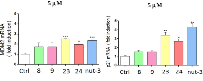
Compounds 13 and 14 resulted practically inactive to decrease the percentage of viable U87MG cells (Data not shown). First of all we have evaluated if compounds 7, 8, 9, 23 and 24 were able to increase the expression levels of p21 and MDM2. For this purpose we have treated tumor cells with each compound at the concentration of 5µΜ.
Compound 7 resulted totally inactive and its activity has not been reported; compounds 8 and 9 have not produced any significant results (Figure 21).
On the contrary compounds 23 and 24, but mainly compound 23, resulte the most effective to activate the function of genic transactivation of p53 (Figure 21).
Figure 21. Increase of mRNA levels for MDM2 or p21 ofter treatment with compounds 8, 9, 23
and 24. Analysis ANOVA and test Bonferroni p<0.05 (*); p<0.0005 (***).
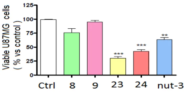
At the same time we have evaluated if compounds 7, 8, 9, 23 and 24 were able to decrease the percentage of viable U87MG cells. An analogue trend was observed indeed only compounds 23 and 24 are able to decrease the percentage of living U87MG cells (Figure 22). Compound 7 is totally inactive and its activity has not been reported.
Figure 22. Percentage of viable cells treated with compound 8, 9, 23, 24, and with theNnutlin-3.
Analysis ANOVA and test Bonferroni p<0.05 (*); p<0.0005 (***). On the basis of these results compounds 7, 8 and 9 have not been tested anymore.
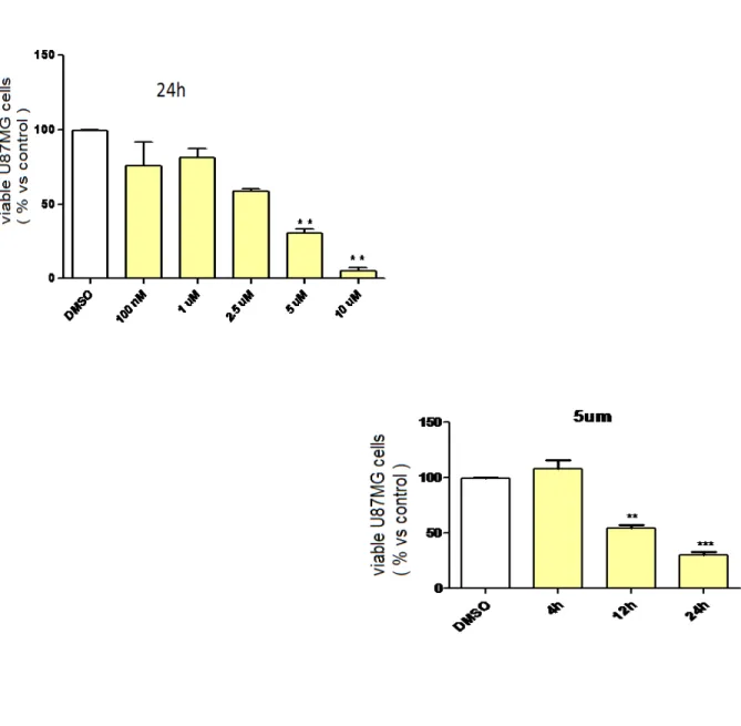
Then we have tested if compounds 23 and 24 activity on cell viability is concentration and time dependent (Figure 23 and 24).
To evaluate the concentration-dependence of antiproliferative effect, U87MG cells have been treated with growing dose of compounds 23 and 24 (from 100 nM to 10µΜ) and after one day of incubation living cells have been counted.
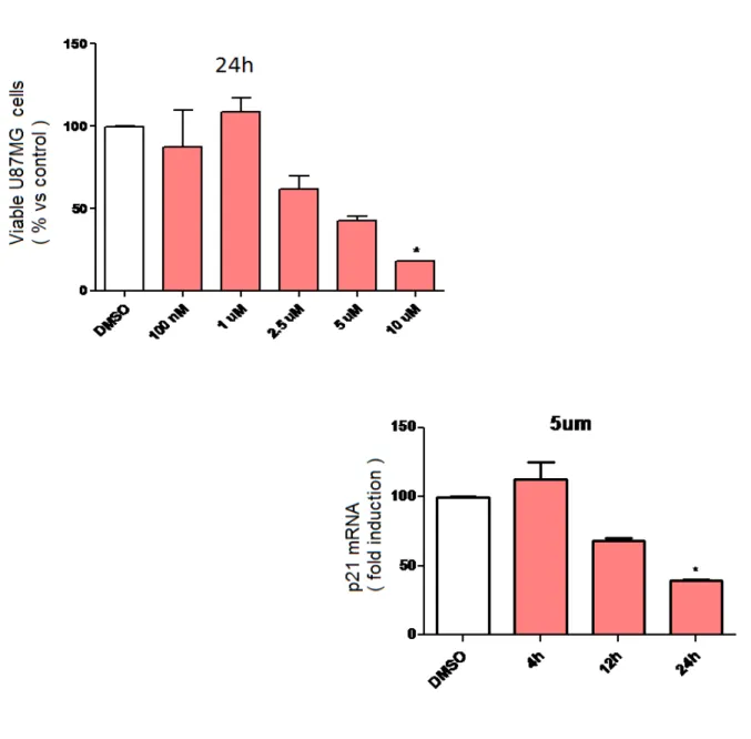
On the contrary to evaluate the time-dependence of anti-proliferative effect, the U87MG cells have been treated at the concentration of 5µΜ of compounds 23 and 24 and after 4, 12 and 24h of incubation living cells have been counted. Results have been shown in the Figure 23 and 24. The Figure 23 and 24 highlight the dose and time-dependence of compounds 23 and 24, suggesting the specificity of the antitumor in vitro effect.
Figure 23. Percentege of viable cells treated with compound 23.
Figura 24. Percentege of viable cells treated with compound 24.
Analysis ANOVA and test Bonferroni p<0.05 (*); p<0.0005 (***).
Subsequently we have evaluated if compounds antitumor effect is due to the irreversible block of cell cycle progression.
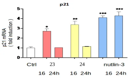
For this purpose we have analyzed if p21 mRNA levels in U87MG cells, after treatment with compounds 23 and 24 at the concentration of 5µΜ, were stably increased during all treatment.
For compounds 23 and 24 there is an increase of mRNA levels for p21 only after 16 h incubation. On the contrary Nutlin-3 induces a significant increase of mRNA for p21 also after 24 h incubation.
Preliminary data showed that all compounds induced block of cell cycle progression (specifically G1/ G2 phases) (Figure 25).
Figure 25. Increase of mRNA levels for p21 after treatment with compounds 23 and 24.
Analysis ANOVA and test Bonferroni p<0.05 (*); p<0.0005 (***).
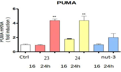
Moreover we have tested if compounds antitumor effect is due to the apoptosis. For this purpose we have evaluated mRNA levels for PUMA, a pro.apoptotic protein encoded by p53, in U87MG cells after treatment with compounds 23 and 24 at the concentration of 5µΜ.
Notably there is an important increase of mRNA levels for PUMA after 24 h incubation. These data suggest an implication of the apoptotic process in the antiproliferative effect of compounds
23 and 24.
Figure 26. Increase of mRNA levels for PUMA after treatment with compound 23 and 24.
Analysis ANOVA and test Bonferroni p<0.05 (*); p<0.0005 (***).