CHAPTER 1.
p73 and the p53 family: a complex network of
proteins
The TP53 tumor suppressor gene: a question of life or death
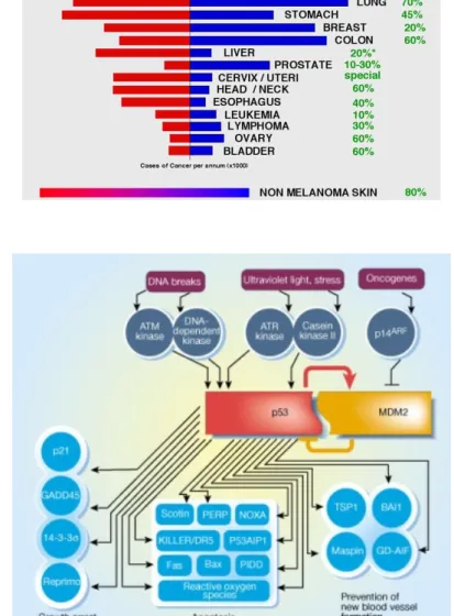
Discovered in the late 1970’s as a cellular protein that efficiently associates with SV40 T antigen, p53 has been the first tumor suppressor gene identified and is now recognized to play a pivotal role in human cancers genesis (Lane DP and Crawford LV, 1979; Linzer DI and Levine AJ, 1979). Virtually every human cancer type displays incidence of mutation, deletion or functional inactivation of p53 (Figure 1a) (Greenblatt MS et al, 1994; Hollstein M et al, 1991; Nigro JM et al, 1989). Indeed p53-/- mice develop tumors at nearly 100% incidence within the first months of age (Donehower LA et al, 1992; Jacks T et al, 1994; Purdie CA et al, 1994).
The p53 protein is a sequence-specific transcription factor whose main function is to regulate the expression of many genes involved in cell cycle arrest and apoptosis in response to genotoxic damage or cell stress (Figure 1b) (el-Deiry WS et al, 1994; Kastan MB et al, 1991; Kastan MB et al, 1992, Kuerbitz SJ et al, 1992). p53 induction of growth arrest or apoptosis prevents the replication of damaged DNA and the division of genetically altered cells. Moreover p53 can determine a tumor-specific response to anti-cancer drugs that trigger apoptosis by inducing DNA damage: thus p53 function is not just to protect from malignancy but also to specifically commit tumor cells to death upon drug treatment (Fisher DE et al, 1994; Lowe SW et al, 1993; Lowe SW et al, 1994).
For almost twenty years p53 was thought to be alone not having any related protein. With p73 and p63 discovery the whole vision of “p53 world” changed prompting researchers to elucidate similarities and differences, in terms of functions and regulation, between p53 family members (Kaghad M et al, 1997; Yang A et al, 1998).
The TP73 gene locus: a variety of isoforms
Structural domains of p53 family members
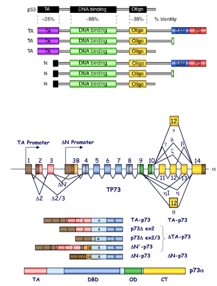
p73 and p63 share with p53 aminoacidic similarity and high structural homology. Each protein contains an amino-terminal Transactivation Domain (TA), a Proline Rich Domain (PR), a core DNA Binding Domain (DBD) and a carboxy-terminal Oligomerization
(a)
(b)
(a)
(b)
Figure 2. p53 family isoforms and gene structure. (a) Comparison of
the structural domains of p53 and the six principal encoded p63/p73 isoforms. TA, Transactivation Domain; DBD, DNA Binding Domain; OD, Oligomerization Domain; SAM, Sterile alpha motif; PS, Post-SAM Domain (Yang A et al, 2002). (b) Schematic representation of TP73 gene. TP73 gene encodes fundamentally two classes of proteins with different functions: TAp73 products that possess the TA domain and are transcriptionally active and ΔTAp73 products that lack the TA domain (Stiewe T et al, 2002).
θ η η1 12 β δ γ ζ ε 12 11 TP73 14 13 10 9 8 7 6 5 4 3B 1 2 3 TA Promoter ΔN Promoter Δ2 Δ2/3 ΔN′ TA DBD OD CT p73α 2 2 4 2 4 3 3B TA-p73 ΔN-p73 p73Δ ex2 p73Δ ex2/3 1 ΔTA-p73 2 3 4 5 1 5 5 5 3B 4 4 1 3 4 5 ΔN'-p73 1 ΔN-p73 TA-p73 1 12 α
Domain (OD) (Figure 2a) (Blandino G and Dobbelstein M, 2004; Levrero M et al, 2000; Melino G et al, 2002; Stiewe T and Putzer, 2002).
p73 and p63 contain also a characteristic carboxy-terminal portion, the Sterile Alpha Motif (SAM) domain, that is not present in p53. SAM is a globular domain composed of four α-helices and a small 310-helix
and its precise function is still not well understood: generally it’s found in proteins that are involved in development and mediates interactions between proteins or between proteins and RNA (Kim CA and Bowie JU, 2003). The p73 SAM domain displays binding to several artificial lipids and the functional mechanism involved in this interaction has yet to be investigated (Barrera FN et al, 2003). Unlike other SAM domains, those present in p73 and p63 seem to be not involved in homo- or hetero-dimerization but could mediate interactions with other not yet identified proteins (Chi SW et al, 1999).
The highest homology of p53 family members is reached within the DNA Binding Domain where the sequence identity between p53 and p73 is almost of 65%. This domain recognizes and interacts with a set of common and unique for each protein promoters. Differences in the subset of target genes controlled could be explained with the different strength by which the transcription factors bind their own consensus sequences. At least in electrophoretic mobility shift assays p53 seems to bind DNA weakly and the binding is strengthened by antibody-induced conformational change. In contrast p73 binds DNA more efficiently than p53 and this binding is not further increased by antibodies (Kartasheva NN et al, 2002). p63 DNA Binding Domain shows higher thermostability and loss of cooperativity in DNA binding in contrast with the homologous p53 domain (Klein C et al, 2001). The different way p53 family members bind DNA can be an additional regulation step required just for p53 but not for p73 and p63.
At the carboxy-terminal side of the DNA Binding Domain all members have a conserved Oligomerization Domain that would suggest the possible formation of hetero-oligomers as well as homo-oligomers. In reality p53 binds to DNA as a homo-tetramer and the other members preferentially form homo-tetramers rather than hetero-tetramers with each other (Davison TS et al, 1999; Marin MC and Kaelin WG Jr, 2000). Essentially wt p53 does not co-precipitate with p73 but some p53 dominant negative mutants bind and inactivate p73 and p63 transactivation function (Bergamaschi D et al, 2003; Di Como CJ et al, 1999; Gaiddon C et al, 2001; Monti P et al, 2003; Strano S et al, 2000; Strano S et al, 2002).
The domain at the extreme carboxy-terminal end of p53, p73 and p63 has activities not yet well understood. In the case of p53 this portion in vitro exerts a negative regulation on p53 binding to DNA
this part of the protein. The C-teminus can also exert an autoinhibitory effect on p73/p63 transactivation (Ozaki T et al, 1999). p73 isoforms generated by alternative splicing
From the very first the study of TP73 and TP63 genes highlighted complex mechanisms of regulation based on alternative splicing and alternative promoters usage. In contrast, alternative splicing of p53, although discovered many years ago, has only recently caught wider attention (Arai N et al, 1986; Courtois S et al, 2004). Murine cells express a carboxy-terminal truncated p53 form more efficient in DNA binding: in human cells this kind of spliced form is absent but, as is emerging, different alternative splicing patterns of p53 may occur.
Most of the p73 splicing processes concerns the carboxy-terminal domain, involves exons 10 to 13 and creates proteins with different C-termini (Figure 2b). At the moment at least six different p73 isoforms, named α, β, γ, δ, ε and ζ, are known to be present in normal cells (De Laurenzi V et al, 1998; De Laurenzi V et al, 1999; Kaghad M et al, 1997; Levrero M et al, 2000; Ueda Y et al, 1999). The β and δ isoforms are truncated forms of the full-length p73α and the δ isoform, lacking the majority of the C-terminus, is the one that more resembles p53. The ζ isoform has an internal deletion (from residue 400 to residue 496) and the γ isoform displays a different C-terminus due to a long alternative reading frame. At the list above have been added other three short isoforms named θ, η and η1. Identified by
RT-PCR this spliced products lack the carboxy-terminal transactivation domain and the Oligomerization domain (Figure 2b) (Scaruffi P et al, 2000). Unlike the other C-truncated isoforms, the η isoform has been detected just in neoplastic cells (Scaruffi P et al, 2000).
The p73 spliced products have been found in different proportions in the several cell species analyzed and are overexpressed in many tumor cell lines (Zaika AI et al, 1999; Zwahlen D et al, 2000). The prevalence in cancer cells of carboxy-terminal deleted forms, lacking a domain involved in transcriptional regulation, suggests that the process of splicing can modulate p73 expression during tumorigenesis. Less is known about the pattern of exon skipping during development or in normal cells.
In addition to the C-terminal side diversity of the human p73 other isoforms, identified in cancer cells and truncated at the amino-terminal side, are also arranged by exon skipping: ΔN’p73, Δ2p73, Δ3p73 and Δ2,3p73 (Fillippovich I et al, 2001; Ishimoto O et al, 2002). These truncated forms, named ΔTAp73, are transactivation-defective, behave as dominant negative isoforms in regards to TAp73 and p53 and act as anti-apoptotic proteins (Pozniak CD et al, 2000).
p73 isoforms generated by the use of alternative promoters
TP73 gene has two major promoters, P1 and P2, with the latter located in the third intron 30kb downstream of the former. These promoters direct expression of two kinds of proteins: the TA-proteins, which contain the Transactivation Domain, encoded by exon 2 and 3, and the ΔN-proteins, which lack this domain (Figure 2b) (Grob TJ et al, 2001; Sayan AE et al, 2001; Yang A et al, 2000).
The Transactivation Domain is important for interactions with several transcription coactivators that allow the enhanced expression of p53 target genes. Thus the TA-p73 isoforms are able to activate transcription of p53-responsive genes, such as p21, GADD45, PUMA, BAX, controlling growth arrest and apoptosis. On the contrary the ΔN variants inhibit the TA-p73 isoforms and p53 in a dominant negative manner: the ΔN isoforms can occupy p53-responsive promoters not recruiting the transcription machinery or can interact with TA-p73 isoforms giving rise to complexes not competent for transcription (Figure 3a) (Pozniak CD et al, 2000; Yang A et al, 2000). The ΔN forms are expressed in all possible carboxy-terminal variants, creating and increasing grade of complexity (Grob TJ et al, 2001; Ishimoto O et al, 2002; Stiewe T et al, 2002).
Recently amino-terminal truncated isoforms of p53 have been discovered but their existence is not the consequence of the use of an internal promoter but rather to the translation from an internal start site (Courtois S et al, 2002; Courtois S et al, 2004).
A the light of what is known about the TP73 gene products it’s possible to delineate a rough classification of the p73 proteins family: we can distinguish “p53-like” proteins, such as TA-p73 proteins, able in activating transcription and with a tumor suppression effect and “p53-nonlike” proteins, Δ Np73 and ΔTAp73, not competent for transcription and acting as dominant negative isoforms with anti-apoptotic and pro-proliferative effect (Figure 3b).
Regulation of p73 activity
Regulation of p73 expression by different promoters activity
As above mentioned, TP73 gene has two promoters that drive the transcription of several p73 isoforms and that can be regulated in a distinct way.
(a)
(b)
Figure 3. The p53 family: a complex network of proteins. (a) Modes
of transcriptional suppression by p73 and p63 ΔN isoforms (Yang A et al, 2002). (b) Crosstalk between N- and C-terminal isoforms in the p53 family (Courtois S et al, 2004).
Sequence analysis and characterization of the P1 promoter, TA-p73 promoter, revealed the presence of at least three potential E2F1 consensus sites and of two sequences that resembles consensus p53-responsive elements (p53RE). E2F1 controls p73 expression under physiological condition, such as in the G1/S transition or in activation-induced cell death of thymocytes (Irwin M et al, 2000; Lissy NA et al, 2000; Stiewe T et al, 2000). It has been also demonstrated that upon DNA damage E2F1 is acetylated and is selectively recruited on the P1p73 promoter to activate it and to promote cellular apoptotic response (Pediconi N et al, 2003). Moreover a recent report suggests a mechanism of p73 induction associated with the removal of transcriptional repression from the TAp73 promoter. In fact, upon treatment with DNA damaging drugs, the transcription factor C-EBPalpha is translocated in the cytoplasm and its direct repressive activity on the transfactor E2F1, mediated by direct DNA binding, is abrogated. These data suggest that in normal condition C-EBPalpha forms a repressor complex with E2F1 on the TAp73 promoter (Marabese M et al, 2003).
Within the first intron of p73, which is positioned immediately upstream to the ATG codon of exon 2, there is a 1-kb regulatory fragment that behaves as a silencer of p73 activity. It contains six consensus binding sites for the transcriptional repressor ZEB and might have an effect in the modulation of p73 expression during differentiation (Fontemaggi G et al, 2001).
The P2 promoter, ΔNp73 promoter, interestingly not contains E2F1 binding site but contains a very efficient p53/p73 responsive element. This promoter is activated in response to non-apoptotic DNA damage in a p53-dependent manner to accumulate ΔNp73 proteins and to determinate p53/p73 induced cell cycle arrest in cells that do not undergo apoptosis (Grob TJ et al, 2001; Kartasheva NN et al, 2002; Nakagawa T et al, 2002; Vossio S et al, 2002). As in the case of the mdm2-p53 loop, the control of p53/TAp73 on Δ Np73 promoter creates a negative autoregulatory feedback loop that can fine regulate p53 family functions. Deregulation of ΔNp73 promoter results in malignant transformation of NIH-3T3 fibroblasts and tumor growth in nude mice (Stiewe T et al, 2002). Thus the loss of ΔNp73 promoter autoregulatory pathway, as happens in cancer or infected cells, would result in an indefinitely increase of ΔNp73 expression that inactivates p53 and p73, contributing to cancer development (Stiewe T et al, 2002; Allart S et al, 2002).
The only creation of selectively knock-out mice for TA promoter and Δ N promoter products will allow to dissect the distinct and specific role that the two promoters exert in controlling the p53 family functions.
Interactions of p73 with regulatory proteins
Since their discovery p73 proteins have been tested for all the several interactions that regulate p53 and in these years a large variety of regulatory proteins have been characterized to interact with and regulate p73. The elucidation and comprehension of the network that controls p73 expression, stabilization and function could be useful to more define its role in many cellular processes.
Mutual interactions with the other p53 members
p53 family members regulate each other by several mechanisms. The most obvious is the competition for the same consensus sequences as happens on the promoter of the p53 responsive genes that are negatively regulated by ΔNp73 isoforms. Another kind of regulation can be possible at the level of protein-protein interaction. The fact that the region involved in oligomerization is conserved in p53 family proteins suggests the possibility of interactions between the whole family members. It’s demonstrated that p63 and p73, unlike p53, can assemble in hetero-oligomers and this add another level of complexity to the mechanisms that govern the regulation of each protein (Davison TS et al, 1999). Although the wild type p53 seems not to be able to bind its homologues, several authors reported that in human tumors p53 mutants physically interact, through their DNA Binding Domain, with p63 and p73. The interaction can vary in strength and specificity with the kind of p53 mutation and the presence of the polymorphism at the P72 of p53 (Marin MC et al, 2000; Strano S et al, 2000; Strano S et al, 2002; Strano S and Blandino G, 2003).
More interestingly the stress-induced apoptosis promoting function of p53 has been demonstrated to be dependent on p63 and p73 presence in the cell: specifically, the p53 homologues work to selectively stabilize p53 interaction with pro-apoptotic gene promoters (Flores ER et al, 2002).
The mechanism of interaction between p53 and its homologues is still unknown but two models are proposed. In one model, “the dynamic exchange model”, p53, p63 and p73 form a large complex that sequester the three proteins nearby the promoter of the target genes. At any time one of them can bind DNA to activate transcription and p63/p73 have the function to stabilize the complex and to favorite active tetramers-DNA binding. The other mechanism, “the dual site stabilization model”, is suggested by the existence, in many gene promoters, of several p53 responsive elements: two transcription factors at a time, such as p53 and p73, are required for an efficient transcription due to a complete recruitment of the transcriptional
(a) Dynamic Exchange Model
(b) Dual Site Stabilization Model
Figure 4. Two hypothetical models of the relations between p53
family members in the stress induced apoptotic response. (a) The dynamic exchange model. The three proteins form, nearby the DNA responsive elements, a large transcriptional complex in which at any time one of the p53 family members can promote transcription. (b) The dual site stabilization model. p53 and p63 and/or p73 bind two different responsive elements on DNA. This simultaneous binding of the two transcription factors stabilizes the recruitment of the
machinery on the responsive elements (Figure 4) (Urist M and Prives C, 2002).
Both the models strongly evocate a potential p53 protein conformational change that allows an interaction with the homologues p63 and p73 directly nearby the DNA binding sequence. How specifically and strongly the different species of hetero-tetramers bind DNA is not well established and is something researchers are seeking after and that could explain the differences in the p53 family members expression in various tissues during development, tumorigenesis and stress response.
MDM2
The protein p53 is not highly expressed in the cell but accumulates upon DNA damage or cellular stresses and its function is mainly regulated at the level of protein stability. The murine mdm2 gene product, such as the product of the human homologue hdm2, is the principal regulator of p53 stability and function and is involved in several types of human cancer. mdm2 binds specifically and tightly p53 within the Transactivation Domain neutralizing its transcriptional activity and mediating its ubiquitination and rapid proteasome degradation (Momand J et al, 1992). Upon DNA damage p53 is phosphorylated in many sites within the amino-terminal domain such as Ser 15, Ser 20, Ser 33 or even Thr 18 and this phosphorylations can disrupt the interaction between p53 and mdm2 allowing p53 accumulation (Ashcroft M and Vousden KH, 1999).
Moreover the MDM2 gene is directly activated by p53 transcriptional function and this establishes a negative autoregulatory feedback loop in which p53 activates its own inhibitor. The restrain of p53 activity within the cells is determinant to terminate p53-mediated responses once the inducing signal is turned off.
Another level of regulation exerted by mdm2 is the interaction with the p53 nascent polypeptide and the induction of translation from two alternative initiation sites. p53 can be translated in two different forms p53 and ΔNp53, a truncated form that lacks the amino-terminal domain responsible of transactivation activity (Courtois S et al, 2002; Yin Y et al, 2002). Although the p53 shorter form cannot bind mdm2 anymore is still able to interact with the non-truncated form and to inhibit its transcriptional activity. The final effect is in any case the abolishment of p53 response in growth arrest or apoptosis by two different mechanisms: one based on p53 synthesis control and another one based on p53 degradation.
A recent discovery changed again the scenario of p53 and mdm2 network. In fact, while mdm2 promote the addition of a single chain of ubiquitin to a cluster of six Lysines located in the p53 C-terminus, p53 can be also polyubiquitinated together by mdm2 and p300/CBP that
functions as an E4 ligase (Grossman SR et al, 2003).
At the light of so many data concerning the multiple levels of mdm2 control of p53 function many researchers have tried to elucidate how the same protein regulates p73. p73α interacts with mdm2 in vivo and in vitro through its amino-terminal region thus the specificity of the binding is not supposed to be modified in the case of the several carboxy-terminal p73 spliced isoforms (Balint E et al, 1999; Ongkeko WM et al, 1999; Zeng X et al, 1999). The p73 protein also interacts with mdmX a protein related to mdm2 (Ongkeko WM et al, 1999).
Although p73 binds mdm2 the association does not result in p73 degradation as happen for p53: actually the two proteins differ just in the C-terminus, the region that could be responsible for mdm2-mediated degradation. Nonetheless interaction of p73 with mdm2 has a functional consequence because reduces TAp73-dependent transcription (Balint E et al, 1999; Dobbelstein M et al, 1999; Lee CW and La Thangue NB, 1999; Zeng X et al, 1999). An explanation of this effect could be the fact that mdm2 binds the cofactor p300/CBP, important for the p73 transcription activity, and sequester it away from p73 transcriptional competent complexes (Zeng X et al, 1999).
The informations available at the moment suggest that the accumulation of p73 seems not to rely in a change of the steady-state protein level by disruption of p73-mdm2 interaction. Furthermore the fact that mdm2 can control p53 protein synthesis and polyubiquitination opens new perspectives on the possible role of MDM2 E3 ligase in the control of the balance between the different p73 isoforms and in facilitating the degradation promoting function of p300/CBP rather than its acetylation function.
YAP
Differences between the p53 family members have been found in the interaction with the potent transcription factor Yes-associated protein (YAP) that binds TAp73 forms but not ΔNp73 and p53 and this different behavior could confer specificity in the p73-mediated apoptosis (Strano S et al, 2001). Indeed impairment of YAP retention in the nucleus after AKT phosphorylation can attenuate p73 apoptosis in response to DNA damage (Basu S et al, 2003).
Viral Oncoproteins
In tumors viruses p53 activity is antagonized by many viral proteins. The adenovirus E1B 55k-Da protein and SV40 T-antigene inactivate p53 protein by sequestering it in not active complexes.
growth suppression and apoptotic functions are not inhibited by the simple presence of the above mentioned oncoproteins (Dobbelstein M and Roth J, 1998; Higashino F et al, 1998; Marin MC et al, 1998; Prabhu NS et al, 1998; Steegenga WT et al, 1999).
The adenovirus E1A protein binds to and inactivates p73 and p53 and another adenovirus protein, E4orf6, inhibits p73 transactivation function and induction of apoptosis, although other studies contradict this assumption. (Higashino F et al, 1998; Querido E et al, 2001; Roth J et al, 1998; Steegenga WT et al, 1999; Wienzek S et al, 2000). The fact that p73 protein, as well as p53 protein, is induced by adenoviral infection strongly suggests that inhibition of p73 is necessary for virus replication and probably adenovirus inhibits the two members of the p53 family using different oncoproteins.
Another viral protein involved in tumorigenesis is the HCV core protein that can induce or inhibit programmed cell death accordingly with the virus life cycle necessities (Otsuka M et al, 2000; Ray RB and Ray R, 2001). Many p73 isoforms interact, through their C-terminus, with the HCV core protein and this results in the translocation of core protein into the nucleus where this particle interferes with the control of cell proliferation modulating p73-dependent transcription (Alisi A et al, 2003).
Further studies are necessary to better understand differences and similarities, between p53 and p73, in viral protein recognition and interaction to discover if the two transcription factors have different roles in virus replication and transformation.
p73 subcellular localization
p73 is mostly localized into the nucleus where mediates the induction of many target genes and its functions, as happens for p53, can be regulated by subcellular localization. The mechanism by which p53 and p73 are localized is not fully understood but some consensus signal inside the protein, the Nuclear Export Signal (NES) and the Nuclear Localization Signal (NLS), mediate the import/export between cytoplasm and nucleus.
p53 NES is masked when the protein is in form of tetramer. The mdm2 mediated ubiquitination of Lysines within the C-terminus of p53 induces a conformational change that exposes the NES and allows p53 to exit from the nucleus. p73 differs the most from p53 exactly in the carboxy-terminal side and the Lysines ubiquitinated on p53 are not conserved in p73. In fact mdm2 does not ubiquitinate p73β and does not promote NES exposure. Indeed p73β aggregates and is retained inside the nucleus (Gu J et al, 2001).
A Nuclear Localization Signal (NLS) has been identified in p73 that is responsible for its translocation into the nucleus. Mutants lacking
the NLS are localized in the cytoplasm and, consequently, have a reduced transcriptional activity (Inoue T et al, 2002). The increased stability of mutants lacking NLS and NES strongly suggests the importance of the proper localization for efficient p73 degradation. Thus p73 localization is controlled by both nuclear import and export and the overall distribution of p73 is likely to result from the balance between these two processes (Inoue T et al, 2002).
A recent report shows that the promyelocytic leukemia (PML) protein modulates p73 half-life by inhibiting its degradation in a PML-nuclear body (NB)-dependent manner. p73 recruitment into the nuclear bodies requires its phosphorylation by p38 and subsequent PML-dependent stabilization (Bernassola F et al, 2004). Also the nuclear protein Daxx, which co-localizes with PML, has been found to interact with p53 and p73 and to inhibit their transcriptional activity. The authors suggest that there is a competitive binding between Daxx, p53 and PML and this regulation pathway could also control p73 (Kim EJ et al, 2003).
p73 transcription factor cytoplasmic retention can be a mechanism that impairs the apoptotic and stress induced response. The amphiphysins are members of a protein superfamily involved in clathrin-mediated endocytosis, particularly of synaptic vescicles. Amphiphysin IIb-1 binds through its SH3 domain the p73β carboxy-terminal end. This interaction results in p73β relocalization in the cytoplasm. The NLS of p73 can be masked by the amphiphysin IIb-1 binding thus preventing the nuclear import and impairing p73 transcriptional activity (Kim KC et al, 2001).
Another mechanism has been recently discovered to be important in sequestering p73 in the cytoplasm. The recently cloned WWOX tumor suppressor gene encodes for a protein that contains two WW domains, generally known to mediate protein-protein interaction. Wwox protein physically interacts via its first WW domain with p73 by a mechanism that is tyrosine kinase Src phosphorylation dependent. Wwox overexpression triggers redistribution of nuclear p73 to the cytoplasm and, hence, suppresses its transcriptional activity although cytoplasmic p73 in turn contributes to the pro-apoptotic activity of Wwox. Wwox expression is altered in several cancer type, thus, it is important to understand how TP73 and WWOX gene products function and how their loss affects malignant transformation. Additional genetic and biochemical approaches will elucidate the biological consequences of this association in normal and cancer cells (Aqeilan RI et al, 2004).
Another protein involved in p53 subcellular localization is the recently discovered Parc, a Parkin-like ubiquitin ligase, that anchors p53-associated protein complexes in the cytoplasm (Nikolaev AY et
and induction of apoptosis. In many neuroblastoma derived cell lines p53 is abnormally localized in the cytoplasm. This condition can be easily released and p53 dependent response activated with siRNA-mediated reduction of endogenous Parc (Nikolaev AY et al, 2003). This novel interaction that regulates p53 localization has not been yet tested for p73 or the other p53 family members.
p73 involvement in development, differentiation and senescence
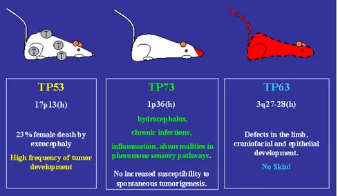
While TP53 gene product is ubiquitously expressed, p63 and p73 proteins show tissue specificity and are involved in defined developmental stage. p73 is mainly found in many regions of the central nervous system and p63 is highly expressed in multi-layered squamous epithelia like skin (Yang A et al, 1999; Yang A et al, 2000). To support the hypothesis of a functional compartmentalization of the p53 homologues, genetic analysis of knockout mice showed profound and distinct developmental defects that suggested a p63 and p73 supposed role in processes that govern the correct development of the organisms.
p63 -/- mice essentially do not develop multilayered epithelia: mice have truncated limbs, have no hair follicles, no teeth, no mammary, lacrimary or salivary glands (Yang A et al, 1999). Thus p63 is important in defining the characteristics of the epidermal stem cell compartment (McKeon F, 2004). Indeed both the TAp73 forms and the ΔNp73 forms seem to be essential for the control of epidermal development (Figure 5) (Koster MI and Roop DR, 2004; Koster MI and Roop DR, 2004; Koster MI et al, 2004).
The unique role for p73 in neurogenesis, sensory pathways and homeostatic control is revealed by p73 -/- mice that display congenital hydrocephalus, hippocampal dysgenesis, loss of peripheral-sympathetic neurons, chronic infections and inflammations and defects in pheromone detection (Figure 5) (Yang A et al, 2000; Pozniak CD et al, 2000). Unlike p53-null mice, however, p73 and p63 knockout mice do not show increased susceptibility to spontaneous tumorigenesis (Yang A et al, 1999; Yang A et al, 2000).
Many of the neurological abnormalities observed in the p73-/- mice are a consequence of neuron absence or loss (Yang A et al, 2000). During nervous system development, neurons are overproduced and cells directly compete, in order to survive or die, for their growth factors in a process known as naturally occurring cell death. The neurons that usually are committed to survive during the developmental apoptosis instead die in the p73 knockout mice (Pozniak CD et al, 2000).
In the p73 -/- mice the absence of Δ Np73, the isoform more expressed in murine fetal nervous system, could be responsible of an
enhanced neuronal apoptosis (Pozniak CD et al, 2000; Porziak CD et al, 2002). At least in cultured wild type sympathetic neurons, that in the organism display a mainly developmental apoptosis, NGF treatment leads to an increase in ΔNp73 levels that counteracts p53-induced apoptosis and promotes survival. ΔNp73 is involved also in a p53-independent mechanism inhibiting events at the mitochondrial apoptotic checkpoint, as the induction of BimEL and the release of mitochondrial cytochrome c, or inhibiting also one very early event in the apoptotic cascade, the activation of c-Jun N-terminal protein kinase (JNK), likely by the direct binding to JNK (Lee AF et al, 2004). The role of ΔNp73 in neuronal survival is not limited to developing neurons because p73-/- mice exhibit continued neuron loss throughout life. Thus ΔNp73, with its anti-apoptotic function, plays a critical role in the pathways that determine the survival and the long-term maintenance of neurons (Pozniak CD et al, 2000; Porziak CD et al, 2002). Therefore, the balance between p53 and the TA and ΔN forms of p73 modulates the level of apoptosis in neurons during development (Jacobs WB et al, 2004).
Interestingly some phenotype observed in the p73 knockout mice resemble human neurodegenerative disorders and preliminary data show p73 altered subcellular distribution in Alzheimer Disease patients-derived brain tissues (Wilson C et al, 2004).
In agreement with the neurological defects found in p73 -/- mice the simple p73 overexpression in undifferentiated neuronal cells as neuroblastoma cells, is sufficient to trigger neuronal differentiation in vitro. Indeed increasing transcription of the TP73 gene was observed in the neuroblastoma cells upon induction of retinoids-mediated differentiation. Indeed p73 transactivation function allows the increase of NCAM, p21 and neurofilaments (De Laurenzi et al, 2000).
Like its homologue p53, p73 has been recently implicated in cellular senescence (Alexander K et al, 2003; Fang L et al, 1999; Narita M et al, 2003).Cellular senescence is a process by which the cells are stably arrested; this mechanism guarantees that damaged cells do not proliferate to prevent cancer progression. In tumor cells lacking functional p53, the induced overexpression of p73 isoforms, p73α and p73β, is sufficient to promote permanent growth arrest with markers of replicative senescence (Fang L et al, 1999). Activation of E2F in senescent cells can result in the use of p73 as a cell death effector. The maintenance of the terminal cell cycle arrest of senescent cells requires continuous pRb-mediated inactivation of E2F activity, the reappearance of which in these irrevocably altered cells triggers a cell death program instead of an inappropriate resumption of cell cycling (Alexander K et al, 2003).
senescence-Figure 5. Main characteristic of p53, p63 and p73 knockout mice.
p53-/- mice develop normally but display a higher frequency of tumor than their wild-type counterparts. p63-deficient mice do not develop multilayered epithelia and have truncated limbs, have no hair follicles, no teeth, no mammary, lacrimary or salivary glands (Yang A et al, 1999). p73 knockout mice show severe neurological abnormalities, chronic infections and inflammations and defects in pheromone detection (Yang et al, 2000). Unlike p53, p63 and p73 deficient mice do not have increased susceptibility to spontaneous tumors (http://www.iarc.fr/p53).
cyc B genes, irreversibly repressed in senescent human fibroblast, are repressed by p73. DNA-binding activity of the NF-Y transcription factor, required for transcription of cdk1 and cyclin B genes and inactivated in senescent fibroblasts, is sensibly decreased by p73 and p53 (Jung MS et al, 2001, Melino G et al, 2003). Similarly, inactivation of pRb and p53 by adenovirus E1A protein also results in the transcriptional silencing of E2F-responsive cell-cycle-related genes due to the binding of heterochromatin proteins to E2F target gene promoters within senescence-associated heterochromatic foci (SAHF) (Narita M et al, 2003).
p73 and malignancy: tumor suppressor or not?
The discovery that p53 in fact belonged to a family of proteins raised the challenging question of the role of the other family members. Assessing that TP73 gene was located in a tumor suppressor locus was accepted with enthusiasm for the correspondence with a possible p73 function in tumor suppression.
The excitement was soon cooled off with the characterization of p73-/- mice. Unlike p53 knockout mice, p73 deficient mice are not prone to cancer and display profound developmental defects (Jacks T et al, 1994; Yang A et al, 2000). Moreover p73 is rarely mutated in human cancer (Stiewe T and Putzer BM, 2002). Taken together all these data suggested that p73 is not a conventional tumor suppressor conformed to Knudson’s two hit model.
This unexpected finding can now be explained with the presence of two antagonistic classes of proteins encoded from the same gene: knocking the gene means exclude the TA-forms and ΔN-forms, not changing the balance of their respectively pro-apoptotic and anti-apoptotic effects. In fact the alteration of TA/ΔN forms balance or the increase of a specific C-terminus isoform can determine the kind of response the cell activates upon growth stimuli, genotoxic stresses or viral infection.
Unfortunately just in the last few years, with the characterization of the whole spectrum of p73 isoforms, reagents are being designed to specifically discriminate between TA, ΔN and ΔTA p73 isoforms.
The great amount of data available at the moment demonstrates how signaling by the entire p73 proteins family is different yet similar to what is known for p53, suggesting a comparable role for p73 network in tumorigenesis and stress response.
melanoma and breast cancer (Kaghad et al, 1997). TP73 gene is localized in a restricted region of overlap (SRO) suggesting that p73 was indeed the 1p36.3 tumor suppressor gene the researchers were seeking after (Perri P et al, 1999). This finding prompted many groups to begin the analysis of several human cancers with the intent to find p73 mutations (Table 1). The fact that all these studies failed in finding p73 mutations could be in part explained by the consideration that TP73 gene maps in a imprinting region: the loss of one allele is sufficient to make a cell p73 null if the other allele is imprinted. Actually, although many studies have demonstrated monoallelical expression in certain tissues and tumors, other studies reported loss of imprinting (LOI), biallelic expression or allele switching of p73 (Table 1). All these confusing data taken together make difficult to understand the role of p73 imprinting in tumorigenesis.
Mutations of p73 in primary cancer
In contrast to the common p53 mutations in cancer, TAp73 mutations are rarely found in human malignancies (Table 1). Two known examples are P405R and P425L mutations, a somatic and a germline mutation, found in primary neuroblastoma. The P405R substitution does not affect p73α or β transcriptional activity. Instead the P425L substitution reduced p73α-dependent growth-suppression but not p73β activity (Naka M et al, 2001).
Overexpression of wild-type p73
Most of the studies on p73 function in tumorigenesis have analyzed p73 expression and have found higher p73 mRNA and protein levels in cancer tissues rather than in the normal surrounding tissues (Table 1). If p73 expression levels are almost undetectable by Western blot or Northern blot analysis of normal cells, this is not the case of tumor-derived cell lines and primary tumors (Stiewe T et al, 2004; Zaika AI et al, 1999). In particular, as shown in Table 1, neuroblastomas, ependymomas, hepatocellular and cholangiocellular carcinomas, lung, prostate, colorectal, gastric, breast, bladder, ovarian and esophageal cancers have p73 overexpressed in respect to the corresponding normal tissues.
Importantly, p73 overexpression correlates with poor prognosis in hepatocellular carcinomas, with advanced tumor grade in meningiomas, with lymph node metastases, vascular invasion and higher pathologic stage in breast cancer and B-CLL (Dominguez G et al, 2001; Novak U et al, 2001; Nozaki M et al, 2001; Tannapfel A et
secondary event but is a step involved in tumorigenesis. Further studies have yet to come to elucidate p73 isoforms pattern and the mechanism at the basis of p73 overexpression in human cancer. Upregulation of p73 by oncogenes
Most human cancers display elevated activity of the cell cycle promoting transcription factor E2F1. Deregulation of E2F1 by mitogenic oncogenes (Ras, c-myc, v-abl and E1A), by the loss of p16, pRb or by overexpression of cyc D, leads to abnormal cell-cycle progression which in turn contributes to unregulated tumor cells growth. E2F1 deregulation also triggers an apoptotic checkpoint, leading to accumulation of p53 and inducing p53-dependent apoptosis (Sherr CJ, 1998). E2F1, in fact, promotes the activation of the tumor suppressor ARF that impairs p53 degradation by mdm2. p73 interacts with and is inactivated by mdm2 but is not committed to degradation by the E3 ubiquitin ligase: thus the oncogenic activation of the ARF/mdm2 pathway results in the accumulation of p53 but not of p73.
E2F1 regulates p73 levels directly, through recognition and transactivation of the TP73 gene promoter that contains several E2F-binding sites (Irwin M et al, 2000; Lissy NA et al, 2000; Pediconi N et al, 2003; Stiewe T and Putzer BM, 2000). E2F1 control of p73 expression regards the steady-state protein level: in fact p73 levels fall in serum starved cells and reaccumulate upon S-phase entry, a period in the cell cycle in which E2F-responsive genes are actively transcribed (Irwin M et al, 2000; Stiewe T and Putzer BM, 2000). In human tumor cells endogenous p73 is activated by E2F1 and induces apoptosis: abrogating p73 function, using a dominant negative p53 mutant or an anti-sense construct, inhibits E2F1-induced apoptosis (Stiewe T and Putzer BM, 2000).
Other oncogenes, including E1A and c-myc, belong to the p73 signaling pathway in vivo and promote p73-dependent apoptosis in tumor cells (Zaika A et al, 2001). E1A induced apoptosis, in a p53-null cellular context, correlates with its ability to bind E2F1 regulators, p300/CBP and RB and it’s therefore conceivable, that at least the effect of E1A on p73, can be attributed to activation of E2F1 (Putzer BM et al, 2000). Increased levels of p73 in cancer tissues might therefore be mostly attributable to increased transactivation of the TP73 gene by tumor-associated deregulation of E2F1 by various oncogenes. However, it still remains possible that E1A and c-myc exert additional effects on p73 unrelated to E2F1. At the moment the interaction between c-myc and p73 is still controversial: some authors report that c-myc induces and activates p73 others show that c-myc interacts with and therefore inactivates p73 transcriptional activity (Watanabe K et al, 2002; Zaika A et al, 2001).
Expression of ΔNp73 and ΔTAp73 isoforms
Recent reports have addressed the question of the specific N-terminus truncated p73 form expression in human cancer: ΔNp73 or ΔTAp73 are overexpressed in a subset of tumors as breast cancer, ovarian cancer, vulval cancer, neuroblastoma and hepatocellular carcinoma (Melino G et al, 2002; Ng SW et al, 2000; Stiewe T and Putzer BM, 2002; Stiewe T et al, 2004). Moreover, consistent with its putative role as an anti-apoptotic oncogene higher expression levels of ΔNp73 have been shown to represent a prognostic factor in several tumors (Bergamaschi D et al, 2003; Casciano I et al, 2002; Zaika AI et al, 2002). These studies together with the data obtained in animal model suggest that ΔNp73 has oncogenic properties because high levels of the protein promote cellular transformation similarly to many classic oncogenes (Petrenko O et al, 2003; Stiewe T et al, 2002). How the balance between TA and ΔN proteins is regulated or dysregulated during tumorigenesis is still not well understood.
Inhibition of p73 by p53 mutants
Although p53 is considered one of the most important tumor suppressors identified, p53 mutants behave as potent oncogene and actively participate at the genesis of human cancers (Bergamaschi D et al, 2003; Irwin MS et al, 2003; Irwin MS and Miller FD, 2004).
Unlike other tumor suppressor genes, inactivated by non-sense or frameshift mutations, p53 gene have mutations that lead mainly to a stable product, lacking its DNA binding activity and accumulating in the nucleus of tumor cells (Soussi T and Beroud C, 2001). The accumulation of this kind of mutations has two consequences: p53 mutants behave as dominant negative forms of wt p53 and gain new potent and not specific functions.
After many years of research directed to understand the potential mechanism of oncogenicity of the p53 mutants and the resultant chemoresistance to anticancer drugs, an explanation can emerge with the discovery of p63 and p73. In particular, recent studies show that p73 apoptotic response is inactivated by p53 mutants (Bergamaschi D et al, 2003; Irwin MS et al, 2003). In fact p53 mutants, trough their DNA Binding Domain, bind directly p73 and p63 (DiComo CJ et al, 1999; Marin MC et al, 2000; Strano S et al, 2002). In particular, in co-transfection assays, the R175H and R248W p53 mutants co-precipitate with p73α and in a p53 reporter assay, transactivation activity of p73α is partially inhibited by the p53 mutants, which correlates with a reduction in p73α -mediated
One characteristic of p53 required for its binding to p73 is recognition by the monoclonal antibody PAb240, which recognizes the p53 core domain in a mutant conformation (Gaiddon C et al, 2001). Thus, the core domain of mutant p53, that cannot bind and activate p53 target genes, is able to promote a protein-protein interaction that might contribute to the gain of function activities of mutant p53 by sequestering and inactivating proteins like p73 (Strano S et al, 2000). Indeed many p53 mutants interact with p73 and recent reports attribute to a conformational change of the p53 central domain the success of this interaction (Bensaad K et al, 2003).
Also the p53 polymorphism in codon 72 (Arg or Pro) influences the interaction with p73: in particular the Arg in the 72 position is required to bind p73 (Marin MC et al, 2000). Moreover Arg-p53 mutants cooperated with Ras to transform cells and inhibit p73-dependent apoptosis more effectively than their Pro-counterparts. Interestingly only p53 mutants in the Arg form are resistant to cytotoxic agents (Bergamaschi D et al, 2003). This observation is also supported by a study on patients with head and neck cancer: the ones expressing mutant associated with Arg polymorphism have a poor prognosis and a short survival.
Thus inactivation of p73 by certain p53 mutants appears to provide a selective advantage in promoting tumorigenesis. However, reduction of p73 apoptotic potential by mutant p53 cannot account for all cases of p73 overexpression because many studies on several primary tumors have failed to find a significant correlation between p53 mutational status and p73 overexpression.
p53 family network and chemoresistance
As reported in many studies p73 is induced and activated by many chemoterapic drugs (Irwin MS et al, 2004). Many of these drugs induce p73 post-translational modifications, as phosphorylation (upon cisplatin and γ irradiation treatment) and acetylation (upon doxorubicin treatment) that affect p73 stability and pro-apoptotic transcriptional activity (Agami R et al, 1999; Costanzo A et al, 2002; Gong JG et al, 1999; Yuan ZM et al, 1999). Interestingly, not all the tumor cells activate p73 apoptotic response upon drugs treatments. The cellular context, the presence of pro- or anti-apoptotic proteins, such as the other p53 family members, is likely important to determine p73 activity in the cell.
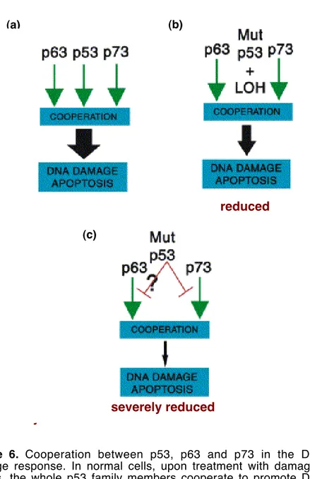
For example interaction of p73 with tumor-derived p53 mutants modulates chemosensitivity (Irwin MS, 2004). Indeed, as above mentioned p53 family proteins collaborate to activate target genes involved in apoptosis following drugs treatment and specific TA forms inactivation in tumor cells leads to chemoresistance (Figure 6) (Bergamaschi D et al, 2003; Flores ER et al, 2002; Irwin MS et al,
severely reduced
Figure 6. Cooperation between p53, p63 and p73 in the DNA
damage response. In normal cells, upon treatment with damaging agents, the whole p53 family members cooperate to promote DNA damage induced apoptosis. In tumor cells p53 can display thee kind of mutants: (a) inactive mutants with no dominant negative function
(b) inactive mutants with a dominant negative function (c) mutants
severely reduced
reduced
(a) (b)
2003). In contrast overexpression of ΔNp73 in tumor cells protect them from drug- induced cell death (Zaika AI et al, 2002). Therefore once again a variation of the balance between TA/ΔNp73 isoforms or the activity of p53 mutants within the tumor cells not only promotes transformation but also determine cell response to chemotherapy.
Evolution of the p53 family
The whole amount of data available at the moment suggests that the different members of the p53 family perform distinct functions in mammalian organisms, accordingly with the distinguishing phenotypes characterized by the analysis of the knockout mice.
Since the discovery of p63 and p73 the challenging question is still the same: have the three genes evolved from a precursor whose function was to protect the cell from malignancy or rather from one involved in development and stem cell control? Alternatively could this ancestor have been able of the all functions?
The sequencing projects of Drosophila melanogaster and Caenorhabditis elegans, organisms with a single p53-like gene, provide new informations about the relationship between p53, p63 and p73.
The fly p53, highly different from mammalian p53, has been involved in mechanisms that protect genome integrity (Brodsky MH et al, 2000; Jin S et al, 2000; Ollmann M et al, 2000). In particular the use of a dominant-negative version of Dm-p53 seems to prevent the DNA damage induction of the pro-apoptotic REPER gene (Brodsky MH et al, 2000). In a similar way p53-deficient worms, although develop normally, display germ cells resistant to radiation-induced cell death and chromosome instability, the both phenotype consequences of the impairment of the “mammalian p53” characteristic functions (Derry WB et al, 2001).
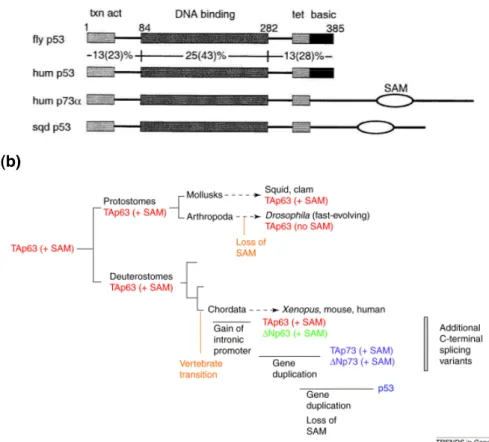
At the beginning p53 was thought to be the elder member of the family because of the genome protecting functions of Drosophila p53 and because of the SAM domain absence. To counteract these arguments is the observation that Dm-p53 and Ce-p53 reveal a gradient of similarities starting with p53 of squid and clams, then the core regions of mammalian p63, p73, and finally p53. In fact the squid p53 contain a SAM domain located inside the carboxy-terminal region and share with p63 high identity. A phylogenetic analysis of the invertebrate and vertebrate p53 gene families reveal that the SAM domain present in the mammalian p63 and p73 proteins is present also in p53 belonging to protostomes (Mollusca, Arthropoda, Annelidia) and deuterostomes (Echinodermata and Chordata), indicating that the ancestral p53 molecule indeed possessed a SAM domain (Figure 7a) (Aguinaldo AM et al, 1997; Gerhard J and
Kirschner M, 1997). These observations taken together suggest that p53, the first to be discovered, is likely the “younger” product of evolution (Figure 7b).
(a)
(b)
Figure 7. Evolution of the p53 family members. (a). Schematic
representation of p53 domains. Percent sequence identities (and similarities) are indicated for the principal regions of Drosophila and human p53. An extended C-terminal tail containing a putative SAM domain is present in squid p53 and human p73 but not Drosophila or human p53. Numbers above the alignments indicate amino acid positions in Drosophila p53 (Brodsky MH et al, 2000). (b) Main events of metazoan evolution of the p53 family members. TAp63 with a SAM domain is present before separation between protostomes and deuterostomes and this suggests that the common ancestor of the family is not a p53-like protein but is more similar to p63 protein. p73 and p53 appeared later in the evolution probably because of gene duplication events (Yang A et al, 2002).
CHAPTER 2.
p73 functions are modulated by post-translational
modifications
Transcriptional activity of p73
The p73 transcription factor is involved in several cellular processes as cell-cycle arrest, apoptosis and neuronal differentiation (De Laurenzi V et al, 1998; De Laurenzi V et al, 2000; Jost CA et al, 1997; Kaghad M et al, 1997). p73 mediates these responses by its transcriptional function that lead to a specific and selective activation of common with p53 or distinct groups of target gene promoters (De Laurenzi V et al, 1998; Fontemaggi G et al, 2002; Kaghad M et al, 1997; Zhu J et al, 1998). How p73 selects its targets and what determines the subset of p53 gene promoters that are specifically regulated by p73 is still unknown. For p53 protein are emerging the features of a functional model in which p53 “chooses” between the response pathways to activate: this model could be extended also to p73 protein (Vousden KH, 2000).
Initially, reports on p53 mutants able to induce growth arrest but not to promote apoptosis-dependent suppression of transformation suggested that p53 functions could be independently regulated (Crook T et al, 1994; Friedlander P et al, 1996; Ludwig RL et al, 1996; Rowan S et al, 1996). Point mutations can induce conformational changes in p53 mutant proteins that result in slightly different capabilities to recognize the responsive elements of low-affinity apoptotic vs high-affinity growth arrest gene promoters. This model is supported by the finding that low levels of p53 activate cell growth arrest and high levels of p53 activate apoptotic response, masking the concomitant cell-cycle arrest response (Chen X et al, 1996). Recent studies examining the binding of p53 to promoters within the context of chromatin in vivo have partially supported this model (Kaeser MD and Iggo RD, 2002; Szak ST et al, 2001).
However, not all known pro-apoptotic target gene promoters display low-affinity p53-binding sites. An example is the promoter of PUMA pro-apoptotic gene that is similar, for what concerns p53 affinity, to the CDKN1A and MDM2 gene promoters. Thus the binding affinity of p53 to different promoters is not sufficient to explain differences in p53 target gene regulation.
A new emerging model involves p53-interacting proteins as important regulators of p53 DNA binding activity. Some cofactors generally contribute to p53 transcriptional function, others selectively enable p53 expression of the apoptotic target gene (Samuels-Lev Y et al, 2001; Shikama N et al, 1999). A similar contribution to apoptotic
target-gene activation by p53 was recently revealed by the unexpected observation that induction of cell death by p53 requires the presence of at least one of the other p53-family members, p63 or p73. In fact the simultaneous recruitment of different members of the p53 family allows the selective transactivation of a class of target genes that are involved in apoptosis (BAX, NOXA and PERP) (Flores ER et al, 2002; Vousden KH and Lu X, 2002). Thus the formation of large transcriptional complexes containing p73 and p53 could confer specificity for pro-apoptotic target genes.
p73 and the DNA damage induced response
The cellular response to DNA damage includes the recognition of injured DNA, cell cycle arrest to assess the damage, and implementation of the appropriate response, i.e., DNA repair or, in the event of irreparable damage, cell death in order to preserve genome integrity (Rich T et al, 2000). Several evidences indicate that the quality of cellular response is influenced by the nature of the DNA-damaging agent used, i.e., IRs, that produce primarily DNA strand breaks, versus base-damaging agents such as UV radiation or DNA alkylation (Holbrook NJ et al, 1996).
Both the tumor suppressor protein p53 and its close relative p73 are activated by DNA damage. However, whereas p53 is induced by a variety of DNA damaging agents as well as by many other forms of cellular stress, p73 is induced and activates apoptosis only upon a subset of agents, including IRs and cisplatin (Agami R et al, 1999; Giaccia AJ and Kastan MB, 1998; Gong JG et al, 1999; Yuan ZM et al, 1999). Moreover, upon DNA damage the p73 transcription factor, like p53, is able to drive the transcription of several apoptotic effectors, including BAX, p53AIP1, NOXA and PUMA (Costanzo A et al, 2002; Flores ER et al, 2002; Miyashita T and Reed JC, 1995; Oda E et al, 2000; Oda K et al, 2000; Steegenga WT et al, 1999; Zhu J et al, 1998). p73 might also contribute indirectly to the activation of apoptosis by modulating p53 function: p73 sequesters mdm2 and allows the increase of p53 levels.
The p53 protein transcriptional function, activated in response to genotoxic stress, is influenced by a complex pattern of post-translational modifications including phosphorylation and acetylation (Meek DW, 1998; Prives C and Hall PA, 1999). Nevertheless, the comprehensive role of p53 phosphorylation and acetylation in the response to DNA damage in vivo is still incomplete.
Although DNA damage mainly induces p73 transcription by E2F1, the activities of p73, like p53, are also regulated by post-translational
(a)
(b)
Figure 8. (a) Structural domains, some modification sites and
co-regulators are shown for human p53 (Wahl GM and Carr AM, 2001).
(b) Known post-translational p73 modifications. The post-translational
modifications that modulate p73 functions are phosphorylation (Y99, T86, S47, S289) acetylation (K321, 327, 331) and SUMO-lation (K627). Abbreviations: TA, Transactivation Domain; DBD, DNA Binding Domain; OD, Oligomerization Domain; SAM, Sterile Alpha Motif; TI, Transcription Inhibitory Domain.
TA DBD OD SAM p73α P-T86 P-Y99 K627 TI P-S47 P-S289 Ac-K321,327,331
Post-translational modifications could determine p73 specificity and direct it through a specific response. Whether Δ Np73 is phosphorylated or acetylated is also uncertain, and it remains possible that post-translational modifications enhance the anti-apoptotic function of Δ Np73 to promote survival. Similarly, informations about the interactions of the different splicing isoforms with known p73 cofactors are not yet available. It seems likely that the response to DNA damage is highly dependent on the cellular context.
Phosphorylation
The first discovered p73 modification was the phosphorylation by the nuclear tyrosine-kinase c-Abl (Agami R et al, 1999; Gong JG et al, 1999; Yuan ZM et al, 1999).
The c-Abl protein tyrosine kinase is distributed in the nucleus and in the cytoplasm of proliferating cells. In the nucleus, c-Abl activity is negatively regulated by the retinoblastoma protein (RB) and positively regulated by DNA damage signals. Activation of the c-Abl kinase by DNA damage requires the function of ATM, which regulates cell cycle checkpoint, DNA repair and apoptosis in response to DNA damage. In addition, c-Abl-dependent inhibition of PI 3-kinase contributes to the induction of apoptosis.
The physiologic function of c-Abl may reside in controlling the cellular response to DNA strand breaks that occur during DNA replication, genetic recombination and gene rearrangements. In response to genotoxic stress, c-Abl functions in determining cell fate and promoting growth arrest and repair or induction of apoptosis. Upon DNA damage c-Abl binds to p53 and induces p53-dependent p21 expression thus controlling a pathway that regulate the G1 arrest response. However if the DNA lesions are irreparable c-Abl promotes the induction of apoptosis by mechanisms that are p53-dependent or not (Kharbanda S et al, 1998; Wang JY, 2000). In fact, although p53-deficient cancer cells are less responsive to chemotherapy, their resistance is not complete, which suggests that another apoptotic pathway may exist and many groups assessed that p73 is involved in this mechanism.
c-Abl binds to p73, through its SH3 domain with the Oligomerization Domain of p73. Upon DNA damage, induced by ionizing radiation (IR) and cisplatin (CDDP), c-Abl phosphorylates p73 on a tyrosine residue at position 99 to promote p73 stabilization and apoptosis-inducing function (Agami R et al, 1999; Gong JG et al, 1999; Yuan ZM et al, 1999). Another effect of c-Abl dependent p73 phosphorylation is that, in response to IR, p73 undergoes nuclear
that are defective in c-Abl kinase activation (Ben-Yehoyada M et al, 2003).
c-Abl also induces p73 phosphorylation in threonine residues adjacent to prolines and the p38 MAP kinase pathway mediates this response (Sanchez-Prieto R et al, 2002). p38 can phosphorylate p73 on threonine directly, without the requirement for c-Abl (Sanchez-Prieto R et al, 2002). Furthermore p38 phosphorylation of p73 is required for p73 stabilization and recruitment into the nuclear bodies. The promyelocytic leukaemia (PML) protein, in fact, modulates p73 half-life by inhibiting its ubiquitin-proteasome degradation in a PML-nuclear body (NB)-dependent manner. PML regulates p73 stability by positively modulating the levels of acetylation, a process that impair p73 ubiquitination. As a result, PML potentiates p73 transcriptional and pro-apoptotic activities that are markedly impaired in Pml-/-primary cells (Bernassola F et al, 2004). Thus, both p38 phosphorylation pathways enhance the stability and transcriptional activity of p73.
Also the checkpoint kinase 1 (Chk1), an essential component of the DNA damage checkpoint, phosphorylates p73α. Endogenous p73α interacts with Chk1 and is phosphorylated on serine 47 by endogenous Chk1 upon DNA damage, which is a mechanism required for the apoptotic-inducing function of p73α (Gonzalez S et al, 2003).
Protein kinase C (PKC) δ is cleaved by caspase-3 to a kinase-active catalytic fragment (PKCδCF) in the DNA damage apoptotic cellular response. PKCδCF associates with p73β and phosphorylates its Transactivation and DNA Binding Domains. One PKCδCF-phosphorylation site has been mapped to serine 289 in the p73β DNA-binding domain. PKCδCF-mediated phosphorylation of p73β is associated with its accumulation and induction of its transactivation activity. Indeed PKCδCF stimulates p73β-mediated apoptosis as this response is attenuated with the p73β (S289A) mutant (Ren J et al, 2002).
Modification by Pin1
Similarly to p53, p73 binds to and is regulated by the prolil isomerase Pin-1 that regulates several phases of the cell cycle, including G1–S and G2–M, as well as the DNA-replication checkpoint (Mantovani F et al, 2004). The p73 conformational change catalyzed by Pin1 is crucial in the DNA damage induced pathway. The loss of Pin1 reduces p73 stability, impairing its accumulation upon genotoxic stress. Indeed, upon treatment with chemotherapeutic drugs c-Abl enhances the phosphorylation-dependent interaction between Pin1 and p73 and this, in turn, promotes p300-mediated p73 acetylation.
Thus Pin 1 is required for the ability of c-Abl and p300 to increase p73 stability and transcriptional activity (Mantovani F et al, 2004). Sumolation
p53 and p73 interact with the small ubiquitin-like modifier (SUMO) protein. Indeed SUMO protein covalently modifies p73α, but not β, on the C-terminal lysine 627 residue (Minty A et al, 2000; Rodriguez MS et al, 1999). This modification seems to potentiate p73α degradation by proteasome and this can explain the differences between α and β isoforms in term of transcriptional activity. In fact p73β is more powerful than p73α in activate transcription because the sumolation of p73α influences the interactions with other proteins, such as c-Abl tyrosine kinase. The involvement of SUMO in PML nuclear bodies has raised the possibility that p73 interacts with PML in the PML bodies (Maisse C et al, 2003).
p73 regulation during the cell cycle
p73 expression is regulated during the cell cycle: in fact p73 is induced by E2F1 at the early S phase when activates the transcription of adenosine deaminase (ADA) gene, an important enzyme involved in nucleotide metabolism, the deficit of which causes the inhibition of DNA synthesis and repair (Tullo A et al, 2003). This finding suggests that the p73 protein might be important in the control of cell growth and DNA replication. Consistent with this hypothesis a recent study reports that p73 interacts with various cyclins (A, B, D, and E). Moreover cyclin A/cdk1/2, cyclin E/cdk2 and cyclin B/cdk1/2 complexes phosphorylate p73 on threonine 86 residue in vitro. Using an antibody that specifically recognizes phosphorylated Thr86, it was observed that in vivo phosphorylation of p73 at Thr86 is cell cycle dependent. Phosphorylation of Thr86 is induced during S phase and is highest in the G2–M phase of the cell cycle. Consistent with this observation inhibitors of cdks, such as p16 and serum starvation, reduce phosphorylation of the p73 Thr86.
Unlike c-Abl and p38MAP kinases, which are activators of p73, cdks act as p73 inhibitors because the T86 phosphorylation represses its ability to induce p21 expression (Gaiddon C et al, 2003).
Moreover a recent report describes the ability of p73β, but not p53, to activate expression of the cyclin-dependent kinase inhibitor p57 (KIP2) and KvLQT1, two genes that are co-regulated in an imprinted region of the genome (Blint E et al, 2002).
RESULTS
p73 acetylation upon DNA damage
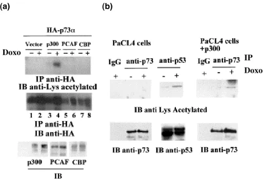
Acetylation is one of the post-translational modifications that can affect the p53-dependent apoptotic response and could also have a crucial role in p53 stability (Ito A et al, 2001; Luo J et al, 2000; Rodriguez MS et al, 2000). The p300/CREB-binding protein (CBP) and p300/CBP-associated factor (PCAF) acetylate the carboxyl terminus of p53 at lysines 373 and 382 (p300/CBP) and at Lys320 (PCAF). Indeed CBP and p300 co-activate p53 (Avantaggiati ML et al, 1997; Gu W and Roeder RG, 1997; Lill NL et al, 1997).
The general consensus seems to be that the recruitment of these coactivators stabilizes p53 and augments sequence-specific DNA binding following DNA damage (Avantaggiati ML et al, 1997; Gu W and Roeder RG, 1997; Lill NL et al, 1997; Luo J et al, 2004). The p300 protein might further regulate the transcriptional activity of p53 through a novel acetylation site, Lys305 (Wang YH et al, 2003). CBP and p300, but not PCAF, might modulate cellular p53 activity not only by modifying p53, but also by inactivating MDM2 through acetylation (Wang X et al, 2004). Furthermore, MDM2 can inhibit acetylation of p53 mediated by either CBP or p300 and conversely deacetylation of p53 is required for its effective degradation by MDM2 (Ito A et al, 2001).
A recent report assessed that mammalian histone deacetylases (HDACs) downregulate p53 transcriptional activity (Juan LJ et al, 2000). Indeed the human homologue of SIR2, SIRT1, has been shown to directly deacetylate p53 at Lys382, resulting in an impaired response to γ-radiation-induced apoptosis and a decreased transcriptional activation of p53 target genes such as CDKN1A (Vaziri H et al, 2001). Also Sir2α expression has been showed to suppress p53 transcriptional activation and p53-mediated apoptosis (Luo J et al, 2001).
Thus p53 acetylation by many co-factors and in many cellular contexts seems to be a key step in the regulation of DNA damage response and we tried to asses if the p73 transcription factor, whose functions often overlap with p53 functions, can be regulated by the same mechanisms (Figure 9).
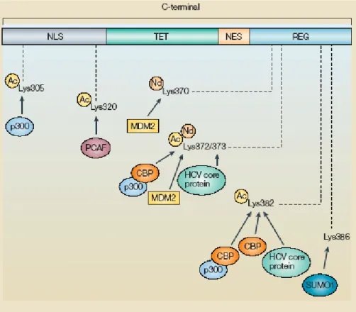
Figure 9. Acetylation sites of human p53. Several lysines have been
reported to be acetylated on p53 and all are located in the carboxy-terminal region. In addition, one lysine residue (Lys386) is sumoylated by small ubiquitin-like modifier 1 (SUMO1) and three lysines (Lys370, Lys372 and Lys373) are neddylated by the addition of NEDD8. Each site and the proteins responsible for the modification are indicated. Ac, acetylation; HCV, hepatitis C virus; Nd, neddylation; NES, nuclear export signal; NLS, nuclear localization signal; REG, C-terminal regulatory domain; TET, tetramerization domain (Bode AM and Dong Z, 2004).