The Pediatric Infectious Disease Journal • Volume 33, Number 5, May 2014 www.pidj.com |
525
I
mmunology
R
epoRts
Abstract: Chronic granulomatous disease is a rare primary immuno-deficiency caused by phagocytic cell defect. We describe the case of 43-month-old boy with chronic granulomatous disease presenting with Salmonella spp brain abscesses, together with a review of the 13 cases reported in the literature.
Key Words: chronic granulomatous disease, brain abscess, Salmonella spp, treatment, review of literature
(Pediatr Infect Dis J 2014;33:525–528)
C
hronic granulomatous disease (CGD) is an inherited immu-nodeficiency disorder which results from the absence or mal-function of nicotinamide adenine dinucleotide phosphate oxidase subunits in phagocytic cells. CGD patients are susceptible to recur-rent life-threatening bacterial or fungal infections as well as granu-loma formations. Brain abscesses are rarely reported in patients with CGD, accounting for <5% of all infections.1 We describe thecase of a child with multiple brain abscesses as the initial presenta-tion of X-linked CGD and review the corresponding literature.
CASE REPORT
This boy is the only child of a non-consanguineous couple. The neonatal history was normal and he had received all the recom-mended immunizations. At the age of 36 months, he was hospital-ized for an enteritis. Vidal-Wright tests resulted positive for
Salmo-nella typhi AgO. The patient was initially treated with ceftriaxone.
In view of the persistence of clinical symptoms and positive stool cultures for multiresistant Salmonella Typhimurium variant Copen-hagen, treatment with imipenem was given for 3 weeks resulting in complete remission. Despite appropriate clinical and labora-tory follow up, he suffered from 2 further episodes of enteritis of unknown etiology.
In January 2013, at the age of 43 months, he was admitted to our Unit with a history of 7 days of fever and headache associated with ataxia, mild ptosis of the right eye and left angle mouth devia-tion. A complete blood count revealed normal white blood cells
count (10,030/μL) and mild thrombocytosis (589,000/μL). Inflam-matory markers such as erythrocyte sedimentation rate (16 mm/h) and C reactive protein (1.32 mg/dL) were slightly elevated. A blood culture obtained on admission, resulted positive for Salmonella spp. computed tomography and magnetic resonance imaging (MRI) of the brain showed multiple ring enhancing lesions in the cerebrum consistent with brain abscesses: a major formation on basal ganglia and 2 minor formations in the left cortical frontal and right tem-poral regions (as shown in Figure, Supplemental Digital Content 1, http://links.lww.com/INF/B829). He was initially treated for 7 days with intravenous meropenem and ceftazidime, according to antibiotic susceptibilities.
Cerebrospinal fluid appeared macroscopically clear, with normal concentrations of protein and glucose. Direct bacterioscopic examinations with Gram stain as well as periodic acid-Schiff and Ziehl-Neelsen stains were negative. Galattomannan also resulted negative. Cerebrospinal fluid cultures resulted sterile. Cytologic examination did not reveal any neoplastic cells.
On the basis of the history of recurrent and resistant Sal-monellosis, a complete immunologic evaluation was performed. Nitroblue tetrazolium test and dihydrorhodamine 1,2,3 Assay dem-onstrated that the patient’s stimulated neutrophils failed to gener-ate reactive oxygen products, leading to the diagnosis of CGD. A definitive diagnosis of X-linked CGD was based on the detection of a nonsense mutation (c.469C>T, p.R157X) in the gene encoding the gp91phox (CYBB). Genetic testing in the family identified the
mother as carrier of the mutation.
Based on the diagnosis of X-linked CGD, intravenous voriconazole (VCZ) was added to the therapy for possible mould co-infection and ceftazidime was replaced by ciprofloxacin. A complete resolution of the clinical picture was obtained. This treatment was continued for 8 weeks, until discharge. Ten months after discharge, MRI scanning showed significant reduction in the size of the brain lesions with no evidence of active disease (as shown in Figure, Supplemental Digital Content 2, http:// links.lww.com/INF/B830). He is currently in good clinical con-dition, receiving standard prophylactic treatment and awaiting an unrelated bone marrow transplantation from a suitably matched donor.
DISCUSSION AND LITERATURE REVIEW
CGD is a rare primary immunodeficiency, occurring in about 1 in 250,000 live births. CGD results from the absence or malfunction of nicotinamide adenine dinucleotide phosphate oxi-dase subunits in phagocytic cells.2 In 65% of all cases, the defect isa consequence of mutation of the CYBB gene (Xp21.1); the remain-ing 35% are inherited in an autosomal recessive manner.3 Because
of a defective innate immune system, the patients suffer not only from serious infections, but also from exuberant inflammatory responses leading to granuloma formation. Life-threatening infec-tions usually become apparent in the first years of life and are typi-cally caused by intracellular catalase-positive microorganisms. The most common infections encountered in CGD are: pneumonia, cutaneous abscess, lymphadenitis, hepatic abscess, osteomyelitis
Copyright © 2014 by Lippincott Williams & Wilkins ISSN: 0891-3668/14/3305-0525
DOI: 10.1097/INF.0000000000000270
Chronic Granulomatous Disease Presenting
With Salmonella Brain Abscesses
Andrea Finocchi, MD, PhD,* Alessia Claps, MD,* Jessica Serafinelli, MD,* Irene Salfa, MD,*
Daniela Longo, MD,† Gigliola Di Matteo, BSc, PhD,‡ Alessandro Aiuti, MD, PhD,*§ and Paolo Rossi, MD, PhD*
Accepted for publication December 31, 2013.
From the *Unit of Immunology and Infectious Diseases, University-Hospital Pediatric Department; †Unit of Radiology, Bambino Gesù Children Hospi-tal, IRCCS; ‡Department of Public Health and Cellular Biology, Tor Vergata University, Rome; and §Pediatric Immunohematology and Bone Mar-row Transplant Unit and San Raffaele Telethon Institute for Gene Therapy (HSR-TIGET), HS Raffaele Scientific Institute, Milan, Italy.
The authors were supported in part by grant CELL PID (HEALTH- F5-2010–261387 to AA) and E-RARE. The authors have no other funding or conflicts of interest to disclose.
Address for correspondence: Dr. Andrea Finocchi, MD, PhD, Unit of Immunol-ogy and Infectious Diseases, University Hospital Department of Pediatrics, Bambino Gesù Children’s Hospital and University of Rome “Tor Vergata,” Piazza Sant’Onofrio, 4 00165 Rome, Italy. E-mail: [email protected].
Finocchi et al The Pediatric Infectious Disease Journal • Volume 33, Number 5, May 2014
526
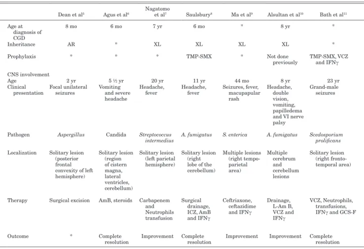
| www.pidj.com © 2014 Lippincott Williams & WilkinsTABLE 1. Clinical-therapeutic Findings in CGD Patients With Brain Abscesses
Dean et al5 Agus et al6 Nagatomo et al7 Saulsbury8 Ma et al9 Alsultan et al10 Bath et al11 Schuetz et al12 Hipolito et al13 Metin et al14 Bukhari and Alrabiaah15 Patiroglu et al16 McNeil et al17 Our patient
Age at diagnosis of CGD 8 mo 6 mo 7 yr 6 mo * 8 yr * 18 mo * 4 yr * 16 yr * 43 mo Inheritance AR * XL XL XL XL * XL-McLeod phenotype * XL * AR * XL
Prophylaxis * * * TMP-SMX * Not done
previously TMP-SMX, VCZ and IFNγ TMP-SMX, ICZ TMP-SMX, ICZ Not done previously * Not done previously TMP-SMX, itraconazole (noncompliance) Not done previously CNS involvement Age 2 yr 5 ½ yr 20 yr 11 yr 44 mo 8 yr 23 yr 6 yr 5 yr 4 yr 5 yr 16 yr 24 yr 43 mo Clinical
presentation Focal unilateral seizures Vomiting and severe headache
Headache,
fever Headache, fever Seizures, fever, macupapular rash Headache, double vision, vomiting, papilledema and VI nerve palsy Grand-male
seizures Incidental diagnosis Focal seizures on right arm, sinutisis, lympho- adenopathy Headache, vomiting, backache, paraplegia Torticollis, upper
back swelling Seizures Tonic clonic seizure, right-sided facial droop, left arm weakness Fever, mild ptosis on right eye, left angle mouth deviation, fever and ataxia Pathogen Aspergillus Candida Streptococcus
intermedius A. fumigatus S. enterica A. fumigatus Scedosporium prolificans Suspected inflammatory etiology
Alternaria
infectoria A. fumigatus A. nidulans A. fumigatus Phaeoacremonium parasiticum Salmonella Spp Localization Solitary lesion
(posterior frontal convexity of left hemisphere) Solitary lesion (region of cistern magna, lateral ventricles, cerebellum) Solitary lesion (left parietal hemisphere) Solitary lesion (right lobe of the cerebellum) Multiple lesions (right tempo- parietal area) Multiple cerebrum and cerebellum lesions Solitary lesion (right fronto- temporal area) Multiple intracerebral lesions Solitary lesion (left parietal dorsally frontal cortex) Multiple lesions (right cerebellar lobe and vermis) Paraverte-bral, epidural and subcutaneous abscess
Solitary lesion (right
frontal lobe) Multiple lesions (right temporal lobe and putamen)
Multiple lesions (basal ganglia and cortical frontal and temporal areas) Therapy Surgical excision AmB, steroids Carbapenem
and Neutrophils transfusion Surgical drainage, ICZ, AmB and IFNγ Ceftriaxone, ceftazidime and IFNγ Drainage, L-Am B, VCZ and IFNγ VCZ, Neutrophils, transfusions, IFNγ and GCS-F BMT L-AmB, IFNγ, meropenem, vancomyci, ceftazidime, VCZ and caspofungin Surgical drainage; TMP-SMX, L-AmB and IFNγ VCZ Surgical excision; VCZ, L-AmB and IFNγ Surgical biopsy; vancomycin, meropenem, caspofungin, L-AmB, VCZ and IFNγ VCZ, ciprofloxacin and meropenem Outcome * Complete
resolution Improvement Complete resolution Improvement Improvement Complete resolution Complete resolution Death Improvement Complete resolution Complete resolution Death Complete resolution
*Unknown.
AR, autosomal recessive; XL, X-linked; TMP-SMX, trimethoprim-sulfamethoxazole; CNS, central nervous system; ICZ, itraconazole; AmB, amphotericin B; L-Am B, liposomal amphotericin B; BMT, bone marrow transplantation.
and perirectal abscesses. Brain abscesses are reported very rarely in patients with CGD, accounting for <5% of all infections.1
The most frequent microorganisms cultured from brain abscesses are Aspergillus spp, Salmonella spp, Klebsiella spp and
Staphylococcus aureus.4 A summary of clinical aspects of reported
patients with brain abscess in CGD is shown in Table 1. In most of the cases, the etiology was represented by fungal pathogens, in par-ticular Aspergillus spp. The number and localization of abscesses are quite variable: about half of the patients presented a single cerebral localization, whereas the other half had multiple lesions within the brain. The frontal lobe is often involved in the case of solitary lesions. One child had extensive spinal cord infection as the primary neurologic manifestation.
Diagnosis of CGD was made at a mean age of 55 months (median 43 months, range 6–192 months). The commonest symp-toms at presentation were seizures, headache and vomiting. Fever is not a common symptom and has been reported in only 4 patients, combined with elevation of inflammatory markers. An asympto-matic presentation is extremely uncommon as reported in the sin-gle case described by Schutz et al.12 Patients with known bacterial
infections frequently showed an acute onset with fever and eleva-tion of inflammatory markers, whereas in the case of fungal infec-tions, most patients showed a subacute onset.
MRI is the most sensitive and specific diagnostic test for brain abscesses. Diffusion-weighted MRI and magnetic resonance
spectroscopy can be helpful in cases where it is difficult to differ-entiate between a brain abscess and a tumor and potentially to dif-ferentiate between fungal and bacterial causes.18
Clinical management of the reported patients has been het-erogeneous. There has been only 1 report of traditional open crani-otomy with complete excision of the mass, which was performed in the earliest case described in 1993 by Dean et al.5 Many patients
recovered with antimicrobial therapy, sometimes combined with a minimally invasive approach such as surgical drainage. A surgical approach is used less frequently because of the significant mor-bidity associated with immunodeficiency and with the invasiveness of the procedure and also because of the great therapeutic success obtained with antimicrobial therapy alone. Improvement or com-plete resolution with no neurologic sequelae has been obtained in 6 patients with antimicrobial therapy alone. However, surgery is often necessary to obtain biopsy for an accurate diagnosis and there are some suggestions that an aggressive surgical approach improves prognosis in central nervous system aspergillosis.14
Management of pediatric brain abscesses is extremely vari-able. The choice, duration and administration route of empiric anti-biotics were not clearly defined, illustrating a lack of consensus guidelines. Ceftriaxone/cefotaxime plus metronidazole has been recently recommended for use in immunocompetent children, whereas meropenem has been proposed as a good first-line choice for immunocompromised patients, in whom outcome is poorer.19
The Pediatric Infectious Disease Journal • Volume 33, Number 5, May 2014 Brain Abscess and CGD
© 2014 Lippincott Williams & Wilkins www.pidj.com |
527
The appropriate duration of antimicrobial therapy is still unclear, ranging from 4 to 6 weeks of antibiotics if the abscess is drained, to at least 6–8 weeks with conservative treatment.20 As infections often
respond slowly in CGD patients, intravenous antibiotic treatment should be followed by prolonged oral treatment and an individu-ally tailored approach guided by clinical improvement, normaliza-tion of inflammatory markers and resolunormaliza-tion on imaging should be considered. In the case of Aspergillus infection, VCZ is currently considered the treatment of choice. In addition to systemic antifun-gal treatment, there has been some suggestion that neurosurgical procedures may improve the outcome in central nervous system aspergillosis.21
Granulocyte transfusions and Interferon gamma (IFNγ) have been used in severely ill CGD patients, especially in those with fungal infections. Their value, however, has not been investi-gated in a controlled study and their clinical use remains somewhat controversial. In addition, with the advent of potent new antifungal drugs, the use of granulocyte transfusions and IFNγ is likely to decrease in the future.
Overall the clinical outcome was benign with only 2 cases of death, both caused by fungal etiology. It should be taken into account that a surgical biopsy was not performed in our patient, in view of the good clinical response to the rapid initiation of anti-biotic/antifungal therapy. Collectively, the identification of
Salmo-nella spp in blood culture, the past medical history with recurrent
Salmonella infections as well as the clinical improvement after
empiric antibiotic therapy, led us to conclude that cerebral lesions were caused by Salmonella infection. Patients with chronic granu-lomatous disease characteristically present a susceptibility to
Sal-monella spp infections.2
Case reports of CGD-associated brain abscesses caused by
Salmonella spp are extremely rare. To our knowledge, only 1 case
has been reported thus far by Ma et al9 in 2003. The patient was
a 44-month-old boy, affected by CGD, who developed multiple parieto-temporal abscesses caused by Salmonella enterica, after a 4-week history of fever and maculopapular rash with skin nod-ules. Similarly to our patient, Salmonella was isolated from blood culture and not from cerebral biopsy. The patient recovered after prolonged antibiotic therapy with third generation cephalosporin plus IFNγ injections. In our case also the resolution of brain infec-tion was induced by a prolonged (8 weeks) antibiotic therapy with meropenem, in association with a broad-spectrum antibiotic such as ciprofloxacin. VCZ was also added for antifungal protection. To our knowledge, our patient is the second case of CGD-associated brain abscesses because of Salmonella spp, but is the first case where the brain infection was the harbinger of CGD. In conclusion, although brain abscesses are not frequently reported in CGD patients, our case emphasizes the importance of investigating a strong clinical suspicion of CGD in the setting of brain abscesses, especially when they occur early in life.
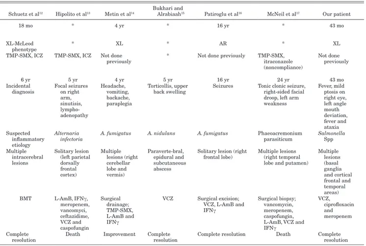
TABLE 1. Clinical-therapeutic Findings in CGD Patients With Brain Abscesses
Dean et al5 Agus et al6 Nagatomo et al7 Saulsbury8 Ma et al9 Alsultan et al10 Bath et al11 Schuetz et al12 Hipolito et al13 Metin et al14 Bukhari and Alrabiaah15 Patiroglu et al16 McNeil et al17 Our patient
Age at diagnosis of CGD 8 mo 6 mo 7 yr 6 mo * 8 yr * 18 mo * 4 yr * 16 yr * 43 mo Inheritance AR * XL XL XL XL * XL-McLeod phenotype * XL * AR * XL
Prophylaxis * * * TMP-SMX * Not done
previously TMP-SMX, VCZ and IFNγ TMP-SMX, ICZ TMP-SMX, ICZ Not done previously * Not done previously TMP-SMX, itraconazole (noncompliance) Not done previously CNS involvement Age 2 yr 5 ½ yr 20 yr 11 yr 44 mo 8 yr 23 yr 6 yr 5 yr 4 yr 5 yr 16 yr 24 yr 43 mo Clinical
presentation Focal unilateral seizures Vomiting and severe headache
Headache,
fever Headache, fever Seizures, fever, macupapular rash Headache, double vision, vomiting, papilledema and VI nerve palsy Grand-male
seizures Incidental diagnosis Focal seizures on right arm, sinutisis, lympho- adenopathy Headache, vomiting, backache, paraplegia Torticollis, upper
back swelling Seizures Tonic clonic seizure, right-sided facial droop, left arm weakness Fever, mild ptosis on right eye, left angle mouth deviation, fever and ataxia Pathogen Aspergillus Candida Streptococcus
intermedius A. fumigatus S. enterica A. fumigatus Scedosporium prolificans Suspected inflammatory etiology
Alternaria
infectoria A. fumigatus A. nidulans A. fumigatus Phaeoacremonium parasiticum Salmonella Spp Localization Solitary lesion
(posterior frontal convexity of left hemisphere) Solitary lesion (region of cistern magna, lateral ventricles, cerebellum) Solitary lesion (left parietal hemisphere) Solitary lesion (right lobe of the cerebellum) Multiple lesions (right tempo- parietal area) Multiple cerebrum and cerebellum lesions Solitary lesion (right fronto- temporal area) Multiple intracerebral lesions Solitary lesion (left parietal dorsally frontal cortex) Multiple lesions (right cerebellar lobe and vermis) Paraverte-bral, epidural and subcutaneous abscess
Solitary lesion (right
frontal lobe) Multiple lesions (right temporal lobe and putamen)
Multiple lesions (basal ganglia and cortical frontal and temporal areas) Therapy Surgical excision AmB, steroids Carbapenem
and Neutrophils transfusion Surgical drainage, ICZ, AmB and IFNγ Ceftriaxone, ceftazidime and IFNγ Drainage, L-Am B, VCZ and IFNγ VCZ, Neutrophils, transfusions, IFNγ and GCS-F BMT L-AmB, IFNγ, meropenem, vancomyci, ceftazidime, VCZ and caspofungin Surgical drainage; TMP-SMX, L-AmB and IFNγ VCZ Surgical excision; VCZ, L-AmB and IFNγ Surgical biopsy; vancomycin, meropenem, caspofungin, L-AmB, VCZ and IFNγ VCZ, ciprofloxacin and meropenem Outcome * Complete
resolution Improvement Complete resolution Improvement Improvement Complete resolution Complete resolution Death Improvement Complete resolution Complete resolution Death Complete resolution
*Unknown.
AR, autosomal recessive; XL, X-linked; TMP-SMX, trimethoprim-sulfamethoxazole; CNS, central nervous system; ICZ, itraconazole; AmB, amphotericin B; L-Am B, liposomal amphotericin B; BMT, bone marrow transplantation.
Finocchi et al The Pediatric Infectious Disease Journal • Volume 33, Number 5, May 2014
528
| www.pidj.com © 2014 Lippincott Williams & WilkinsREFERENCES
1. Roos D, Kuijpers TW, Curnutte JT. Chronic granulomatous disease. In: Ochs HD, Smith CIE, Puck JM, eds. Primary immunodeficiency diseases a molecular and genetic approach. 2nd ed: Oxford: Oxford University Press, 2007:525–549.
2. Holland SM. Chronic granulomatous disease. Clin Rev Allergy Immunol. 2010;38:3–10.
3. Di Matteo G, Giordani L, Finocchi A, et al.; IPINET (Italian Network for Primary Immunodeficiencies). Molecular characterization of a large cohort of patients with Chronic Granulomatous Disease and identifica-tion of novel CYBB mutaidentifica-tions: an Italian multicenter study. Mol Immunol. 2009;46:1935–1941.
4. Van de Berg JM, van Koppen E, Åhlin A, et al. Chronic granulomatous disease: the European experience. PLoS One. 2009;4:e5234.
5. Dean AF, Janota I, Thrasher A, et al. Cerebral aspergilloma in a child with autosomal recessive chronic granulomatous disease. Arch Dis Child. 1993;68:412–414.
6. Agus S, Spektor S, Israel Z. CNS granulomatosis in a child with chronic granulomatous disease. Br J Neurosurg. 2000;14:59–61.
7. Nagatomo T, Ohga S, Saito M, et al. Streptococcus intermedius-brain abscess in chronic granulomatous disease. Eur J Pediatr. 1999;158: 872–873.
8. Saulsbury FT. Successful treatment of aspergillus brain abscess with itra-conazole and interferon-gamma in a patient with chronic granulomatous disease. Clin Infect Dis. 2001;32:E137–E139.
9. Ma JS, Chen PY, Lau YJ, et al. Brain abscess caused by Salmonella enter-ica subspecies houtenae in a patient with chronic granulomatous disease. J Microbiol Immunol Infect. 2003;36:282–284.
10. Alsultan A, Williams MS, Lubner S, et al. Chronic granulomatous dis-ease presenting with disseminated intracranial aspergillosis. Pediatr Blood Cancer. 2006;47:107–110.
11. Bhat SV, Paterson DL, Rinaldi MG, et al. Scedosporium prolificans brain abscess in a patient with chronic granulomatous disease: successful com-bination therapy with voriconazole and terbinafine. Scand J Infect Dis. 2007;39:87–90.
12. Schuetz C, Hoenig M, Schulz A, et al. Successful unrelated bone marrow trans-plantation in a child with chronic granulomatous disease complicated by pul-monary and cerebral granuloma formation. Eur J Pediatr. 2007;166:785–788. 13. Hipolito E, Faria E, Alves AF, et al. Alternaria infectoria brain abscess in a
child with chronic granulomatous disease. Eur J Clin Microbiol Infect Dis. 2009;28:377–380.
14. Metin A, Koker MY, Ozturk MH, et al. Multiple cerebellar Aspergillus abscess in an X-CGD patient. Neurosciences (Riyadh). 2009;14:94–95. 15. Bukhari E, Alrabiaah A. First case of extensive spinal cord infection with
Aspergillus nidulans in a child with chronic granulomatous disease. J Infect Dev Ctries. 2009;3:321–323.
16. Patiroglu T, Unal E, Yikilmaz A, et al. Atypical presentation of chronic gran-ulomatous disease in an adolescent boy with frontal lobe located Aspergillus abscess mimicking intracranial tumor. Childs Nerv Syst. 2010;26:149–154. 17. McNeil CJ, Luo RF, Vogel H, et al. Brain abscess caused by
Phaeoacremonium parasiticum in an immunocompromised patient. J Clin Microbiol. 2011;49:1171–1174.
18. Sheehan JP, Jane JA, Ray DK, et al. Brain abscess in children. Neurosurg Focus. 2008;24:E6.
19. Felsenstein S, Williams B, Shingadia D, et al. Clinical and microbiologic features guiding treatment recommendations for brain abscesses in children. Pediatr Infect Dis J. 2013;32:129–135.
20. De Louvois J, Brown EM, Bayston R, et al. The rational use of antibiotics in the treatment of brain abscess. Br J Neurosurg. 2000;14: 525–530. 21. Giacchino M, Chiapello N, Riva C, et al. Intracranial aspergillosis in
chil-dren successfully treated with antifungal therapy and surgical intervention. Pediatr Infect Dis J. 2006;25:379–381.