UNIVERSITÀ DEGLI STUDI DI MESSINA Dipartimento di Scienze Chimiche, Biologiche,
Farmaceutiche ed Ambientali
DOTTORATO DI RICERCA IN SCIENZE CHIMICHE XXX CICLO
The supramolecular chemistry of water soluble
calixarenes, cyclodextrins and pillararenes
Dott. Lucia Barbera
Supervisor: Coordinator:
Prof. Anna Notti Prof. S. Campagna
Index
Overview
IChapter 1: Introduction
1.1. Supramolecular Chemistry: a brief overview 1 1.1.1. Historical Background of Macrocyclic Compounds 4 1.2. The Supramolecular Chemistry in water 8
1.2.1. Cyclodextrins 10 1.2.2. Calix[n]arenes 12 1.2.3. Pillar[n]arenes 15 References 20 Chapter 2: Macrocyclic amphiphiles 2.1. Introduction 25
2.2. Macrocyclic amphiphiles based on calixarenes 26 2.2.1. Our contribution to anionic calixarene-based surfactants:
results and discussion 29
2.2.2. Self-assembly of amphiphilic anionic calix[4]arene
and drug solubilisation 31
2.2.3. PEGylated-Calix[5]arenes: results and discussion 48 2.3. Macrocyclic amphiphiles based on cyclodextrins 55
2.3.1. Results and discussion 57
2.4. Conclusions 68
2.5.1. Materials 69 2.5.2. Experimental methods 69 2.5.3. Synthetic procedures 74 References 78 Chapter 3: Calix[5]arene-based Supra-amphiphiles
3.1. Supramolecular amphiphiles (supra-amphiphiles) 82 3.2. Calix[5]arenes and n-alkylammonium ions 87 3.3. A calix[5]arene-based supra-amphiphile 89
3.4. Results and discussion 92
3.4.1. Insight into the aggregation properties of calix[5]arene 2
and structures of the supra-amphiphiles 92
3.4.2. Determination of the cmc 104
3.4.3. Comparison of DOSY NMR and DLS data 106
3.4.5. Drug solubilising studies 111
3.5. Conclusions 113
3.6. Experimental section 114
3.6.1. General experimental methods 114
3.6.2. Synthetic procedure 116
3.6.3. Determination of cmc 116
3.6.4. Drug solubilisation 117
References 118
Chapter 4:
Self-sorting of supramolecular systems in water
4.1. Introduction 122
4.2. Results and discussion 126
4.4. Experimental section 144
References 145
Chapter 5:
Drug carriers based on a water soluble pillar[5]arene
5.1. Introduction: synthesis and properties of pillar[n]arenes 147 5.1.1. Water soluble Pillar[5]arenes 151
5.2. Results and discussion 155
5.2.1. WP5 and Amikacin 155
5.2.2. WP5 and Amikacin: antimicrobial activity in vitro and release 162 5.2.3. WP5 and Levofloxacin: preliminary experimental data 166
5.3.WP5 and layer-by-layer assembly 169
5.3.1. Introduction 169
5.3.2. Results and discussion 171
5.4. Conclusions 177
5.5. Experimental section 178
5.5.1. Materials and Methods 178
5.5.2. General Experimental methods 178 5.5.3. Determination of the minimum inhibitory concentration (MIC) 179 5.5.4. Antibacterial activity assessment 180 5.5.5. Antibacterial activity of LbL system 180
I
Overview
The main focus of this PhD thesis is the synthesis of new water soluble macrocycles (calix[n]arenes, cyclodextrins and pillar[n]arenes) and the investigation of their supramolecular chemistry. After a brief introduction about the principles, perspectives, and recent developments in the field of water-soluble synthetic receptors (Chapter 1) the following sections describe the experimental results of my PhD project.
Chapter 2 reports the synthesis and the aggregation properties of new amphiphilic macrocycles based on anionic and neutral p-alkyl-calix[n]arenes and a mono-substituted cationic cyclodextrin by means of different techniques (1D and 2D 1H NMR, DOSY, DLS, AFM, Cryo-Tem) together with different applications in the recognition and/or encapsulation of different substrates.
Chapter 3 describes the aggregation features of supramolecular amphiphilic systems (supra-amphiphiles) based on the water soluble p-tert-butylcalix[5]arene-penta-O-4-butylsulfonato and gemini ,-alkanediyldiammonium ions, demonstrating that the aggregation phenomena can be efficiently modulated by changing the length of the spacer in the gemini guest and/or the host-guest ratio. Furthermore, this
II
chapter demonstrated the great potential of calixarene-based supra-amphiphiles in the solubilisation of water insoluble drugs by using as a model the anticancer drug tamoxifen.
Chapter 4 provides a fascinating example of social (non integrative) self-sorting based on a four component system consisting of two water-soluble calix[5]arene derivatives differently substituted at the upper rim and two alkanediyldiammonium ions of different lengths (H3N+-(CH2)n-NH3+, n = 8, 10). The obtained results demonstrated that among the ten possible complexes with mixed/different stoichiometry, only two capsular-complexes are formed.
Finally, Chapter 5 proposes a potential pillararene (WP5) based drug-transport system where WP5 and the antibiotic drugs amikacin and levofloxacin interact with each other forming stable inclusion complexes in aqueous solution. Furthermore, WP5 was employed for the preparation of layer-by-layer (LbL) thin solid films on glass surfaces loaded with the above mentioned antibiotics, with the ultimate goal of fabricating antibacterial multilayered coatings for controlled drug release.
1
Chapter 1
Introduction
1.1. Supramolecular Chemistry: a brief overview
Supramolecular chemistry has obtained tremendous attention in all branches of science (chemistry, biology, material science) since 1987, when Lehn, Cram, and Pedersen won the Chemistry Nobel Prize on account of their discoveries in the host-guest systems.1 Different noncovalent interactions, such as hydrogen-bonding, van der Waals force, stacking, electrostatic and hydrophobic/hydrophilic etc., can be involved to obtain supramolecular assemblies.2 During the past decades supramolecular chemistry has been widely explored in various areas, including catalysis, functional materials, electronic devices, sensors, molecular machines and nanomedicine,3 as finally demonstrated by the award of the 2016 Nobel Prize in Chemistry to Jean-Pierre Sauvage, Sir J. Fraser Stoddart and Bernard L. Feringa for the "Design and synthesis of molecular machines".4
In 1983 Jean-Pierre Sauvage succeeded in linking two ring-shaped molecules together in form of a chain, called catenanes.5 The two interlocked rings were able to move relative to each other when gaining energy, which is a basic requirement for any machine to be able to perform a task. Then, in 1991, Fraser Stoddart developed a rotaxane, a molecular ring around a thin molecular axle, and
2
proved that the ring was able to move along the axle.6 Later in 1999, Bernard Feringa was the first person to develop a molecular motor, and also designed a nano-car with dimensions 1000 times thinner than a single hair strand (Figure 1.1.1).7
Figure 1.1.1. From catenanes to nano-cars.
Non-covalent interactions present several advantages when compared to covalent ones. First, the noncovalent strategies are easier than multi-step synthesis and are often low-cost and environmentally friendly. Furthermore, supramolecular systems can be synthesized by taking advantage of the spontaneous self-assembly process of suitable building blocks in solution at ambient conditions.8 Upon external stimuli, supramolecular materials can rearrange their structure or morphology toward more stable states driven by the decrease of the Gibbs free energy. Therefore, this behaviour can be used to design stimuli-responsive functional materials based on supramolecular architectures. Finally, the possibility of manipulating molecules as scaffolds for building supramolecular architectures (i.e. the "bottom-up" approach) allow us to control accurately both the size and the morphology of the
3
resulting new system. Moreover, the fabrication of supramolecular materials with a size ranging in the nanometer domain has become a hot research topic, particularly, in the field of nanomedicine. Because of the aforementioned advantages, together with an acceptable biocompatibility or low toxicity of certain molecules and materials, supramolecular systems have been widely exploited in the biological field. Many authors reported polymeric supramolecular systems developed for drug delivery, which include polymeric micelles, vesicles, and polymeric hydrogels.9,10 Among various noncovalent interactions, host-guest interactions have been extensively investigated. The host molecule usually contains a large cavity volume to accommodate guest molecules on the basis of shape and size complementarity. The high selectivity between the host and guest molecules provides strong dynamic interactions in molecular self-assemblies and offers vast possibilities in the construction of novel supramolecular biomaterials with high degree of structural complexity and programmable functions.11
During the past few decades, a series of macrocyclic molecules and their derivatives have been developed, including calix[n]arenes (CAs), crown ethers, cyclodextrins (CDs), cyclophanes, cucurbit[n]urils, pillar[n]arenes, and so on. These macrocyclic molecules have gained increasing popularity, especially for their applications in biomedical field. One major reason is that they are basically friendly to the biological environment and exhibit good biocompatibility.12 Another reason is that the host-guest complex
4
formation is a facile way to design stimuli-responsive supramolecular systems.
1.1.1. Historical background of macrocyclic compounds
Historically, the synthesis or discovery of new macrocyclic molecules was in many cases by chance.
Figure 1.1.1.1. Chemical structure of cyclodextrins, crown ethers, calixarenes and
pillararenes.
Cyclodextrins were discovered in 1891, when the French scientist Villiers13 described the isolation of 3 g of a crystalline substance
Villiers 1891 Pedersen 1967
5
from bacterial digest of 1000 g of starch; cyclodextrin derivatives were later characterized in the first half of the last century but only came available as highly purified excipients during the past forty years.14 As shown in Figure 1.1.1.1, CDs are cyclic oligosaccharides consisting of glucopyranose units attached by -1,4-linkages. The most common and commercially available CDs named , , and CD consist of six, seven or eight glucopyranose units respectively, and their cavity size increases by increasing the number of repeating units (4.9 Å – 9.5 Å). Since their discovery, extensive work has been conducted exploring the molecular recognition and encapsulation of substrates by CDs and their derivatives for a wide variety of scientific and commercial applications.15 Because of their chirality, CDs have attracted considerable interest in the field of chiral separation16 and can be used as a chiral scaffold to afford moderate to high diastereomeric excesses for several diastereo-differentiating photoreactions.17 Crown ethers were first serendipitously synthesized by Pedersen18 in 1967. He observed that the etherification between bi-functional catechol and 1,2-bis(2-chloroethoxy)ethane afforded dibenzo-18-crown-6 as a minor product. Focusing on these minor products, Pedersen developed a general synthetic protocol for differently sized crown ethers by using metal cation templates, which can be selectively accommodated inside the macrocycle.18,19 Despite the selective recognition of ions is a fundamental function in living systems, ion-recognition by synthetic compounds had not been reported until the discovery of crown ethers. Therefore, this
6
discovery was a starting point not only in the fields of molecular recognition and supramolecular chemistry, but also in the field of biomimetic chemistry.
Back in 1872, Adolph von Baeyer20 reacted phenols with formaldehyde under strongly acidic conditions, producing a hard resin-like product which he was unable to characterise at that time. Nearly three decades later, Leo Baekeland21 re-examined this process and, as a result, produced phenol/formaldehyde-based resins or heavily cross-linked polymers, which he commercialised under the name "Bakelite". During the 1940s and 1950s, Zinke and co-workers22 modified the reaction conditions, heating various
para-substituted phenols with aldehydes in strongly basic solutions
to yield cyclic tetramers instead of polymeric resins. These cyclic tetramers did not become popular until David Gutsche23—who coined the term "calixarenes"—reported an optimised synthesis24 in the late 1970s, leading to their convenient preparation in larger quantities. Calix[n]arenes (CAs) are composed of phenolic units linked by methylene bridges at their 2- and 6-positions (meta positions). Gutsche et al. discovered that calix[n]arenes (n = 4–9) were selectively obtained by tuning the reaction conditions. Among CAs, the structures of the odd-numbered members (n = 5, 7, 9) and of the large calix[n]arene homologous, formed in low yields, were fully elucidated only after the discovery of the even-numbered calix[n]arenes.25 The host–guest/supramolecular chemistry26 of CAs has been investigated since the 1970s revealing their ability to interact with anionic, cationic and neutral guests.27
7
During last two decade, the research on CAs has been focused on the development of anion receptors,28 fluorescent sensors,29 colorimetric chiral recognition,30 multivalent ligands,31 supramolecular nanostructures,32 polymers,33 and functional nanomaterials.34
Finally, the more recent discovery of pillar[n]arenes was also by chance. Ogoshi and co-workers35 investigated in 2008 the reaction of 1,4-dimethoxybenzene with paraformaldehyde to synthesise new phenolic resins and, surprisingly, the obtained product was not a polymer with a wide distribution but rather a highly symmetrical cyclic molecule. The chemical structure of this new species is very similar to that of a calix[n]arenes. However, one of the main differences is the position of the methylene bridges. More in details, the units of any calix[n]arenes are bound through methylene bridges at the meta position (2,6-positions) of phenolic units while those of pillar[n]arenes show the same connection but at the para position (2,5-positions). The different positions of the methylene bridges dramatically affect the properties of such structures. Ogoshi was inspired by the Parthenon at Athens in Greece in defining the motif of this macrocycle, therefore he decided to name the new cyclic pentamer pillar[5]arene. Synthesis and yields of pillar[n]arenes are superior to those of other typical host molecules.36 Owing to the cylindrical structure of pillar[n]arenes, guest molecules can access the cavity from both rims. As a relatively new class of synthetic macrocycles, pillar[n]arenes have become a focus of particular attention since
8
they can provide different sizes of rigid -electronrich cavities to interact strongly with various electron-deficient species, including pyridinium, imidazolium, and alkylammonium cations as well as some neutral molecules.37 The diverse and facile functionalisation of pillar[n]arenes can provide various modified versatile derivatives with anticipated chemical or physical properties for a wide range of applications in supramolecular chemistry,38 materials science,39 and biology.40
1.2. The Supramolecular host-guest Chemistry in water
Water is a unique molecule that provides an environment for life and regulates many processes occurring in nature. Water is more and more exploited as reaction medium, because it is an inexpensive “green” solvent and its usage has minimal ecological impact. Furthermore, its unique properties provide an enhancement of both the rate and selectivity of many reactions.41 The concepts of molecular recognition and self-organization in synthetic supramolecular architectures rely on inspiration from natural systems. Natural receptors such as enzymes and antibodies show strong and selective host–guest complexation through multiple weak, non-covalent interactions between the functional groups on the binding partners. Concomitantly, the design, synthesis, and study of supramolecular assemblies in water are intriguing goals.42 First, the host needs to be soluble in water. This severely limits the type of building blocks which can be used for its9
construction. Second, special interactions and approaches have to be chosen to overcome the competitive influence of water. Another important feature of large water-soluble receptors is the encapsulation of several guests. This facility allows molecular interactions to be studied within a confined space and to carry out chemical reactions in aqueous media. The main driving force for self-assembly in water of most supramolecular architectures, either biological or synthetic, is the hydrophobic effect. Additionally, strengthening and directing polar interactions such as hydrogen bonding, ion–ion, and ion–dipole interactions can take place. It is important to note that self-assembly processes in aqueous solutions depend on the concentration and the type of salts present in solution.43 For example, salt effects are considerable for self-assembly systems based on ion–ion interactions, but of much lesser influence on self-assembly processes mainly driven by hydrophobic interactions. As a result, most of the supramolecular systems applied to biological targets feature a strong hydrophobic assembly component, combined with structuring polar interactions.
Recently various micelles, vesicles, and supramolecular nanoparticles have been developed for the construction of supramolecular nanocarriers. The combination of supramolecular chemistry with biology thus offers a wealth of new possibilities to study and influence biological processes.
10
1.2.1. Cyclodextrins
A lot of research has demonstrated the significant role of CDs in supramolecular chemistry, especially for the preparation of CD based supramolecular assemblies for biological applications.44 Cyclodextrins exhibit good water solubility, good biocompatibility, and non-toxicity towards biological systems.45 For instance, many pioneer researchers, like Bender,46 Breslow,47 Tabushi,48 and Saenger,49 demonstrated the use of CDs as enzyme models. From then on, numerous studies showed that, after associating catalytically active groups with CDs, the resultant functionalized CDs can be used as artificial enzyme models to catalyse many biomimetic reactions.50 Furthermore, by forming the host-guest complexes between drugs and CDs or CD derivatives, the water solubility of hydrophobic drugs can be greatly increased, thereby enhancing the drug availability in biological systems. This is the direct application of CDs for drug delivery.51 Wang et al. developed a nanovector for gene delivery by integrating a pH-responsive cyclodextrin material and low molecular weight polyethylenimine (Figure 1.2.1.1).52 However, for better pharmaceutical uses, chemical modifications on CDs were carried out to further improve their solubility and drug encapsulation/realising ability, while minimizing the toxicity of the CDs. In this aspect, a variety of functional groups were inserted onto CDs. Cationic CDs were synthesized by introducing amino groups onto the primary side of
11
the CDs,53 whereas anionic CDs were produced by attaching carboxylmethyl groups or sulfonic groups.54
Figure 1.2.1.1. Synthesis of acetated-α-CD by kinetically controlled acetalation
and fabrication of hybrid nanoparticles. (Adapted from ref. 52).
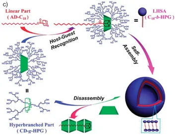
The inclusion complexation abilities of natural CDs and simply modified CDs are usually limited, which is inevitably unfavourable for the bioavailability and bioactivity of CDs. To avoid this disadvantage, scientists focused on the construction of CD-based supramolecular assemblies, a class of CD aggregates of nanometer size. Jiang and co-authors discussed the construction strategy of supramolecular self-assemblies based on CDs, which includes the formation of micelles by CD modified polymers, layer-by-layer hollow microcapsules, reversible micelles and vesicles, as well as polymer hydrogels formed by host-guest recognition.55 Zhou et al. conjugated hydrophilic hyperbranched polyglycerol (HPG) onto
β-12
CD (CD-HPG), and hydrophobic alkyl chain (C18) was coupled with Adamantane (Ad-C18). Through the host-guest complexation between β-CD and Ad, supramolecular amphiphiles were constructed, which could further self-assemble into core-shell structured vesicles (Figure 1.2.1.2).56
Figure 1.2.1.2. Preparation, self-assembly, and disassembly of the vesicle by a
linear hyperbranched supramolecular amphiphile. (Adapted from ref. 56).
1.2.2. Calix[n]arenes
During recent years CAs-based supramolecular systems have shown promising applications in biomedical field.57 As compared to CDs, a reduced number of studies have been reported for the biomedical applications of CAs,58 mainly because of their high hydrophobicity and poor water solubility. To avoid this problem, researchers have designed hydrophilic and water-soluble CAs. Sulfonation at the upper rim provided a common strategy to
13
prepare water soluble CAs.59 In addition, the conjugation of carboxylic acid groups onto the lower rim60 and the functionalization of both rims with polar groups were also carried out for the same purpose.61 By using click chemistry, cationic, anionic, and non-ionic CAs derivatives with a good water solubility were efficiently synthesized.62 For instance, poly(pyridinium)-functionalized CAs were obtained for biosensing applications (Figure 1.2.2.1a).63 Consoli et al. modified both the lower and the upper rims of CAs to obtain a folic acid-calix[4]arene conjugate that is able to encapsulate the hydrophobic drug model indomethacin, and enhance its solubility in water (Figure 1.2.2.1b).64
Figure 1.2.2.1. (a) Chemical structure of poly(pyridinium) salt containing
calix[4]arene unit (left) and its interaction with DNA studied by TEM (right). (Adapted from ref. 63). (b) Folic acid conjugated-Calix[4]arene for drug delivery by forming the host−guest inclusion with indomethacin. (Adapted from ref. 64).
14
In terms of biocompatibility, the water-soluble
p-sulfonatocalix[n]arenes exhibit low toxicity, and the in vivo dosage can reach up to 100 mg kg-1 without a toxic effect in mice.65
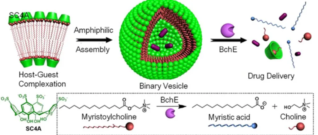
In 2012 Liu e co-workers developed an enzyme-responsive supramolecular vesicle by employing biocompatible p-sulfonatocalix[4]arene (SC4A) to deliver the drug tacrine, specifically for the treatment of Alzheimer’s disease.66 The binary vesicle formed by the host-guest complexation between SC4A and myristoylcholine exhibited highly specific and efficient responsiveness to cholinesterase enzyme that could break the hydrophilic-hydrophobic balance, leading to the disassembly of the binary vesicle and thus the release of loaded drugs. Such kind of binary vesicles could be achieved using other host molecules (Figure 1.2.2.2.).
Figure 1.2.2.2. Amphiphilic assembly of myristoylcholine in the presence of SC4A for responsive drug release. (Adapted from ref. 66).
15
Thus far, water soluble CAs have found wide applications in the solubilization of hydrophobic drugs, biomimic of ion channels, biochemical recognition, drug/gene delivery, and enzyme-like activity. In addition to their host-guest chemistry, some CAs have also shown interesting inhibition activities toward virus,67 bacterial,68 fungus,69 and some cancers.70
1.2.3. Pillar[n]arenes
Pillar[n]arene assemblies based on host-guest interactions have shown great potential in biology, especially in recognition of biomolecules, cell imaging, drug delivery, and biomedicine.40a The parent pillar[n]arenes are totally insoluble in water, for this reason many strategies have been proposed to improve water solubility by introducing water-soluble functional groups such as phosphates, carboxylates, oligoethers, polyethers, amines, and ammoniums. Ogoshi et al. reported the synthesis of the first water soluble pillar[5]arene derivative, known as carboxylato-pillar[5]arene (WP5), along with its ability to form strong 1:1 inclusion complex with paraquat in aqueous medium.71 Yang et
al.72 recently designed tryptophan-pillar[5]arene, which resulted highly water-soluble. Hou and co-workers73 reported a cation transport system based on pillar[5]arene capable of voltage-driven reversible insertion in a lipid bilayer (Figure 1.2.3.1).
16
Figure 1.2.3.1. Schematic representation of the artificial voltage-gated ion channel
prepared by the incorporation of positively charged Arg units to pillar[5]arene side chains. Cation transport through the ion channel is regulated by voltage-dependent reversible insertion of the artificial ion channel in the lipid bilayer. (Adapted from ref. 73).
Recently, Schalley et al.74 presented a versatile, simplified hydrophobic guest transport system based on the water soluble pillararene WP5 (Figure 1.2.3.2). The supramolecular interaction between WP5 derivative and the drug Norharmane (NHM) exhibited an improvement of the water solubility of the bioactive molecule and, compared with the individual NHM, the NHM/
WP5 supramolecular transport showed a significant reduction of
17
Figure 1.2.3.2. Schematic description of the supramolecular hydrophobic guest
transport system based on pillar[5]arene (WP5). (Adapted from ref. 74).
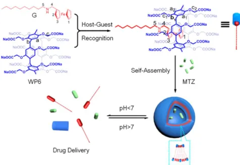
In 2013, Wang and co-workers managed to construct a novel type of supramolecular vesicles with a thin thickness of about 7 nm, where the self-assembly process was driven by the host-guest inclusion between a water soluble pillar[6]arene (WP6) and hydrophobic ferrocene derivative N-1-decylferrocenylmethylamine (G), in aqueous environment (Figure 1.2.3.3.).75 Then the anticancer drug mitoxantrone (MTZ) was encapsulated in the vesicles. The rapid MTZ release under acidic pH indicates the potential application of this vesicle carrier for controlled drug release inside the cancer cells. The drug delivery system was tested against SMMC-7721 cancer cell and NIH3T3 normal cell, respectively. Very recently, Wang and co-workers reported another multiple
stimuli-18
responsive supramolecular vesicle by using WP6; in this system the anticancer drug Doxorubicin (Dox) was loaded and then released in a controllable manner.76
Figure 1.2.3.3. Formation of a supramolecular vesicle and its pH-responsive drug
release. (Adapted from ref. 75).
Pei and co-workers77 synthesized a Ferrocenium-capped amphiphilic-pillar[5]arene (FCAP) able to self-assemble into cationic vesicles with high loading efficiency for polyanionic siRNA. When exposed to a reducing agent (glutathione), the disassembly of the cationic vesicles was achieved, and up to 92% of the model drug Dox was released to cancer cells.
19
Figure 1.2.3.4. formation of cationic vesicles, and their redox-responsive
drug/siRNA release. (Adapted from ref. 77).
Considering the advancements on pillar[n]arenes over such as a short time span and their ever-expanding applications, pillar[n]arenes will undoubtedly emerge as indispensable cyclophanes in biological applications.
20
References
1 (a) Lehn J.M., Angew. Chem., Int. Ed. Engl., 1988, 27, 89; (b) Cram, D.J., Angew.
Chem., Int. Ed. Engl., 1988, 27, 1009; (c) Pedersen, C.J., Angew. Chem., Int. Ed. Engl.,
1988, 27, 1021.
2 (a) Sijbesma R.P., Beijer F.H., Brunsveld L., Folmer B.J.B., Hirschberg J.H.K.K.,
Lange R.F.M., Lowe J.K.L., Meijer E.W., Science, 1997, 278, 1601; (b) Park T., Zimmerman S.C., Nakashima S., J. Am. Chem. Soc., 2005, 127, 6520; (c) Ryu J.H., Hong D.J., Lee M., Chem. Commun., 2008, 1043; (d) Hartgerink J.D., Beniash E., Stupp S.I., Science, 2001, 294, 1684; (e) Zhang W., Jin W., Fukushima T., Saeki A., Seki S., Aida T., Science, 2011, 334, 340.
3 (a) Ariga K., Kunitake T., Supramolecular Chemistry-Fundamentals and
Applications, 2006; (b) Wang F., Zhang J., Ding X., Dong S., Liu M., Zheng B., Li S.,
Wu L., Yu Y., Gibson H.W., Huang F., Angew. Chem., Int. Ed., 2010, 49,1090; (c) Zhang Z., Luo Y., Chen J., Dong S., Yu Y., Ma Z., Huang F., Angew. Chem., Int.
Ed., 2011, 50, 1397; (d) Wang F., Han C., He C., Zhou Q., Zhang J., Wang C., Li N.,
Huang F., J. Am. Chem. Soc., 2008, 130, 11254.
4 (a) Feringa B.L., Angew. Chem., Int. Ed., 2017, 56, 11060; (b) Stoddart J.F., Angew.
Chem., Int. Ed., 2017, 56, 11094; (c) Sauvage, J.P., Angew. Chem., Int. Ed., 2017, 56,
11080.
5 Dietrich-Buchecker C.O., Sauvage J.P., Kintzinger J.P., Tet. Lett., 1983, 24, 5095. 6 Anelli P.L., Spencer N., Stoddart J.F., J. Am. Chem. Soc., 1991, 113 5131.
7 Koumura N., Zijlstra R.W.J., Delden R.A., Harada N., Feringa B.L., Nature, 1999,
401, 152.
8 Liu K., Kang Y., Wang Z., Zhang X., Adv. Mater., 2013, 25, 5530. 9 Yoon H.J., Jang W.D., J. Mater. Chem., 2010, 20, 211.
10 Zhao Y., Sakai F., Su L., Liu Y., Wei K., Chen G., Jiang M., Adv. Mater., 2013, 25,
5215.
11 (a) Harada A., Takashima Y., Nakahata M., Acc. Chem. Res., 2014, 47, 2128; (b)
21
12 (a) Challa R., Ahuja A., Ali J., Khar R.K., AAPS Pharm. Sci. Tech., 2005, 6, 329;
(b) Stella V., Rajewski R., Pharm. Res., 1997, 14, 556; (c) Uzunova V.D., Cullinane C., Brix K., Nau W.M., Day A.I., Org. Biomol. Chem., 2010, 8, 2037; (d) Wang L., Li L.L., Fan Y.S., Wang H., Adv. Mater., 2013, 25, 3888.
13 Villiers A., Compt. Rend. Acad. Sci. ,1891, 112, 536.
14 Loftsson T., Duchêne D., International Journal of Pharmaceutics 2007, 329, 1. 15 (a) Breslow R., Dong S.D., Chem. Rev., 1998, 98, 1997; (b) Cramer F.,
Cyclodextrins and their Industrial Uses, ed. D. Duchêne, Editions de Santé, Paris, 1987;
(c) Szejtli J., Chem. Rev., 1998, 98, 1743; (d) Crini G., Chem. Rev., 2014, 114, 10940.
16 (a) Zhou J., Tan J., Tang W., Trends in Analytical Chemistry, 2015, 65, 22; (b)
Shpigun O.A., Ananieva I.A., BudanovaN.Y., ShapovalovaE.N., Russ. Chem. Rev.,
2003, 72, 1035.
17 (a) Nakamura A., Inoue Y., J. Am. Chem. Soc., 2003, 125, 966; (b) Nakamura A.,
Inoue Y., J. Am. Chem. Soc., 2005, 127, 5338; (c) Rekharsky M.V., Inoue Y., J. Am.
Chem. Soc., 2002, 124, 813; (d) Yang C., Mori T., Origane Y., Ko Y.K., Selvapalam
N., Kim K., Inoue Y., J. Am. Chem. Soc., 2008, 130, 8574.
18 Pedersen C.J., J. Am. Chem. Soc., 1967, 89, 7017.
19 Gokel G.W., Leevy W.M, Weber M.E., Chem. Rev., 2004, 104, 2723. 20 Baeyer A., Ber. Dtsch. Chem. Ges., 1872, 5, 280.
21 Baekeland L.H., US Pat., US942699, 1909.
22 Zinke A., Kretz R., Leggewie E., Hössinger K., Hoffmann G., Weber P.,
Ostwalden V., Wiesenberger E., Sobotka M., Kretz R., Monatsh. Chem. Verw. Teile
Anderer Wiss., 1952, 83, 1213.
23 Gutsche C.D., Acc. Chem. Res., 1983, 16, 161.
24 (a) Gutsche C.D., Muthukrishnan R., J. Org. Chem., 1978, 43, 4905; (b) Gutsche
C.D., Dhawan B., No K.H., Muthukrishnan R., J. Am. Chem. Soc., 1981, 103, 3782.
25 (a) Ninagawa A., Matsuda H., Makromol. Chem., Rapid Commun., 1982, 3, 65; (b)
Stewart D.R., Gutsche C.D., J. Am. Chem. Soc., 1999, 121, 4136.
26 Michinori T., Seiji S., Bull. Chem. Soc. Jpn., 1995, 68, 1088.
22
28 P. Lhoták, in Anion Sensing, ed. I. Stibor, Springer Berlin Heidelberg, Berlin,
Heidelberg, 2005, 65.
29 Kim J.S., Quang D.T., Chem. Rev., 2007, 107, 3780.
30 Kubo Y., Maeda S.Y., Tokita S., Kubo M., Nature, 1996, 382, 522.
31 Baldini L., Casnati A., Sansone F., Ungaro R., Chem. Soc. Rev., 2007, 36, 254. 32 Prins L.J., Timmerman P., Reinhoudt D.N., Pure Appl. Chem., 1998, 70, 1459. 33 (a) Guo D.S., Liu Y., Chem. Soc. Rev., 2012, 41, 5907; (b) Sun R., Xue C., Ma X.,
Gao M., Tian H., Li Q., J. Am. Chem. Soc., 2013, 135, 5990.
34 Wei A., Chem. Commun., 2006, 1581.
35 Ogoshi T., Kanai S., Fujinami S., Yamagishi T., Nakamoto Y., J. Am. Chem. Soc.,
2008, 130, 5022.
36 Ogoshi T., Pillararenes (Monographs in Supramolecular Chemistry), Royal
Society of Chemistry, 2015
37 (a) Zhang Z., Luo Y., Chen J., Dong S., Yu Y., Ma Z., Huang F., Angew. Chem.,
Int. Ed., 2011, 50, 1397; (b) Strutt N.L., Forgan R.S., Spruell J.M., Botros Y.Y.,
Stoddart J.F., J. Am. Chem. Soc., 2011, 133, 5668.
38 Zhang H., Strutt N.L., Stoll R.S., Li H., Zhu Z., Stoddart J.F., Chem. Commun.,
2011, 47, 11420.
39 (a) Tan L.L., Li H., Tao Y., Zhang S.X., Wang B., Yang Y.W., Adv. Mater., 2014,
26, 7027; (b) Wang W., Chen L.J., Wang X.Q., Sun B., Li X., Zhang Y., Shi J., Yu Y., Zhang L., Liu M., Yang H.B., Proc. Natl. Acad. Sci. U. S. A., 2015, 112, 5597.
40 (a) Sathiyajith C., Shaikh R.R., Han Q., Zhang Y., Meguellati K., Yang Y.W.,
Chem. Commun., 2017, 53, 677; (b) Si W., Xin P., Li Z.T., Hou J.L., Acc. Chem. Res.,
2015, 48, 1612; (c) Si C.W., Zhang L.,. Tang G., Li Z.T., Hou J.L., J. Am. Chem. Soc., 2013, 135, 2152.
41 (a) Blandamer M.J., Engberts J.B.F.N., Gleeson P.T., Reis J.C.R., Chem. Soc. Rev.,
2005, 34, 440; (b) Li C.J., Chem. Rev., 2005, 105, 3095; (c) Otto S., Engberts J.B.F.N.,
Org. Biomol. Chem., 2003, 1, 2809; (d) Lindstrom U.M., Chem. Rev., 2002, 102, 2751;
(e) Akiya N., Savage P.E., Chem. Rev., 2002, 102, 2725; (f) Engberts J.B.F.N., Blandamer M.J., Chem. Commun., 2001, 1701.
23
42 (a) Schrader T., Hamilton A.D., Functional Synthetic Receptors Wiley-VCH,
Weinheim, 2005; (b) Ariga K., Kunitake T., Supramolecular Chemistry— Fundamentalsand Applications, Springer, Berlin, 2005. (c) Kubik S., Reyheller C.,
Stuwe S., J. Inclusion Phenom. Macrocyclic Chem., 2005, 52, 137.
43 Schneider H.J., Kramer R., Simova S., Schneider U., J. Am. Chem. Soc., 1988, 110,
6442.
44 Zang J., Ma P.X., Advanced Drug Delivery Reviews, 2013, 65, 1215. 45 Irie T., Uekama K., J. Pharm. Sci., 1997, 86, 147.
46 D’Souza V.T., Bender M.L., Acc. Chem. Res., 1987, 20, 146. 47 Breslow R., Science, 1982, 218, 532.
48 Tabushi I., Acc. Chem. Res.,1982, 15, 66.
49 Saenger W., Angew. Chem., Int. Ed. Engl., 1980, 19, 344.
50 Murakami Y., Kikuchi J.I., Hisaeda Y., Hayashida O., Chem. Rev., 1996, 96, 721. 51 Brewster M.E., Loftsson T., Adv. Drug Delivery Rev., 2007, 59, 645.
52 Chen H., Liu X., Dou Y., He B., Liu L., Wei Z., Li J., Wang C., Mao C., Zhang J.,
Wang G., Biomaterials, 2013, 34, 4159.
53 (a) Donohue R., Mazzaglia A., Ravoo B.J., Darcy R., Chem. Commun., 2002, 2864;
(b) Zhao Y.L., Li Z., Kabehie S., Botros Y.Y., Stoddart J.F., Zink J.I., J. Am. Chem.
Soc., 2010, 132, 13016.
54 (a) Dubes A., Bouchu D., Lamartine R., Parrot-Lopez H., Tetrahedron Lett., 2001,
42, 9147; (b) Sukegawa T., Furuike T., Niikura K., Yamagishi A., Monde K., Nishimura S.I., Chem. Commun., 2002, 430.
55 Chen G., Jiang M., Chem. Soc. Rev., 2011, 40, 2254.
56 Tao W., Liu Y., Jiang B., Yu S., Huang W., Zhou Y., Yan D., J. Am. Chem. Soc.,
2011, 134, 762.
57 Nimse S.B., Kim T., Chem. Soc. Rev., 2013, 42, 366. 58 Guo D.S., Liu Y., Acc. Chem. Res., 2014, 47, 1925.
59 Perret F., Lazar A.N., Coleman A.W., Chem. Commun., 2006, 23, 2425.
60 Arduini A., Pochini A., Raverberi S., Ungaro R., J. Chem. Soc. Chem. Commun.,
24
61 Shahgaldian P., Coleman A.W., Kalchenko V.I., Tetrahedron Lett., 2001, 42, 577. 62 Ryu E.H., Zhao Y., Org. Lett., 2005, 7, 1035.
63 Lu Y., Xiao C., Yu Z., Zeng X., Ren Y., Li C., J. Mater. Chem., 2009, 19, 8796. 64 Consoli G.M.L., Granata G., Geraci C., Org. Biomol. Chem., 2011, 9, 6491.
65 Coleman A.W., Jebors S., Cecillon S., Perret P., Garin D., Marti-Battle D.,
Moulin M., New J. Chem., 2008, 32, 780.
66 Guo D.S., Wang K., Wang Y.X., Liu Y., J. Am. Chem. Soc., 2012, 134, 10244. 67 Hwang K.M., Liu S.Y., Qi Y.M., Method of Treating Viral Infections with Aryl
Macrocyclic Compounds, Google Patents, 1994.
68 (a) Casnati A., Fabbi M., Pelizzi N., Pochini A., Sansone F., Unguro R., Di
Modugno E., Tarzia G., Bioorg. Med. Chem. Lett., 1996, 6, 2699; (b) Colston M.J., Hailes H.C., Stavropoulos E., Herve A.C., Herve G., Goodworth K.J., Hill A.M., Jenner P., Hart P.D., Tascon R.E., Infect. Immun., 2004, 72, 6318.
69 Hart P.D., Armstrong J.A., Brodaty E., Infect. Immun., 1996, 64, 1491.
70 Menger F.M., Bian J., Sizova E., Martinson D.E., Seredyuk V.A., Org. Lett., 2003,
6, 261.
71 Ogoshi T., Hashizume M., Yamagishi T.A., Nakamoto Y., Chem. Commun.,
2010, 46, 3708.
72 Yang K., Chang Y., Wen J., Lu Y., Pei Y., Cao S., Wang F., Pei Z., Chem. Mater.,
2016, 28, 1990.
73 Si W., Li Z.T., Hou J.L., Angew. Chem., Int. Ed., 2014, 53, 4578.
74 Qi Z., Achazi K., Haag R., Dong S., Schalley C.A., Chem. Commun., 2015, 51,
10326.
75 Duan Q., Cao Y., Li Y., Hu X., Xiao T., Lin C., Pan Y., Wang L., J. Am. Chem.
Soc., 2013, 135, 10542.
76 Cao Y., Zou X., Xiong S., Li Y., Shen Y., Hu X., Wang L., Chin. J. Chem., 2015, 33,
329.
77 Chang Y., Yang K., Wei P., Huang S., Pei Y., Zhao W., Pei Z., Angew. Chem., Int.
25
Chapter 2
Macrocyclic amphiphiles
2.1. Introduction
In the field of self-assembly systems, amphiphiles are a class of interesting molecules containing both hydrophilic and hydrophobic portions connected by covalent bonds.
Figure 2.1.1. Conventional amphiphiles.
When dissolved in aqueous media, the hydrophilic component of an amphiphile tends to interact with the aqueous phase while its hydrophobic part prefers staying in the nonpolar solvent or residing in the air.1 Thus they are able to self-assemble in water into various well-defined structures, ranging from simple micelles and vesicles to highly organized nanofibers, nanohelices, nanotubes, nanorods and nanosheets. Furthermore, after an external stimuli, such as temperature, concentration, pH and ionic strength,2 they are also able to reorganize their structure.
26
Amphiphiles play a significant role in nature at many different levels, from peptides, proteins and genes (e.g. DNA, RNA) to viruses and cell membrane.3 Inspired by nature, researchers engineered and produced artificial self-assembly structures which have been widely applied in many fields including nanodevices, drug/gene delivery and cell imaging.4 Over the past decades, among all kinds of synthetic amphiphiles, macrocyclic amphiphiles obtained from macrocyclic scaffolds have captured much attention due to their unique superiority in the self-assembly process. Compared with traditional linear amphiphiles, macrocyclic amphiphiles can be designed to promote their self-assembly into various well-defined architectures by locating hydrophilic and hydrophobic chains on the respective sides of the macrocyclic frameworks.
This chapter will describe the experimental results on the synthesis and aggregation properties of new amphiphilic macrocycles based on anionic and neutral calixarenes and a cationic cyclodextrin, together with different applications in the recognition and/or encapsulation of different substrates.
2.2. Macrocyclic amphiphiles based on calixarenes
Calix[n]arenes are macrocyclic oligomers composed of n phenolic units linked by methylene groups at the meta-positions, forming a unique basket shape.5 Due to their unique structure and facile functionalization of both upper and lower rims, calix[n]arenes have been widely used to construct macrocyclic amphiphiles. The most obvious feature that distinguishes these amphiphiles from conventional linear surfactants is
27
the presence of a cavity that can be used to include guest molecules. In fact, calixarene-based surfactants were defined by Shinkai as “surfactants with a host–guest recognition site”.6
Figure 2.2.1. Cartoon and chemical structure of an amphiphilic calix[4]arene. The parent calixarenes show effectively zero solubility in aqueous solution, and undoubtedly this property is the one which delayed the biopharmaceutical application of calixarene derivatives as compared to cyclodextrins and crown ethers. Various methods have been developed since the 1980s to obtain water-soluble calixarenes through functionalization at three possible sites: at the phenolic functions, para to the phenolic groups, and by modification of the methylene bridges. Ungaro and co-workers reported the first example of such a compound in 1984, with carboxylic acid groups coupled to the lower rim of a calix[4]arene.7 More recently, functions such as phosphates, ammonium groups or sulfonates functions have been used. Regen et al., in 1989, prepared vesicles by injecting a tetrahydrofuran solution of an unmodified calix[6]arene into water.8 From then on, various kinds of amphiphilic calix[n]arenes were synthesised to investigate their
self-28
assembly behaviors. In 2001, Shahgaldian et al. demonstrated that further modification of the acyl-calix[4]arenes could be used to generate calixarene analogues of the natural phospholipids.9
Among the several calixarene-based surfactants described in the literature, p-sulfonatocalix[n]arenes bearing alkyl groups at the lower rim are probably the most studied.10 Conversely, apart from Shinkai’s pioneering work,11 very few examples of lower rim O-alkylsulfonato derivatives have been thoroughly investigated.12 Upon micelle formation, unlike their p-sulfonato analogues, O-alkylsulfonato-calixarenes direct their hydrophobic cavities towards the inner core of the micelle, exposing their charged moieties to bulk water. As a result, they are expected to display different recognition, solubilisation and aggregation propertiesthan their upper rim analogues.
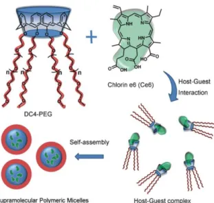
Among neutral calixarene-based surfactants, a series of large-ring PEGylatedcalix[6,7,8]arene analogues have been synthesised and are able to modify the growth of M. tuberculosis in infected cells.13 In 2011, Zhu et al.14 took advantage of the host-guest interaction between a PEGylated calix[4]arene and the hydrophobic chlorin e6 to form supramolecular polymeric micelles, which exhibited more efficient photodynamic therapy efficacy than the free chlorin e6 (Figure 2.2.2). Raston et al. established new micellular delivery systems based on phospholipid calix[4]arenes with potent antioxidant activity.15
29
Figure 2.2.2. Representation of supramolecular polymeric micelles based on host–guest
interaction. (Adapted from ref. 14).
2.2.1. Our contribution to anionic calixarene-based surfactants: results and discussion
During 2013 my research group reported on the synthesis of a new water-soluble p-tert-butylcalix[5]arene 1, bearing O-4-butylsulfonato groups at the lower rim, and its aggregation properties in water.16
30
According to the method previously reported for calixarene 1, two new amphiphilic calixarenes bearing O-4-butylsulfonato pendant groups at the lower rim (p-tert-calix[4]arene 2 and p-methyl-calix[5]arene 3; Scheme 2.2.1.1) were synthesized and their aggregation properties investigated.
Scheme 2.2.1.1. Synthesis of p-tert-butylcalix[4]arene 2 and p-methyl-calix[5]arene 3.
Amphiphilic calixarene 2 and 3 were obtained (in 64% and 56% yields, respectively) from p-tert-butylcalix[4]arene and p-methylcalix[5]arene, by reaction with 1,4-butanesultone (3 equiv. x OH) and NaH (3 equiv. x OH) in refluxing anhydrous THF for 48 hours. The white powder obtained after addition of MeOH was collected, washed with EtOH, redissolved in water and finally precipitated by the salting-out method using a solution of sodium acetate. As expected, both derivates 2 and 3
31
showed an amphiphilic nature. The detailed amphiphilic features of derivatives 2 and 3 will be reported in the next section and in Chapter 4 respectively.
2.2.2. Self-assembly of amphiphilic anionic calix[4]arene 2 and drug solubilisation.‡
Here I will describe a deep insight on the aggregation properties of the amphiphilic p-tert-butylcalix[4]arene tetra-O-butylsulfonate (2) by means of complementary techniques, 1D and 2D NMR, Dynamic light scattering (DLS) and atomic force microscopy (AFM). Furthermore the ability of the micellar aggregates of calixarene 2 to increase the solubility of poorly water soluble drugs was also studied.
The amphiphilic nature of derivative 2 was immediately tested through preliminary NMR experiments. The 1H NMR spectrum of 2, recorded at a low concentration ([2] = 0.2 mM, D2O), displays a set of sharp resonances typical of calixarene species adopting a cone conformation (Figure 2.2.2.1a).17 In particular the presence of singlets at 6.87 and 0.97 ppm (relative to the aromatic and tert-butyl hydrogens, respectively) together with one single AX system ( 4.31 and 3.13 ppm, J = 12.5 Hz) for the axial and equatorial hydrogens of the bridging methylenes, experiencing different magnetic anisotropy generated by aryl residues, confirm the cone conformation of derivative 2. Upon increasing the concentration to 1.4 mM (Figure 2.2.2.1b), the resonances broaden, indicating that an aggregation process is taking place, and thus
‡The experimental results reported in this section have been already published: Barbera L. et al.,
32
confirming the surfactant nature of 2. A comparison of traces (a) and (d) in Figure 2.2.2.1 revealed that the resonances relative to the aromatic and the tert-butyl hydrogens underwent an upfield shift upon concentration. This can be explained considering that in the micelle, calixarene molecules are forced to stay closer and, as a consequence, some resonances resulted affected by the anisotropic shielding of the aromatic rings of surrounding calixarenes.
A closer inspection of the aromatic region in traces (b) and (c) revealed a second broad signal for the aromatic protons at 6.78 ppm (see arrows in Figures 2.2.2.1b and 2.2.2.1c), whose intensity increases until it becomes the predominant species in solution (for [2] ≥ 4.0 mM, Figure 2.2.2.1d) and can be assigned to the formation of micellar aggregates. The concomitant presence of two close but distinct sets of aromatic signals indicates a slow exchange on the NMR time-scale between the two species (monomer and micelle). This phenomenon is quite unusual in conventional surfactants but has been already reported for some amphiphilic calixarenes.18
33
Figure 2.2.2.1. 1H NMR (500 MHz, 298 K, D2O) spectra of calix[4]arene 2 at different
concentrations: (a) 0.2 mM, (b) 1.4 mM, (c) 2.3 mM and (d) 4 mM.
The critical micellar concentration (cmc) for 2 in D2O was determined by diffusion-ordered NMR (DOSY)19 studies, using calixarene solutions in the 10.0–0.10 mM concentration range. In table 2.2.2.1 are reported the Diffusion Coefficients (D2,obs) for selected resonances (SCH2) which were
found isochronous in the concentration range investigated and therefore represent the weighted average Diffusion Coefficients for monomers and micelles present in solutions. As can be observed, the values of D2,obs
decreases (from 2.52 10-10 to 0.80 10-10 m2/s) by increasing the concentration of derivative 2. This result is consistent with the formation of bigger, and therefore slower diffusing species (micelle). A cmc of 1.15 mM was estimated from the intersection point of the two lines best fitting the experimental data points obtained by plotting the self-diffusion coefficient (D2,obs) vs. the inverse of the concentration of
34
calixarene 2 (Figure 2.2.2.2). Another research group determined for the same calixarene a cmc of 1.76 mM by surface tension analysis.20
[2] (mM) D2,obs 10–10 (m2/s) 10 0.80 ± 0.03 7.5 0.88 ± 0.03 5 0.98 ± 0.01 4 1.14 ± 0.03 3 1.33 ± 0.08 2.8 1.38 ± 0.01 2.6 1.43 ± 0.03 2.5 1.48 ± 0.01 2.4 1.49 ± 0.01 2.3 1.54 ± 0.02 2.2 1.58 ± 0.02 2.1 1.62 ± 0.03 2.0 1.70 ± 0.03 1.9 1.76 ± 0.01 1.8 1.82 ± 0.02 1.7 1.88 ± 0.04 1.6 1.94 ± 0.04 1.5 2.01 ± 0.03 1.4 2.08 ± 0.02 1.3 2.16 ± 0.01 1.2 2.20 ± 0.02 1.1 2.28 ± 0.03 1.0 2.35 ± 0.02 0.9 2.39 ± 0.03 0.6 2.47 ± 0.02 0.4 2.48 ± 0.01 0.2 2.52 ± 0.01 0.1 2.52 ± 0.02
Table 2.2.2.1. Diffusion coefficients (D2,obs) of the species present at 298 K in D2O
solutions of calixarene 2 at different concentrations. Data were calculated from DOSY experiments, using the SCH2 resonance ( = 2.89 ppm) as the probe signal.
35
A closer inspection of the concentration ranges slightly below and above the cmc (see the insert in Figure 2.2.2.2) showed the presence of two distinct break points (at 0.83 mM and 1.41 mM, respectively), reasonably ascribable to two distinct aggregation phenomena of 2. The former (0.83 mM) likely refers to the initial formation of small oligomers (premicelles)21 seen – on the NMR time-scale – in a fast-exchange regime with the monomers. The latter (1.41 mM), on the other hand, most probably indicates the onset of micellar aggregation (Figure 2.2.2.1b). As the concentration of 2 increases further (above 3.0 mM), new peaks appear in the 1H NMR spectrum (see arrows in Figure 2.2.2.1d) and, accordingly, new aggregates form as a result of an additional structural transition (micelle–micelle or micelle–vesicle). These newly-formed aggregates could not be studied by NMR because of the poor solubility of 2 at concentration above 10 mM.
Figure 2.2.2.2. Plot of the self-diffusion coefficients (D2,obs) vs. the inverse of the concentration of calixarene 2 in D2O at 298 K.
36
The hydrodynamic radii (Rh) of the monomer (7.9 Å) and the micelle (24.9 Å) were conveniently obtained from the D2,obs values determined
below ([2] = 0.1 mM) and above ([2] = 10.0 mM) the cmc, using the Stokes–Einstein equation (Dobs = kBT/6Rh) which correlates Diffusion Coefficient and hydrodynamic radii assuming a spherical shape of the objects under investigation.
Additional evidence corroborating micelle formation was obtained from 2D NOESY studies (Figure 2.2.2.3.). The spectrum of a 5.0 mM solution of 2 shows NOE cross-peaks for all the calixarene resonances. Interestingly, there are cross-peaks indicating the proximity of the upper-rim tert-butyl hydrogen atoms and the lower-rim butanesulfonate residues of adjacent calixarene molecules. The absence of cross-peaks in the spectrum of 2 in the monomeric form (0.5 mM) demonstrates the intermolecular origin of the NOEs mentioned earlier (i.e., [2] = 5.0 mM), suggesting that in the micellar aggregate the calixarene units are probably arranged in a staggered fashion (Figure 2.2.2.4).22
37
Figure 2.2.2.4. Schematic representation of staggered calixarene molecules within a
micelle. Arrows indicate relevant NOE cross-peaks.
As already observed for the homologous calix[5]arene,23 NMR quantitative analysis of D2O solutions of calixarene 2 below the cmc, carried out using the quantitative qNMR software Varian Vnmrj 3.2, showed an apparent discrepancy between the nominal and the detectable concentrations of monomers in solution (for example, for [2] = 0.2 mM, a concentration of [2] = 0.1 mM was calculated), indicating that NMR “invisible” aggregates of 2 were present even at low concentrations. This finding is in line with previous observations on the tendency of amphiphilic calixarene-type surfactants to aggregate below the cmc in large infinite structures (“open model”24). In order to confirm the presence of such big aggregates, dynamic light scattering (DLS) experiments were performed on solutions of calixarene 2 below the estimated cmc value. The DLS experiments (Figure 2.2.2.5) revealed the presence of two populations of very large aggregates, with hydrodynamic radii of approximately 100 and 300 nm. The number fraction of the 300 nm aggregates was found to be about 0.03.25
38
When 0.5 and 5.0 mM aqueous solutions of 2 were pre-filtered through a 0.45 m Millipore membrane, the 100 nm aggregates became even more predominant, with the number fraction of the larger aggregates (300 nm) decreasing to about 0.01 and becoming almost negligible in the two solutions, respectively (Figure 2.2.2.5b). Remarkably, no smaller micellar aggregates (i.e., in the 2 nm range) were visible by DLS either before or after filtration. The obtained results demonstrated that a combined use of DOSY – for the smaller aggregates – and DLS – for the larger ones, “transparent” to NMR26– is necessary to get a full picture of the aggregation behaviour of amphiphilic molecules such as 2.27
Figure 2.2.2.5. Size distributions of the calixarene 2 aggregates in aqueous solution: (a)
[2] = 0.5 mM; (b) [2] = 0.5 and 5.0 mM (black and red trace, respectively) after filtration through a 0.45 μm Millipore filter.
Further information on the morphology of the aggregates were finally provided by atomic force microscopy (AFM) measurements. As showed
39
in figure 2.2.2.6, derivative 2 tends to form complex structures, most of them having an average diameter of 100–200 nm, though many smaller particles (ca. 50 nm) are also visible (Figure 2.2.2.6.).
Figure 2.2.2.6. AFM topography image of calixarene 2.
All these data indicate that calixarene 2 self-assembles in premicelles and very large aggregates below the cmc, as schematically depicted in Figure 2.2.2.7.
40
Micellar systems find medical and pharmacological applications as drug delivery systems, especially thanks to their ability to solubilise in aqueous media poor-water soluble drugs. Moreover their reduced size, loading and releasing abilities, governed by their cmc, make micelle-based drug delivery systems more advantageous than other systems such as water soluble polymers or liposomes.
In order to test the potential of amphiphilic calix[4]arene 2 to act as a molecular carrier upon micelle formation, we decided to investigate its ability to increase the water solubility of the nonsteroidal anti-inflammatory hydrophobic drugs naproxen (4) and flurbiprofen (5).
Figure 2.2.2.8. Structures of naproxen (4) and flurbiprofen (5).
In these experiments, a fixed amount of solid 4 or 5 (5 mol) was added to 1 mL of the surfactant solution above the cmc ([2]= 5 mM, D2O) previously filtered through a 0.1 m Millipore filter, and the samples were stirred at room temperature overnight. The solutions were then centrifugated to remove undissolved solid. The 1H NMR spectra of naproxen and flurbiprofen in D2O prior to and after segregation within the 5.0 mM micellar solution of 2 are shown in Figure 2.2.2.9. In both cases under examination, all the resonances of 4 and 5 underwent significant complexation-induced shifts, as a result of an interaction
41
between the drug and the micellar environment provided by 2. In order to rule out the formation of inclusion complexes between 2 (as a monomer) and the two drug molecules, parallel extraction experiments, were performed below the cmc (Figure 2.2.2.10.).
Figure 2.2.2.9.1H NMR (500 MHz, 298 K, D2O) spectra of: (a) [2] = 5.0 mM; (b) 4 after
extraction with a 5.0 mM solution of 2; (c) [4] = 0.14 mM; (d) 5 after extraction with a 5.0 mM solution of 2 and (e) [5] = 0.13 mM.
As shown in Figure 2.2.2.10 for drug 5, adding increasing amount of 2 (in the 0.25 – 0.75 mM range, that is below the cmc) to a 0.13 mM solution of flurbiprofen (5) did not produce any detectable complexation-induced shifts of the drugs’ resonances. These data confirm that the drug molecule was not encapsulated inside the calixarene cavity, but rather located in the lipophilic palisade of the micellar aggregate.
42
Figure 2.2.2.10. Flurbiprofen (5) extraction experiments with 2 below the cmc: a) [5] =
0.13 mM; b) [2] = 0.25 mM, [5] = 0.13mM; c) [2] = 0.5 mM, [5] = 0.13 mM; d) [2] = 0.75 mM, [5] = 0.13 mM.
To confirm this hypothesis 2D NOESY NMR experiments were used to shed light on the location and orientation of the drugs within the micelles.
Figure 2.2.2.11. Section of the 2D NOESY spectrum (500 MHz, 298 K) of a D2O solution
43
In the case of 4, the α-CH3 group shows NOE interactions with the hydrogen atoms of both the aromatic and the aliphatic moieties of 2 (Figure 2.2.2.11.)
In the case of Flurbiprofen (5) stronger correlation peaks of the aromatic signal at 7.19 ppm with all the calixarene resonances (red circles in Figure 2.2.2.12.), demonstrated the presence of NOE contacts. Cross-peaks are consistent with the aromatic unit of 5 and the lower-rim aliphatic moieties of 2 being in close proximity. All these findings suggest that both naproxen and flurbiprofen molecules most likely reside within the micellar palisade of 2, oriented in such a way that the aromatic ring of the drug points towards the interior of the micelle, where they can experience favourable interactions with the calixarene outer faces, while the carboxylic acid moiety points in the opposite direction, towards the bulk.
Figure 2.2.2.12. Section of the 2D NOESY spectrum (500 MHz, 298 K) of a D2O solution
44
Our next objective was to estimate the degree of solubilisation of the two drugs by the calixarene micellar solutions. To this purpose, three different solutions of 2 with concentrations above the cmc (2.5, 5.0 and 10.0 mM) were employed for solid–liquid extraction experiments with a fixed aliquot of drug samples (5 moles). After 24 h stirring at room temperature, the resulting suspensions were centrifuged to remove the undissolved 4 or 5, as well as any large calixarene/drug aggregates formed, and then the concentrations of the calixarene and naproxen/flurbiprofen present in the supernatant were determined by a quantitative NMR protocol.
Data in Figure 2.2.2.13 show the amount of naproxen and flurbiprofen solubilised as a function of the surfactant concentration. The linear correlation observed is a clear evidence that the water solubility of these drugs28 significantly improves with increasing calixarene concentration. Molar solubilisation capacities of 0.20 and 0.51, for naproxen and flurbiprofen respectively, were derived from the slope of the linear regression. To the best of our knowledge, these results compare well with those reported for other anionic surfactants.29
Figure 2.2.2.13. Plots of drug vs. calixarene concentrations determined by quantitative
45
It is a matter of fact that the extent of segregation of molecules bearing acid moieties inside anionic micelles depends on the pKa of the target molecule, that is, the lower the pKa of the target molecule, the stronger the repulsion between the acidic guest (in the deprotonated form) and the anionic head-groups of the micelles is expected to be, thus decreasing the solubilisation efficiency.30 On the other hand, it is known that the pKa of lipophilic carboxyl acids may increase in the hydrophobic medium of micelles with respect to aqueous solution.31 Naproxen and flurbiprofen (pKa ca. 4.2–4.8)32 are not fully ionized in an aqueous solution of calixarene 2 (pH 5.4) and, as a result, the undissociated form can easily be incorporated inside the anionic micelles.
Being aware that quantitative NMR measurements cannot indicate the molar fraction of drug molecules segregated inside very large aggregates (Rh = 100–300 nm) – as these are “NMR invisible”– the total concentration of naproxen dissolved in an aqueous 5.0 mM micellar solution of 2 was also investigated by UV spectroscopy. To this end, to avoid the potential overlapping of calixarene and naproxen absorbance bands during the analysis in aqueous solution, naproxen was extracted in CHCl3 and its concentration was spectrophotometrically determined, in the absence of 2, before and after filtration of the aqueous solutions through 0.1 m Millipore filters.
46
UV (prior filtration) UV (after filtration) NMR Dissolved Naproxen mM mg/mL 0.85 ± 0.03 0.196 ± 0.007 0.63 ± 0.03 0.145 ± 0.07 0.63 ± 0.05 0.145 ± 0.011
Table 2.2.2.2. UV and NMR determination of naproxen (4) solubility in a 5.0 mM
micellar solution of 2.
Data obtained (Table 2.2.2.2.) for the filtered solutions ([4] = 0.63 ± 0.03 mM) are in excellent agreement with the NMR measurements, whereas slightly higher values were found for the unfiltered ones ([4] = 0.85 ± 0.03 mM), thus indicating that the bigger aggregates of 2 are also able to encapsulate the drugs under investigation (Figure 2.2.2.14).
Figure 2.2.2.14. Cartoons of the drugs encapsulation inside the aggregates.
The affinity of drugs 4 and 5 for the micellar environment of calixarene 2 was also demonstrated by means of DOSY measurements. Data in Table 2.2.2.3 show that the diffusion coefficients of naproxen and flurbiprofen decrease from 5.47–5.66 10–10 m2/s (for the free species) to values closer to those of the calixarene micellar aggregates. These results clearly
![Figure 2.3.1.6. Size distributions of the cyclodextrin aggregates in aqueous solution: (a) [8] = 10 mM (red line 298 K; green line 338 K); (b) [8] = 10 mM (298 K) after filtration through a 0.45 μm Millipore filter](https://thumb-eu.123doks.com/thumbv2/123dokorg/4581634.38720/69.773.109.674.109.325/figure-distributions-cyclodextrin-aggregates-aqueous-solution-filtration-millipore.webp)
![Figure 3.4.1.2. SEM images of [2] = 0.1 mM.](https://thumb-eu.123doks.com/thumbv2/123dokorg/4581634.38720/100.773.135.665.721.913/figure-sem-images-of-mm.webp)
