XXIII Ciclo
Anno Accademico 2009-2010 Università degli Studi di CataniaMetal Nanoparticles Produced
by Pulsed Laser Ablation
in Liquid Environment
Elena Messina
Tutor:
Chiar.mo Prof. G. Compagnini
Coordinatore: Chiar.mo Prof. A. Licciardello
A Roberto e alla mia famiglia
Table of contents
1 SCOPE OF THE THESIS ……….. 1
1.1 METAL NANOPARTICLES ………4
1.2 UV-Vis CHARACTERIZATION OF METAL NANOPARTICLES USING MIE THEORY ………...6
1.3 THERMODYNAMIC PROPERTIES ……….14
1.4 CHEMICAL PROPERTIES ………17
REFERENCES ...20
2 SYNTHESIS……….23
2.1 FUNDAMENTAL ASPECT OF PLAL ………..23
2.2 PLAL OF SILVER NANOPARTICLES………..41
2.3 PLAL OF GOLD NANOPARTICLES……….54
2.4 PLAL OF COPPER NANOPARTICLES……….66
2.5 GENERATION OF AgCl CUBES BY PLAL OF BULK Ag IN AQUEOUS NaCl SOLUTIONS ..………68
REFERENCES .……….76
3 METAL/CARBON STRUCTURES ………81
3.1 SYNTHESIS AND CHARACTERIZATION OF LINEAR CARBON CHAINS (LCCs) ………82
3.2 LCCs OBTAINED BY PLAL IN WATER ………86
3.3 LCCs PREPARED IN THE PRESENCE OF METAL COLLOIDS: AGGREGATION AND COAGULATION PHENOMENA ……..93
3.4 LCCs MIXED WITH METAL COLLOIDS: AGGREGATION AND COAGULATION PHENOMENA …….100
3.5 RAMAN AND MS CHARACTERIZATION OF LCC- PROTECTED METAL NANOPARTICLES ……….103
3.6 SERS HOT SPOTS ………116
4 OPTICAL TWEEZERS ………..125
4.1 OPTICAL TRAPPING ………..127
REFERENCES ……….139
Particles in the nanometer size range have attracted increasing attention with the growing interest in nanoscience and nanotechnology. They hold potential as basic components for sub-wavelength optical devices, for surface-enhanced spectroscopy, for biological labelling and sensing, and for cancer therapy. For such applications, it is crucial to prepare metal nanoparticles with desired shape, and size distribution. The intense research in this field is also motivated by the search for new multifunctional materials that will allow designing of the modern miniature electronic and optical devices for ultra fast data communication and optical data storage. In this regard, the interaction of light with small particles depends strongly on the size, shape and composition of the particles, as well as on the composition of the medium in which the particles are embedded. A large number of chemical methods have been developed for the synthesis of silver and gold nanostructures that have well-controlled shapes, including triangular plates, cubes, wires and rods either in the form of colloidal dispersion or nanostructured films.
In this context, Pulsed Laser Ablation in Liquids (PLAL) has become a key method for synthesis of nanoparticles with controlled geometry and size. The ablation of metal targets in liquid environments is considered as a unfailing alternative to traditional chemical reduction methods for obtaining noble metal colloids, since such a strategy is considered
environmental friendly (“green” technique) with products which frequently do not need stabilizing molecules or other chemicals. Laser ablation-based synthesis can be implemented in pure deionized water or even in biologically-compatible aqueous solutions and can be coupled with well established protocols to enhance the sensitivity of classical vibrational spectroscopies such as in the case of Surface Enhanced Raman (SER) phenomena
In the past few years, the interest toward metals doped or bonded with carbon nanostructures has grown enormously, thanks to their wide use in optics and microelectronics applications.
Many recent publications deal with the possibility to incorporate heteroatom into carbon allotropes to significantly improve most of their physical properties.
On the other hand, metal nanoparticles are often susceptible to oxidation and aggregation (thus reducing their free energy), leading to a loss of their peculiar properties. Therefore, several efforts are devoted to protect metal nanoparticles with inert shells, to preserve them from surface modifications and to keep their main characteristics unchanged.
Finally, Gold nanoparticles are proven to be excellent candidate for in
vivo micro-manipulation using Optical Tweezers.
Optical tweezers(OT), instruments based on a strongly focused laser beam, have been recently used to trap, manipulate, control and assemble metal and semiconducting nanostructures and their latest combination with Raman spectroscopy enables a thorough investigation of trapped samples. Historically, optical trapping of 36nm gold nanoparticles was first demonstrated by Svoboda & Block. More recently, the trapping range of gold spherical nanoparticles was expanded up to 250 nm, and very accurate measurements of optical forces have become possible30. A complete theory
was shown to be in very good agreement with experiments. The optical trapping of non-functionalized gold nanoparticles obtained by chemical methods can also originate particles agglomeration due to the observation of reversible electrical conductivity changes of the solution of the nanoparticles upon laser illumination or direct heating. Particles agglomeration during trapping experiments can be an obstacle for the accurate determination of the optical forces generated by the trapping beam and for the investigation of the effect of the plasmonic properties of nanoparticles on the trapping process.
In details, this PhD thesis pass through the synthesis of gold and silver nanoparticles, with final aim of studying optical properties and SERS applications.
Three fundamental aspect of metal nanoparticles were studied: (1) synthesis of nanostructures by PLAL using both the first (1064 nm) and the second harmonic (532 nm) wavelengths, (2) the interaction between LCCs (polyynes) and different metal nanoparticles (Cu, Au, Ag) to provide an insight into the factors influencing chemical (reactivity) and physical (optical) properties of the metal/LCCs core/shell systems produced at different experimental conditions, (3) optical tweezers working in the near-infrared of gold nanoaggregates.
The study of these nanostructures was performed by using transmission electron microscopy high resolution (HRTEM). TEM analysis allows accurate measurement of particles average size and is useful to investigate their crystalline or defective structure.
The optical properties were studied by UV-visible near infrared absorption spectroscopy. This analysis is particularly helpful to obtain raw information about size, structure and composition.
X-ray photoelectron spectroscopy (XPS) analysis is used to quantity the gold or silver to check oxidation state of metal atoms.
1.1 Metal nanoparticles
The term nanocluster refers to an aggregate of atoms or molecules from several tens to thousands. They are generally organized in structures having dimension ranging between 1 nm and 100 nm, and showing a large number of surface and interfaces.
Furthermore, metal nanoclusters must posses some requirement such as very well defined composition and monodisperse size distribution (size dispersion < 15%).
Nanostructures materials can be obtained by a large number of synthesis methods and their properties often depend on the fabrication route. Some commonly used deposition processes of metals nanoparticles are: electrodeposition, sputtering, vacuum deposition (Physical Vapour Deposition (PVD), Chemical Vapour Deposition (CVD)), diffusion processes. These deposition methods give some undesired effects, such as carrier gas in sputtered films, residual gas in the imperfect vacuum-evaporated films, solute molecules in vapour-deposited films and in film produced by diffusion process. There is evidence that the presence of electrons or ions in the condensing beam may influence structure in a marked way.
These technique operates in completely different conditions and can produce nanostructured thin films having very diverse structures and physical-chemical properties. Comprehensive information is seldom available to enable detailed comparisons between different films present problems related to the adhesion of the metals to the host matrix.
The growing interest of scientific community and industry in the area of the nanostructured materials led to develop and optimize further synthesis methods. There is not a universal method for production of small particles applicable to every element. In many cases the preparation
methods must be chosen according to constraints imposed by the experimental measurement techniques. For example, the measurement of the heat capacity requires good thermal contact between particles and a large amount of material, while optical absorption experiments can be more easily interpreted if the particle are widely dispersed in a matrix, The most exploited synthetic strategies can be divided in two different categories: chemical routes and physical routes.
Chemical synthesis is often the easiest and most economic route for the preparation of small metal particles. Chemical reactions for nanoparticles synthesis can be carried out in solid, liquid or gaseous state.
Chemical synthesis of metal powders can be performed in aqueous or no-aqueous media by introducing in solution some reducing agents. Nanostructured metals show a high reactivity due to their large surface area; for this reason the washing , filtration and drying processes, which follow the chemical synthesis, must be performed with great care in order to avoid hydrolysis or oxidation reactions.
One of the most conventional approaches in solid state chemistry for the synthesis of nanoclusters, is the use of solid precursors of the metal, like metal oxides or organometallic compounds.
An organometallic compound is one which has a direct metal to carbon bond.
Advantages of using organometallic compounds are that precursors can be synthesized that have the constituents in molecular proximity to each other and that can be decomposed at relatively low temperatures to yield the delivered final product. These reactions can be used in fine chemicals synthesis.
In some of the earliest systematic studies for the production of small particles, the evaporation of the material under a relatively high pressure of an inert gas, such as helium, neon or argon was exploited. This
methods can be used to create a fairly narrow size distribution of most metal, semiconductor particles and many compounds.
In these last years advanced methods to produce any kind of cluster assembled materials in a wide range of size have been developed.
In this regards , it has been shown that the intense and ultra-short linearly polarized laser pulses are able to ablate a metal surface immersed in a liquid, producing metal nanoparticles.
This research work was focused on the nanostructured materials prepared by laser ablation in liquid.
As already mentioned, the great interest on nanocluster science is due to their characteristic to exist in an intermediate state between the nuclear and the bulk state. This allowed advanced materials to be synthetized, having unique properties with respect to the traditional materials.
In the following, some peculiar properties of these materials are reviewed.
1.2-Uv-Vis characterization of metal nanoparticles using Mie
theory
Among all nanomaterials, the nanoparticles are very attractive due to their physical and chemical properties and to their applications in a wide range of fields. In nature, nanoparticles are present almost everywhere and can be found as solids (all crystals are “nanocrystal” in their early existence), liquids (droplets) or gases (gas bubbles in nanoporous materials).
Metal nanoparticles are of great interest because of their size- and shape-dependent properties. Among those, the nanoparticles made of noble
metals like gold and silver started to be intensively studied in the last decades, because of their unique properties which make them useful for applications in several rapidly developing fields like photonics, information technology, cancer treatment and in vivo Raman spectroscopy [1]. The unique optical properties of small metallic particles are exploited in the manufacturing of optical filters, labels for bio-macromolecules, in reversible photosensitive monochromatic glasses, for optical switching based on their large, ultrafast nonlinear optical response, and for optical trapping due to their high polarizability.
For the beauty and resilience of their colour, since the time of the Romans, the metal nanoparticles (at that time known as gold powder) are used as decorative pigments. Maybe the most interesting and precious evidence of this use of the metal nanoparticles is the Lycurgus Cup (Fig.1.1), famous for its unique feature of changing colour depending on the light in which is viewed. The glass analysis reviled that it contains a small amount of ~ 70nm metal crystals containing Ag and Au in a molar ratio of 14:1.
Even if their were used in stained glasses very early, the first systematic study of the synthesis and colors of colloidal gold was done only in 1857 by Michael Faraday [2]. He attributed the beautiful reds, burgundy, or purples colors of the stained-glasses to the presence of “very finely divided dispersed gold” (known today as gold nanoparticles). Since then thousands of scientific papers have been published on the synthesis, modification, properties, and assembly of metal nanoparticles, all this leading not only to reliable procedures for the preparation of metal nanoparticles of any desired size and shape, but also to the understanding of many of their physic-chemical features.
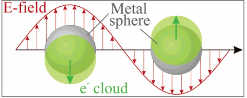
Modifying the size or shape of the nanoparticles results in a change of their colour. This effect is due to the occurrence of the surface plasmon resonance [3], the frequency at which conduction electrons oscillate in response to the alternating electric field of incident electromagnetic radiation (Fig. 1.2).
Fig 1.2: Schematic of plasmon oscillation for a sphere, showing the displacement of the
Therefore, the interest in the optical spectra of metal hydrosols and nanocomposites in these last decades is essentially motivated by the fruitful information expected to be gained on the electronic structure and the dynamics of the delocalized conduction electrons. Most works have focused on the surface plasmon excitation (the dipolar Mie resonance) which dominate the photoabsorption spectra in the near UV and visible range for metal particles of diameter much lower than the wavelength of light.
So the colloidal suspension of noble metal particles were subjected to spectral analysis using a Perkin Elmer Lambda 2 spectrometer in the region 190-1100 nm, with a 1 nm resolution power.
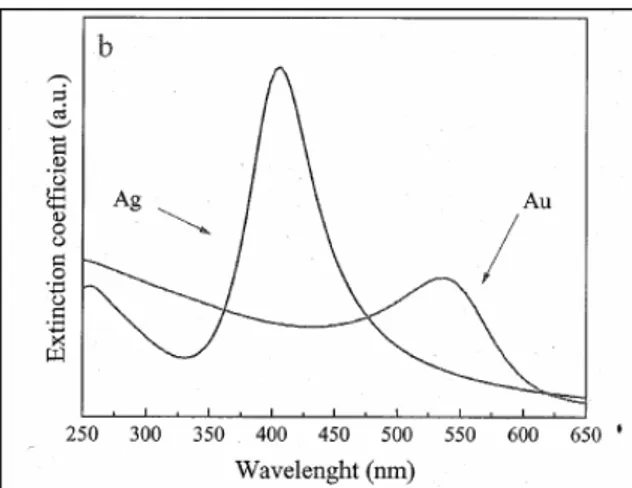
Fig1.3: Surface plasmonic resonance spectra of silver and gold nanoparticles. The inset
shows the best fit of experimental spectra (dot) with the Mie-Gans model we have adopted (continuous lines).
The extinction of each sample was monitored in the relevant plasmonic resonance (PR) region that is around 400 nm for silver and around 520 for gold.
Typical PR spectra of these samples are shown in figure 1.3. The reader may see the noticeable intensity difference between gold and silver PR spectra . The role played by the complex dielectric constant of two metals in producing these differences has been analysed.
The visible absorption (PR) of these colloids is instead interpreted at the ligh of the Mie theory [4], introducing the Gans approximation [5], to take into account the role played by the dielectric function of liquid medium in which the colloids are sunspended.
Therefore, our spectra have been modelled, in the framework of Mie’s theory, with the equation given by Papavassiliou [6], for the extinction coefficient K defined for N particles of volume V.
In this regard, if one consider that the extinction coefficient of a well dispersed collection of small particles, it is possible to obtain a simple expression in the cases in which εm is real and frequency independent, so that the extinction coefficient for N particles of volume V is given by the following equation:
In this equation E is the photon energy in eV, V is the particle volume in m3, ε1 and ε2 are the real and imaginary components of complex dielectric function. It is interesting to note that k has the dimensions of a cross-section (m2), which can be directly considered for a valuation of the total
amount of metallic atoms present in the sample, through the use of the atomic density.
In this regard, we would like to stress that HR-TEM data are of relevant importance for a check of the sample shape or size.
In fact, for particles smaller than the bulk electronic mean-free path, the dielectric function of the metal must be corrected for quantum size effects.
This point must be considered with much attention, because it directly influences the particles’ optical response and may be taken into account if one considers a classic Drude model.
In this case the dielectric function of each metallic cluster can be expressed as
In this equation λ, correlated with the experimental width of the Mie absorption, can be considered as a damping factor to make terms on the right-hand side of equation, excluding the εb(E) term, when E = Ep, i.e.
at resonance conditions. It measures also the mean collision rate of conduction electrons at optical frequencies. εb(E) is the contribution
coming from interband transitions and from all other non-conduction electrons to the dielectric function of the metal, Ep is the plasma energy
of free electrons, i.e. the PR energy at the absorption maximum.
When the particles are not too small, one can correct the dielectric function approximately by modifying the λ value. For very small particles the quantum size effect corrections can be complicated and may result in band narrowing, band splitting and frequency shifts in either directions from the PR band centre of bulk particles.
The range of size for this correction on the PR spectra has been detailed in ref. [7], and this is the case in our experiments.
The general expression which takes into account a classic quantum size effect relatively to λ quantity is to following:
Recently is has been shown [8,9] that it is possible to fit, by the use of very few fitting parameters, the surface plasmon spectra of gold colloids starting from the known bulk optical constants of the metal and correcting them for the above-mentioned quantum size effects.
Figure 1.4 shows the simulated PR spectra of silver and gold, when the simulation was performed by taking into account the role of the complex dielectric function of the system constituted by water and the relevant metal colloids.
In our laboratory either SERS [10] and catalytic application [11] have been carried out in there last years by the use of silver and gold nanoclusters. Especially in the latter case, an estimative measure of gold particle dimensions have been made by low-frequency Raman- modes (LOFIS modes) and their behaviour under different polarization conditions of incident light [12]. It is well known fact, since the pioneer work by Lamb [13], that surface acoustic phonons propagate in assumed spherical nanometric objects, producing dilatational and shear motions that scatter quasi elastically the visible light as a consequence of density fluctuations [14-16].
The Raman frequency shift, with respect to the elastic peak, is inversely correlated to the radius of the particles through the sound propagation speed and a dimensionless constant whose value is assumed to vary within a restricted range between 0.7 and 0.9, depending on the material [14-16].
Due to acoustic nature of these features in the low-frequency Raman spectra of the nano-objects, one can retrieve analogies with the hydrodynamic triplet of the Rayleigh-Brillouin spectra [17], where the Brillouin frequency shift is proportional to the sound propagation speed in the relevant medium through the transferred wave vector. On the other hand, in the equation describing the transferred wave vector, one has at the denominator the wavelength of the scattered light, which plays the role of length as that of the particle size in the LOFIS spectrum counterpart.
1.3-Thermodynamic Properties
Almost any properties in a solid in associated with a particular length scale, below this length, the properties vary. As the size decreases to the nanometer scale, the thermodynamic properties are significantly different form those of the bulk. This is mainly related to a very increased number of atoms or molecular units that lie on the surface.
An interesting physical property is the melting point. It is well know the melting point of small particles may differ form their bulk value. It has been reported that nanoparticles melt at temperature significantly below their equilibrium melting points. This reduction of melting point with size is not restricted to any particular material but it was found in a wide variety of materials: metals, semiconductors, and molecular organic crystals. It seems that the melting process beings with the formation of a liquid “skin” layer around the particles. The depression of the melting point of small particles can be explained in term of a higher surface energy contribution to the Gibbs free energy of the particles, as a significant fraction of atoms lie in the surface. In order to explain the experimental data on the lowering of the melting temperature for small nanoparticles, some theoretical models have been proposed. The first one was presented by Palow in 1909 [18].
Theoretical phenomenological considerations Pelow to develop a quantitative relationship between the melting temperature, Tm, and the
particle size. The Palow correlation is not quantitatively accurate, but it is still often used to estimate the size dependence of Tm .Afterwards, other
models have been proposed, based on various assumptions [19-22]. The general result of most of the experimental, theoretical, and computer simulation studies suggest that the melting temperature, Tm, depends on
(3) Tm =Tb- R C
Where R in the radius of a spherical nanoparticles, Tb is the bulk melting
temperature, and C is a constant depending on the material in specified conditions.
One of the most famous relations between the particle size and the melting point was proposed by Buffat and Borel [23].
Their version of the modified Palow relation is:
(4) =1 T r T b m( ) -
{
s s γ λ ρ 4 -γl(ρs/ ρl)2/3}
1/Dwhere D is the particle diameter, λ is the heat of fusion and ρ and γ are the density and surface free energy of the solid (s) and the liquid (l). The equation (4) gives a good descriptions of the experimental observations [24].
Zhao et al. studied the melting properties of size-selected isolated silver nanoparticles by using molecular dynamics simulation. They based their studies on phenomenological models that predict, at the melting point, the coexistence of a solid sphere core and a concentric liquid shell of a critical thickness, t0.
The relation proposed by Zhao is reported below:
where γsl is the interfacial surface tension between the solid and the liquid, while γlv is the one between the liquid and its vapour. By substituting the following values for silver nanoparticles:
Tb = 960.7 ºC λ = 1.06 x 10⁹ erg/g ρs = 10.49 g/cm3 ρl = 9.35 g/cm3 γsl = 184 dyn/cm γlv = 910 dyn/cm equation (5) becomes: (6) Tm= 960.7-2463 (1/0.603(R-t0) + 1/R)
Other later theoretical treatments have improved the accordance with the experimental data, but they all predict the inverse dependence. In addition large depressions of the melting point, about 500 ºC, were observed in 5 nm gold nanoparticles and about 140 ºC for 4 nm size aluminum particles.
There is also a large change in the vapour pressure of small particles due to the high curvature. This is called the “ Gibbs-Thompson effect” and can be described by the “Kelvin equation”, originally proposed for small liquid droplets and later extended to the phase transition of nanoparticles. The Kelvin equation is reported below:
(7) P= P* e(-2γ/vm r RT)
where P* is the pressure of the liquid, γ is the surface tension, vm is the molar volume, and r is the radius of the droplet.
1.4-Chemical Properties
As mentioned, the surface atom/volume atom ratio is mainly responsible of the peculiar properties of nanoparticles. This becomes particularly important in the chemical reactivity areas. It is well known that when matter is subdivided in such a way that the surface area is large, it becomes more reactive. Therefore, the use of nanoparticles is a very attractive area to develop matrices for any chemical reaction, this could be particularly interesting for example in pollution clean-up, and it is being seriously pursued, e.g. to destroy chlorinated hydrocarbons.
Another area in chemistry where the high surface area of nanoparticles has been exploited for many years is in the heterogeneous catalysis. In this case the active catalytic component may be a metal or a metal oxide, which is usually present in small quantities. The heterogeneous catalysis are prepared in the form of highly dispersed particulate material, often in conjunction with molecular sieve, and in the form of porous materials for either chemical processing to synthesize compounds or to clean up pollutants by catalyze action. An example of this is the use of TiO2 nanoparticles that catalyze the formation of free radicals, which can be used to destroy bacteria, viruses or hydrocarbon contaminants [25]. Fundamentally, catalysis involves a cyclic process in which a site on a catalyst forms a complex with reactants from which products are desorbed, thereby restoring the original sites and continuing the cycle. In nanostructured materials the active site of the reaction may be a croup of cluster of neighboring atoms on the catalyst surface, or a species absorbed onto the catalyst. These sites are associated with surface structures, lattice defects, and edges or corners of crystallites. Using a number of different synthesis process it is now some control over aspects such at the lattice parameters and the amount or type of internal defects
with a meta-stable structure quite different from what is expected with a normal equilibrium process. Furthermore, as the crystallite size decreases below 10 nm, and even below 5 nm, surface atoms will start to dominate while, at the same time, other structures, such as triple junctions, will be more and more present. The surface design of materials, as well as the increased presence of these structures could have a large impact on the properties of catalytic materials.
In catalytic reactions, metal clusters must allow the migration of coordinated ligands from a metallic atom to another one (CO, H etc.). This mobility is important in catalysis because it allows atoms or molecules to approach each other. For example, CO and H are common ligands on metal clusters, so they give the opportunity for the development of selective catalyst for synthesis gas conversion.
In addition, unusual coordination modes of a ligand to more than one metal atom were observed. In fact, some ligands are mostly found in clusters because they need stabilization by bonding to several metal atoms (CCO, BCO, CS, SO, CH, PO, etc.). Therefore, it is possible to stabilize unstable or highly reactive intermediate species in solution by supporting them on a cluster. Isolation of such metal clusters containing rare ligand, by multi-metal attachment allowed the study the elementary reaction steps involved in catalysis.
Metallic nanoparticles with an average grain size between 1 to 20 nm, such as Pt or Rh, dispersed on various substrates like SiO2 or Al2O3 have been often used in heterogeneous catalysis [26].
These materials have been used extensively in many industries, such as petrochemical production, automobile emission control and fine chemical synthesis.
Recent studies showed that surface diffusivity can be significantly higher than in the bulk. This suggest that the atomic transport in a nanometric cluster is faster, since most of the atoms are on the surface.
Therefore, all cluster chemical reaction are greatly affected by several factors such as the atomic structure, the steric effects, the atom bond energy, etc., and a strong dependence on the number of atoms which form the cluster was observed. Studies on the reactivity of the clusters were conducted by saturating such nanometric aggregates with different types of adsorbed molecules. From the number of adsorbed molecules, the nature of the absorption sites was deducted, thus suggesting the possible structure of the clusters. Usually, metal clusters have a roughly spherical shape. In addition, it was observed that the ratio of the absorbed species and the surface metal atoms is less than 1 for metals of first transition series.
In many cases, for cluster having specific dimension, a very narrow distribution of absorbed species was detected, suggesting a quite rigid conformation with only one structure. In those cases when saturation was reached, bimodal distributions were detected and the presence of two or more isomeric species was suggested.
Studies on the absorption thermodynamic of clusters suggested that the [27] absorbate-cluster bond energies are significantly higher than in bulk materials, and , as a consequence, they decrease by increasing the particle dimensions.
Recently, an experimental procedure was developed, which combines thermodynamic measurements with reactivity and composition measurements at the saturation.
References
[1] Kreibig, U. and Vollmer, M., Optical properties of metal clusters, Springer-Verlag Berlin.
[2] Faraday, M., Philos. Trans. Royal Soc. London, (1857), 147, 145. [3] Bohren, C.F. and Huffman, D. R., Absorption and Scattering of Light
by Small Particles, Wiley New York, (1998).
[4] Mie G., Ann. Phys., (1908), 25, 377.
[5] Compagnini, G.; Messina, E.; Puglisi, O. and Nicolosi, V., Appl.
Surf. Sci., (2007), 254, 1007.
[6] Papavassiliou, G.C., Prog. Solid State Chem. , (1980), 12, 185. [7] Link, S. and El-Sayed, M. A., J. Phys. Chem. B, (1999), 103, 8410 [8] Amendola V.; Polizzi, S. and Meneghetti, M., J. Phys. Chem. B., (2006), 110, 7232.
[9] Link, S.; Wang, Z.L. and El-Sayed, M. A., J. Phys. Chem. B, (1999), 103, 3529.
[10] Compagnini, G.; Patanè, G.; D’Urso, L.; Puglisi, O.; Cataliotti, R.S. and Pignataro, B., J. Phys. Chem. C.; (2008), 112, 20301.
[11] Cataliotti, R.S.; Compagnini, G.; Crisafulli, C.; Minicò S.; Pignataro, B., Sassi, P. And Scirè, S., Surf. Sci. , (2001), 494, 75.
[12] Cataliotti, R.S., Compagnini, G.; Morresi, A.; Ombelli, M. and Sassi, P., Phys. Chem. Chem. Phys. (2002), 4, 2774.
[13] Lamb, H., Proc. London Math. Soc., (1882), 12, 189.
[14] Duval, E.; Boukenter, A. and Champagnon, B., Phys. Rev. Lett., (1986), 56, 2052
[15] Montagna, M. and Dusi, R., Phys. Rev. B., (1995), 52, 10080.
[16] Fujii, M.; Nagareda, T.; Hayashi, S. and Yamamoto, K., Phys. Rev.
B, (1991), 44, 6243.
[18] Palow, P., Z. Phys. Chem., (1909), 65,1.
[19] Couchman, P.R. and Ryan, C.L., Phil. Mag. A, (1978), 37, 369. [20] Matsubara, T. and Nataniski, A., J. Phys. Soc. Japan., (1975), 39 1415.
[21] Hoshino, K. and Shimamura S., Phil Mag. A , (1979), 40, 137 [22] Wautelet, M., Solid State Comm., (1990), 74,1237.
[23] Buffat, P. and Borel, J. P., Phys Rev. A, (1976), 13, 2287.
[24] Zhao, S.J.; Wang, S.Q.; Cheng, D.Y. and Ye, H.Q., J. Phys. Chem., (2001), 105, 12857.
[25] Hayashi, C. and Oda, M., J. Aerosol Science, (1998), 29, 757. [26] Davis, S.C.; Klabunde, K.J., Chem. Rev., (1982), 82, 153 [27] Andres, R.P., J. Mater. Res. (1989),4.
2
SYNTHESIS
2.1 Fundamental aspect of PLAL
Pulsed Laser Ablation in Liquids (PLAL) has become a key method for synthesis of nanoparticles with controlled geometry and size. Pulsed laser ablation was first developed in the 1960s, shortly after the invention of the pulsed ruby laser. Since then, laser ablation in a vacuum or dilute gas has been studied by many researchers. By using different target materials and background gases, and varying parameters such as the laser wavelength, fluence, and pulse duration, it is possible to produce a wide variety of thin films. These include high temperature superconductors [1], metals, semiconductors, oxides, and other ceramics [2], and diamond-like carbon [3].
These studies clearly indicate that PLAL has become a successful material fabrication technique, allowing versatile design through choosing suitable solid targets and confining liquids.
Compared to the conventional physical methods (including chemical vapour deposition [4], vapour phase transport [5], and pulsed laser ablation in vacuum [6], and chemical methods (including hydrothermal methods [7], soft-template [8] and use of various surfactants [9,10]), the technique of PLAL has many distinct advantages.
These include (i) a chemically ‘simple and clean’ synthesis, the final product is usually obtained without by-products and no need for further purification; (ii) low cost of experimental setup and easily controlled parameters; (iii) the extreme confined conditions and induced high temperature, high pressure region favour the formation of unusual metastable phases.
In experiments of pulsed laser ablation of solid materials, submerged in liquid environments (PLAL), colloidal suspensions of objects at a nanometer-size scale are obtained. The nature of these nanoparticles and the characteristics of the plasma plume that is surrounding the struck point of the solid target, present a strong dependence on the experimental conditions and diverse a clear description in their physical implications. This because it is possible to modulate the shape and the size of the ablated materials, only acting with the pulsed laser beam and regulating its fluence, i.e. the amount of energy of the laser electromagnetic field releaser on the target, expressed in J/cm2 of surface [11].
In our laboratory, the PLAL technique is commonly carried out with the aim of obtaining nanometer-sized metal particles, which are ideal supports for SERS measurement [12].
In fig. 2.1 the simple set-up we used to perform PLAL experiments in shown. Briefly, it consist of a pulsed laser source, which is a Nd-YAG by Continuum, model Surrelite II, operating at 1064 nm in the first harmonics and 532 nm in the secondharmonics.
The laser beam has been focused on the target by a lens of 250 nm focal length and the target was submerged in the liquid medium.
Typical irradiation conditions has been: pulsed repetition was set at 10 Hz with 3 ns pulse duration; the irradiation period has been protracted for twenty min.
The laser spot size on the surface has been varied in the range 1-3 mm of diameter to get always as energy density on the target fluence of around 2 J/cm2.
Fig.2.1: A block scheme of the set-up used in our experiments to produce nanoparticles
in liquid media. The irradiation condition are described in the text.
It was observed that, when we varied the asset of experiments by suspending vertically the target in the liquid medium, instead to put it in the vessel bottom, and irradiate sending the laser beam at 90 degrees to sample, a clear double layer having particles suspended on the top was observed in the irradiated zone only.
This phenomena was more clearly visible with gold that with silver, due to intense red colour of gold colloids. Suspending the irradiation and allowing the system to restore equilibrium conditions, diffusion of Brownian particles in the whole submerging fluid was observed in a few minutes.[13]
Liquid
Target
Lens
During a irradiation a typical plasma plume was observed and its appearance is that shown in Fig. 2.2.
Fig.2.2: Laser ablation of a noble metal target vertically submerged in liquid water and
ablated by a pulsed laser beam impinging at 90 degrees.
The strong luminosity is due to electromagnetic radiation emission by the ionic states of metal atoms in the plasma. At the end of irradiation, we observed a coloured colloidal suspension of gold or silver in water, that appeared red or yellow, respectively. The aqueous suspension was transparent and just ready for optical measurements in the visible spectral region.
Analysis of the plasma plume was performed observing the propagation of sound waves in the liquid confined region around the struck point, whereas the shock waves in the solid target were studied at the light of Courant [14] and Cole [15] treatments, that consider the failure of the Navier-Stokes equations to treat this matter.
Fig.2.3: A 1000 times magnification of the plasma plume generated in water by pulsed
ablation of solid noble metal targets.
When a laser ablation experiment is carried out in a liquid medium, the confinement action operated by the liquid in which the ablated solid target is submerged determines the formation of a shock wave in the plasma plume [16-18]. The laser produced plasma tends to expand at a supersonic velocity, but this expansion creates the shock waves because the liquid confines the expansion itself. The laser energy of pulses, impinging on the solid target, produced a continuous supply of ablated material in the plume; this is the source of the plasma intense luminosity. In fact, the vaporizing species are highly excited ionic particles, that incoherently relax towards fundamental quantum states emitting electromagnetic radiations. The emission process is however different from that occurring in a LASER resonator, since the light emission from the plume is spontaneous and it is not coherently stimulated as in the
laser effect. As shown in fig. 2.3, the plasma appears strongly illuminated due to these incoherent emission processes.
Thermodynamic and kinetic factors may influence the formation of different phases due to the evolution of the laser-induced plasma. As reported by Yang [19], three are the thermodynamic factor which are of importance in determining the nature of phases in the plasma. There are: density of ablated species, temperature and pressure in the plasma. The first parameter, i.e. the density of ablated species, may be evaluated by measurements of the expansion volume of the plasma plume itself. In practice, this expansion volume and the amount of the ablated species here contained, is measured trough the volume of the hole which remains on the target surface after the ablation.
As shown in fig. 2.4, taken form Yang’s review [19], the expansion volume of the plasma is measured through the images of the light emitting region on the surface. In this case the image refers to a graphite target submerged in water [20]. In such an experiment, the volume of the plasma plume, being a hemisphere of diameter corresponding to the FWHM intensity of emitted light, is estimated as 9.9×10⁻⁷cm3.
Fig. 2.4: Image (taken from ref.[20]) of the expansion volume of the plasma plume in
Assuming that the hole volume on the surface of target is linearly increased by a number pulses, the ablation volume for a single pulse is determined to be 7.4×10⁻⁸cm3. Therefore, the density in the plasma plume of carbon species, coming form graphite ablated with pulses of a Nd-YAG laser, resulted 6.7×1021cm-3.
The second thermodynamic parameter is the extra temperature of the plasma plume. This is measured, as in a pyrometer, through the optical emission spectra of the ablated species form the plasma confined in the liquid medium. Again, using a graphite target as ablation source, a plume temperature of ca. 5000 K has been determined when the optical emission spectra of C2 molecules as measured. These species were obtained in water, ablating the target with a Nd-YAG laser at 1064nm, and pulses having 20ns duration and energy fluence of 10 J/cm2 [20]. Finally we must talk of the third parameter, i.e. pressure. By conducting in the liquid state the ablation of a solid target with a pulsed laser, this parameter depends on the sum of two contributions.
The first one is the adiabatic expansion of the plasma under the confined action of the liquid. In this medium an acoustic wave propagates longitudinally under the action of the expanding plasma, whilst a shock wave will propagate in the bulk of the solid target. Such situation is schematically depicted in fig. 2.5, taken from ref. [19]. Thus, the shock wave inside the solid target is the origin of the second contribution to the extra pressure generated in the system, when the plume is obtained for the ablation in a liquid. Many experimental techniques have been developed, especially by Fabbro and co-workers [21-26], to measure the pressure generated by the shock wave propagating inside the solid target. These experiments were also accompanied by the development of theoretical models for the extra pressure due to the shock waves, done by the same author and his school.
Fig.2.5: A scheme (taken from ref. [20]) showing the acoustic waves propagation in the
confining liquid (water, and the shock waves generation inside the solid target.
The shock wave induce in the plasma plume extra conditions of pressure, temperature and density. As to the pressure increase, for instance, Berthe [25] reported a pressure of 2-2.5 GPa, when pulses of 50 ns form the wavelength 0.308 μm of a XeCl excimer laser were employed. With shorter pulse durations, as for instance 3 ns, pressure up to 10 GPa have been reported. Concerning density and temperature values, densities up to 1022-1023 atom/cm3 have been reported for the ablated species in the plasma, and the plume temperature reaches values of 4000-5000 K [20]. From Fabbro’s studies [21-26], a relationship for the extra pressure in the plasma can be given as
), ( ( 3 01 . 0 ) ( 2 0 1 2 − − − + = Z gcm s I GWcm GPa P α α
α being the internal energy fraction which is of thermal nature (α ~ 0.25),
I0 the intensity of incident power, and Z the reduced shock impedance
between the confining liquid medium and the solid target. In the case of water as liquid medium, Z is defined by the formula
2/Z= 1/Zwater +1/Ztarget
To give an exemplum of the Z values [21-26], for aluminium and silicon targets, ablated in water as submerging medium, these are:
Zwater = 0.165 ×10⁶, and Z silicon = 2.1 × 10⁶, being the units g cm-2s-1. In our case, the corresponding values have been measured to be Zgold= 1.96 ×10⁶, and Z silver= 1.33 × 10⁶ g cm-2s-1, respectively.
With these values of Z, and assuming for α the value 0.25, pressures in the range 2.8-5.2 Gpa were obtained as a function of our I0 values. It
must be noted that the plasma state is very far from the thermodynamic equilibrium, so tat the application of any type of state equation to evaluate the real pressure form the temperature value is not possible. For instance, if one would assume the plasma state as an ideal gas in thermodynamic equilibrium and would apply the relevant state equation
V
T
K
nN
P
=
a B/
Na and KB being the Avogadro and Boltzmann constants, respectively,
the estimated pressure by such equation will result much lower than the measured in the plasma plume in experiments of pulsed laser ablation in liquids.
Under equilibrium conditions, the propagation velocity may be expressed with the formula
The quantity c is a thermodynamic property of the system and constitutes the speed of propagation of the sound waves. In the perfect gas approximation
being
The linearised equation of motion becomes
Introducing the continuity equation , where the term (υ·δρ/δr) is a very small quantity of the second order, we get the linearized equation of
Regarding the kinetic aspects, there are some peculiarities of PLA in liquid confined targets. Surprisingly, the velocity of ablation of a solid target submerged in a liquid medium is much higher than the corresponding velocity in vacuum or in a diluted inert gas. In fact, due to the confining action operated by the submerging liquid , the plasma having the above said characteristics of high temperature, pressure, and density, can continuously etch the solid target at the interface solid-plasma, not expanding very fat from the etched target as happens in the vacuum, or in diluted gas environments [19] .
In fig. 2.6 we show the effect of laser ablation of a silicon target with an UV laser, having pulses in the range of 20-30 ns, when two different confining media, i.e. water and air, are submerging the target [28-31]. It is also shown as the thickness of the submerging water has a maximum efficiency at a definite depth, but it decays more than linearly when the thickness of submerging layer overcomes a certain value.
Although the layer thickness of the submerging liquid is very important for the mechanisms of energy dissipation, the maximum observed at a certain value of thickness in the plot ablation rate vs. layer thickness (Fig.2.6 b) may be explained with the minor possibility given to the plasma plume to expand when the layer of the submerging medium is less. Therefore, a more confined plasma, around the etched point in the target, will concentrate better the energy due to the high temperature and high pressure of the plume, to promote more easy etching of the solid sample.
The mechanism seems to be controlled by the shock waves generated in the target under the laser pulses having an opportune fluency, because they decay into acoustic mechanical waves in the liquid medium that are called ablative piston. The action exerted by the ablative piston enhances
that one due to extra pressure and high temperature of the plume increasing the ablation rate.
Fig.2.6: The effects, on the ablated particle amounts, of different confining media(a)
and of the layer thickness (b) in PLAL experiments.
Because the confining liquid absorbs the laser energy in the beam crossed layer [13], the etching action by the plasma plume and the depth of the liquid layer must reach a right compromise. The absorption of the laser beam energy by the submerging liquid produces so high temperature
increase in the crossed layer, that the density of heated portion of liquid may be strongly modified respect to the equilibrium value [11], especially if the density vs. temperature curve of the relevant submerging liquid has a great slope.
On the other side, the depth of the liquid layer can reduce the ablation threshold of the solid target surface and the dissipation of the plasma temperature at the plasma-liquid interface. In conclusion, high production of ablated particles by PLAL in confining liquid media is possible either for thin films deposition that for small particles synthesis [19].
An important kinetic aspect of PLAL in liquid media is the rapid dissipation of the plasma plume energy at the boundary on the confining liquid. Such a quenching action determines, from one side, the possibility to repetitive shoot on the surface target by the pulsed laser beam and this fact increases the amount of produced ablated species, but, from another site, determines the heating of the layer strictly in contact with the plume and crossed by the laser beam; we noticed that this fact affects the density variations such layer [13]. The rapid quenching action operated by the presence of liquid medium can be seen in fig. 2.7, taken from ref. [19].
Here the plasma light emission in water, of a solid graphite target is generated in PLAL experiments with different pulse duration.
One can see, from the dimension of emitting plume volume, that with longer duration of pulse, the confining action of the liquid in dissipating the energy of system is more efficient.
In fact, in the case of liquid confined plasma, the plume is confined near to the target surface, thus providing a continuous supplying of ablated material which , in state of plasma , enforces the luminosity of the emission [19].
Finally, the cooling effect operated by the liquid confining medium should enhance the formation in the plasma plume of metastable structures and phases.
Fig.2.7: Influence, on the expansion volume of the plasma plume. Of the laser pulses
duration (taken from ref.[20])
There rapidly convert to the stable phases due to the short quenching times that the confining liquid exerts at the plasma-liquid interface. These metastable phases are the results of the reactions occurring inside the
plasma and at the plasma-liquid interface due to the high temperature and high pressure conditions.
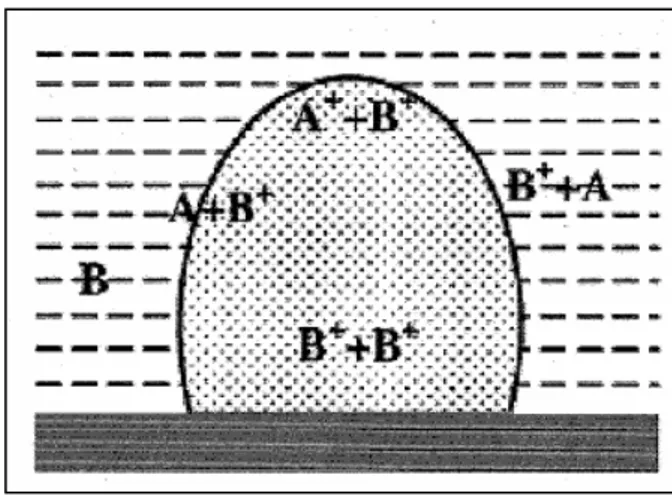
In fig. 2.8, a series of chemical reactions between B (solvent) and A( solid target, and among their ions, are schematically reported. Of course, changing either A or B, these reactions will strongly change, also because the possibility of having ionic species in the plume and at the interface depends on the ionisation energies of the relevant species. In our laboratory, it has been shown that the nature of ablated species coming from the same target will change in different liquid media [33]. The plasmonic resonance spectra, have been used to reveal these differences, depending on the used solvent.
Fig. 2.8: A scheme showing the chemical reactions occurring between solute (A),
solvent (B) and among their ions, inside the plasma plume and at the plume-solvent interface.
Of course, a compressed treatment like this, does not allow to speak of many other physical aspect that are important in plasma states. There are,
for instance, the role played by the discontinuities in the distribution function of plasma electrons, and as these discontinuities can be modelled with approaches deriving from the continua mechanics.
Moreover, it should be important to introduce the effect of the electric fields inside the plasma due to the plasma electrons and to the ions, on the mechanical waves propagating in the plasma plume. The effect is in practice a wave-breaking one, when the intensity of the electric field inside the plasma plume overcomes certain critical values.
Shifting again our attention on the shock waves, we want to describe the hydrodynamic processes accompanying the waves propagation[27]. First of all, we will define a shock wave a system in which the properties of a fluid phase change sharply within a very short distance in the space. There is in the gas phase, a certain tendency of any compression-wave to transform into a shock wave.
Neglecting in the hydrodynamic equations terms involving thermal conductivity, diffusion, and viscosity, the shock wave manifests itself as a mathematical discontinuity in the solutions.
However, including these dissipative term, the effect is that to change the discontinuity in a transition slightly more gradual, which for instance, in a gas at TPS conditions, occur within a distance of few mean free paths,
i.e. approximately within 10-5 cm [27].
Due such sharp gradients in the macroscopic properties o a gas subjected to a shock wave, the Navier-Stokes equations are not sufficiently accurate to describe both structure and thickness of a wave.
We tried to apply at our system of plasma plume the Hugoniot equation for a steady-state one-dimensional problem [14-15]. Assuming that the derivates at x = -∞ are zero, we obtain the relations below between the variables in the two sides of the shock wave:
M = ρ0υ0 = ρ∞ υ∞ ,
M υ0 + p0 =M υ∞ + p∞ ,
H0 + (1/2) υ02= H∞ + (1/2) υ∞ 2
These relations, called Hugoniot equation, correlate the eight quantities
ρ0, υ0, p0, H0, ρ∞, υ∞, p∞ and H∞. However, using the thermodynamic
dependence of H on ρ and p, the number of variable is reduced to six [27]. Thus, the equations are sufficient to express three of the variables in terms of the remaining three. Usually, we will specify the density and pressure on the low pressure side of the shock wave, ρ0 and p0, together
with an additional parameter which indicates the strength of shock, for instance the pressure in the high side, p∞ . The υ0 value represents the
velocity of propagation of the shock wave into the expanding plasma, on the low pressure side. υ∞ is instead the velocity of hot gases away from
the wave front of the plasma.
We rearranged the Hugoniot equation in a different form, and from the equation continuity and motion we obtained
Moreover, because ½ (υ∞2 - υ02) = ½ (υ∞ - υ0) (υ∞ + υ0), using the same
couple of equations, we may obtain [28]:
Now, combining this result with the conservation of energy equation and using the relation H= U +ρ/p we have got the relation
This equations can be considered the Hugoniot equations in a more convenient form.
As we cannot solve explicitly these equations, we have introduced the equation of the state and the thermodynamic condition for the gas in the plasma plume. These are of course large but necessary approximations, and allowed us to calculate values of pressure in the plasma going from ca. 2.8 GPa to ca. GPa, depending on the laser energy we released over the target, whilst the temperature was estimated by the plume luminosity to be around 4200K. With the same above equations and the ideal gas approximations, we obtained energy values of the order of 2.5 J in the plasma, values that are well above those of the impinging pulses of the laser. Therefore, the role played by the energetic of chemical reactions in the plume, and at confining liquid-plume interface, to enhance the energy well above the threshold given by the laser pulses is undoubtedly dominant.
2.2 PLAL of silver nanoparticles
Shape control of metal nanoparticles has received considerable attention in recent years because of the strong correlation between the shape and the chemical, physical, electronic, optical, magnetic and catalytic properties of a nanoparticle.
Wet chemical methods are widely accepted as practical and versatile approaches that produce nanoparticles in cube[34] rod [35] triangular[36] and wire[37] shapes. However, very few of them have been successful in controlling simultaneously both the size and shape of the products.
Photochemical methods have provided alternative approaches to modulate the size and shape of the nanoparticles synchronously. In recent years, numerous reports have focused on photoinduced or laser-induced growth of nanoparticles with definite shape and size.
Zheng et al, observed that the photoinduced method can be used for converting large quantities o silver nanospheres into triangular nanoprisms [36].
Lombardi et al [38] present a method for the tunable production of monodisperse silver nanoparticles. Using monochromatic light of different laser wavelengths to irradiate an initial solution of seed crystals, the size and shape of the products can be controlled.
Trace quantities of Au and Ag nanoprism have been observed as by products of method that predominately produce spheres [39].
In the last few years the growing interest for “plasmonics”, even due to the possibility to produce surfactant-free nanoparticles by pulsed laser ablation in liquid [40] has led to the development of new “nano-photonic” concepts with applications in extremely advanced fields [41].
Interesting studies on the surface chemistry of surfactant-free noble metal nanoparticle obtained by PLAL was reported by Sylvestre et al[42] and Muto et al[43]. Metal nanoparticles produced by PLAL present negative charges on the surface, that stabilize the colloidal solution due to the presence of electrostatic repulsion forces, and therefore do not require the addition of stabilizers or binders in order to control aggregation process.
It was demonstrated that the negative charge is induced by the presence of oxidized metal atoms on the surface nanoparticle. Furthermore, several studies conducted by Liao et al.[44] associated aggregation phenomena of noble metal nanoparticles in water to strong dipole/dipole interactions generated by an asymmetric charge distribution onto the cluster surface. Changes in the surface charge state and therefore on aggregation phenomena can be photo-induced by laser irradiation at different wavelengths.
Therefore, silver colloidal sols were prepared using laser ablation in water. A Nd:YAG laser beam was focused through a lens on a pure metal target submerged in Millipore grade water (5 mL). Both the first (1064 nm) and the second harmonic (532 nm) wavelengths have been used. The laser (Surelite II model by Continuum) has a pulse duration of 5 ns and a repetition rate of 10 Hz. Plasma (plume) generated by the beam- target interaction generally contains ablated species as well as ions and radicals coming from the water [45].
Synthesis and properties of silver colloids have already been investigated through spectroscopic and microscopic techniques.
Each colloidal system have been immediately characterized by performing Uv-Vis extinction spectra using a Perkin Elmer Lambda 2 spectrometer in the range 300-1100 nm.
It is well known that, among the most important features of metal nanoparticles, optical properties play a dominant role. Absorption spectra of silver colloidal dispersions exhibit broad bands in the UV–Vis range due to the excitation of plasma resonances and interband transitions. The existence of a plasmon resonance associated to the nanosized nature and spherical shape of the metal is confirmed by the presence of a peak at about 400 nm, while the interband transitions from the filled d band to the Fermi level is detected in the UV–Vis spectrum in the range from 200 to 350 nm.
The extinction of each colloid has been monitored for a few days and in this time interval the sol seems to be stable and does not show any tendency towards aggregation.
Dynamic Light Scattering (DLS) has been employed to obtain an ‘in liquid’ size distribution of the obtained particles by using a Horiba LB-550 system. Some drops of each colloid have also been deposited onto a single crystalline silicon substrate in order to perform SEM analyses using a ZEISS SUPRA 35 FE-SEM system with a field emission electron gun, while copper grids have been used to obtain TEM images using a JEM 2010F JEOL microscope.
The same procedure has been used to deposit some particles onto a silicon substrate for XPS measurements.
These were obtained using an AXIS-ULTRA spectrometer with a basic pressure in the range of 10-9 Torr. The X-ray radiation was generated by an Al Kα line decay (1486 eV) at operating conditions of 10 KV and 15 mA. The emitted photoelectrons were analyzed with a hemispherical electron energy analyzer. The detailed spectra have been acquired with a resolution below 1 eV.
A first attempt to determine the size distribution of the produced nanoparticles has been done by using dynamic light scattering (Fig. 2.9).
Fig. 2.9: DLS of silver nanoparticles produced by laser ablation in water using both
1064 nm and 532 nm irradiation wavelengths.
The technique exploits the Brownian motion of the particles which causes a Doppler shift in the incident light frequency.
The amount of frequency shift is related to the frequency of the Brownian motion, which is related to the size of the particles. In the case of silver sols obtained by using 532 and 1064 radiations the size distribution as obtained by DLS is reported in Figure2.8.
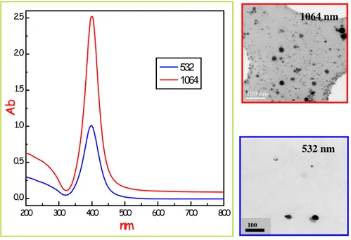
It is straightforward to observe two well separated components at sizes (diameters) of few tens and few hundreds of nanometers. The inset shows a zoom of the size distribution in the range 0-200 nm in which it is clear that that infrared radiation is able to produce smaller particles (20-50 nm), while the ablation performed at 532 nm gives clusters with sizes extending up to 100-120 nm. 0 250 500 750 1000 1250 1500 0 2 4 6 0 50 100 150 200 250 300 0 1 2 3 4 5 6 7 Number of p ar ticl es (% ) 532nm 1064nm Particle size (nm)
The first part of the size distribution is generally attributed to the presence of almost spherical metal nanoparticles which are produced by nucleation during the plasma plume cooling followed by nuclei growth and coalescence.
Three experimental findings support this observation.
• First, transmission electron microscopy (TEM) and UV-Vis analysis evidenced the polycrystalline structure of the silver nanoparticles obtained by PLA as reported in Figure 2.10.
• Second, nanoparticles obtained by LP-PLA in the presence of ligands or stabilizing agents have a smaller average size with respect to particles obtained in pure solvents[46].
• Third, LP-PLA in reactive solvents did not originate pure metal nanoparticles. [47]
As a matter of fact the driving force for the nucleation of metal embryos in the plasma plume is the supersaturation, given by the ratio between the actual vapour pressure to the equilibrium vapour pressure at the system temperature. The nucleation process is fast and is followed by a diffusion limited growth process of the nuclei, which continues for hundreds of nanoseconds after the laser pulse.
During the growth, free metal atoms condense on nuclei which can also coalesce together, originating the typical polycrystalline structure of the metallic nanoparticles. Mafunè and co workers observed that the growth of ligand-free nanoparticles can continue for several days after the synthesis, because metal ions can last in solution for a long time after the formation of the plasma plume in the case of high affinity with the solvent, as for Pt+ and Ag+ in water[46].
Fig. 2.10: Uv-Vis spectra (on the left) and TEM images (on the right) of silver
nanoparticles prepared by two different irradiation wavelengths.
The origin and the specific nature of the larger particles observed in the size distribution of Figure 2.9 is much more intriguing and need a specific investigation.
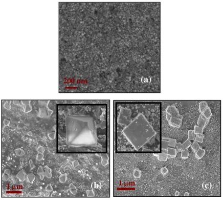
For this reason we performed a series of SEM analysis on both filtered and unfiltered solutions. Filtering has been done by using 200 nm pore size filters in order to separate the two distributions. Figure 2.11 shows the results of such an analysis.
In particular Figure 2.11a reports a plenty of spherical like silver particles after filtering which represents the low size distribution already discussed, while Figure 2.11b and c (large size distribution) give a clear
200 300 400 500 600 700 800 0.0 0.5 1.0 1.5 2.0 2.5 532 1064 Ab nm 100 532 nm 1064 nm
indication of the clusters obtained after the ablation with 1064 and 532 nm radiation respectively.
The nature of the micrometer sized silver aggregate is now clear. They consist in well defined regular solids, namely cubes in the case of the ablation at 532 nm and octahedra when 1064 nm radiation is used. The faces of these silver nanosolids are smooth and their average length varies between 200 and 500 nm. The images also evidences that the corners and edges of some particles are truncated.
Several previous paper have accounted for the formation and size control of silver nanoparticles with well defined shapes.
Fig. 2.11: SEM images of silver nanoparticles
200 nm (a)
Two approaches are generally followed. In the first case chemical effects, like polymer mediated processes or the presence of halide ions, have been proposed during a classical reduction of AgNO3[48].
A second approach has been considered with the so called photoinduced nanoparticles conversion[47]. In this case nanoseeds in the growth solution undergo a photochemical rearrangement to give birth to spherical nanoparticles of smaller and uniform size, while in a following step, the growth process moves into coalescence, where the newly formed nanoseeds aggregate to form rudimentary species which generally consists of silver platelets. The platelets then could evolve towards more complex structures depending on the irradiation wavelength as demonstrated by the already reported micrographs.
In our study we have conducted all experiments in pure water thus we can exclude any chemical effect.
On the contrary it is obvious to consider that the silver particles, which are initially produced during the ablation, are continuously irradiated by the laser beam itself during the overall process. This means that the effect of the laser beam is twofold. First the ablation produces spherical clusters, slightly different in size if infrared or visible radiations are used. These seeds are further induced to grow once the initial colloidal suspension continues to be irradiated.
In this regard, we performed some XPS analysis on the as-produced silver clusters deposited on silicon substrates.
It is well known that the sampling depth of XPS is a few angstrom. Considering that the particles size is about 10-20 nm, then we obtain information confined to few atomic layers around nanoparticles. Moreover the binding energies and line widths determined by XPS are strongly influenced by the nanometric size. In order to detect differences in the chemical state, Ag 3d core level spectra have been analyzed for the
two Ag systems and reported in figure 2.12. XPS analysis clearly indicate that the Ag3d binding energies of Ag@532 aggregates (dashed line) are located at lower (0.93 eV ) energy respect to the Ag@1064 peaks (solid line).
This is one indication that oxidized silver atoms (AgO and/or Ag2O) are present on the at Ag@532 nm cluster surface.
These species are probably formed during or immediately after the nanoparticles formation. In the case of Ag@1064nm the amount of oxide is negligible and the surface can be considered “metallic”.
Figure2.12 also shows the position of metallic silver and the increase of the line width for the oxide signal, in agreement with literature data in which AgO is considered a mixed oxide with the presence of both Ag+ and Ag3+ ions [49].
Fig.2.12: XPS spectra of Ag@532 nm nanoparticle(upper part of the figure) and
In view of XPS results we suggest that during laser ablation the Ag clusters are partially oxidized forming an Ag2O layer on the surface. Indeed, Yan an co-workers [50] reported a laser-based approach to fabricate Ag-Ag2O micro-cubes, octahedral and truncated octahedra directly from bulk Ag in polysorbate 80 aqueous solution at room temperature.
So, Yan results are further confirmation of what suppose by XPS analysis.
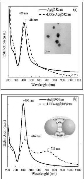
Additional confirmation can be explaining by the different interaction with LCCs. The two as prepared silver colloids (solid lines in figure 2.13) show a moderate difference in the SPR signal and position and width seem to be not influenced by the laser wavelength used for the ablation.
It is to be observed that while at 1064 nm the pulsed beam do not interact with already formed particles in water (at this wavelength the light absorption is negligible), the radiation at 532 nm can be considered partially in overlap with the tail of the plasmonic signal thus locally increasing the colloidal temperature [51].
Once LCCs solution is added, the two colloidal suspensions behave in a deeply different way.
Slight variations were detected in the SPR for LCC-Ag@ 532nm samples; after the interaction a red shift of 14 nm and a certain increase of the signal width is observed and it was already associated to aggregation phenomena [52].
On the contrary in LCCs-Ag@1064nm samples a second plasmon component appears at about 713 nm, while the component at lower wavelength remains positioned at about 416 nm.
Several works reported in literature [53]explain the split of SPR (λ - λ0 )
An exponential decay is reported as function of the distance (g) between two interacting nanoparticles of diameter D.
Fig. 2.13: shows extinction spectra in the surface plasmon resonances (SPR) region for
either silver colloidal sols prepared by ablation at 532 (Ag@532nm) (a) and 1064 (Ag@1064nm) (b) before and after the interaction with LCCs.

![Fig. 2.4: Image (taken from ref.[20]) of the expansion volume of the plasma plume in](https://thumb-eu.123doks.com/thumbv2/123dokorg/4516984.34768/34.892.389.612.653.835/fig-image-taken-ref-expansion-volume-plasma-plume.webp)

![Figure 2.12 also shows the position of metallic silver and the increase of the line width for the oxide signal, in agreement with literature data in which AgO is considered a mixed oxide with the presence of both Ag + and Ag 3+ ions [49]](https://thumb-eu.123doks.com/thumbv2/123dokorg/4516984.34768/55.892.212.577.502.797/figure-position-metallic-increase-agreement-literature-considered-presence.webp)