Epidemiology, pathophysiology, diagnosis, and
management of intracranial artery dissection
Stéphanie Debette*, Annette Compter*, Marc-Antoine Labeyrie, Maarten Uyttenboogaart, Tina M Metso, Jennifer J Majersik,
Barbara Goeggel-Simonetti, Stefan T Engelter, Alessandro Pezzini, Philippe Bijlenga, Andrew M Southerland, Olivier Naggara, Yannick Béjot, John W Cole, Anne Ducros, Giacomo Giacalone, Sabrina Schilling, Peggy Reiner, Hakan Sarikaya, Janna C Welleweerd, L Jaap Kappelle, Gert Jan de Borst, Leo H Bonati, Simon Jung, Vincent Thijs, Juan J Martin, Tobias Brandt, Caspar Grond-Ginsbach, Manja Kloss, Tohru Mizutani, Kazuo Minematsu, James F Meschia, Vitor M Pereira, Anna Bersano, Emmanuel Touzé, Philippe A Lyrer, Didier Leys, Hugues Chabriat, Hugh S Markus, Bradford B Worrall, Stéphane Chabrier, Ralph Baumgartner, Christian Stapf, Turgut Tatlisumak, Marcel Arnold, Marie-Germaine Bousser
Spontaneous intracranial artery dissection is an uncommon and probably underdiagnosed cause of stroke that is defi ned by the occurrence of a haematoma in the wall of an intracranial artery. Patients can present with headache, ischaemic stroke, subarachnoid haemorrhage, or symptoms associated with mass eff ect, mostly on the brainstem. Although intracranial artery dissection is less common than cervical artery dissection in adults of European ethnic origin, intracranial artery dissection is reportedly more common in children and in Asian populations. Risk factors and mechanisms are poorly understood, and diagnosis is challenging because characteristic imaging features can be diffi cult to detect in view of the small size of intracranial arteries. Therefore, multimodal follow-up imaging is often needed to confi rm the diagnosis. Treatment of intracranial artery dissections is empirical in the absence of data from randomised controlled trials. Most patients with subarachnoid haemorrhage undergo surgical or endovascular treatment to prevent rebleeding, whereas patients with intracranial artery dissection and cerebral ischaemia are treated with antithrombotics. Prognosis seems worse in patients with subarachnoid haemorrhage than in those without.
Introduction
Cervico cephalic artery dissection, which corresponds with a haematoma in the wall of a cervical or an intra-cranial artery, is an important cause of stroke in children and young and middle-aged adults.1–3 Although dissection
of the extracranial cervical arteries has been extensively studied and described,4–12 less information is available
about pure intracranial artery dissection (ie, not including the cervical portion of the artery).4 Early reports were
exclusively based on autopsy series, hence biased towards the most severe cases of intracranial artery dissection.13,14
Several possible reasons are available for the absence of information about intracranial artery dissections. First, intracranial artery dissection happens less frequently than cervical artery dissection in non-Asian countries, where the largest series of patients who had cervical artery dissection have been reported so far.9–12 Second,
patients who have cervical artery dissection and mainly present with headache, cervical pain, and ischaemic stroke are mostly seen by neurologists, whereas patients with intracranial artery dissection can also develop a subarachnoid haemorrhage and are therefore managed not only by neurologists, but also by neurosurgeons and interventional neuroradiologists, all of whom might have an incomplete picture of the disease. As a result, no consensus is agreed on for the diagnostic criteria and optimum treatment of patients with intracranial artery dissections.
In this Review we provide a comprehensive overview of reported studies into the epidemiology, patho-physiology, diagnosis, management, and outcome of spontaneous intracranial artery dissections, in addition
to proposing a consensus statement by a group of inter-national experts from various specialties and countries about the diagnosis and management of intracranial artery dissections.
Epidemiology
The incidence of intracranial artery dissections is unknown, but is probably lower than that of symptomatic cervical artery dissection (2∙6–3∙0 per 100 000 people per
year15,16) in populations of European ethnic origin. The
proportion of intracranial artery dissections in all cervicocephalic dissections substantially varies between ethnic origin and age groups, and also depends on study recruitment strategies and ascertainment methods used. Recruitment of patients for studies through neurology departments is biased towards those with cervical artery dissection or intracranial artery dissection without subarachnoid haemorrhage, whereas patient recruitment through depart ments of neurosurgery or interventional neuro radiology is biased towards intra cranial artery dissection with subarachnoid haemorrhage. In a series of 195 patients with vertebral artery dissections who were recruited in neurology departments in France and Switzerland, only 11% of dissections were located exclusively in the intracranial portion of the artery.17 In a
Mexican study18 of 100 patients admitted to a neurology
department for vertebral artery dissection with ischaemic stroke and without subarachnoid haemor rhage, 27 (27%) patients had intracranial artery dissection. In studies undertaken in east Asia,19–23 in which patients were mostly
recruited through neuro surgery and interventional neuroradiology departments, intracranial artery dissection
Lancet Neurol 2015; 14: 640–54
*Authors contributed equally
Department of Neurology, Lariboisière Hospital, Paris 7 University, DHU Neurovasc Sorbonne Paris Cité, Paris, France (Prof S Debette MD,
P Reiner MD, Prof H Chabriat MD, Prof C Stapf MD, Prof M-G Bousser MD); Inserm
U897, Bordeaux University, France (Prof S Debette,
S Schilling MSc); Department of
Neurology and Neurosurgery, Brain Centre Rudolf Magnus, University Medical Centre Utrecht, Utrecht, Netherlands
(A Compter MD, Prof L J Kappelle MD);
Department of Neuroradiology, Lariboisière Hospital, Paris 7 University, DHU Neurovasc Sorbonne Paris Cité, Paris, France (M-A Labeyrie MD); Departments of Neurology and Radiology, University Medical Centre Groningen, Groningen, Netherlands
(M Uyttenboogaart MD);
Department of Neurology, Helsinki University Central Hospital, Helsinki, Finland
(T M Metso MD, Prof T Tatlisumak MD);
Department of Neurology, University of Utah, Salt Lake City, UT, USA (J J Majersik MD); Department of Neurology, University Hospital Inselspital and University of Bern, Bern, Switzerland
(B Goeggel-Simonetti MD, H Sarikaya MD, S Jung MD, Prof M Arnold MD); Department
of Neurology and Stroke Centre, University Hospital of Basel, Basel, Switzerland
(Prof S T Engelter MD, L H Bonati MD, Prof P A Lyrer MD);
Neurorehabilitation Unit, University Centre for Medicine of Aging and Rehabilitation Basel, Felix Platter Hospital, Basel, Switzerland
accounted for up to 67–78% of all cervicocephalic artery dissections.19,20 Most reported series of patients with
intracranial artery dissection are from Asia (95% of studies including >40 patients with intracranial artery dissection, and 61% of studies including 20–39 patients with this disorder). Whether this suggests publication bias,
diff erences in disease prevalence across ethnic origin groups, or both, is unclear.
Intracranial artery dissections can also aff ect children, but a dearth of scientifi c literature exists about this problem in children, given the rarity of childhood stroke.
In a North American single-centre series24 of
(Prof S T Engelter); Department
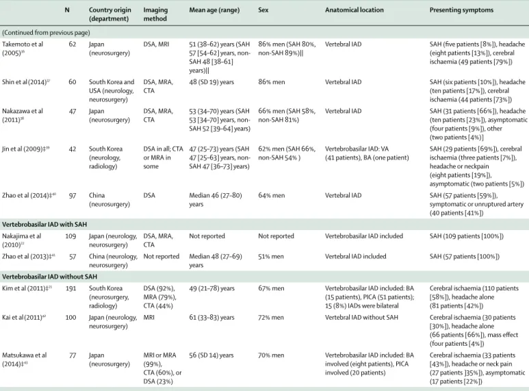
of Clinical and Experimental Sciences, Neurology Clinic, Brescia University Hospital, Brescia, Italy (A Pezzini MD); Neurosurgery Division, Department of Clinical N Country origin (department) Imaging method
Mean age(range) Sex Anatomical location Presenting symptoms
All types Yamaura et al (2000)23 357 Japan (neurosurgery survey)
DSA 51 (8–86) years (SAH 53 years, non-SAH 49 years)
Ratio of men to women 2:1; with non-SAH 2·6:1
3% anterior circulation (SAH 2%, non-SAH 5%); 97% posterior circulation (SAH 98%, non-SAH 95%), in VA (261 patients), BA (22 patients), ICA (ten patients), or other artery (29 patients)*
SAH (206 patients [58%]), cerebral ischaemia (112 patients [31%]), headache alone (26 patients [7%]), other (13 patients [4%]) Mizutani(2011)29 190 Japan (neurosurgery, radiology) MRA, DSA, or CTA 49 (0–74) years (SAH 52 [0–65] years, non-SAH 45 [22–47] years) 69% men (SAH 62%, non-SAH 77%)†
14% anterior circulation (SAH 11%, non-SAH 14%); 88% posterior circulation (SAH 89%, non-SAH 86%), VA (155 dissections), PICA (11 dissections), ACA
(11 dissections), BA (ten dissections), MCA (eight dissections)†
SAH (108 dissections [52%]), headache, or cerebral ischaemia (98 dissections [48%])† Ono et al(2013)30 143 Japan (neurosurgery) DSA and CT in all patients, MRI in some patients 51 (7–82) years (SAH 53 [31–83] years, non-SAH 48 [10–74] years) 59% men (SAH 58%, non-SAH 61%)
22% anterior circulation (SAH 15%, non-SAH 32%); 78% posterior circulation (SAH 85%, non-SAH 68%), VA (99 patients; 16 [11%] IADs in the VA were bilateral), ACA (11 patients), MCA (11 patients), ICA (eight patients), BA (seven patients), PICA
(fi ve patients), PCoA (one patient), PCA (one patient)
SAH (86 patients [60%]), headache, or cerebral ischaemia (57 patients [40%])
Kwak et al (2011)‡19 92 South Korea
(radiology)
DSA 51 years§ 58% men§ 24% anterior circulation (SAH 7%,
non-SAH 44%); 76% posterior circulation (SAH 93%, non-SAH 57%)
SAH (25 patients [27%]), SAH and ischaemia (three patients [3%]), infarction (20 patients [22%]), other cerebrovascular symptoms (44 patients [48%]) Metso et al (2007)31 45¶ Finland (neurology, neurosurgery) SAH: DSA (50%) or CTA (50%); non-SAH: MRA (100%), MRI (96%), US (39%), or CTA (9%) 46 (21–67) years (SAH 51 [32–67] years, non-SAH 42 [21–56] years) 58% men (SAH 50%, non-SAH 65%)
16% anterior circulation (SAH 14%, non-SAH 22%); 84% posterior circulation (SAH 86%, non-SAH 78%), VA (28 patients; one [2%] IAD in the VA was bilateral), ICA (fi ve patients), BA (four patients), PICA (three patients), ACA (two patients), SCA (one patient), PCA (one patient), pericallosal artery (one patient)
SAH (22 patients [49%]), headache, or cerebral ischaemia (23 patients [51%])¶
Vertebrobasilar IAD
Ahn et al (2012)‡32 210 South Korea
(neurosurgery, radiology) DSA in all; CTA, MRI, or MRA in some Median 47 (21–80) years (SAH 45 years, non-SAH 48 years)
61% men Vertebrobasilar IAD included: 20 (10%) IAD in the VA were bilateral
SAH (48 patients [21%]), non-SAH (182 patients [79%]; ischaemia frequency unknown) Kim et al (2011)‡33 111 South Korea
(neurosurgery, radiology)
DSA 45 (24–78) years 63% men Vertebrobasilar IAD included: BA involved (ten), PICA involved (47), eight (7%) IADs were bilateral
SAH (73 patients [66%]), ischaemia, or headache (38 patients [34%]) Matsukawa et al (2012)‡34 103 Japan (neurosurgery) MRI, MRA, CTA, DSA 53 (IQR 45–66) years (SAH 50 [46–59] years, non-SAH 54 [45–69] years) 69% men (SAH 77%, non-SAH 67%)
Vertebral IAD included: three (3%) IADs were bilateral
SAH (22 patients [21%]), ischaemia, or headache (81 patients [79%])
Kashiwasaki et al (2013)35
73 Japan (neurosurgery)
Not specifi ed 52 (SD 9) years 55% men Vertebral IAD without PICA involvement
SAH (45 patients [62%]), non-SAH (28 patients [38%], asymptomatic, or headache)
Neurosciences, Faculty of Medicine, Geneva University Medical Center, Geneva, Switzerland (P Bijlenga MD); Departments of Neurology and Public Health Sciences, University of Virginia, Charlottesville, VA, USA
(A M Southerland MD, Prof B B Worrall MD);
Department of Neuroradiology, Université Paris-Descartes, INSERM UMR 894, Center Hospitalier Sainte-Anne, DHU Neurovasc Paris Sorbonne, Paris, France
(O Naggara MD); Department of
263 consecutive patients with cervicocephalic dissections, 18 (7%) occurred in children, of which 11 (61%) were intracranial. Similar proportions were noted in other studies,24–28 including in non-Asian populations.
In most series, intracranial artery dissections aff ect the posterior circulation more frequently than the anterior circulation (76–93%; table 1; appendix). By contrast, cervical artery dissections and saccular intracranial aneurysms most commonly aff ect the anterior blood circulation.16,44 The relative frequency of intracranial
artery dissections in the diff erent intracranial segments varies between studies (table 1; appendix), but in most series the vertebral artery (intradural portion [V4 segment]) is the most common site. Bilateral
intracranial artery dissection seems to happen less often than bilateral cervical artery dissection does (<11% in most intracranial artery dissection series21,30,32,34 vs 15% in
cervical artery dissection series11,17,45), with bilateral
intracranial artery dissection mostly reported in the V4 segment.
As stated in initial reports by neurologists, paediatric intracranial artery dissection occurs mostly in the anterior circulation, by contrast with adults, in whom it mostly aff ects posterior circulation.24–26,28 However, these
reports are probably biased towards cases without subarachnoid haemorrhage. Nowadays, reports by interventional neuroradiologists frequently note posterior circulation involve ment in children with
N Country origin (department)
Imaging method
Mean age(range) Sex Anatomical location Presenting symptoms
(Continued from previous page) Takemoto et al
(2005)36
62 Japan (neurosurgery)
DSA, MRI 51 (38–62) years (SAH 57 [54–62] years, non-SAH 48 [38–61] years)||
86% men (SAH 80%, non-SAH 89%)||
Vertebral IAD SAH (fi ve patients [8%]), headache (eight patients [13%]), cerebral ischaemia (49 patients [79%])
Shin et al(2014)37 60 South Korea and
USA (neurology, neurosurgery)
DSA, MRA, CTA
48 (SD 19) years 86% men Vertebral IAD SAH (six patients [10%]), headache
(ten patients [17%]), cerebral ischaemia (44 patients [73%]) Nakazawa et al (2011)38 47 Japan (neurosurgery) DSA, MRA, CTA 53 (34–70) years (SAH 53 [34–70] years, non-SAH 52 [39–64] years) 66% men (SAH 58%, non-SAH 81%)
Vertebral IAD SAH (31 patients [66%]), headache (ten patients [23%]), asymptomatic (four patients [9%]), other (two patients [4%)] Jin et al (2009)‡39 42 South Korea
(neurology, radiology)
DSA in all; CTA or MRA in some 47 (25–73) years (SAH 47 [25–63] years, non-SAH 47 [36–73] years) 62% men (SAH 66%, non-SAH 54% ) Vertebrobasilar IAD: VA (41 patients), BA (one patient)
SAH (29 patients [69%]), cerebral ischaemia (three patients [7%]), headache or neckpain (eight patients [19%]), asymptomatic (two patients [5%]) Zhao et al (2014)‡40 97 China
(neurosurgery)
DSA Median 46 (27–80) years
64% men Vertebral IAD SAH (57 patients [59%]),
symptomatic or unruptured artery (40 patients [41%])
Vertebrobasilar IAD with SAH
Nakajima et al (2010)22 109 Japan (neurology, neurosurgery) DSA, MRA, CTA
Not reported Not reported Vertebrobasilar IAD included SAH (109 patients [100%])
Zhao et al (2013)‡41 57 China (neurology,
neurosurgery)
Not reported Median 48 (27–69) years
51% men Vertebral IAD included SAH (57 patients [100%])
Vertebrobasilar IAD without SAH
Kim et al (2011)‡21 191 South Korea
(neurosurgery, radiology)
DSA (92%), MRA (79%), CTA (44%)
49 (21–78) years 67% men Vertebrobasilar IAD included: BA (15 patients), PICA (51 patients); 15 (8%) IADs were bilateral
Cerebral ischaemia (110 patients [58%]), headache alone (81 patients [42%]) Kai et al(2011)42 100 Japan (neurology,
neurosurgery)
MRI 61 (33–83) years 72% men Vertebral IAD without SAH Cerebral ischaemia (30 patients [30%]), headache alone (66 patients [66%]), mass eff ect (four patients [4%]) Matsukawa et al (2014)‡43 77 Japan (neurosurgery) MRI or MRA (99%), CTA (60%), or DSA (23%)
56 (SD 14) years 70% men Vertebrobasilar IAD included: BA involved (eight patients), PICA involved (20 patients)
Cerebral ischaemia (33 patients [43%]), headache or neck pain (27 patients ]35%]), asymptomatic (17 patients [22%])
N=number of patients. SAH=subarachnoid haemorrhage. VA=vertebral artery. BA=basilar artery. ICA=internal carotid artery. MRA=MR angiography. DSA=digital subtraction angiography. CTA=CT angiography. PICA=posterior inferior cerebellar artery. ACA=anterior cerebral artery. MCA=middle cerebral artery. IAD=intracranial artery dissection. PCoA=posterior communicating artery. PCA=posterior cerebral artery. SCA=superior cerebellar artery. US=ultrasound. *Information about IAD site is missing in 29 patients and there are discrepancies between text and tables. †Numbers and percentages of dissected arteries (206) are presented, not numbers and percentages of patients (190 patients). ‡These series partly overlap. §Numbers and percentages only reported all patients with intracranial and extracranial dissection (133 patients), and not for subgroup of patients with IAD (92 patients). ¶103 patients in total were included in the study, but only 45 patients had pure IAD (the remaining 58 patients had cervical artery dissection with intracranial extension). ||Numbers and percentages reported only for 14 patients with aneurysm and surgical treatment.
Neurology, Dijon University Hospital, Dijon, France
(Prof Y Béjot MD); Department
of Neurology, University of Maryland School of Medicine, Baltimore, MD, USA
(J W Cole MD); Department of
Neurology, Gui de Chauliac Hospital, Montpellier I University, Montpellier, France
(Prof A Ducros MD);
Department of Neurology, Institute of Experimental Neurology (INSPE), IRCCS San Raff aele, Milano, Italy
(G Giacalone MD); Department
of Neurology, University Hospital of ZÜrich, ZÜrich, Switzerland (H Sarikaya); Department of Vascular Surgery, University Medical Centre Utrecht, Utrecht, Netherlands (J C Welleweerd MD, G J de Borst MD); Department of Neurosciences, Experimental Neurology, Laboratory of Neurobiology, KU Leuven University of Leuven, Leuven, Belgium (Prof V Thijs MD); VIB—Vesalius Research Center, Leuven, Belgium (Prof V Thijs); Department of Neurology, University Hospitals Leuven, Leuven, Belgium (Prof V Thijs); Department of Neurology, Sanatorio Allende, Cordoba, Argentina (J J Martin MD); Clinics for Neurologic Rehabilitation, Kliniken Schmieder, Heidelberg, Germany (T Brandt MD); Department of Neurology, University of Heidelberg, Heidelberg, Germany (C Grond-Ginsbach PhD, M Kloss MD); Department of Neurosurgery, Showa University, Tokyo, Japan
(Prof T Mizutani MD);
Department of Cerebrovascular Medicine, National Cerebral and Cardiovascular Centre, Suita, Japan
(Prof K Minematsu MD);
Department of Neurology, Mayo Clinic, Jacksonville, FL, USA (Prof J F Meschia MD); Division of Neuroradiology, Department of Medical Imaging, and Division of Neurosurgery, Department of Surgery, Toronto Western Hospital, University Health Network, Toronto, ON, Canada
(V M Pereira MD);
Cerebrovascular Disease Unit, IRCCS Foundation C Besta Neurological Institute, Milan, Italy (A Bersano MD); Université Caen Basse Normandie, Inserm
intracranial artery dis section, a location probably more prone to present with sub arachnoid haemorrhage than the anterior circulation.27
In a study that recruited patients with intracranial artery dissection through both neurosurgery and neurology departments during the same period, investi-gators reported that the proportion of people with pure intracranial artery dissection (ie, without dissection of the cervical portion of the artery) leading to subarachnoid
haemorrhage was 54%.31 Subarachnoid haemorrhage
from intracranial artery dissection is much less common than subarachnoid haemorrhage from ruptured intra-cranial saccular aneurysms. Autopsy series from Japan46,47
have reported that between 4∙5% and 10∙5% of fatal non-traumatic cases of subarachnoid haemorrhage had ruptured intracranial artery dissection. In a study48
combining data from interventional neuroradiology and neurosurgery departments in two hospitals during 6 years, a total of 756 (568 ruptured [75%] and 188 unruptured [25%]) saccular aneurysms, and 14 (1∙8%) symptomatic, intradural, dissecting vertebral aneurysms were treated.
Similarly, in the multicentre aneurIST study,49
17 intracranial artery dissections among 1834 ruptured and unruptured aneurysms (1∙5%) were reported in 1135 patients.
In adults, a male preponderance was noted in Asian populations with intracranial artery dissection, but not in non-Asian populations. Mean age at occurrence of intracranial artery dissection was 50∙4 years (range 47–61 years),19,21–23,29–43 and patients with intracranial artery
dissection with subarachnoid haemorrhage tend to be older than those with intracranial artery dissection without subarachnoid haemorrhage (table 1; appendix). Studies into paediatric intracranial artery dissections have consistently shown a substantial male preponderance, as with cervical artery dissection in children.26,27
Pathophysiology
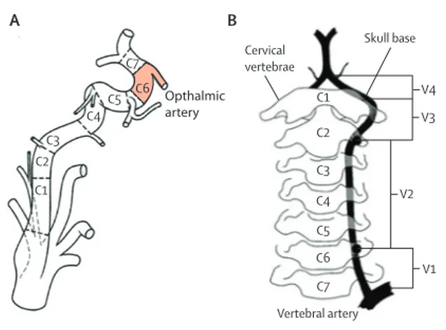
Anatomy of the intracranial carotid and vertebral arteries The intradural portion of the internal carotid artery starts at the clinoid segment of the artery (C6), from which the ophthalmic artery originates in most patients. The intradural portion of the vertebral artery is called the V4 segment, from which the anterior spinal artery and posterior inferior cerebellar artery originate (fi gure 1).
By contrast with cervical arteries, intradural arteries are characterised by a well developed internal elastic lamina, a paucity of elastic fi bres in the media, little adventitial tissue, and no external elastic lamina.52,53
These features, and weaker supporting tissues than cervical arteries,54 probably make intra cranial arteries
increasingly prone to subadventitial dissection and subsequent subarachnoid haemorrhage.13,55 In internal
carotid arteries, the external elastic lamina is present in the petrous portion (the portion where the internal carotid artery enters the canal in the petrous portion of the temporal bone; C3), but disappears in the horizontal
segment of the cavernous portion (C5) when the artery is situated between the layers of the dura mater, forming the cavernous sinus.56,57 Hence dissections starting in the
intrapetrous portion of the internal carotid artery mimic cervical artery dissections, whereas dissections in the intradural portion of the internal carotid artery—ie, starting in C6—can lead to subarachnoid haemorrhage. In vertebral arteries, the reduction of elastic fi bres in the tunica media and external elastic lamina is most pronounced in the last 0∙5 cm before the intradural portion, but is not complete until 0∙5 cm after the point of dural perforation.55 Sometimes to distinguish between
a dissection of the distal extracranial segment (V3) and a dissection in the V4 segment can be challenging due to blood fl ow changes immediately proximal and distal of the dissection site.
Mechanisms and pathological features
Little is known about the pathophysiology of intracranial artery dissection. Although available neuropathological specimens have generally shown a disruption of the internal elastic lamina and the media,30,47 whether direct
bleeding of vasa vasorum (small blood vessels in the wall of larger blood vessels) in the arterial wall can be the initial event is unclear.58 Vasa vasorum are not always
seen in intracranial arteries and seem to predominate in the tunica adventitia and proximal intracranial arteries.59
In a study30 where tissue samples were obtained by
surgery or autopsy at diff erent timepoints after symptom onset, the intramural haemorrhage was replaced by granulation tissue after 14 days from onset, followed by compensatory intimal thickening around the pseudo-lumen. In samples obtained after more than 30 days from symptom onset, neo vascularisation in the thickened
Figure 1: Anatomy of (A) carotid and (B) vertebral arteries, delineating cervical and intracranial segments
(A) Segments of the internal carotid artery. The intradural portion starts at the carotid artery segment (C6 [clinoid segment; highlighted in red]), with the ophthalmic artery arising in the intradural portion, except for some anatomical variants. The fi gure is adapted from Lasjaunias,50 by permission of Interventional
Neuroradiology. (B) Segments of the vertebral artery. The fi gure is reproduced from Khan and colleagues,51 by permission of Journal of Neurology, Neurosurgery,
and Psychiatry. C1 C2 C3 C4C5 C7 Cervical vertebrae Skull base Vertebral artery V1 V2 V3 V4 C1 C2 C3 C4 C5 C6 C7 A B Opthalmic artery C6
intima was reported, leading to chronic fusiform aneurysm formation,30 possibly aided by repetitive intramural
haemorrhage with the rupture of fragile neovessels.60
Diff erent patterns of intimal injury in intracranial artery dissection have been reported. A mural haematoma might be caused by one entrance in the pseudolumen (so-called entry-only lesions) or an entrance and an exit in the pseudolumen (so-called entry-exit lesions). Entry-only lesions can have a higher occurrence of subarachnoid haemorrhage than entry-exit lesions.61
The pathophysiological overlap of intracranial artery dissection with giant fusiform aneurysms and blood blister-like aneurysms is controversial, but they should probably be regarded as distinct entities.62–65 Mycotic or
oncological giant fusiform aneurysms are non-dissecting and are caused by the release of proteases by bacteria or tumour cells that break down the vessel wall. Blood blister-like aneurysms located at non-branching sites of intracranial arteries are caused by a degeneration of the internal elastic lamina and media without associated arterial dissection (no mural haematoma or double lumen on pathological examination).66
Risk factors and predisposing conditions
Risk factors for intracranial artery dissections are unknown. No comparisons exist between putative risk factors in patients with intracranial artery dissection and healthy controls. In the few studies that included both patients with cervical artery dissection and those with intracranial artery dissection, distribution of vascular risk factors did not diff er between the two groups,31
except for one study37 showing a higher prevalence of
hypertension in patients with intracranial artery dissection. However, this fi nding37 might be accounted
for by the older age of patients with intracranial artery dissection than control participants in that study (mean age 48 years vs 37 years).
Whether cervicocerebral trauma is a risk factor for intracranial artery dissection, as it is for cervical artery dissection,67 is unclear. In two studies25,37 that compared
patients with cervical artery dissection and patients with intracranial artery dissection, a history of minor trauma was more often present in patients with cervical artery dissection, both in children and adults. In our experience, sudden physical movements that lead to a sudden stretch of the arteries are sometimes reported before the event, but this association has not been systematically analysed in large patient series. Some instances of intracranial artery dissection in children, in our experience, have been associated with intracranial or systemic infections.
Diff erences in prevalence and characteristics of
intracranial artery dissections between ethnic origins, and the more frequent occurrence of intracranial artery dissection in children than in adults, suggest that genetic risk factors could contribute to the occurrence of intracranial artery dissection. However, genetic contribution to intracranial artery dissections has so far
not been explored. Exceptionally, intracranial artery dissection might be a complication of rare monogenic disorders of connective tissue, such as Loeys-Dietz syndrome.68,69 Whether carotid and vertebral artery
dissections noted in patients with vascular Ehlers-Danlos syndrome include intracranial artery dissections is not detailed in reported large series.70,71 Isolated cases of
suspected intracranial artery dissections have been noted in patients with Marfan’s syndrome.72
Patients with fi bromuscular dysplasia (a
non-atherosclerotic, non-infl ammatory vascular disease that mainly aff ects the renal and cervical arteries) have an increased risk of cervical artery dissection and intracranial aneurysms; whether these patients also have an increased prevalence of intracranial artery dissection is unknown.73–75 Only isolated instances of intracranial
artery dissection and fi bromuscular dysplasia have been reported,76,77 and patients with fi bromuscular dysplasia
were excluded from many reported series of patients with intracranial artery dissection. Overlap between intracranial artery dissection and segmental arterial mediolysis (a rare arterial disease that presents with life-threatening haemorrhages through ruptured aneurysms in the abdominal cavity), the retroperitoneum, and more seldom the base of the brain, is unclear.78,79
Clinical presentation and radiological features
Clinical presentation of intracranial artery dissections is not specifi c. The two main manifestations are sub-arachnoid haemorrhage and cerebral ischaemia.31 In mostreported series (table 1), intracranial artery dissections with subarachnoid haemorrhage represent 50–60% of all intracranial artery dissections. Subarachnoid haemor rhage occurs if the arterial wall of an intracranial artery dissection in the intradural portion ruptures. Between 30% and 78% of patients with intracranial artery dissection present with cerebral ischaemia (ischaemic stroke or transient ischaemic attack), without subarachnoid haemorrhage. No specifi c pattern of brain infarction emerged from our Review, and underlying stroke mechanisms could be either haemodynamic, thromboembolic, or due to occlusion of a perforating artery by the mural haematoma. Rarely, both subarachnoid haemorrhage and ischaemic stroke can be present in combination.17 About 80% of
patients with intracranial artery dissection have prodromal headache, before a subarachnoid haemorrhage or cerebral ischaemia, with subarachnoid haemorrhage occurring within 3 days after onset of headache in 96% of patients.29,47,80 Onset of prodromal headache was described
as sudden in only a few patients (in 13% of patients with intracranial artery dissection without subarachnoid haemorrhage and in 17% with intracranial artery dissection with subarachnoid haemorrhage).29
Other uncommon manifestations of intracranial artery dissections include isolated headache and symptoms associated with mass eff ect, which mostly aff ects the brainstem or cranial nerves (table 1; appendix). Rarely,
U919, Department of Neurology, CHU Côte de Nacre, Caen, France (Prof E Touzé MD); Department of Neurology, Lille University Hospital, Lille, France (Prof D Leys MD); Department of Clinical Neurosciences, University of Cambridge, Cambridge, UK
(Prof H S Markus MD); French
Centre for Paediatric Stroke and EA3065, Saint-Etienne University Hospital, Saint-Etienne, France
(Prof S Chabrier MD);
NeuroCentre, Clinic Hirslanden Zürich, Zürich, Switzerland
(Prof R Baumgartner MD) Correspondence to: Prof Stéphanie Debette, INSERM
U897, Bordeaux University, 33000 Bordeaux, France
stephanie. [email protected]
intracerebral haemorrhage has been noted in patients with intracranial artery dissection and, in our experience, even sometimes without subarachnoid haemorrhage.
Radiological diagnosis of intracranial artery dissection can be a challenge in view of the small size of intracranial arteries and the subtle and non-specifi c radiological signs, which tend to develop with time. Table 2 lists possible diff erential diagnoses to consider, along with features in favour of intracranial artery dissection diagnosis.
Pathognomonic radiological fi ndings of intracranial artery dissection include mural haematoma, intimal fl ap, and double lumen. In one study81 a dissection fl ap could be
identifi ed on MRI in more than 90% of patients with clinical symptoms and CT angiography fi ndings of a possible intracranial artery dissection. A mural haematoma was identifi ed in more than 50% of these patients. A mural haematoma usually leads to a regular crescent-shaped thickening of the arterial wall with enlargement of the external diameter of the dissected artery and often a reduced and eccentric arterial lumen. On T1-weighted MRI a haematoma is spontaneously hyperintense 48–72 h after onset. Detection of a mural haematoma can be improved by use of high resolution 3 Tesla imaging and three-dimensional acquisition of fat-suppressed sequences with black-blood eff ect that increase sensitivity and specifi city of images (fi gure 2).82–84
Other conditions, such as a partly recanalised thrombus or a haemorrhagic atherosclerotic plaque, might mimic this pattern and decrease specifi city of recognition, but these are not associated with a focal enlargement of the external diameter. In our experience, the presence of a mural haematoma is particularly rare in aneurysmal
forms of intracranial artery dissections. The presence of an intimal fl ap, with or without a double lumen, is a subtle sign, which is mainly observed in proximal arterial segments, and is probably best detected by digital subtraction angiography (fi gure 3).
Intracranial artery dissection can present with aneurysmal dilatation, segmental stenosis, or occlusion, with the distribution of these radiological subtypes widely varying between studies. Some studies23,29,31,32 reported
that aneurysmal dilations were more common in intracranial artery dissection with subarachnoid haemorrhage than in intracranial artery dissection without subarachnoid haemorrhage. Both segmental stenosis and occlusion in subarachnoid haemorrhage are highly suggestive of intracranial artery dissection. However, in intracranial artery dissection without subarachnoid haemorrhage, these fi ndings of segmental stenosis and occlusion are non-specifi c. Likewise, a fusiform or irregular aneurysmal dilation located at a non-branching site on an artery is very suggestive of intracranial artery dissection if associated with a segmental stenosis, but fusiform or irregular aneurysmal dilations are not specifi c for intracranial artery dissection in isolation.85 Additional
radiological elements are needed to confi rm the diagnosis of intracranial artery dissection, including rapid change in morphology. Whether the shallow and broad-based, blood blister-like intracranial aneurysms at the supraclinoid internal carotid artery are caused by intra cranial artery dissections is controversial.65,86 As a result, some of the
criteria used to defi ne intracranial artery dissection in studies included in our Review, such as fl ame-shaped occlusion or irregular stenosis, are not specifi c for
Features that favour intracranial arterial dissection
Atherosclerotic stenosis Isolated or unusual location of arterial stenosis; absence of other features of atherosclerosis (such as calcifi cations or plaques in other arteries); serial dynamic physical change of lesion shape (especially improvement of stenosis) on follow-up examinations; of a young age (<65 years) without traditional vascular risk factors
Vasospasm due to subarachnoid haemorrhage
Focal narrowing in intracranial artery seen on the day of onset (vasospasm occurs between 4 days and 3 weeks after subarachnoid haemorrhage)
Reversible cerebral vasoconstriction syndrome
Focal narrowing in one rather than many intracranial arteries; absence of classical triggers for reversible cerebral vasoconstriction syndrome (post-partum period, sympathomimetic or vasoconstrictive drugs); residual stenosis persisting for more than 3 months or serial dynamic physical change of lesion shape on follow-up examinations, especially if developing towards an aneurysm
Vasculitis Focal narrowing in one rather than many intracranial arteries; absence of diff use vessel wall infl ammatory imaging signs; absence of systemic infl ammatory disorder
Fibromuscular dysplasia Acute symptoms; single so-called pearl-and-string sign and not so-called string-of-beads (medial fi broplasia); long stenosis, string sign, and not focal band-like constriction or tubular stenosis (intimal fi broplasia); dynamic change of lesion shape on follow-up examination
Fusiform aneurysm without dissection Acute symptoms; mural haematoma, intimal fl ap, or double lumen; dynamic change of lesion shape on follow-up examination Dolichoectasia Acute symptoms; mural haematoma, intimal fl ap, or double lumen; dynamic change of lesion shape on follow-up examination
Thromboembolic occlusion Concurrent visualisation of a mural haematoma or subsequent recanalisation showing a long fi liform stenosis, a fusiform aneurysm, a pearl-and-string sign, or an intimal fl ap or a double lumen
Transient cerebral arteriopathy* Intracranial artery dissection is mainly seen in teenagers; parenchymal infarct often has a large size; arterial lesions are irregular; and an arterial wall haematoma can be seen on T1-weighted fat-saturated sequences
Fenestration (anatomical variant) Acute symptoms; no distinct adventitial layers (one vessel with two lumina and not two vessels); dynamic change of lesion shape on follow-up examinations
In transient cerebral arteriopathy the infarct is often restricted to the deep territory and associated with a smooth and focal arterial stenosis. All of these conditions can coexist with or might cause intracranial arterial dissection. *Present in children, mainly due to infl ammation, parainfectious angiopathy (eg, post-varicella angiopathy), or idiopathic arteriopathy (eg, focal cerebral arteriopathy).
intracranial artery dissections (appendix). These non-specifi c criteria are an important limitation of most studies.
On the basis of a multidisciplinary expert consensus we have compiled terminology standards and grading of imaging diagnostic criteria for the diagnosis of intracranial artery dissection (panel).
To detect a mural haematoma of the arterial wall, high resolution 3 Tesla MRI that includes three-dimensional fat-suppressed T1-weighted images with black-blood eff ect is regarded as optimum imaging method. Imaging of the arterial lumen to detect an occlusion, stenosis, aneurysm, or intimal fl ap, with or without double lumen, can be done with CT angiography or MR angiography. Digital subtraction angiography is the gold standard for luminal imaging but, because of its invasive nature this method, is mainly used if CT or MR imaging is inconclusive, if patients present with subarachnoid haemorrhage, or if surgical or endovascular treatment is being considered.
The defi nite diagnosis of intracranial artery dissection often needs the combination of arterial wall and lumen imaging, and also the comparison between baseline and follow-up imaging (fi gure 2).
Management and outcome
Treatment options
Optimum treatment for patients with intracranial artery dissections is unknown. No randomised trials exist and only observational studies with small sample sizes are available, thus providing a very low level of evidence.
Patients with intracranial artery dissection with subarachnoid haemorrhage are usually treated with surgical or endovascular procedures because up to 40% of patients have rebleeding within the fi rst days after the event.30,87 If patients are in very poor clinical health or the
proposed treatment has an unacceptably high risk of complications, a decision can be made to withhold surgical or endovascular treatment.
In earlier reported series, patients with intracranial artery dissection without subarachnoid haemorrhage and with aneurysmal dilation were often off ered surgical or endovascular treatment because of concern that the dissecting aneurysm would rupture.21 However,
in recent years, most patients with intracranial artery dissection without subarachnoid haemorrhage have been treated medically, and off ered acute stroke treatment and long-term prevention of ischaemic stroke. Endovascular treatment is undertaken only in patients with recurrent ischaemic symptoms despite receiving optimum medical treatment. Sometimes, endovascular treatment is undertaken if the dissecting aneurysm has increased in size, to prevent rupture, or more rarely to reduce signs of brainstem com pression.21,35,88,89 In children, the
preferred and widespread practice is surgical or endovascular treatment in patients with intracranial artery dissection with subarachnoid haemorrhage and those without subarachnoid haemorrhage and mass-eff ect, whereas patients with intracranial artery dissection without subarachnoid haemorrhage and cerebral ischaemia tend to be given medical treatment.
Figure 2: Example of intracranial artery dissection with mural haematoma changing over time
(A) Ischaemic stroke in the left occipital lobe and thalamus on diff usion-weighted MRI. (B) Fusiform aneurysmal dilatation of the P2 segment of the left posterior cerebral artery, directly arising from the internal carotid artery on time-of-fl ight MR angiography (double arrows). (C) Clear hyperintense mural haematoma with eccentric superior lumen of posterior cerebral artery on sagittal cervical and intracranial view of a three-dimensional fat-suppressed T1-weighted sequence, shown on (D), magnifi ed image of C, with arrow.(E) Arrows show normalisation of lumen of the posterior cerebral artery at 3 months follow-up, in MR angiography.
A B C D E
Figure 3: Detection of intracranial artery dissection with diff erent imaging modalities
(A) Three-dimensional digital subtraction angiography (DSA) of a patient with subarachnoid haemorrhage. Arrows point to two typical intracranial arterial dissections of the right vertebral artery (bottom arrow)and the basilar artery (top arrow) with associated stenosis and aneurysmal dilatation. (B) DSA of right internal carotid artery with dissecting aneurysm and irregular stenosis resulting in a so-called pearl-and-string sign (arrow). Arrow head points to intimal fl ap visible in the M1 segment of the right, middle cerebral artery. (C) CT angiography of basilar artery showing dissecting aneurysm (bottom arrow)and intimal fl ap (top arrow).
Surgical and endovascular treatment
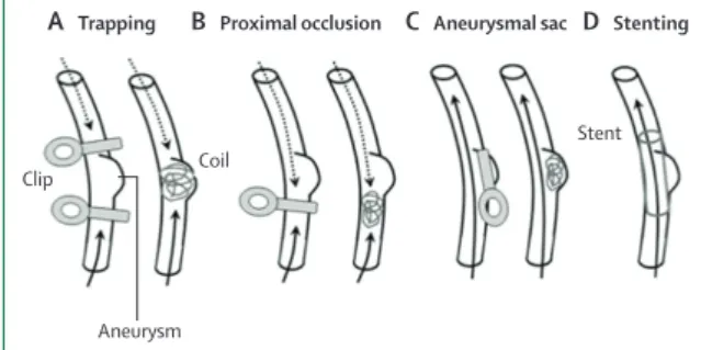
Various surgical and endovascular treatment methods have been proposed for intracranial dissecting aneurysms (fi gure 4).22 All treatment methods aim to reduce blood
fl ow in the dissected region. Deconstructive techniques sacrifi ce the parent artery, whereas reconstructive techniques aim to maintain a parent artery.
Parent artery occlusion is a deconstructive technique in which blood fl ow into the dissected segment of the artery is stopped by occlusion either surgically or through an endovascular approach. Preferably, the dissected segment is occluded both proximally and distally to prevent rerupture through retrograde fi lling of a dissecting aneurysm. Parent artery occlusion has a risk of brain infarct in case of insuffi cient collateral supply. Before permanent occlusion, the collateral supply can be assessed with temporary balloon-occlusion or amobarbital infusion during digital subtraction angio graphy (with simultaneous monitoring of the patient’s neurological function).
Reconstructive techniques, such as selective aneurysmal sac occlusion through clipping (surgical) or coiling (endovascular), are often diffi cult in intracranial artery dissection given the non-saccular shape of the dissecting aneurysm. In endovascular treatment, stent-assisted coiling can be used. Stenting of the dissected artery without any coiling by fl ow-diverter stents or conventional close-cell stents has been reported in small series;85,90,91 however, several days to months can pass
before the dissecting aneurysm is thrombosed. Moreover, stenting needs dual antiplatelet treatment for several months after the procedure, thereby exposing patients to an increased risk of haemorrhagic complications.21,41
Seldom, a bypass surgery between extracranial and intracranial arteries can be considered if the risk of infarction due to parent artery occlusion is unacceptably high and stenting is impossible.22,42 In very rare instances,
mostly in middle-cerebral artery dissections (M2 branches), the dissected segment can be excised and arterial stumps reanastomosed.92
As for saccular intracranial aneurysms, endovascular treatment is currently more frequently undertaken than is surgical treatment in most patients with intracranial artery dissection with subarachnoid haemor rhage in scientifi c literature. A com parison between surgical and endovascular treatment has not been made in a randomised trial.
Some observational studies have reported peri-procedural complications after surgical or endovascular treatment for intracranial artery dissection (table 3; appendix). Recurrent bleeding was reported in 0–11% of patients with intracranial artery dissection with sub-arachnoid haemorrhage after surgical or endovascular treatment, and after treatment for ischaemia in 0–22% of patients.19,21–23,29–43 Cranial nerve palsies and spinal cord
infarctions were seldom reported.35,41,93 Overall, of
813 endovascular procedures, 50 (6∙2%) cerebral or spinal cord ischaemia, 15 (1∙8%) rupture and rebleeding, and
seven (0∙9%) cases of cranial nerve palsies were reported; of 125 surgical procedures, 23 (18∙4%) cerebral or spinal cord ischaemia, one (0∙8%) rupture and rebleeding, and one (0∙8%) case of cranial nerve palsy were reported. As previously emphasised, these percentages are likely to be an underestimation due to reporting and publication bias. Medical treatment
Medical treatment of intracranial artery dissection without subarachnoid haemorrhage encompasses both acute stroke treatment (recanalisation) and long-term
Panel: Proposed terminology and grading of imaging diagnostic criteria for intracranial artery dissection
Proposed terminology for imaging diagnostic criteria of intracranial artery dissection At least one of the three following features should be present when diagnosing an intracranial artery dissection:
• Fusiform or irregular aneurysmal dilation at a non-branching site of an intracranial artery, with at least one of the following criteria
• Intramural haematoma (hyperintense rim on images with T1-weighted MRI), intimal fl ap, or double lumen*
• Rapid change in morphology on repeated imaging (increase or reduction in size, subsequent appearance of stenosis)
• Association with a focal stenosis (so-called pearl-and-string sign)
• Long fi liform or irregular stenosis of an intracranial artery, with at least one of the following criteria:
• Intramural haematoma (hyperintense rim on images with T1-weighted MRI), intimal fl ap, or double lumen*
• Rapid change in morphology on repeated imaging (increase or reduction in size, or subsequent appearance of aneurysmal dilation)
• Association with a fusiform or irregular aneurysmal dilation (so-called pearl-and-string sign)
• Occlusion of an intracranial artery that recanalises in either a fusiform or irregular aneurysmal dilation at a non-branching site, or a long fi liform or irregular stenosis Proposed grading of imaging diagnostic criteria for evidence of intracranial artery dissection
• Defi nite intracranial artery dissection
• Stenosis or occlusion of an intracranial artery secondarily developing towards a fusiform or irregular aneurysmal dilation at a non-branching site
• Intramural haematoma, intimal fl ap, or double lumen • Pathological confi rmation of intracranial artery dissection • Probable intracranial artery dissection
• Fusiform or irregular aneurysmal dilation and focal, long fi liform, or irregular stenosis (so-called pearl-and-string sign) without subarachnoid haemorrhage, or still present >1 month after subarachnoid haemorrhage
• Fusiform or irregular aneurysmal dilation at non-branching site with rapid change in morphology (increase or reduction in size, or subsequent appearance of stenosis)
• Possible intracranial artery dissection
• Fusiform or irregular aneurysmal dilation at non-branching site without change in morphology on repeated imaging within 6–12 months after fi rst imaging • Long fi liform or irregular stenosis of an intracranial artery, with reduction in size or
disappearance over time
prevention of ischaemic stroke. The safety and eff ectiveness of intravenous and intra-arterial thrombolysis in patients with intracranial artery dissection without subarachnoid haemorrhage are unknown, because only case reports have been published.94–97 The choice of
anti-thrombotic treatment (anticoagulants or antiplatelets) in patients with intracranial artery dissection without subarachnoid haemorrhage with cerebral ischaemia has not been assessed in randomised controlled trials or in systematic reviews and meta-analyses of observational data. By assuming that the mechanisms of cerebral ischaemia in intracranial artery dissection resemble those of cerebral ischaemia in cervical artery dissection, examining studies on antithrombotic treatment in cervical artery dissection might be helpful. Results from randomised controlled trials of antithrombotic treatment in cervical artery dissection are also missing. A pilot trial98
on 250 patients has been completed, showing no diff erence in effi cacy of antiplatelet and anticoagulant drugs at preventing stroke and death in patients with cervical artery dissection, but stroke was rare in both groups. Several meta-analyses5,99 of obser vational studies
have not shown any signifi cant diff erence in clinical outcome between patients treated with anticoagulants and those treated with antiplatelets, mostly aspirin. Findings from a meta-analysis100 using a Bayesian approach
suggested a treatment eff ect in favour of antiplatelet drugs, the advantage of which was less obvious when the analysis was restricted to studies of higher methodological quality. Although no haemor rhagic complication was reported in a small series of patients with intracranial artery dissection without subarachnoid haemorrhage who were treated with anticoagulants,31 the risk of subarachnoid
haemorrhage is greater in intracranial artery dissection than in cervical artery dissection. Several studies reported patients with intracranial artery dissection with initial ischaemic manifestations who then subsequently or
concurrently developed subarachnoid haemorrhage, prompting caution.30
In patients with intracranial artery dissection without subarachnoid haemorrhage and no signs of cerebral ischaemia, or in rare cases when both subarachnoid haemorrhage and cerebral ischaemia are present, no antithrombotic treatment, but close monitoring has been proposed.42 Studies investigating the predictors of
subarachnoid haemorrhage and cerebral ischaemia in patients with unruptured intracranial artery dissection are warranted to optimise management strategies.
Outcome
Because of treatment and publication biases, little is known about the natural history of intracranial artery dissection. Overall, intracranial artery dissection has a more severe course than cervical artery dissection, with a more ominous outcome in patients with subarachnoid haemorrhage than in those without subarachnoid haemorrhage.30,101 Table 3 and the appendix summarise
outcomes reported in individual studies. Mortality
Mortality outcome for patients with intracranial artery dissection and subarachnoid haemorrhage ranges between 19% and 50%. In studies including only patients who qualifi ed for endovascular treatment, lower mortality rates (5–9%) were reported, which is not surprising given that these studies excluded the most severe patients from the outset.35,41 Mortality outcome in patients without
subarachnoid haemorrhage is low and similar to that in patients with cervical artery dissection, ranging between none and 3% in reported series.
Recurrent haemorrhagic or ischaemic events
Although recurrences are poorly defi ned in most studies, overall, recurrent haemorrhagic or ischaemic events seem to follow the pattern of the initial event in more than 90% of patients.30 In patients with subarachnoid haemorrhage,
recurrence of subarachnoid haemorrhage in up to 40% of patients has been reported,30 with the highest rates being
noted in patients treated conservatively.22 Most haemorrhagic
recurrences cluster within the days after the initial event.22,30
In one study,30 recurrent subarachnoid haemorrhage
occurred more frequently in patients with intracranial artery dissection older than 50 years and less frequently in patients with carotid intracranial artery dissection.
In patients without subarachnoid haemorrhage, recurrence rates of ischaemic stroke ranged between 2% and 14%, with a mean follow-up spanning from 3 months to 8 years. In one study,42 38% of patients had recurrent
cerebral ischaemia during a mean follow-up of 24 months, but antiplatelet drugs were not prescribed after the initial ischaemic event. Extension of the intracranial artery dissection into the basilar artery and involvement of the posterior inferior cerebellar artery were reported as risk factors for recurrent ischaemic stroke.43
Figure 4: Four types of surgical and endovascular treatments
(A) Trapping aims to exclude blood fl ow from the dissected region and aneurysm. (B) Proximal occlusion reduces blood fl ow in the dissected region and aneurysm. Both trapping and proximal occlusion can be done by clipping or coiling. (C) Aneurysmal sac occlusion can be done by clipping or stenting and selectively occludes the aneurysm, but does not change blood fl ow in the vessel. (D) Stenting aims to cover the dissected region and aneurysm, leaving blood fl ow in the vessel unchanged. Solid arrows represent antegrade blood fl ow. Dotted arrows represent retrograde blood fl ow. Figure reproduced from Nakajima and colleagues,22 by
permission of Acta Neurochirurgica.
Stent
Clip Coil
A Trapping B Proximal occlusion C Aneurysmal sac D Stenting
Very few patients seem to have diff erent types of events associated with the intracranial artery dissection over time—ie, subarachnoid haemorrhage after initially unruptured, intracranial artery dissection (described between 4 days and 11 days after the initial diagnosis29,30)
or ischaemic events attributed to residual arterial lesions several months or years after an initial subarachnoid haemorrhage.30 In some patients, signs of brainstem
compression were noted several years after a dissecting aneurysm.21,30
Recurrent dissections
Little information is available about the risk of recurrent intracranial artery dissection. One study29 of 190 patients
with regular follow-up imaging after intracranial artery
dissection reported recurrent intracranial artery dis-section in 18 patients (9%) during a mean follow-up of 3∙4 years.29 Of the recurrent dissections, 12 (67%)
occurred within 1 month after the initial event. This rate of recurrent dissections seems in line with the recurrence rate of cervical artery dissection, which is estimated between 0% and 8% in most studies.7
Functional outcome
Functional outcome is not always reported in published series and has been rated on diff erent scales. Overall,
more than 79% of patients with intracranial artery
dissection and without subarachnoid haemor rhage had a favourable functional outcome (modifi ed Rankin Scale ≤1 or ≤2, or equivalent).19,21–23,29–43 One study21 showed that
N Treatment Follow-up time Deaths during
follow-up Good functional outcome Recurrences or complications All types Yamauraet al (2000)23
357 SAH: 125 (61%) surgical or endovascular treatment, 80 (39%) treated conservatively (no details); non-SAH: 26 (17%) surgical or endovascular treatment, 125 (83%) treated conservatively (no details; one [<1%] not described)
≥3 months 17% (SAH 27%, non-SAH 3%)
75% GOS 5 (SAH 63%, non-SAH 90%)
34 (10%) recurrence (no details on type; SAH: 29 recurrences [14%; no details on type of recurrence]; non-SAH: fi ve recurrences [4%; no details on type of recurrence])
Mizutani (2011)29
190 SAH: 71 (71%) surgical treatment, 31 (29%) treated conservatively (no details); non-SAH: mild-volume expansion plus free radical scavengers for infarction (occasionally antiplatelet drugs or anticoagulants for infarction plus stenosis ), three (3%) surgical treatment (in patients who had aneurysm extension) Mean 3·4 years (range 2–20·4 years) in non-SAH 11% (SAH 68%,* non-SAH 2%)
Not reported 18 (10%) recurrence (new IADs, all in diff erent arteries, 12 recurrent IADs occurred within 1 month, six recurrent IADs occurred after >1 year); one (1%) patient had a SAH at day 11 after onset
Ono et al (2013)30 143 SAH: 54 (63%) surgical treatment,
32 (37%) treated conservatively (no details); non-SAH: 12 (21%) surgical treatment, 45 (79%) treated conservatively (no details)
Mean 8·2 years (range 1 day–25 years) 18% (SAH 29%, non-SAH none) 69% independent (SAH 55%, non-SAH 90%)
36 (33%) SAH (SAH: 35 patients [41%] rebleeding at mean 4·8 days [range 0–26 days]; non-SAH: one [2%] SAH 4 days after initial ischaemic event), 10 (7%) ischaemic stroke (SAH one [1%] ischaemic stroke at 85 months, non-SAH nine [16%] ischaemic stroke); non-SAH group one (2%) hemifacial spasm at 21 months due to compression by enlarged aneurysm Kwak et al(2011)†19 92 Not reported Not reported Not reported Not reported Four (4%) SAH (SAH: four [14%] rebleeding)
Metso et al (2007)31 45 SAH: 19 (86%) surgical or endovascular
treatment (no details), three (14%) treated conservatively (no details); non-SAH: 23 (100%) treated with anticoagulation
Mean 1·3 years (range 1 day–8 years)
16% (SAH 32%, non-SAH none)
58% mRS≤2 (SAH 32%, non-SAH 83%)
No SAH for patients without SAH; not reported for patients with SAH
Vertebrobasilar IAD
Ahn et al (2012)†32 210 SAH: 48 (100%) endovascular treatment;
non-SAH: 59 (32%) endovascular treatment
Median47 months (range 8–105 months)
Not reported Not reported No SAH
Kim et al(2011)†33 111 Endovascular treatment Mean 35 months
(range 15–84 months)
8% 85% mRS≤2 (SAH
77%, non-SAH 100%)
Six (5%) SAH (SAH: six [8%] rebleeding, of which one [1%] on day of onset, four [5%] 3–4 days after treatment, and one [1%] 15 days after treatment), four 4% unruptured angiographic recurrent dissection in 100 patients with radiological follow-up, seven (10%) ischaemic stroke (SAH: fi ve [11% ischaemic stroke]; non-SAH: two [7%] ischaemic stroke)
Matsukawa et al (2012)†34
103 Not reported Not reported Not reported Not reported Not reported
N Treatment Follow-up time Deaths during follow-up
Good functional outcome
Recurrences or complications
(Continued from previous page) Kashiwasaki et al
(2013)35
73 Endovascular treatment Mean 55·6 months‡
(range 6–145 months) 8% (SAH 13%, non-SAH none) 91% mRS≤2 (SAH 86%, non-SAH 100%)
One (1%) SAH (SAH: one (2%) rebleeding periprocedural), seven (10%) ischaemic stroke (SAH: fi ve [11%] ischaemic stroke; non-SAH: two [7%] ischaemic stroke), two (3%) spinal cord infarction, six (8%) cranial nerve palsy, two (3%) asymptomatic recurrences Takemoto et al
(2005)36
62 SAH: fi ve (100%) surgical treatment; non-SAH: nine (16%) surgical treatment, 48 (84%) treatment not specifi ed
Not reported None§ 79% GOS 5§
(SAH 40%, non-SAH 100%)
No SAH; two (14%) cerebral infarction (SAH: one [20%] periprocedural cerebral infarction; non-SAH: one [11%] cerebral infarction 2 weeks after treatment)§
Shin et al (2014)37 60 Not reported 3 months SAH 50%, non-SAH
not reported Favourable outcome (SAH 50%, non-SAH not reported) Not reported Nakazawa et al (2011)38
47 SAH: 31 (100%) endovascular treatment; non-SAH: four (25%) endovascular treatment, 12 (75%) surgical treatment
Not reported 15% (SAH 23%, non-SAH none)
81% good recovery (SAH 71%, non-SAH 100%)
One (2%) SAH (SAH: one [3%] rebleeding after treatment), two (4%) ischaemic stroke (SAH: two [6%] periprocedural ischaemic stroke)
Jin et al (2009)†39 42 Endovascular treatment Mean 21·1 months
(range 1–44 months) 10% (SAH 14%, non-SAH none) 69% GOS 5 (SAH 55%, non-SAH 100%)
Three (7%) SAH (SAH: three [10%] rebleeding after treatment), nine (21%) cerebral infarction (SAH: nine [31%] periprocedural cerebral infarction)
Zhao et al(2014)†40 97 Endovascular treatment Mean 58 months¶
(range 12–132 months)
7% 84% mRS≤1 Three (3%) SAH (SAH: one [2%] rebleeding; non-SAH: two [5%] SAH), seven (7%) angiographic recurrent dissection or aneurysm
Vertebrobasilar intracranial artery dissection with SAH
Nakajima et al (2010)22
109 88 (81%) surgical treatment, 21 (19%) treated conservatively (no details)
Not reported Not reported Not reported Ten (9%) rebleeding (eight [38%] in patients treated conservatively, two [7%] rebleeding after surgery)
Zhao et al (2013)†41 57 Endovascular treatment Mean 62 months
(range 12–78 months)
5% 83% mRS≤1 Two (4%) rebleeding after treatment (one not confi rmed by imaging),
fi ve (9%) angiographic recurrence
Vertebrobasilar intracranial artery dissection without SAH
Kim et al (2011)†21 191 46 (24%) endovascular treatment,
49 (26%) anticoagulants (all with ischaemic events), 48 (25%) antiplatelet drugs, 48 (25%) analgesics only (all without ischaemic events)
Mean46 months (range 15–102 months||)
1% 94% mRS≤1 Four (2%) recurrent cerebral ischaemia within 6 months, one (1%) brainstem compression symptoms at 3 years due to compression by enlarged aneurysm
Kai et al (2011)42 100 Four (4%) initial surgery or endovascular
treatment, five (5%) endovascular treatment during follow-up (due to lesion progression [three], new ischaemia [one], or mass-effect [one patient], despite medical treatment); 91 (91%) treated conservatively (if progressive ischaemia, antiplatelet drugs given [after second ischaemic attack]; if progressive mass-effect, given steroids; if headache only, non-progressive ischaemia, or mass-effect, systolic blood pressure controlled [<140 mm Hg]; if progression despite medical treatment, further surgical or endovascular treatment)
24 months Not reported Not reported No SAH, 38 (38%) recurrent or de-novo cerebral ischaemia (initially headache [18 patients] or ischaemia [20 patients])
Matsukawa et al (2014)†43
77 75 (97%) treated conservatively (analgesics and blood pressure control in all; if ischaemic stroke give aspirin [fi ve patients]), two (3%) endovascular treatment due to prominent aneurysmal dilation
Mean 17 months (range 3–38 months)
None Not reported Three (4%) cerebral ischaemia, three (4%) vertebrobasilar insuffi ciency (not specifi ed), 19 (25%) morphological worsening of IAD (ie, aneurysmal enlargement, worsening of stenosis, or occlusion)
N=number of patients. SAH=subarachnoid haemorrhage. GOS 5=Glasgow outcome scale version 5. MCA=middle cerebral artery. mRS=modifi ed Rankin Scale score. *Follow-up only available for 31 patients without surgical treatment. †Series partly overlap. ‡Duration of follow-up was calculated only for 61 survivors not lost during follow-up. §Numbers and percentages only reported for 14 patients with aneurysm and surgical treatment. ¶Duration of follow-up was calculated only for 90 survivors. ||178 patients.
being older and basilar artery involve ment were independent predictors of unfavourable functional outcome. Between 24% and 86% of patients with vertebral subarachnoid haemorrhage were reported to reach a good functional outcome after treatment, with the highest rates in patients who were preselected for endovascular treatment. Older age and unfavourable Hunt-Hess scale scores at admission to hospital were independent predictors of unfavourable functional outcome.41
Recanalisation rates with conservative treatment The timeframe of changes seen in imaging characteristics in patients with intracranial artery dissection and the rate of recanalisation for conservative treatment are unknown. In patients without subarachnoid haemorrhage, one study29 reported that major changes in vessel geometry
are almost completed within the fi rst 2 months after dissection, with minor changes still happening after 2 months. After a mean follow-up of 15 months, in another study32 of 114 patients with vertebrobasilar
intracranial artery dissection without subarachnoid haemorrhage, the imaging results showed improvement in 66 (58%), no change in 34 (30%), and worsening in 14 (12%) patients. In another independent series of 91 patients who were treated conservatively, partial or complete normalisation was reported in 18 (20%), no change was seen in 70 (77%), and secondary occlusion occurred in three (3%) patients.42 In patients with
intracranial artery dissection and subarachnoid haemorrhage, the natural timecourse of structural arterial changes is unknown, because most patients undergo an operation.29
Conclusions and future directions
Intracranial artery dissection is an uncommon and presumably underdiagnosed cause of both ischaemic stroke and subarachnoid haemorrhage. Diagnosis of intracranial artery dissection is often diffi cult because of non-specifi c clinical presentation; low sensitivity of radiological methods for pathognomonical signs, such as a mural haematoma, intimal fl ap, or double lumen, in view of the small size of the arteries; and the dynamic nature of the disease. We propose terminology and grading of imaging diagnostic criteria for intracranial artery dissections (panel). The defi nite diagnosis of intracranial artery dissection often needs the combination of arterial wall and lumen imaging and also the comparison between baseline and follow-up imaging.
In view of the absence of randomised trials, suggestions for treatment of intracranial artery dissection are general and empirical. Hence, we propose a multidisciplinary expert consensus statement on the management of intracranial artery dissection. In patients with an acute ischaemic stroke suspected to be caused by intracranial artery dissection (diagnosis is seldom defi nite within the
short time window for thrombolysis), intravenous thrombolysis should probably not be withheld in the absence of associated haemorrhage on initial brain imaging. Outside the time window for thrombolysis, before initiation of antithrombotic treatment in patients with intracranial artery dissection and cerebral ischaemia, a lumbar puncture can be done if neuro imaging cannot formally rule out minor subarachnoid haemorrhage. The higher theoretical risk of subarachnoid haemorrhage than cervical artery dissection and the superiority of aspirin over anticoagulants in the acute phase of ischaemic stroke in general82 are empirical arguments in favour of prescription
of aspirin rather than anticoagulants. In case of recurrent thromboembolic events despite aspirin, dual antiplatelet treatment or anticoagulants could be considered. Endovascular or surgical treatment might be an option if additional embolic events happen or if a progressive increase in aneurysmal size is reported, particularly if it causes a mass-eff ect.
Risk of rebleeding in patients presenting with subarachnoid haemorrhage probably justifi es endo-vascular or surgical intervention in most instances. Treatment indications and options should be discussed in multidisciplinary teams before implementation. Many centres consider endovascular parent artery occlusion as the fi rst treatment choice. Stent placement or stent-assisted coiling or, in some instances, surgical repair or bypass are mostly regarded as a second treatment choice, in case of insuffi cient collateral blood supply or important side branches stemming from the parent artery.
Despite providing important information about the characteristics, treatment, and outcome of intracranial artery dissections, reported studies have important limitations. First, all studies were retrospective and included quite small cohorts (<400 patients), because of the low frequency of the disease. Second, the defi nition of intracranial artery dissection was often non-specifi c
Search strategy and selection criteria
References for this Review were identifi ed through searches of PubMed with the terms “intracranial”, “intradural”, “intracranial aneurysm”, or “intracranial artery diseases” in combination with “dissection”, “vertebral artery dissection”, “carotid artery, internal, dissection”, or “aneurysm, dissecting” between PubMed inception and Dec 1, 2014. We also identifi ed scientifi c papers by reviewing reference lists of relevant articles and through searches of the authors’ personal fi les. We considered articles published in English, French, German, Dutch, Italian, Turkish, Finnish, Swedish, and Spanish. Abstracts published only at meetings were excluded from our search. Only original articles describing at least one aspect of clinical characteristics and radiological features and outcome, in series of at least 20 patients, were chosen. If several studies into overlapping samples had been reported, only the largest and most recent were included, except if the overlap was only partial or if diff erent parameters were assessed. Autopsy series and papers describing iatrogenic dissections or dissections secondary to penetrating trauma were not included. Additionally, cervical artery dissection with an intracranial extension, intracranial arterial dissection secondary to penetrating trauma, and iatrogenic intracranial arterial dissection are not discussed in this Review.