UNIVERSITY OF PISA
School of Graduate Studies
“Scienza del Farmaco e delle Sostanze Bioattive
”
(SSD Bio 10)
PhD THESIS
2009-2011
Enzymes and activation of intracellular signalling in
cancer and neurodegenerative diseases
Candidate:
Tutor:
INDEX
Abstract
Chapter 1: Introduction
1. Glia:the other half of the brain 1
1.1. Glia in health and diseases 2
1.1.1 Microglia 3
1.1.2 Macroglia 3
1.2. Glia roles in brain diseases: astroglial cells 5
1.2.1 Neuroinflammation and neurodegenerative diseases 6
1.2.1.1 Neuroinflammation 6
1.2.1.2 Neurodegenerative diseases: Amyotrophic lateral sclerosis 9
1.2.2 Glioblastoma multiforme 13
Chapter 2: Neuroinflammation
Translocator Protein (TSPO) 16
Experimental section 20
Chapter 3: Neurodegenerative diseases
Nrf2-ARE pathway: implications in Amyotrophic lateral sclerosis 32Experimental section 36
Chapter 4: Glioblastoma multiforme I
Matrix Metalloproteinases 49Experimental section 51
Matrix Metalloproteinase Inhibitors 4.1 Inhibition of metalloproteinases derived from tumours: new insights in the treatment of human glioblastoma 55
4.2 Biological Evaluation in U87MG Glioma Cells of (Ethynylthiophene)Sulfonamido-Based Hydroxamates as Matrix Metalloproteinase Inhibitors 71
Chapter 5: Glioblastoma multiforme II
p53 reactivation by the new small-molecule ISA27 MDM2 inhibitor is highly effective in inducing apoptosis of U87MG human glioblastoma multiforme cells 79Experimental section 81
Chapter 1: Introduction
1.Glia: The Other Half of the Brain
The recent book, “Driving Mr. Albert” tells the true story of pathologist Thomas Harvey, who performed the autopsy of Albert Einstein in 1955. After finishing, Harvey irreverently took Einstein's brain home where he kept it preserved in a plastic container for the next 40 years. From time to time Harvey doled out small brain slices to scientists and pseudoscientists around the world who probed the tissue for clues to Einstein's genius. But when Harvey reached his 80’s, he placed what was left of the brain in the trunk of his Buick Skylark and embarked on a road trip across the country to return it to Einstein's granddaughter (Paterniti M, 2001).
One of the respected scientists who examined sections of the prized brain was Marian C. Diamond, of the University of California at Berkeley. She could unfortunately find nothing unusual about the number or size of the neurons in her samples. Although on closer examination of the association cortex which is responsible for higher-level cognition, she discovered a surprisingly large number of non-neuronal cells known as glia at a much greater concentration than that found in the average brain. An odd curiosity? Perhaps not. A growing body of evidence suggests that glial cells play a far more important role than historically presumed. For decades, physiologists focused on neurons as the brain's prime communicators. Glia were thought to serve only a maintenance role: bringing nutrients from blood vessels to neurons, maintaining a healthy balance of ions in the brain, and warding off pathogens that evaded the immune system. Supported by glia, neurons were free to communicate across tiny contact points called synapses and to establish a web of connections that allow us to think, remember and jump for joy.
This long-held model of brain function could change dramatically if new findings regarding glia prove to be correct. Recent advances in microscopy have shown that neurons and glia engage in a two-way dialogue from embryonic development
assigning new prominence to glia too quickly, yet they are excited by the prospect that more than half the brain has gone largely unexplored and may contain a trove of information about how the mind works.
Ben Barres’s lab has shown that brains from patients with various neurological diseases or injuries show altered glial cell phenotypes (Barres BA et al., 2008). There are many questions unanswered regarding the role of glia in the central nervous system (CNS): what is the normal function of glial cells, and what is their role in disease? Might glial cells be important drug targets? If so, this has the opportuinity to open a new avenue of drug therapies for patients suffering from brain injuries and neurological disease.
1.1.Glia in health and disease
Figure 1. Astrocytes can be identified in culture because, unlike other mature glia, they express glial fibrillary acidic protein (GFAP).
Glial cells, sometimes referred to as neuroglia or simply glia, are non-neuronal cells that maintain homeostasis, form myelin, and provide support and protection for neurons in the both the CNS and parts of the nervous system such as in the autonomic nervous system (ANS) (Jessen KR and Mirsky R, 1980). In the human brain, there is roughly one glia for every neuron with a ratio of about two neurons for every glia cell in the cerebral gray matter (Azevedo FA et al., 2009).
As the Greek name implies, glia are commonly thought of as the “glue” in the nervous system; however this is not fully accurate. Currently there are four known functions of glia cells. The first is to surround neurons and provide support. Secondly,
they supply a steady stream of nutrients and oxygen to the neurons in order for the cells to maintain proper homeostasis. Thirdly, to eliminate cross-talk it is important for glia to insulate one neuron from another. The fourth and final known function of glial cells is to destroy pathogens and remove dead neurons. For over a century, it was believed that they did not play any role in neurotransmission. That idea is now discredited; they do modulate neurotransmission, although the mechanisms are not yet well understood (Gourine AV et al., 2010, Wolosker H et al., 2008).
1.1.1.Microglia
Microglia are specialized macrophages capable of phagocytosis, protecting neurons of the CNS from foreign pathogens. They are derived from hematopoietic precursors rather than ectodermal tissue and are commonly categorized as glia because of their supportive role for neurons.
They comprise approximately 15% of the total cells in the CNS and are found in all regions of the brain and spinal cord. Microglia cells are small relative to macroglial cells, with changing shapes and oblong nuclei. Notably, they are mobile and can undergo division following brain injury. In a healthy CNS, microglia processes constantly monitor all aspects of their environment including neurons, macroglia and blood vessels.
1.1.2.Macroglia
The most abundant type of macroglial cell, astrocytes (also called astroglia) have numerous projections that anchor neurons to their blood supply. They regulate the external chemical environment of neurons by removing excess ions, notably potassium, and recycling neurotransmitters released during synaptic transmission. The current theory suggests that astrocytes may be the predominant "building blocks" of the blood-brain barrier. Astrocytes may regulate vasoconstriction and vasodilation by producing substances such as arachidonic acid, whose metabolites are vasoactive.
and consequent activation of purinergic receptors on other astrocytes, may also mediate calcium waves in some cases.
In general, there are two types of astrocytes; protoplasmic and fibrous. Both are similar in function but distinct in morphology and distribution. Protoplasmic astrocytes have short, thick, highly branched processes and are typically found in gray matter. Fibrous astrocytes have long, thin, less branched processes and are more commonly found in white matter.
Oligodendrocytes are cells that wrap around axons in the CNS with their cell
membrane forming a specialized structure referred to as myelin. This produces a myelin sheath which provides insulation to the axon and allows electrical signals to propagate more efficiently (Baumann N and Pham-Dinh D, 2001).
Ependymal cells, also named ependymocytes, line the cavities of the CNS and
compose the walls of the ventricles. These cells create and secrete cerebrospinal fluid (CSF) which is then circulated with the help of their beating cilia. They also make up the Blood-CSF barrier and are thought to act as neural stem cells (Johansson CB et al., 1996).
Radial glia cells arise from neuroepithelial cells after the onset of
neurogenesis. Their ability to differeniate is more restricted than neuroepithelial cells. In the developing nervous system, radial glia function both as neuronal progenitors and as a scaffold upon which newborn neurons migrate. In the mature brain, the cerebellum and retina retain characteristic radial glial cells. In the cerebellum, these are Bergmann glia, which regulate synaptic plasticity. In the retina, the radial Müller cell is the principal glial cell, and participates in a bidirectional communication with neurons (Campbell K and Götz M, 2002).
Similar in function to oligodendrocytes, Schwann cells provide myelination to axons in the peripheral nervous system (PNS). They also have phagocytotic activity and clear cellular debris that allows for regrowth of PNS neurons (Jessen KR and Mirsky R, 2005).
Satellite glial cells are small cells that surround neurons in sensory,
sympathetic and parasympathetic ganglia (Hanani M, 2008). These cells help regulate the external chemical environment. Like astrocytes, they are interconnected by gap junctions and respond to ATP by elevating intracellular concentration of calcium ions. They are highly sensitive to injury and inflammation, and appear to contribute to pathological states, such as chronic pain (Ohara PT et al., 2008).
Enteric glial cells are found in the intrinsic ganglia of the digestive system.
They are thought to have many roles in the enteric system, some related to homeostasis and muscular digestive processes (Barsotti G et al., 2007)
1.2. Glia roles in brain diseases: astroglial cells
Over the past 25 years it has become clear that astrocytes are responsible for a wide variety of complex and essential functions in the healthy CNS, including primary roles in synaptic transmission and information processing by neural circuit functions. A growing body of evidence suggests that the loss of normal function or gain of abnormal effects in astrocytes contributes to disease progression. There are now numerous examples of how astrocytes contribute to both clinical and pathological mechanisms in neurological diseases (Barres BA 2008, De Keyser J et al., 2008, Seifert G et al., 2006, Sofroniev MV, 2000).
Because astrocytes constitute nearly half of the cells in the human brain, there is no CNS disease that does not substantially involve astrocytes. Even though astrocyte swelling is a dramatic and very harmful component of any acute neurological injury including stroke and brain trauma, we still do not understand well why astrocytes are more likely to swell than neurons and how this swelling can be lessened. Neurological diseases, including dysmyelinating diseases and epilepsy can result from mutations of astrocyte genes. Reactive gliosis (astrocytosis) also accompanies every neurological disease. Although reactive astrocytosis clearly is beneficial in that it can encapsulate infections and help seal a damaged blood-brain barrier, there are many ways in which it has been found to be harmful. Glial scarring contributes substantially to the glial cues that inhibit severed CNS axons from regenerating (Silver and Miller, 2004). Reactive astrocytes upregulate synapse-inducing genes such as thrombospondins, which have the potential to help repair the brain (Liauw et al., 2008) but may also induce unwanted synapses that can cause epilepsy or neuropathic pain (Boroujerdi et al., 2008). In
The importance of future CNS disease therapies that not only address dysfunctional neurons but also glial-cell-controlled inflammatory processes is crucial.
The involvement of glial cells has been clearly shown in CNS disorders including multiple sclerosis (MS), Alzheimer’s disease (AD) (Lue LF et al., 2001), stroke (Wang X, 2005), Parkinson’s disease (Hald A, 2005) (PD), Amyothropic lateral sclerosis (ALS) (Di Giorgio FP et al., 2008), gliomas, and inflammation (Sofroniew MV and Vinters HV, 2010). Development of potential drug targets and therapies would be beneficial in treating CNS diseases such as these, particularly those targeted at astrocytes which play a central role in these diseases.
1.2.1. Neuroinflammation and neurodegenerative diseases.
1.2.1.1. Neuroinflammation
Features of inflammation in many CNS diseases such for example as AD, have been studied over many years, and this topic has been reviewed extensively (Schwab C et al., 2008, Rozemuller AJ et al., 2005). There are differences in the cellular components and the ways in which inflammation is mediated in the brain (neuro-inflammation) compared with the periphery. A complex network of cells, signaling molecules and molecular mediators of inflammatory responses interact within the brain. Astrocytes undergo activation in the areas involved in the disease progress and may release a variety of signaling molecules.
A host of secreted molecules enable communication between astrocytes, microglia and neurons. Many of these have been implicated as inflammatory mediators in AD and other CNS diseases. Examples include: s100B and α1-anti-chymotrypsin (α1-ACT), which are produced by astrocytes; cytokines such as TNF-α, IL-1β and IL-6; and chemokines such as macrophage colonystimulating factor and macrophage inflammatory proteins, which can promote proliferation and trophic support of specific types of inflammatory cells. Astrocytes, microglia and neurons are all able to produce complement proteins, which may act as direct mediators of inflammation and have been detected in neuropathologic studies of various CNS diseases. Together they can produce an environmental niche that may be stressful or damaging to surrounding cells.
In various CNS diseases, it has recently been proposed as an imaging target the mitochondrial protein called Traslocator Protein (TSPO) (Chen MK and Guilarte RT, 2008). TSPO basal expression is up-regulated in a number of human pathologies, including a variety of tumors and neuropathologies, such as gliomas and neurodegenerative diseases (Huntington’s and Alzheimer’s diseases), as well as various forms of brain injury and inflammation (Papadopulus V and Lecanu L, 2009, Chen MK and Guilarte RT, 2008).
In this view, the evaluation of TSPO expression and distribution may represent a promising diagnostic marker in pathological conditions, prompting the development of new synthetic molecular probes specific for this protein receptor (Doorduin J et al., 2008). These compounds are generally derivatives of selected ligands that may be chemically modified in such a way to retain high receptor affinity and selectivity while permitting the visualization of the receptor protein.
Over the last decade, identification of biomarkers that can reliably detect the early stages of CNS diseases has been important in both the design of clinical trials and in the accurate identification of research patient populations. Markers that change with disease progression may be useful in assessing rates of disease progression and the efficacy of potential therapeutics on the pathology.
Biomarkers used to assess disease severity should be measurable, reproducible, and demonstrate changes with disease progression in longitudinal studies. The use of such markers in the trials of disease modifying therapies will help to identify the appropriate dosage, measure drug efficacy in proof-of-concept trials, and to improve safety assessments.
In recent years, research into biomarker discovery has successfully utilized genomics, proteomics, and metabolomics for the identification of several promising markers. Targets have been identified in blood, CSF and cells of individuals affected by different CNS diseases. These include proteins involved in inflammation, oxidative stress, apolipoproteins, and markers of neurodegeneration (Maes et al., 2007).
hybridization, or autoradiography, unless fluorochromes are used that are detectable without further manipulation of the labeled cells.
An ideal marker should label cells efficiently and stably allowing for the specific and sensitive detection of single cells. Most markers only partially fulfill these criteria and, therefore, the choice of a certain marker depends on the experimental setup, the grade of tissue manipulation needed for marker detection, and the stability of the marker in the experimental conditions.
The use of fluorochromes as probes for the study of proteins has been recently considered a valid alternative to traditional methods of study and research in biology. They provide a wide range of information on the receptors to which they are related, their location and on the interaction of these receptors in viable cells. In addition, fluorescent ligands may represent a useful alternative compared to radiolabeled ligands. Indeed, fluorescent ligands have higher sensitivity and security and are more cost effective compared to radiolabeled ligands.
Recent studies employing fluorescent compounds capable of specifically binding cell surface receptors whose density is altered in CNS diseases, have highlighted the possibility of using these new ligands as probes for diagnostic imaging (Daly CJ et al., 2003, Taliani S et al., 2007). The success of these observations depends on the ability of the probe to bind the receptor specifically at low concentrations during both positron emission tomography (PET) and single photon emission tomography (SPECT). The same probes, when used in techniques such as computed tomography (CT) and magnetic resonance imaging tomography (MRT), however, require higher concentrations. Generally, to be used as a probe, the ligands must have a high affinity for the receptor. In addition it should be possible to remove the ligand in excess from the tissue under examination simply washing the sample. This may represent a limit for the studies because experiments conducted on tissues have shown that the techniques used for washing cause often an alteration of the chemical equilibrium with consequent detachment of the fluorescent ligand from the receptor. Finally, the probe must display a good resolution image even at low concentrations.
Another versatile tool for the study of receptors are irreversible ligands. (Newman AH, 1991). These agents have been invaluable in the characterization, isolation and purification of a number of receptor systems, including TSPO. This has allowed for a greater understanding of their physiological and pharmacological properties, which aid in designing better therapeutic compounds. The strategy is to
introduce on a high affinity ligand a chemical moiety that permits the covalent binding to the receptor protein.
In chapter 2 the biological characterization of two novel irreversible ligands as well as an irreversible fluorescent ligand to TSPO is described in detail.
1.2.1.2. Neurodegenerative diseases: Amyotrophic lateral
sclerosis
ALS is characterized by premature death of upper and lower motor neurons starting in adulthood. The pathology of ALS is characterized by abnormal accumulation of insoluble and misfolded proteins in degenerating motor neurons. Neuronal death results in progressive paralysis, which typically is fatal 2–5 years after the onset due to respiratory failure. Ten percent of ALS cases are inherited, while the rest are considered sporadic and the cause has not been discovered yet. Twenty percent of inherited ALS cases are caused by mutations in the gene encoding for superoxide dismutase 1 (SOD1) (Rosen DR et al., 1993, Pasinelli P and Brown RH, 2006). SOD1, a ubiquitously expressed enzyme, catalytically converts reactive superoxide to oxygen and hydrogen peroxide. It is now recognized that all mutations of the SOD1 gene (both enzymatically active and inactive mutants) uniformly cause toxicity in cells not by loss but rather by gain of function where accumulation of protein in neurons and glia causes toxicity. However, the exact mechanisms and nature of toxicity is still unknown (Bruijn LI et al., 2004, Turner BJ and Talbot K, 2008). Currently, numerous mechanisms of toxicity have been proposed that could mediate pathology in mutant SOD1-mediated ALS. The most important mechanisms are thought to be excitotoxicity from glutamate, failure of protein degradation machinery, endoplasmic reticulum stress, damage to mitochondria, superoxide generation through neuroinflammation, axonal transport disruption, etc. (Rothstein JD et al., 1995, Yang Y et al., 2009, Kikuchi H et al., 2006, Liu J et al., 2004, Harraz MM, 2008, Williamson TL and Cleveland DW, 1999, Zhong Z et al 2008).
There is good evidence for all of these mechanisms to be at play, and most likely it is a combination of different events that contribute to the overall development
Figure 2. Molecular mechanisms of motorn neuron injury in ALS. ALS is a complex disease involving activation of several cellular pathways in motor neurons, and in dysregulated interaction with neighboring glial cells. Microglia activate an inflammatory cascade via secretion of MCP-1 and other cytochines. Astrocytes contribute to motor neuron injury through various mechanisms, including release of inflammatory ediators such as NO and PGE2; reduced expression and activity of the glutamate reuptake transporter EAAT2; reduced lactate release; and activation of pro-NGF-p75 receptor signaling. Motor neurons might also undergo transcriptional dysregulation and abnormal RNA processing which, together with overproduction of ROS, contribute to aberrant protein folding. Aberrant proteins can form aggregates, leading to proteasome impairment and ER stress, and ultimately activating autophagy and apopthotic pathways. Mitochondrial impairment and dysregulation of calcium handling are two major components of motor neuron injury that also lead to activation of the apoptotic cascade that is observed in ALS. Motor neurons can produce and secrete complement subunits that are important signals of cellular stress to neighboring cells. Abbreviations: ALS, Amyotrophic lateral sclerosis; EAAT2, excitatory amino acid transporter 2, ER, endoplasmic reticulum; IL, interleukin; MCP-1, monocyte chemoattractant protein; NGF, nerve growth factor; NO, nitric oxide; PGE2, prostaglandin E2; ROS, reactive oxygen species.
The discovery of SOD1 mutations led to the development of animal models that recapitulate ALS-like disease. Overproduction of mutated human SOD1 protein in these mouse models leads to a progressive neurodegenerative disease that closely resembles human pathology with a selective motor neuron death and gliosis accompanied by accumulation of misfolded proteins (Gurney ME et al.,1994.).
Originally, the selective death of motor neurons was believed to be caused by a cell autonomous mechanism. However, through genetic and chimeric mice studies there is indication that it may actually be non-cell autonomous mechanisms. These studies showed that when expression of SOD1 mutations was restricted to either motor neurons or astrocytes, but not in both simultaneously, it did not lead to the development of ALS (Lino MM et al., 2002, Pramatarova A et al., 2001, Gong YH et al., 2000.). Other labs have succeeded in producing very late onset disease in mice when mutant is expressed in only neurons (Jaarsma D et al., 2008). However, the severity and speed at which the disease progressed in the mice was much more modest when compared to mice expressing the same mutant gene ubiquitously.
Next wild-type neurons were analysed in chimeric mice containing both wildtype and mutant SOD1-expressing cells. They showed that neurons surrounded by glial cells bearing SOD1 mutation acquired an ALS phenotype (Clement AM et al., 2003). When they excised the mutant floxed SOD1 gene in either astrocytes or microglia through the use of a Cre recombinase, they saw a slowing in the disease progression and an extension in life expectancy (Yamanaka K et al., 2008, Boill´ee S et al., 2006, Wang L et al., 2009, Wang L et al., 2011). Immunohistological studies also showed glial cell involvement in ALS pathology where astrogliosis and microgliosis are considerable hallmarks of the disease (Hall ED et al., 1998, Alexianu ME et al., 2001). Among the non-neuronal cell types, a role of glial cells, and in particular of astrocytes, in ALS has been most intensively investigated.
Evidence is available suggesting two quite different potential roles for astrocytes in ALS or motor neuron disease, through either the loss of a neuroprotective function or the gain of a neurotoxic effect. Sporadic ALS is characterized by selective
A high-throughput screen of small molecules has identified that certain β-lactam antibiotics can stimulate the expression of glutamate transporters in astrocytes and thereby enhance glutamate uptake sufficiently enough to reduce excitotoxicity, and provide neuroprotection in animal models of stroke and ALS (Rothstein JD et al., 2005). The β-lactam antibiotic, Ceftriaxone, began stage 3 clinical trials in May 2009 to determine efficacy in reducing excitotoxicity and neurodegeneration in ALS. Focal grafts of healthy astrocytes are reported to be neuroprotective in an animal model of ALS, suggesting that transplantation of astrocytes may be a potential therapeutic strategy (Lepore AC et al., 2008).
A second role that can be attributed to deleterious astrocytic behavior in ALS is an insufficient release of neurotrophic factors that are important in maintaining neuronal health. Glial-derived neurotrophic factor, brain derived neurotrophic factor, ciliary neurotrophic factor, and vascular endothelial growth factor are all released by astrocytes and can rescue motor neurons (Ekestern E, 2004, Dewil M et al., 2007). A loss of neurotrophins if not directly, then indirectly, might be a cause of neuronal death. In addition to neurotrophins, astrocytes may release hazardous factors. In vitro studies confirm that factors released by SOD1 astrocytes in culture media can induce apoptosis in motor neuron cultures. One of the identified toxic factors is neurotrophic growth factor (NGF) (Pehar M et al., 2004). Similarly, wild-type embryonic stem (ES) cell-derived motor neurons co-cultured with mutant SOD1-expressing glial fibrillary acidic protein (GFAP) positive astrocytes are induced to degenerate and die indicating non-cell autonomous degeneration mechanism (Nagai M et al., 2007, Di Giorgio FP et al., 2008, Di Giorgio FP et al., 2007, Marchetto MCN et al., 2008).
The role of astrocytes in ALS pathology has been widely recognized and appreciated. Astrocytes have become an interesting therapeutic target, and many new studies are looking at various forms of intervention.
The transcription nuclear factor erythroid 2-related factor 2 (Nrf2) regulates the expression of genes containing antioxidant response element (ARE), which are preferentially activated in astrocytes. An attempt to activate Nrf2-ARE in astrocytes has been successful in protecting neighboring neurons in vitro, and extends the survival in ALS mice (Vargas MR et al., 2008). Increased level of glutathione [ɣ-l-glutamyl-l-cysteinylglycine (GSH)] seems to be a major component of the protection conferred by Nrf2 activation. Glutathione is synthesized by the consecutive action of two enzymes,
glutamate-cysteine ligase (GCL) and glutathione synthetase. Nrf2 regulates both enzymes and GCL is the rate-limiting enzyme for glutathione synthesis.
Increased production and secretion of glutathione by astrocytes is known to improve the antioxidant status of co-cultured neurons and protect them from oxidative insults (Dringen et al., 2000).
In this view, the identification of activators of Nrf2-ARE pathway in astrocytes may represent a promising strategy to extend the survival of patients suffering from ALS.
Chapter 3 will describe the experiments I performed during the period I spent at the Sheffield Institute for Translational Neuroscience, SITraN, (Sheffield University, UK). There I screened two libraries of small molecules on different cell lines (as C6 rat astrocyte cell line) in order to identify activators of Nrf2-mediated transcription.
1.2.2.Glioblastoma multiforme
Glioblastoma multiforme (GBM, WHO grade IV) is the most malignant common cancer of the central nervous system and is particularly resistant to chemotherapy currently in use. The GBM represents approximately 60% of all primary malignant brain tumors, and although the techniques in neurosurgery, radiotherapy and the introduction of new cancer agents have improved, the overall survival is still low, particularly in patients with recurrent or refractory GBM.
Histologically it is characterized by the proliferation of vascular endothelial (neoangiogenesis) and large areas of necrosis with consequent alteration of the blood-brain barrier. The GBM mainly affects adults and occurs more often in the subcortical white matter of the cerebral hemispheres. The sites most commonly affected are the temporal lobe (31%), the parietal lobe (24%), the front (23%) and occipital lobes (16%) with the typical combination being fronto-temporal (Kleihues P et al., 2007). Less frequently they are localized to the cerebellum, brainstem and spinal cord. Except in
Figure 3.
The GBM cells of origin, whether potentially astrocytes, glial precursors, or stem cells, are the subject of intense investigation (Furnari FB et al., 2007). Consistent with the cancer stem cell hypothesis, there is considerable evidence that only minor populations of cells in primary gliomas are capable of forming a tumor, (Furnari FB et al., 2007, Singh SK et al., 2004). The GBM contains small numbers of cells that exhibit stem cell-like properties, called brain tumor stem-like cells (BTSC) (Charles NA et al., 2011), in that they self-renew, are multipotent and form neurospheres in vitro and constitutively produce the different types of cells found within the parent tumors (Nakano I and Kornblum HI, 2009). Growth properties of glioma-derived neurospheres
in vitro were found to be significant predictors of tumor progression in vivo and of
clinical outcome (Laks DR et al., 2009). Nevertheless, the origins of gliomas are not yet understood and could be heterogeneous.
Figure 4. The glioblastoma microenvironment is composed of several stromal cell types, each of which are believed to make distinct contributions to tumor progression and invasion. These cells include but are not limited to astrocytes, macrophages, pericytes, fibroblasts, and endothelial cells.
BTSCs are refractory to conventional therapy that includes radiation therapy and chemotherapy with alkylating agents of DNA (Salmaggi A et al., 2006), also the blood-brain barrier provides extra resistance to chemotherapy because it makes difficult the distribution of drugs from the blood to the brain tissue.
Alkylating agents are the most widely used chemotherapeutic agents to treat GBM. Among the chemotherapeutic compounds used is temozolomide (TMZ), a cytotoxic alkylating agent, has shown activity in recurrent glioblastomas (Brada M et al., 1999; Conrad CA et al., 1995; Hirose Y et al., 2001; Levin VA et al., 2001).
It has delayed tumour progression and prolonged patient survival; however, many patients develop resistance to this drug, have tumour recurrence, and typically survive only 12-15 months after diagnosis.
Therefore, new approaches are essential for the treatment of these patients, especially because the occurrence of gliomas is increasing (Hess KR et al., 2004, Johannesen TB et al., 2004).
In chapters 4 and 5 the biological characterization of novel promising molecules acting on different interesting targets for glioma therapy is reported.
Chapter 2: Neuroinflammation
Translocator Protein (TSPO)
Previously named the peripheral benzodiazepine receptor (PBR), the translocator protein is an 18 kDa protein located primarily in the outer membrane of mitochondria. The earliest term, PBR, was widely accepted in the scientific community even though multiple other names have been used to identify this protein, including mitochondrial benzodiazepine receptor, mitochondrial diazepam-binding inhibitor receptor complex, isoquinoline-binding protein, pk18 and ω3 (Papadopoulos V et al., 2006). Although each name identified specific properties, none of them completely reflects the nature and function of TSPO. For this reason, the scientific community has progressively supported renaming of the protein, from PBR to TSPO, with the aim of accurately representing the subcellular roles and putative tissue-specific functions.
TSPO was first identified as a benzodiazepine-binding site outside the central nervous system (Braestrup C et al., 1977).
In addition to benzodiazepine derivatives (i.e., Ro5-4864), TSPO binds high-affinity endogenous ligands, such as protoporphyrin IX, diazepam-binding inhibitor, triakontatetraneuropeptide, phospholipase A2 and cholesterol (Ferrero P et al., 1986, Slobodyansky E et al., 1989, Snyder MJ et al., 1998, Lacapère JJ et al., 2003). It is also able to bind synthetic ligands, including isoquinolines (i.e., PK11195), imidazopyridines (i.e., Alpidem), indole derivatives (i.e., FGIN-1-27 and SSR180575), pyrrolobenzoxazepines and phenoxyphenyl acetamide derivatives (i.e., DAA1106). TSPO contains 169 amino acids that are arranged in five transmembrane segments, each of which is composed of approximately 21 amino acids in an α-helical structure (Joseph-Liauzun E et al., 1998). Potential binding sites for ligands have been described in the literature. For instance, the first cytoplasmic loop (L1) seems to be essential for the binding of PK11195, Ro5-4864 and benzodiazepines. The TSPO-binding site for carboxamide derivatives involves two loops (L1 and L3). Finally, the C-terminal region, located on the cytoplasmic side of the membrane, has been shown to be part of the binding site for cholesterol (Joseph-Liauzun E et al., 1998).
TSPO has been found in the entire animal kingdom, including insects, molluscs, amphibians, aves and mammals. Apart from its abundance in peripheral
tissues (Braestrup C et al., 1977, Verma A et al., 1989), TSPO is also present in glial cells (Syapin PJ et al., 1979, Schoemaker H, et al., 1983).
At the subcellular level, TSPO is reported to be located primarily in mitochondria, at the contact sites between the outer (OMM) and inner (IMM) mitochondrial membranes (Anholt RR et al., 1986, Antkiewicz-Michaluk L et al., 1988, Culty M et al, 1999).
The topography and organization of TSPO have been investigated by transmission electron and atomic force microscopy performed on mitochondrial preparations; these images showed that TSPO forms clusters containing four to six molecules. Notably, after hormonal treatment, the formation of 15–25 gold particle clusters, larger than the previous clusters, has been described (Papadopoulos V, et al, 1999). Furthermore, UV photoirradiation of recombinant TSPO stimulates polymer formation, probably due to the generation of reactive oxygen species, and spectroscopic analysis revealed the formation of dityrosines as covalent cross-linkers between TSPO monomers (Delavoie F et al., 2003, Papadopoulos V et al., 1999).
Many cellular functions are directly or indirectly associated with TSPO, including regulation of cholesterol transport, synthesis of steroid hormones, porphyrin transport, heme synthesis, anion transport, regulation of mitochondrial functions, immunomodulation, cell proliferation and apoptosis. In particular, TSPO is largely involved in the complicated apoptotic mechanism, forming a mitochondrial protein complex in association with the voltage-dependent anion channel (VDAC) and the adenine nucleotide transporter (ANT). These three proteins are the core components of the mitochondrial permeability transition pore (MPTP) (Maaser K et al., 2001) (see Figure 1).
Figure 1. The mithocondrial protein complex (Taken from Papadopoulos et al., 2006).
As previously reported, clinical investigations have revealed that PBR basal expression is up-regulated in a number of human pathologies, including a variety of tumors. Furthermore, significantly enhanced PBR expression has been observed in neurodegenerative diseases (Huntington’s and Alzheimer diseases and Multiple sclerosis), as well as in various forms of brain injury and inflammation (Chen MK and
Guilarte RT, 2008). All these findings have stimulated the development of new ligands targeting TSPO as powerful tools to image and measure the expression level of this protein in both humans and animals (Fookes C J et al., 2008, Briard E et al, 2008, Dollè F et al, 2009, Taliani S et al., 2010).
Fluorescent ligands represent a safer faster and less-expensive alternative to radioligands in probing the ligand-receptor complex. Fluorescent agents with high specificity and attractive spettroscopic properties are therefore needed in the field of biomedical research.
Experimental section
Novel irreversible fluorescent probes targeting the 18kDa
translocator protein: biological characterization
In this field, Taliani et al. have recently developed novel highly potent and selective fluorescent probes targeting TSPO with the general formula II, designed as derivatives of the highly potent N,N-dialkyl-2-phenylindol-3-ylglyoxylamide ligands I, (Primofiore G et al., 2004, Da Settimo F et al., 2008) featuring the fluorescent moiety linked to the N-alkyl chain (see Figure 2) (Taliani S et al., 2007). For these compounds, the researchers selected the well-known 7-nitrobenz-2-oxa-1,3-diazol-4-yl (NBD) group as the fluorophore, because its small size does not generally affect affinity of the parent ligand (Taliani S et al., 2007).
Tissue experimental procedures using a labelled ligand often cause the alteration of the chemical equilibrium and the subsequent loss of the bound fluorescent ligand. To overcome this problem, some high affinity ligands which bind covalently to the receptor protein have been developed (Taliani S et al., 2007).
In this chapter the biological characterization of a series of 2-phenylindol-3-ylglyoxylamides, and the test of their validity as new TSPO probes is reported. All of these compounds are characterized by the presence of a chemoreactive isothiocyanate group, able to bind the receptor protein irreversibly and covalently (compounds 7-8, Figure 2). Moreover, compound 18 (Figure 2) featuring the NBD-fluorescent moiety was synthesized to develop an irreversible fluorescent probe. The presence of these two devices (a chemoreactive group and a fluorescent chromophore) on a single molecule may offer a multiplicity of advantages, both in protein purification/characterization, and in protein cell visualization/density determination.
N O N R1 R2 R3 R4 O N,N-Dialkyl-2-phenylindolylglyoxylamides I H1 L1 L4 L3 H N O HN (CH2)n X O H N ON NO2 II N N O O H N R R C S 7 R = n-propyl 8 R = n-hexyl N HN O O N 6 H N O N NO2 N H C S 18
Figure 2. Structures of Known (I and II) and Novel (7, 8, and 18) TSPO Ligands
Materials and methods
Materials
[3H]Ro 5-4864 (s.a. 70.0 Ci/mmol) was purchased from Perkin-Elmer Life Sciences. Ro 5-4864 powder was obtained from Sigma-Aldrich.
Compounds 7, 8 and 18 were synthesized in the laboratory of Professor Da Settimo at the University of Pisa.
Cell culture
The human GBM cell line U87MG was obtained from the National Institute for 6
L-glutamine, 100 U/ml penicillin, 100mg/ml streptomycin and 1% of non-essential amino acids.
[3H]Ro 5-4864 binding to Rat Kidney Mitochondrial
Membranes: reversible binding
The binding studies were carried out essentially as previously described (Taliani S et al., 2007, Selleri, S et al., 2005).
Time course of binding of the new ligands to the TSPO
receptor: irreversible binding
The binding studies were carried on essentially as previously described (Martini C et al., 1987). Briefly, rat kidney membranes were incubated with each compound (concentration range: 1 nM - 1 µM) for different times (0, 10, 30, 60, 90 and 180 min), in binding assay buffer. Incubations were stopped by the addition of 1 ml of ice-cold 50 mM Tris/HCl buffer (pH 7.4), and then the samples were centrifuged for 15 min at 13,000 x g at 4 °C. Pellets were washed twice with cold binding buffer. Then, samples were processed for determination of protein content. [3H]Ro 5-4864 binding assays were performed in a final volume of 500 µl of binding assay buffer containing membranes (65 µg of protein/tube), as mentioned above.
Fluorescent labelling of human glioma cells
U87MG cells were cultured in 96-well plates (4x10⁴ cells/well) in RPMI 1640 complete medium. After 24 h, the cells were incubated with different concentrations of compound 18 (100 nM, 250 nM and 500 nM) in cell culture medium, for 20 and 90 minutes, under 5% CO2 at 37 °C. In parallel, some samples were incubated with Ro 5-4864 10 µM prior to add compound 18. To remove the excess of unbound compound, the cells were washed four times with cell medium with cysteine, a molecule able to react with the isothiocyanate group. After washing, PBS was added to the cells and the fluorescence intensity (Ex:485 nm; Em: 535 nm) from each sample was measured by Victor Wallac 2 (Perkin Elmer, Boston, USA). All data are presented as means ± SEM, derived from at least three independent experiments done in duplicate.
Data Analyses
Data were analysed by use of the GraphPad Prism software (GraphPad Software, version 4.0; San Diego, CA). Statistical analyses were performed by one-way ANOVA (with post hoc Bonferroni test).
Results
As a first step, the equilibrium binding parameters (IC50 and Ki values) of the test compounds were derived by competition experiments against [3H]Ro 5-4864 in rat kidney membranes. Ro 5-4864 was used as the reference standard, showing a Ki value of 23 nM. The curves of [3H]Ro 5-4864 displacement by the irreversible ligands 7 and
8, and by the fluorescent ligand 18 are shown in Figure 3. Binding data for the three
ligands 7, 8, and 18 are listed in Table 1: all compounds showed affinity values in the nanomolar range, comparable to that of the reference standard Ro 5-4864 (Ki from 37.6 nM to 49.5 nM).
b)
c)
Figure 3. Displacement of [3H]Ro 5-4864 by compound 7 (a), 8 (b), and 18 (c) in rat kidney mitochondria membranes. Bound radioactivity is expressed as a percentage of specific binding in the presence of competitor molecule. Points represent the mean values ± SEM of triplicate determinations pooled from three independent experiments.
Compound IC50 (nM) Ki (nM)
7 45.6±0.4 37.9±0.4
8 59.7±0.6 49.5±0.5
18 42.2±0.4 37.6±0.4
Ro 5-4864 - 23±3.1
Table 1. Values of IC50 and Ki for compounds 7, 8, and 18 obtained from three independent
equilibrium binding assays. The IC50 values were converted to an absolute inhibition constant Ki, using
the Cheng-Prusoff equation (Cheng Y and Prusoff WH, 1973). Data are shown as values ± SEM.
In order to investigate the potential covalent binding of compounds 7, 8, and 18 to TSPO, the samples derived from competition radiobinding assays were diluted more then ten times with fresh buffer at the end of the incubation time: data did not markedly change with respect to those obtained in the undiluted samples (data not shown). This observation indicates that the TSPO-ligand interaction is irreversible, and it immediately disqualifies the application of the Cheng-Prusoff equation (Cheng Y and Prusoff WH, 1973) in binding parameter analysis.
With the aim of evaluating the rate order of the binding reaction, a kinetic analysis method was developed, as reported in literature (Liu-Chen LY et al., 1975, Taliani S et al., 2007). Results showed that mitochondrial membrane pre-treatment with newly synthesized TSPO ligands inhibited the [3H]Ro 5-4864 binding to TSPO. This inhibition was time- and ligand concentration-dependent, as showed in Figure 4, where B is binding after pre-treatment of membranes with different concentrations of TSPO ligands for several times, and B0 is control binding. When the B and B0 ratio was plotted on a logarithmic scale versus incubation times, the decrease in the ln B/B0 values appeared to be linearly related to the pre-incubation time. Therefore, inactivation proceeded according to apparent first-order kinetics.
a)
b)
c)
Figure 4. Time course of compounds 7 (a), 8 (b) and 18 (c) binding to receptor. The semi logarithmic representation of the decrease in [3H]Ro 5-4864 binding to TSPO by pre-treatment (0, 10, 30, 60, 90 and 180 min) with various concentrations of compound 18 is shown. Points represent the mean values ± SEM of duplicate determinations pooled from three independent experiments.
The apparent first-order rate constant (Kobs) was defined by equation 1:
[eq. 1]
Thus, a mathematical analysis was conducted to calculate the dissociation constant (Kd) of the reversible receptor-ligand complex, and the pseudo first-order rate constant (K2) for the irreversible interaction between the receptor and the irreversible ligand, as previously reported by Liu-Chen and colleagues (Liu-Chen LY et al., 1975). The mathematical equation describing the process is shown below (eq. 2):
[eq. 2]
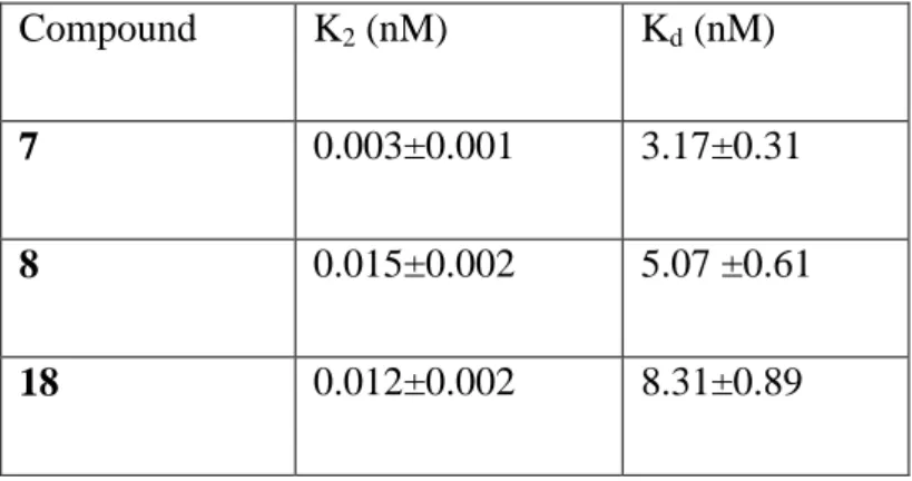
Kobs is the observed first-order rate constant for irreversible binding, and it is derived from the slope of the different straight lines shown in Figure 4. The plotting of 1/Kobs versus 1/[D] (where D is the ligand concentration) gave a straight line that intercepted the ordinate and abscissa axes at 1/K2 and -1/Kd, respectively. The slope of the line was the ratio Kd/K2 (Figure 5). K2 and Kd values of each compound are listed in Table 2. Interestingly, all the Kd values are in the nanomolar range (Kd from 3.17 to 8.31 nM), confirming an effective interaction with the receptor.
t
K
B
B
obs
0ln
2 21
1
1
K
D
K
K
K
d obs
a)
b)
c)
Figure 5. Calculation of K2 and Kd for ligand 7 (a), 8 (b) and 18 (c). On the basis of data
obtained by three different experiments carried out in duplicate, the values were calculated in accordance with equation 2. Data are shown as mean values ± SEM.
Compound K2 (nM) Kd (nM)
7 0.003±0.001 3.17±0.31
8 0.015±0.002 5.07 ±0.61
18 0.012±0.002 8.31±0.89
Table 2. Values of K2 and Kd. for the irreversible binding of compounds 7, 8, and 18 to TSPO.
Data, obtained from 3 independent kinetic assays, are shown as values ± SEM.
To estimate the ability of compound 18 to permeate cell membranes, a number of its physicochemical properties were calculated and reported in Table 3. Values obtained were consistent with an adequate distribution of 18 into the cell.
a
Calculated n-octanol/water partition coefficient. b Total Solvent Accessible Surface Area. c Polar Surface Area. d Number of atoms. e Number of rotatable bonds. f Molecular weight.
Table 3. Physicochemical predicted properties of compound 18.
The ability of the probe to specifically and irreversibly label TSPO was evaluated in live cells of human glioma, using fluorescence spectroscopy. For this purpose, U87MG glioma cells were incubated with different concentrations of 18 (100, 250 and 500 nM) after pre-treatment with buffer (control) or with 10 µM Ro 5-4864. Incubation was performed at 90 min, a time in which the irreversible bound is formed (see equations 2 and 3), and at a shorter time (20 min). As expected, at the shortest time
Conversely, in the experiments performed at the longest time, Ro 5-4864 showed to be unable to displace 18, demonstrating the irreversibility of the bond.
Figure 6. Human glioma cell staining with the irreversible fluorescent TSPO ligand 18. The cells were incubated with compound 18 for 20 min (A) and 90 min (B), in the absence or presence of 10 µM Ro 5-4864 (grey bars). Data were obtained from 3 independent assays and are shown as mean values of Fluorescence Intensity ± SEM.
Conclusion
In conclusion, we biologically characterized novel irreversible TSPO ligands featuring the 2-phenylindol-3-ylglyoxylamide scaffold. The binding of these compounds to the receptor was investigated in detail, characterizing both order rate and kinetic constants. Experimental data showed that all the new derivatives effectively bind the receptor by means of an irreversible covalent interaction. Fluorescence spectroscopy experiments performed on derivative 18, featuring an NBD fluorophore group, revealed its ability to specifically and irreversibly label TSPO in live U87MG glioma cells. All these novel irreversible TSPO ligands represent useful tools in the investigation of the physiological role of TSPO and in the detection of its expression levels.
In particular, the fluorescent probe 18 may be employed as diagnostic marker to evaluate the TSPO expression in peripheral cells, post-mortem and biopsy tissues, from patients affected by diseases in which the TSPO density is altered. Actually, irreversible probes offer the advantage of a lasting detection in visualization techniques that generally require multiple washes to remove the non-specific signal, steps in which reversible probes may be lost.
Complementary investigations using irreversible and reversible fluorescent TSPO ligands, in in vitro and in vivo studies, respectively, may offer a useful platform for the development of new diagnostic approaches in many CNS diseases such as neuroinflammation and neurodegeneration.
Chapter 3: Neurodegenerative diseases
Nrf2-ARE pathway: implications in Amyotrophic lateral sclerosis
Oxidative damage seems to play a major role in the pathogenesis of chronic motor neuron death in ALS based on the following lines of evidence: i) markers of oxidative damage of proteins, lipids, and DNA are elevated in brain and spinal cord in both mutant superoxide dismutase 1 (SOD1) ALS-mouse models (Gurney ME et al., 1994) and in human familial and sporadic ALS (Aguirre N et al., 2005, Beal MF et al. 1997, Ferrante RJ et al. 1997, Perluigi M et al., 2005), and ii) a large number of antioxidant compounds have been shown to be neuroprotective in ALS mouse models.
A major transcriptional modulator of the cellular antioxidant response is the transcription nuclear factor erythroid 2-related factor 2, NRf2. After activation and translocation to the nucleus, NRf2 binds to the ARE, a regulatory enhancer region within gene promoters, thus regulating the expression of more than 200 genes involved in the cellular antioxidant and anti-inflammatory defense. Among these are the classical phase 2 detoxification enzymes such as NAD(P)H quinone oxidoreductase and glutathione, enzymes that are necessary for glutathione biosynthesis, extracellular superoxide dismutase, glutamate-6-phosphatedehydrogenase, heat shock proteins, and ferritin (Figure 1). Furthermore, pro-inflammatory and anti-inflammatory enzymes such as cyclooxygenase 2, inducible nitric oxide synthase, and heme oxygenase-1 are also regulated in this manner (van Muiswinkel FL et al. 2005, Shih AY et al., 2005).
In addition to its role in inducing ARE-dependent gene transcription, Nrf2 also has direct cytoprotective effects via the inhibition of Fas-triggered apoptotic pathways (Kotlo KU et al., 2003).
Neuronal and astroglial cells from Nrf2 knockout mice are more vulnerable to oxidative stress than wild-type cells in vitro, and overexpression of Nrf2 increases resistance against oxidative and excitotoxic stimuli (Shih AY et al., 2005, Kraft AD et al., 2004, Lee JM et al., 2003).
The translocation of Nrf2 to the nucleus is inhibited by the actin-bound cytoskeletal zinc metalloprotein Kelch-like ECH-associated protein1 (Keap1), which interacts with Nrf2 and leads to its sequestration and subsequent degradation by the ubiquitin-proteasome system. Decreased Keap- Nrf2 binding (via oxidation of sulfhydryl groups or phosphorylation) results in intranuclear shuttling of Nrf2 and subsequent
transcription of ARE-driven genes (van Muiswinkel FL et al. 2005). Therefore, Keap1 and Nrf2 constitute a mechanism by which cells can sense damage caused by free oxygen radicals (Itoh K et al., 1999) (see Figure 2).
Recent evidence has emerged that Nrf2–ARE signaling may be dysregulated in mSOD1 models of ALS, and in the CNS of patients with ALS.
Multiple studies have shown that oxidative stress interacts with, and potentially exacerbates, other pathophysiological processes that contribute to motor neuron injury, including excitotoxicity, mitochondrial impairment, protein aggregation, ER stress, and alterations in signaling from astrocytes and microglia. Thus, effective alleviation of oxidative stress could potentially ameliorate multiple facets of the pathobiology of motor neuron degeneration. A meta-analysis of therapeutic interventions tested in mSOD1 mice up to 2007 concluded that antioxidant therapies were the most effective class of drug at improving survival (Benatar M, 2007). Antioxidants have not yet shown benefit in patients with ALS, although the reported trials have often been of suboptimal design (Orrell R et al., 2007).
On these bases, is evident the need for molecules able to activate the Nrf2-ARE pathway and exert an antioxidant activity in ALS therapy.
Figure. 1. Coordinated upregulation of cytoprotective genes occurs as a result of Nrf2 activation. Nrf2-dependent genes identified by microarray are listed in bold italics. These genes function together in the production and utilization of glutathione. Genes involved in cellular uptake of glutathione-constituent amino acids glycine and glutamate are increased. Additionally, the rate-limiting step of glutathione synthesis is the formation of the -glutamyl cysteine moiety by -glutamyl cysteine ligase. Transcription of both subunits of this enzyme is induced. Moreover, enzymes responsible for glutathione utilization, reduction, and conjugation are all represented in microarray data. Similarly, production and use of the common detoxification enzyme cofactor NADPH is stimulated. The pentose phosphate pathway is a common cellular mechanism whose two main functions are the production of reduction equivalents in the form of NADPH and the production of ribulose-5-phosphate for nucleotide and nucleic acid synthesis. In the case of Nrf2 activation, the pathway is focused on NADPH production, as the fructose-5-phosphate molecules are recycled back to glucose-6-phosphate by transaldolase and transketolase. NADPH also may be synthesized by malic enzyme oxidation of malate to pyruvate. NADPH is the major cofactor used in cellular reductions and is used especially frequently by detoxification enzymes like NQO1, P450s, and GSTs. Furthermore, Nrf2-dependent genes function together to detoxify superoxide, and meanwhile to prevent Fenton chemistry on the SOD-produced intermediate H2O2. This occurs by two methods. First, ferretin sequesters free iron by binding it in the ferric form. Second, catalase, thioredoxin, and peroxiredoxins reduce H2O2 to water. Thus, Nrf2-driven genes reduce superoxide to water and prevent production of hydroxyl radicals.
Figure 2. General scheme for the induction of gene expression through the Keap-Nrf2-ARE signaling pathway. Small molecules of endogenous and exogenous origin lead to activation of Nrf2-regulated genes. These agents disrupt the association of Nrf2 with Keap1,leading to diminished rates of proteolysis of Nrf2 and enhanced nuclear accumulation. Phosphorylation of Nrf2 by a series of kinases also affects its fate and distribution. Interaction of Nrf2 with other transcription factors and proteins of the transcriptional complexallows for transactivation of ARE-responsive genes. Induction of these genes, wich include prototypic coniugating and antioxidative genes, results in an adaptive response that enhances te resistance of cells to environmental stresses mediated electrophiles and free radicals.
Experimental section
Nrf2-ARE pathway as an attractive target for neuroprotection in
ALS: identification of small molecule activators of Nrf2-mediated
transcription in two libraries of drugs
Mounting evidence suggests that ALS is not simply an event that occurs within neurons, distinct from influence by surrounding cells. This concept may be best illustrated in the context of the familial form of ALS, in which several groups have found major astrocyte and microglial involvement in disease progression. Researchers studying this disease have found that expression of the mutated allele in astrocytes alone is sufficient to cause motor neuron degeneration in vitro (Nagai M et al., 2007, Vargas MR et al., 2006). Furthermore, knocking down expression of the mutated allele in astrocytes leads to diminished microglial activation and overall disease progression (Yamanaka K et al., 2008). The mechanism by which astrocyte dysfunction leads to neural degeneration in ALS was explored by Barbieto (Barbieto LH et al., 2004). Several potential routes exist by which reactive astrocytes may potentiate neurodegeneration, including downregulation of glutamate transporters, release of reactive nitrogen species, or active release of proapoptotic proteins such as Fas-ligand or NGF. Thus, targeting non-neuronal cells may be a valuable option for therapeutic intervention, not only to prevent toxicity mediated by glia, but also to stimulate glial protective responses. Interestingly this hypothesis has been extended and tested in cell culture by using the ALS model with Nrf2 as a protective agent. The therapeutic potential of Nrf2 was explored in a model of ALS in isolated astrocytes from mice overexpressing the gene coding for superoxide dismutase-1 that was mutated from glycine to alanine at position 93 (G93A-SOD). Wild-type motor neurons were plated on top of the astrocytes. It was previously found that this co-culture scheme leads to motor neuron apoptosis that is not observed when the astrocytes were derived from wild-type mice (Vargas MR et al., 2006). The investigators coupled this knowledge to the observation that astrocyte-induced apoptosis in similar systems could be mediated by NGF and nitric oxide (Pehar M et al., 2004) and hypothesized that decreasing reactive nitrogen species could rescue the motor neurons. Follow-up experiments showed that activation of Nrf2 in the G93A-SOD–containing astrocytes was able to prevent apoptotic signaling (Vargas MR et al., 2006).
Based on this data, the multifaceted response of Nrf2 makes it an attractive target for prevention of neurodegeneration in ALS, and its activation in astrocytes may rescue motor neurons from death.
For these reasons, during the period I spent at the SITraN in United Kingdom, I screened two libraries of small molecules on different reporter cell lines in order to identify activators of Nrf2-mediated transcription. The cell lines I used were CHO cells and C6 cells, both cell lines were efficiently used in a previous library screening conducted in the SITraN laboratories. The two libraries I screened were from two different commercial sources, Tocris and Prestwick libraries, and each of them was composed of almost 1000 molecules.
Most of the molecules from Prestwick are already used in human therapy and are employed in different pathologies, but their activity on Nrf2-ARE pathway is unknown. Moreover, many molecules in this library have known human safety data as they are that did not make it to clinic. This can be simply because they were no better than other drugs on the market rather than negative reasons.
Tocris library is designed to identify pathways, as it has inhibitors and activators of known pathways. As for molecules from Prestwick library, also the activity of Tocris molecules on Nrf2-ARE pathway is unknown.
Materials and Methods
Cell Culture
Chinese Hamster Ovary (CHO) and C6 (rat) astrocyte cell lines were routinely maintained in DMEM supplemented with 10% FCS and penicillin/streptomicyn. The ARE-TK-GFP and the ARE-TK-GFP reporter constructs were a kind gift of the laboratory of cancer research of the University of Wisconsin. The TK-EGFP reporter construct consists of a 123bp thymidine kinase promoter inserted in the multiple cloning site of pEGFP and the ARE-TK-EGFP also contains 4 repeats of a 41bp GST ARE motif 3’ to the TK promoter. These plasmids were transfected into CHO and C6 cell lines and after selection in 0.5 mg/ml G418 they were expanded and selected for basal expression using fluorescence activated cell sorting (BD, FACSAria). These mixed populations of stable transfectants with basal eGFP expression were used in subsequent assays and designated 4xARE-TK-GFP for the ARE containing line and TK-4xARE-TK-GFP for the control cell line.
Andrographolide EC 10, EC 50 and EC 90 determination
Andrographolide is a diterpen lactone and the principal active component in extracts of the herb Andrographis paniculata, which is widely used in Indian herbal medicine as an anti-infective, anti-inflammatory and hepatoprotective agent. From a previous library screening conducted in the SITraN laboratories, andrographolide was identified as an activator of the Nrf2-ARE pathway in CHO and C6 cells. For these reason andrographolide was used as positive control that represented our current best and its EC10, EC50 and EC90 on CHO and C6 transfected cell lines was determined.
ARE reporter assay-library screening validation
In order to screen the two libraries of 2000 small molecules the TK-GFP CHO ARE reporter cell line and the TK-GFP C6 ARE reporter cell line were subjected to a Z’ score assay in a 384 well plate using different methods of seeding and treating ("z' score determination based on "Assay Validation", Eli Lilly and Company and the National Institutes of Health Chemical Genomics Center). In brief, 15,000 cells/well were seeded in a 384 well plate and after 24 hours alternate wells were incubated with andrographolide at different concentrations corresponding to its EC90, EC50 and EC10 respectively. GFP fluorescence (ARE induction) was then measured at Ex485/Em530 using a fusion plate reader (Packard Bioscience). The Z’ score was calculated as follows:
Acceptable Z’ scores were >= 0.4
The Z’ score assay was performed for both the CHO and C6 transfected cell lines.
Library screening
For the library screening, CHO and C6 transfected cell lines were plated at a density of 15000 cells for each well in a 384 multiwell in normal DMEM containing 10% FBS on day -1 and on day 0, media was replaced and cells were incubated for 24 hours with drug (1 compound/well). The cell seeding was performed using the cell dispenser Matrix Control Mate, (Thermo Scientific). After the seeding, drugs were delivered to the assay using the Echo® Liquid Handling System (Labcyte Inc., USA). Echo® 550 was able to dispense the compounds directly from the 96 well of the library through the use of acoustic energy to transfer liquids. Sound waves ejected precisely-sized droplets from the source liquid into the 384 well plate suspended above the source. The Echo liquid handling platform used no tips, pin tools, or nozzles, so there was no contact between the instrument and the liquid. The Drop transfer volume for Echo® is 2.5 nL and the volume transfer range is 2.5 to 10,000 nL (source-plate specific), moreover its transfer accuracy is <10% of the deviation from target volume. Echo® 550 offered many advantages in respect to hand pipetting, indeed we could deliver tiny volumes, maximize the use of the library and limit contaminations of our cells.
GFP fluorescence (ARE induction, Ex 485nm/Em 530) were then measured. The TK- GFP CHO ARE cell line was tested twice in a single point assay at 10µM, and once at 500nM.
The control TK-GFP CHO cell line was screened once at 10µM and once at 500nM to eliminate false positives.
similarly in CHO an C6 cell lines stabily transfected with 4xARE-TK-GFP and TK-GFP constructs.
Results
Andrographolide EC 10, EC 50 and EC 90 determination
From a previous library screening conducted in the SITraN laboratories, andrographolide was identified as an activator of the Nrf2-ARE pathway in CHO and C6 cells. For this reason, in this study andrographolide was used as positive control that represented our current best and its EC10, EC50 and EC90 on CHO an C6 cell lines stably transfected with 4xARE-TK-GFP was determined.
As reported in Figure 1, andrographolide showed similar activity on both C6 and CHO cell lines (C6 EC50=1.8±0.2µM; CHO EC50=1.4±0.15µM).
-12 -10 -8 -6 -4 -2 0 5000 10000 15000 20000 25000 EC10= 0.31±0.03M EC50= 1.8±0.2M EC90= 10.5±1.0M EC10= 0.4±0.038M EC50= 1.2± 0.10M EC90= 3.5±0.4M C6 CHO log10[compound] G FP f lo u re s c e n c e ( R FU )
Figure 1. Data obtained by three different experiments carried out in duplicate. Data are shown as mean values ± SEM.
ARE reporter assay-library screening validation
The Z’ score assay was performed in order to determine working conditions of confidence and reproducibility necessary to perform the drug screening. Although we set the assay up with a known activator with a dose response already determined, using a single dose assay we had a good chance of identifying a positive response.
The drug screen logistically had to be done at a single dose, therefore it was unlikely that the hits would have been at optimal dose, so we simply needed to know at a dose that we could identify any activation, not simply good activation. Even more, we did not consider any difference between hits until we did the secondary assays of dose response, however big or little the response in the primary screen, they were simply above or below the threshold set by us.
The Z' score determination was run using different combined methods, as reported in table 1 and 2. The best Z' score determined for the CHO cells was equal to 0.773; the best Z' score determined for the C6 cells was 0.300. Both these Z' scores were obtained using a cell dispenser for the seeding and the Echo® Liquid Handling System for the treatments.
CHOcells Seeding Teatments Z score
1st By hand By hand 2,153
2nd Cell dispenser Plate mate 0,681
3rd Cell dispenser Echo 0,473
Table 1.
C6 cells Seeding Treatments Z score


![Figure 3. Displacement of [ 3 H]Ro 5-4864 by compound 7 (a), 8 (b), and 18 (c) in rat kidney mitochondria membranes](https://thumb-eu.123doks.com/thumbv2/123dokorg/7556201.109957/26.892.235.666.113.879/figure-displacement-ro-compound-rat-kidney-mitochondria-membranes.webp)