Matthew Traylor, PhD*
Cathy R. Zhang, MA*
Poneh Adib-Samii, MBBS
William J. Devan, BS
Owen E. Parsons, MSc
Silvia Lanfranconi, MD
Sarah Gregory, PhD
Lisa Cloonan, BA
Guido J. Falcone, MD
Farid Radmanesh, MD
Kaitlin Fitzpatrick, BSc
Allison Kanakis, MD
Thomas R. Barrick, PhD
Barry Moynihan, MD
Cathryn M. Lewis, PhD
Giorgio B. Boncoraglio,
MD
Robin Lemmens, MD
Vincent Thijs, MD
Cathie Sudlow, MD, PhD
Joanna Wardlaw,
FMedSci
Peter M. Rothwell,
FMedSci
James F. Meschia, MD
Bradford B. Worrall, MD
Christopher Levi, MD
Steve Bevan, PhD
Karen L. Furie, MD
Martin Dichgans, MD
Jonathan Rosand, MD
Hugh S. Markus, DM
*
Natalia Rost, MD
*
On behalf of the
International Stroke
Genetics Consortium
Correspondence to Dr. Traylor: [email protected] Supplemental data at Neurology.orgGenome-wide meta-analysis of cerebral
white matter hyperintensities in patients
with stroke
ABSTRACT
Objective:
For 3,670 stroke patients from the United Kingdom, United States, Australia, Belgium, and
Italy, we performed a genome-wide meta-analysis of white matter hyperintensity volumes (WMHV) on
data imputed to the 1000 Genomes reference dataset to provide insights into disease mechanisms.
Methods:
We first sought to identify genetic associations with white matter hyperintensities in a
stroke population, and then examined whether genetic loci previously linked to WMHV in community
populations are also associated in stroke patients. Having established that genetic associations are
shared between the 2 populations, we performed a meta-analysis testing which associations with
WMHV in stroke-free populations are associated overall when combined with stroke populations.
Results:
There were no associations at genome-wide significance with WMHV in stroke patients. All
previously reported genome-wide significant associations with WMHV in community populations shared
direction of effect in stroke patients. In a meta-analysis of the genome-wide significant and suggestive
loci (p , 5 3 10
26) from community populations (15 single nucleotide polymorphisms in total) and from
stroke patients, 6 independent loci were associated with WMHV in both populations. Four of these are
novel associations at the genome-wide level (rs72934505 [NBEAL1], p 5 2.2 3 10
28; rs941898
[EVL], p 5 4.0 3 10
28; rs962888 [C1QL1], p 5 1.1 3 10
28; rs9515201 [COL4A2], p 5 6.9 3 10
29).
Conclusions:
Genetic associations with WMHV are shared in otherwise healthy individuals and
pa-tients with stroke, indicating common genetic susceptibility in cerebral small vessel disease.
Neurology®2016;86:146–153 GLOSSARY
FLAIR5 fluid-attenuated inversion recovery; ROI 5 region of interest; SNP 5 single nucleotide polymorphism; TICV 5 total intracranial volume; WMH5 white matter hyperintensities; WMHV 5 white matter hyperintensity volume; WTCCC2 5 Well-come Trust Case Control Consortium–2.
White matter hyperintensities (WMH) on T2-weighted MRI are associated with increasing age
and cardiovascular risk factors, particularly hypertension, and are predictive of both stroke and
dementia in prospective community populations.
1Severe confluent WMH are often found in
patients presenting with stroke, and are more common in patients with the small vessel stroke
subtype.
2Furthermore, in these patients, WMH burden is linked to poor clinical outcomes after
stroke.
3,4Understanding disease mechanisms that contribute to WMH could lead to advances in
prevention, treatment, and rehabilitation of disability related to vascular cognitive impairment,
age-related functional decline, and stroke.
Twin and family history studies suggest a significant genetic component to WMH.
Herita-bility estimates range from 55% to 80%,
5–8suggesting that a moderate to large proportion of the
disease risk can be attributed to genetic effects. The heritability attributed to common single
nucleotide polymorphisms (SNPs) has been estimated to be between 13% and 45%.
9Previous
genome-wide analyses have focused on the genetic influence on WMH in community
popu-lations,
10,11and a recent meta-analysis identified 8 regions associated with the disease.
12One
*These authors contributed equally to this work.Authors’ affiliations are listed at the end of the article.
Coinvestigators are listed on the Neurology®Web site at Neurology.org.
Go to Neurology.org for full disclosures. Funding information and disclosures deemed relevant by the authors, if any, are provided at the end of the article. The Article Processing Charge was paid by Wellcome Trust.
This is an open access article distributed under the terms of the Creative Commons Attribution License 4.0 (CC BY), which permits unrestricted use, distribution, and reproduction in any medium, provided the original work is properly cited.
might expect genetic risk factors for WMH in
community populations to be similar to those
that confer increased risk of WMH in stroke
patients. However, the underlying pathology
of WMH is heterogeneous, with small
punc-tate lesions being associated with mixed
causes, whereas more confluent areas often
seen in stroke patients correspond primarily
to small vessel disease.
13Therefore, it is
unclear whether the lesions underlying
WMH in the general population are
patholog-ically distinct from the confluent lesions
fre-quently observed in patients with stroke.
In this analysis, we investigated the role of
the genetic contribution to WMH volumes
(WMHV) in patients with ischemic stroke.
We initially performed a genome-wide
meta-analysis of WMHV in stroke patients with
the aim of identifying novel associations.
Sec-ond, we determined whether similar genetic
factors contributed to WMHV in community
populations and stroke patients. Finally,
hav-ing established shared genetic factors in the 2
datasets, we performed a meta-analysis of the
published associations from community
popu-lations with our dataset to identify genetic
as-sociations that are in common in the 2
populations.
METHODS Study populations.Ischemic stroke populations were enrolled through hospital-based studies between 1995 and 2013. Characteristics of the study populations are given in table 1; full details are given in the supplementary material on the Neurology®Web site at Neurology.org. Patients with cerebral
autosomal dominant arteriopathy with subcortical infarcts and leukoencephalopathy or any other suspected monogenic cause of stroke, vasculitis, or any other nonischemic cause of WMH including demyelinating and mitochondrial disorders were excluded from analyses.
Standard protocol approvals, registrations, and patient consents.An institutional review board or regional review board approved the use of human subjects in each of the study popula-tions. All patients gave informed consent.
Neuroimaging analysis.MRI scans were acquired as part of routine clinical practice for evaluation of ischemic stroke (table e-1). Fluid-attenuated inversion recovery (FLAIR) sequences were primarily used for WMH volumetric analysis; however, in their absence, T2-weighted sequences were used (Wellcome Trust Case Control Consortium–2 [WTCCC2], Oxford, and WTCCC2, Munich, only). In all scans, to avoid confounding by hyperintense signal due to acute stroke, WMHV was assessed quantitatively in the hemisphere contralateral to the acute infarction. Chronic lacunar infarcts were identified using standard criteria as low signal on T1 or FLAIR images and were excluded from WMHV estimates.14Trained raters blinded
to all patient information analyzed anonymized MRI scans. All supratentorial white matter and deep gray matter lesions were included in WMHV with the exception of WMH corresponding to infarcts, both lacunar and territorial.2MRIs
with excessive movement artefact, incomplete brain coverage, or bihemispheric infarcts (other than lacunar) were excluded.
To account for interindividual variability in head size, an esti-mate of total intracranial volume (TICV) was derived using site-specific volumetric methodology, as follows. MRIs from the Massachusetts General Hospital, Ischemic Stroke Genetics Study, and Australian Stroke Genetics Collaborative studies were analyzed in Boston. Scans from the Siblings with Ischaemic Stroke Study were analyzed in the same way at the University of Virginia by the Boston-trained rater. FLAIR sequences were analyzed using an MRIcro semiautomated method as previously described.2Using
operator-mediated quality assurances, overlapping regions of inter-est (ROIs) corresponding to WMH produced the final maps for WMHV calculation. Intracranial area was derived as a validated marker of TICV as the average of 2 midsagittal slices traced using anatomical landmarks on T1 sequences.15
The WTCCC2, GENESIS, SGUL, Leuven, and Milan co-horts were analyzed in London using DISPunc semiautomated lesion drawing software.16A seed at the lesion border was first
marked manually, and then outlined automatically based on the signal intensity gradient. Each WMH ROI was visually inspected and manually corrected as required. To estimate TICV, T2-weighted and, in their absence, FLAIR sequences were analyzed using an automated segmentation program, SIENAX,17which
calculates the total volume of CSF and gray and white matter volumes.
WMHV quantification agreement across the 2 main rating centers was performed for 50 randomly selected scans; agreement was very good (intraclass correlation coefficient 0.95, confidence interval 0.91–0.97, n 5 50).
Phenotype definition.To calculate the phenotype used in the genetic analysis, WMHV were doubled to obtain a whole brain estimate. This volume was then multiplied by the ratio of TICV (or intracranial area) to the mean TICV (or intracranial area) for the study, thereby correcting for natural differences in head size. The values were natural log transformed and the resulting ln (WMHV) values were entered into a linear regression model including age, sex, and the first 2 ancestry-informative principal components. To ensure the phenotype was normally distributed, the residuals from the model were then z-transformed and used as the WMHV phenotype in the genetic analysis.
Genome-wide genotyping and imputation.Genotyping of all cohorts was performed on commercially available arrays from Affymetrix (Santa Clara, CA) or Illumina (San Diego, CA) (table e-2). All cohorts performed extensive quality control steps prior to imputation, removing SNPs showing significant departure from Hardy-Weinberg equilibrium, high levels of missingness, or low minor allele frequency. Individuals were removed who did not segregate with Hapmap II European populations based on ancestry informative principal component analysis using EIGENSTRAT or multidimensional scaling in PLINK.18,19
Additionally, individuals showing cryptic relatedness or having high levels of missingness or heterozygosity were excluded. All datasets were imputed to 1000 Genomes integrated variant set (March 2012) using IMPUTE v2.20
Genome-wide association analysis of WMHV in stroke patients.To discover novel associations between WMHV and each autosomal SNP, we performed linear regression of WMHV
on genotype dosages using PLINK v1.07.19SNPs with PLINK
INFO,0.7 or MAF ,0.01 were removed from further analyses. We used genomic inflation to evaluate inflation of test statistics in each study group.21Results across all study groups were combined
using a fixed-effects inverse variance weighted method using METAL.22To control for any excess signal that might result
from study-wise inflation of p values, we performed genomic control correction, multiplying the standard errors from each study by the square root of the genomic inflation factor.21
Heterogeneity was assessed using Cochran q statistic. Following the meta-analysis, we considered only SNPs present in more than 12 study groups, and with heterogeneity p. 0.001, for analysis. We set the significance threshold to p, 5 3 1028. We used
l1000to evaluate inflation at the meta-analysis level.23We had
80% power to detect a variant explaining 1.1% of the trait variance (figure e-1).
Analysis of SNPs associated with WMH in community-based populations.To determine whether SNPs contributing to WMHV in community populations were associated with WMHV in stroke patients, we evaluated each SNP reported as being associated with WMH in healthy adults in a recent publi-cation,12testing if the SNP was associated with WMHV in
ische-mic stroke patients. All 17,936 individuals in the previous study were stroke-free and nonoverlapping with the samples studied here. We performed this analysis first for all genome-wide associated loci from the publication, and second for loci reported at p, 1 31025in European populations or overall.
We set a significance threshold at p 5 0.0033, Bonferroni correcting for the 15 SNPs analyzed. We had 80% power to detect any associations that explain 0.4% of the trait variance. In addition, we tested whether there was evidence overall that genetic susceptibility factors were shared between the 2 populations. We used a binomial test to evaluate whether an excess of the 8 genome-wide significant SNPs shared direction of effect in community populations and stroke patients, and then extended this to the 15 genome-wide significant loci and loci reported at p, 1 3 1025in European populations or overall.
Meta-analysis of stroke samples and published population-based samples. Having established that genetic factors were shared between community populations and stroke patients, we evaluated the overall evidence that each of the 15 previously re-ported SNPs (8 genome-wide significant, 7 suggestive) were associated with WMHV in both populations. We combined p values from the 2 sources using Stouffer z-score weighted method with equal weights, classifying SNPs with p, 0.05 in both populations and reaching p , 5 3 1028 overall as
significantly associated with WMH in both populations. We were not able to perform the reciprocal analysis, testing if suggestive associations with WMH in stroke patients were associated with WMH in stroke-free individuals, due to restrictions on access to the required summary level data. We then evaluated novel genome-wide associations in available databases to test for evidence that affects regulation of genes (RegulomeDB)24or directly affects gene expression (GTEx).25
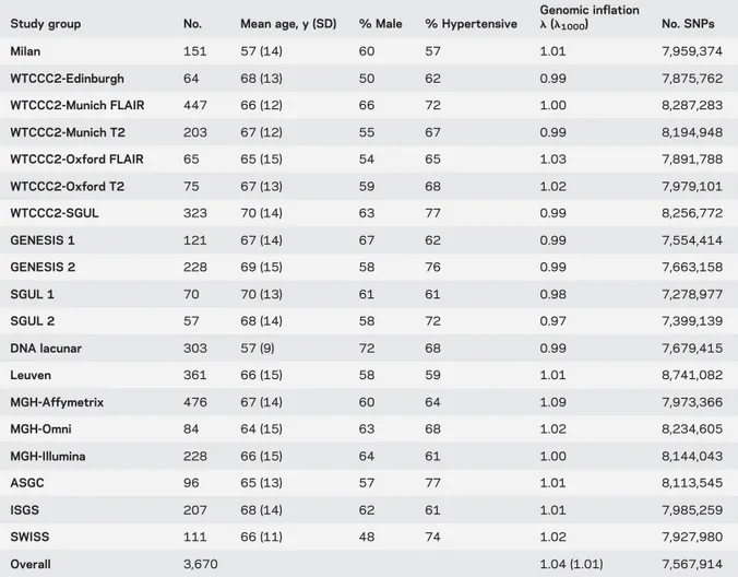
Table 1 Cohort characteristics
Study group No. Mean age, y (SD) % Male % Hypertensive
Genomic inflation l (l1000) No. SNPs Milan 151 57 (14) 60 57 1.01 7,959,374 WTCCC2-Edinburgh 64 68 (13) 50 62 0.99 7,875,762 WTCCC2-Munich FLAIR 447 66 (12) 66 72 1.00 8,287,283 WTCCC2-Munich T2 203 67 (12) 55 67 0.99 8,194,948 WTCCC2-Oxford FLAIR 65 65 (15) 54 65 1.03 7,891,788 WTCCC2-Oxford T2 75 67 (13) 59 68 1.02 7,979,101 WTCCC2-SGUL 323 70 (14) 63 77 0.99 8,256,772 GENESIS 1 121 67 (14) 67 62 0.99 7,554,414 GENESIS 2 228 69 (15) 58 76 0.99 7,663,158 SGUL 1 70 70 (13) 61 61 0.98 7,278,977 SGUL 2 57 68 (14) 58 72 0.97 7,399,139 DNA lacunar 303 57 (9) 72 68 0.99 7,679,415 Leuven 361 66 (15) 58 59 1.01 8,741,082 MGH-Affymetrix 476 67 (14) 60 64 1.09 7,973,366 MGH-Omni 84 64 (15) 63 68 1.02 8,234,605 MGH-Illumina 228 66 (15) 64 61 1.00 8,144,043 ASGC 96 65 (13) 57 77 1.01 8,113,545 ISGS 207 68 (14) 62 61 1.01 7,985,259 SWISS 111 66 (11) 48 74 1.02 7,927,980 Overall 3,670 1.04 (1.01) 7,567,914
Abbreviations:l 5 genomic inflation level; ASGC 5 Australian Stroke Genetics Collaborative; FLAIR 5 fluid-attenuated inversion recovery; ISGS 5 Ischemic Stroke Genetics Study; MGH 5 Massachusetts General Hospital; SGUL 5 St. George’s University of London; SNP 5 single nucleotide polymorphism; SWISS 5 Siblings With Ischaemic Stroke Study; WTCCC25 Wellcome Trust Case Control Consortium–2.
RESULTS Study populations.
Clinical characteristics
of all participating cohorts are given in table 1. In
total, 3,670 individuals of European ancestry were
included in the 19 study groups.
Genome-wide association analysis of WMHV in stroke patients.
With the exception of one study group,
genomic inflation was well-controlled (
l # 1.03,
table 1). Following quality control procedures,
7,567,914 autosomal SNPs remained for analysis.
Genomic inflation was well-controlled at the
meta-analysis level (
l 5 1.04, l
10005 1.01; figure e-2).
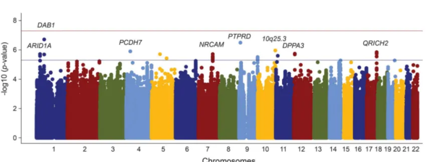
No SNP reached the significance level (figure 1),
although a number of loci reached p
, 5 3 10
26.
These are detailed in table e-3, and regional plots of
these loci are provided in figure e-3. All odds ratios
reported are per 1 SD change in normally distributed
WMHV after accounting for age, sex, and
ancestry-informative principal components.
Analysis of SNPs associated with WMH in community-based populations.
Eight independent SNPs have been
associated with WMH in community populations.
12We evaluated each of these in our dataset of stroke
patients. The direction of effect of all 8 associations
was consistent with the direction in our study. This
alone is unlikely to be due to chance (p
5 7.8 3 10
23from binomial test). For specific SNPs, no
genome-wide associations from community populations
reached our significance threshold, although all had
p
# 0.24 for association with WMH in stroke
patients, and 3 loci reached a nominal significance
level (p
, 0.05) in stroke patients (rs7214628
[TRIM65], p
5 0.015; rs78857879 [EFEMP1], p 5
0.0056; rs2984613 [PMF1-BGLAP], p
5 0.017).
Additionally, 10 loci were reported as suggestively
significant in the same recent publication,
12with
p
, 1 3 10
25in Europeans or overall. Three of these
were rare (MAF
# 0.02), and were not imputed with
enough accuracy to be analyzed in our dataset
(rs186314186, rs150695384, rs117126031). We
evaluated each of the 7 remaining associations in
our population. Of these, 4 passed our significance
threshold (table 2). One locus was nonsignificant and
in the opposite direction in our study (rs2883428,
p
5 0.17). In total, 14 of the 15 genome-wide and
suggestively significant loci shared direction between
community individuals and stroke patients (p
5
9.8
3 10
24from binomial test).
In addition, we searched for other publications
describing associations with any of the SNPs or genes
studied using the following search terms in PubMed:
(SNP or gene) and (white matter or leukoaraiosis
or small vessel disease). No relevant publications were
identified.
Meta-analysis of stroke samples and published population-based samples.
When combining our results in stroke
patients with the 15 previously reported associations
using Stouffer z-score meta-analysis, 6 associations
reached genome-wide significance overall and had
p
, 0.05 in both studies (table 2). Four of these are
novel associations
at genome-wide significance
(rs72934505 [NBEAL1], p
5 2.2 3 10
28; rs941898
[EVL], p
5 4.0 3 10
28; rs962888 [C1QL1], p
5 1.1
3 10
28; rs9515201 [COL4A2], p
5 6.9 3 10
29), all
of which showed good consistency across the 19
cohorts (figure e-4). The same 6 associations reached
genome-wide significance using an alternative
meta-analysis approach (Fisher method). The association
with COL4A2 (rs9515201) is in strong LD (r
2.
0.8) with SNPs previously reported to be associated
with cerebral small vessel disease, and is therefore
likely to represent the same locus.
26Figure 1 Association of genome-wide single nucleotide polymorphisms with white matter hyperintensity volume in ischemic stroke patients by genomic position
For each of these 4 novel associations, we queried
the RegulomeDB database and GTEx portal for
evi-dence that the SNPs affect DNA binding or
expres-sion of any mRNA molecule (figure e-5).
24,25rs962888 lies 25 Kb downstream from C1QL1;
how-ever, interrogation of GTEx portal showed that the
common allele (G, risk allele) of the SNP decreases
expression of elongation factor tu GTP binding
domain containing 2 (EFTUD2) in tibial arteries,
100 kb away (p
5 5.3 3 10
26). Data from
Regulo-meDB support this observation, as the SNP overlies
DNase-seq, FAIRE-seq, and CHIP-seq peaks in
numerous tissues from ENCODE.
27Similarly, the
common allele (T, risk allele) of rs72934505 increases
expression of the nearby gene NBEAL1 in tibial
arter-ies in GTEx (p
5 2.5 3 10
211), and also decreases
expression of islet cell autoantigen 1.69 kDa-Like
(ICA1L) in the thyroid (p
5 6.6 3 10
26), 200 kb
away. No significant eQTLs were identified for
rs941898 or rs9515201, but both overlap numerous
CHIP-seq and DNAse-seq peaks from ENCODE,
indicating they may have a regulatory function.
DISCUSSION
We report the first phase of a
collabo-rative genome-wide meta-analysis of WMHV in
stroke patients. We did not identify any associations
with WMHV in ischemic stroke patients at the
genome-wide significance level. The most likely
explanation for this is lack of power. We had 80%
power to identify a variant explaining 1.1% of the
trait variance (supplementary material), suggesting
that it is unlikely that any common variants explain
more than this proportion of the variance of WMH
in stroke patients. However, we cannot rule out the
existence of rare variants conferring a considerable
proportion of disease risk.
We found strong evidence that many of the same
genome-wide associations with WMHV in healthy
individuals influence WMHV in stroke patients. All
genome-wide significant associations with WMHV
shared direction of effect in our study and 3 reached
a nominal significance threshold. More convincing is
that of the 7 suggestive associations reported with
WMH in healthy individuals, 4 were significantly
associated with WMH in stroke patients. A
meta-analysis of these SNPs in 21,606 subjects suggests
that 4 of these loci are linked to WMH in community
populations and stroke patients at genome-wide
sig-nificance. Two of these associations influence
expression of nearby gene products (NBEAL1/ICA1L
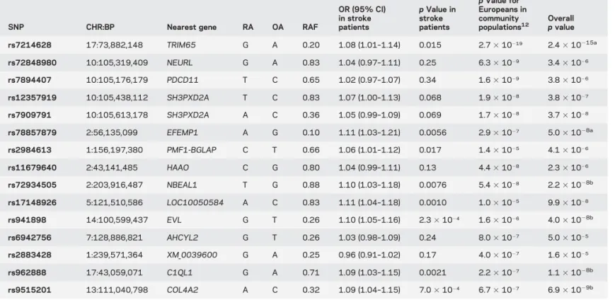
Table 2 Association of WMH-associated SNPs from community populations in stroke patients
SNP CHR:BP Nearest gene RA OA RAF
OR (95% CI) in stroke patients p Value in stroke patients p Value for Europeans in community populations12 Overall p value rs7214628 17:73,882,148 TRIM65 G A 0.20 1.08 (1.01–1.14) 0.015 2.73 10219 2.43 10215a rs72848980 10:105,319,409 NEURL G A 0.83 1.04 (0.97–1.11) 0.25 6.33 1029 3.43 1026 rs7894407 10:105,176,179 PDCD11 T C 0.65 1.02 (0.97–1.07) 0.34 1.63 1029 3.83 1026 rs12357919 10:105,438,112 SH3PXD2A T C 0.83 1.07 (1.00–1.13) 0.068 1.93 1028 3.83 1027 rs7909791 10:105,613,178 SH3PXD2A A C 0.36 1.05 (0.99–1.09) 0.069 1.73 1028 3.73 1028 rs78857879 2:56,135,099 EFEMP1 A G 0.10 1.11 (1.03–1.21) 0.0056 2.93 1027 5.03 1028a rs2984613 1:156,197,380 PMF1-BGLAP C T 0.66 1.06 (1.01–1.12) 0.017 1.43 1025 4.13 1026 rs11679640 2:43,141,485 HAAO C G 0.80 1.04 (0.99–1.11) 0.13 4.43 1028 2.33 1026 rs72934505 2:203,916,487 NBEAL1 T G 0.88 1.10 (1.03–1.18) 0.0076 5.43 1028 2.23 1028b rs17148926 5:121,510,586 LOC10050584 A C 0.83 1.11 (1.04–1.18) 0.0010 1.03 1025 9.93 1028 rs941898 14:100,599,437 EVL G T 0.26 1.10 (1.05–1.16) 2.33 1024 1.63 1026 4.03 1028b rs6942756 7:128,886,821 AHCYL2 G T 0.26 1.03 (0.98–1.09) 0.24 8.03 1027 5.03 1025 rs2883428 1:239,571,364 XM_0039600 G A 0.25 0.96 (0.91–1.02) 0.17 4.03 1027 1.63 1025 rs962888 17:43,059,071 C1QL1 G A 0.71 1.09 (1.03–1.15) 0.0021 2.23 1027 1.13 1028b rs9515201 13:111,040,798 COL4A2 A C 0.32 1.09 (1.04–1.15) 7.03 1024 6.73 1027 6.93 1029b Abbreviations: BP5 base position (hg19); CHR 5 chromosome; CI 5 confidence interval; OA 5 other allele; OR 5 odds ratio; RA 5 reference allele; RAF 5 reference allele frequency; SNP5 single nucleotide polymorphism; WMH 5 white matter hyperintensities.
The top 8 SNPs are genome-wide significant in community populations overall (including all ancestries) in a previous publication,12while the bottom 7 are suggestively significant in community populations. Thep values reported here are for Caucasian populations only. The reported reference allele is the effect allele in community populations.
aAssociated at
p , 5 3 1028overall and withp , 0.05 in both populations.
bNovel association at genome-wide level. The overall
p value gives the results of Fisher meta-analysis of the p values from community populations and stroke patients.
[rs72934505] and EFTUD2 [rs962888]). A
genome-wide significant association with rs9515201, located
in an intron of COL4A2, which encodes collagen 4
subunit 2, was also identified. This association is
par-ticularly interesting as rare mutations in COL4A2 and
the closely related COL4A1 protein lead to small
ves-sel disease and hemorrhagic stroke,
28–30and common
variants in close LD with this SNP (r
2. 0.8) have
been linked to sporadic small vessel disease.
26The observation that genetic risk factors for
WMH in community populations also influence
WMH in stroke patients has implications. It suggests
that the white matter changes seen on the brain MRI
scans of otherwise healthy elderly reflect a similar
dis-ease process as the more severe forms that underlie
cerebral small vessel disease in patients with stroke.
Previous studies have indicated heterogeneity in
WMH pathology: our results do not preclude this
possibility, but suggest that many of the same genetic
factors contribute to both pathologies.
Our study has several strengths. Protocols were
uniformly employed across analyses, including
imputation to the same reference build across all
study groups, using the same software. Similarly,
analyses were performed using the same software
on the same phenotype, derived in the same way.
We performed volumetric analysis of all MRI scans
to quantify WMHV, which has strengths over rating
scales, which are known to have ceiling effects.
14Inter-rater agreement between the 2 coordinating
centers was shown to be good. WMHV was
quan-tified using semiautomated volumetric protocols
val-idated for use in patients with stroke and clinical
grade MRI scans.
Our study also has limitations. Large-scale
collabo-rative GWAS such as that undertaken here necessarily
combine studies with some degree of phenotypic
vari-ability. Differences in environmental exposures,
possi-bly resulting in epigenetic modifications, may
contribute to such variability, which could alter the
results. We identified 4 novel associations at
genome-wide significance when combined with previous
publi-cations. However, we have not provided replication of
these findings and therefore further evidence will be
necessary to verify these associations with WMHV.
MRI used in the analyses were drawn from a number
of centers, with varying image quality. Therefore,
to minimize bias arising from differing image quality,
we quantified WMHV per study group and
meta-analyzed the results. This approach may limit our
abil-ity to detect associations with low frequency variants
due to small sample sizes in some study groups. The
majority of MRI scans used were from FLAIR
sequen-ces. However, where these were unavailable, we used
T2-weighted images, which are less sensitive to white
matter changes. Such differences in sensitivity may
affect quantification of WMHV across study groups,
although future studies that involve centralized
volu-metric MRI analysis pipelines, such as those currently
in development, may account for this variability. In
this analysis, we considered all subtypes of stroke
together as we were underpowered to investigate
subtype-specific influences on WMH. It is possible
that causes of WMH may differ by stroke subtype,
but larger studies with sufficient power will be required
before this issue can be addressed adequately. Similarly,
it has been hypothesized that periventricular and deep
WMH might have distinct underlying
pathophysiol-ogy. In this analysis, we considered total WMHV,
rather than treating these regions separately; our lesion
volume analysis did not differentiate into these 2
re-gions. Further analyses should address this area.
We have shown that the age-related white matter
changes seen in otherwise healthy populations share
genetic susceptibility with the extensive lesions that
underlie cerebral small vessel disease. We report 6
independent loci that are associated with WMHV
in healthy individuals as well as stroke patients, 4 of
which are novel associations at the genome-wide level.
Our results suggest that a full genome-wide
meta-analysis of available cohorts of WMH in ischemic
stroke patients and community populations is likely
to uncover further associations.
AUTHOR AFFILIATIONS
From the Department of Clinical Neurosciences (M.T., H.S.M.) and the Autism Research Centre (O.E.P.), University of Cambridge, UK; the J. Philip Kistler Stroke Research Center (C.R.Z., W.J.D., L.C., G.J.F., K.F., A.K., N.R.) and the Center for Human Genetic Research (W.J.D., G.J.F., F.R., J.R., N.R.), Department of Neurology, Massachu-setts General Hospital, Boston; the Neurosciences Research Centre (P.A.-S., S.G., T.R.B., B.M.), St George’s, University of London, UK; the Neurology Unit (S.L., J.R.), IRCCS Ca’ Granda Ospedale Maggiore Policlinico, Milan, Italy; Program in Medical and Population Genetics (F.R.), Broad Institute, Cambridge, MA; the Department of Medical & Molecular Genetics (C.M.L.) and the Social, Genetic and Developmental Psychiatry Centre, Institute of Psychiatry (C.M.L.), King’s College Lon-don, UK; the Department of Cerebrovascular Diseases (G.B.B.), Fonda-zione IRCCS Istituto Neurologico “Carlo Besta,” Milan, Italy; KU Leuven, University of Leuven, the Department of Neurosciences, Exper-imental Neurology and Leuven Research Institute for Neuroscience and Disease (R.L., V.T.); University Hospitals Leuven, the Department of Neurology (R.L., V.T.); VIB, Vesalius Research Center, Laboratory of Neurobiology (R.L., V.T.), Leuven, Belgium; Austin Health and Mel-bourne Brain Center (V.T.), Florey Institute of Neuroscience and Mental Health, University of Melbourne, Heidelberg, Victoria, Australia; the Division of Clinical Neurosciences, Neuroimaging Sciences and Institute of Genetics and Molecular Medicine (C.S.), and Neuroimaging Sciences, Centre for Clinical Brain Sciences (J.W.), University of Edinburgh, UK; the Stroke Prevention Research Unit (P.M.R.), Nuffield Department of Neuroscience, University of Oxford, UK; the Department of Neurology (J.F.M.), Mayo Clinic, Jacksonville, FL; the Departments of Neurology and Public Health Sciences (B.B.W.), University of Virginia, Charlottes-ville; the Centre for Clinical Epidemiology and Biostatistics (C.L.), Hunter Medical Research Institute and School of Medicine and Public Health, University of Newcastle, NSW, Australia; School of Life Science (S.B.), University of Lincoln, Lincoln, UK; the Department of Neurology (K.L.F.), Rhode Island Hospital, Alpert Medical School of Brown University, Providence; the Institute for Stroke and Dementia
Research (M.D.), Klinikum der Universität München, Ludwig-Maximilians-University Munich; and the Munich Cluster for Systems Neurology (SyNergy) (M.D.), Munich, Germany.
AUTHOR CONTRIBUTIONS
Matthew Traylor: drafting/revising the manuscript for content, study concept or design, analysis or interpretation of data, statistical analysis, ac-cepts responsibility for conduct of research and final approval. Cathy Zhang: drafting/revising the manuscript for content, study concept or design, analysis or interpretation of data, statistical analysis, accepts respon-sibility for conduct of research and final approval. Poneh Adib-Samii: drafting/revising the manuscript for content, study concept or design, anal-ysis or interpretation of data, statistical analanal-ysis, accepts responsibility for conduct of research and final approval. William Devan: drafting/revising the manuscript for content, study concept or design, analysis or interpre-tation of data, statistical analysis, accepts responsibility for conduct of research and final approval. Owen Parsons: drafting/revising the manu-script for content, analysis or interpretation of data, accepts responsibility for conduct of research and final approval. Silvia Lanfranconi: drafting/ revising the manuscript for content, analysis or interpretation of data, ac-cepts responsibility for conduct of research and final approval. Sarah Greg-ory: drafting/revising the manuscript for content, analysis or interpretation of data, accepts responsibility for conduct of research and final approval. Lisa Cloonan: drafting/revising the manuscript for content, analysis or interpretation of data, accepts responsibility for conduct of research and final approval. Guido Falcone: drafting/revising the manuscript for con-tent, accepts responsibility for conduct of research and final approval. Farid Radmanesh: drafting/revising the manuscript for content, accepts respon-sibility for conduct of research and final approval. Kaitlin Fitzpatrick: drafting/revising the manuscript for content, accepts responsibility for con-duct of research and final approval. Alison Kanakis: drafting/revising the manuscript for content, accepts responsibility for conduct of research and final approval. Tom Barrick: drafting/revising the manuscript for con-tent, accepts responsibility for conduct of research and final approval. Barry Moynihan: drafting/revising the manuscript for content, acquisition of data, accepts responsibility for conduct of research and final approval. Cathryn Lewis: drafting/revising the manuscript for content, analysis or interpretation of data, accepts responsibility for conduct of research and final approval. Giorgio Boncoraglio: drafting/revising the manuscript for content, acquisition of data, accepts responsibility for conduct of research and final approval. Robin Lemmens: drafting/revising the manuscript for content, acquisition of data, accepts responsibility for conduct of research and final approval. Vincent Thijs: drafting/revising the manuscript for con-tent, acquisition of data, accepts responsibility for conduct of research and final approval. Cathie Sudlow: drafting/revising the manuscript for con-tent, acquisition of data, accepts responsibility for conduct of research and final approval. Joanna Wardlaw: drafting/revising the manuscript for content, acquisition of data, accepts responsibility for conduct of research and final approval. Peter Rothwell: drafting/revising the manuscript for content, acquisition of data, accepts responsibility for conduct of research and final approval. James Meschia: drafting/revising the manuscript for content, acquisition of data, accepts responsibility for conduct of research and final approval. Bradford Worall: drafting/revising the manuscript for content, acquisition of data, accepts responsibility for conduct of research and final approval. Chris Levi: drafting/revising the manuscript for con-tent, acquisition of data, accepts responsibility for conduct of research and final approval. Karen Furie: drafting/revising the manuscript for con-tent, acquisition of data, accepts responsibility for conduct of research and final approval. Steve Bevan: drafting/revising the manuscript for content, analysis or interpretation of data, accepts responsibility for conduct of research and final approval. Martin Dichgans: drafting/revising the manu-script for content, acquisition of data, accepts responsibility for conduct of research and final approval. Jonathan Rosand: drafting/revising the manu-script for content, study concept or design, acquisition of data, analysis or interpretation of data, study supervision or coordination, accepts responsi-bility for conduct of research and final approval. Hugh Markus: drafting/ revising the manuscript for content, study concept or design, acquisition of data, analysis or interpretation of data, study supervision or coordination, accepts responsibility for conduct of research and final approval. Natalia Rost: drafting/revising the manuscript for content, study concept or design, acquisition of data, analysis or interpretation of data, study
supervision or coordination, accepts responsibility for conduct of research and final approval.
ACKNOWLEDGMENT
The authors thank Kristiina Rannikmae for comments on the manuscript; and Wellcome Trust Case Control Consortium 2.
STUDY FUNDING
Funding for collection, genotyping, and analysis of stroke samples was provided by Wellcome Trust Case Control Consortium–2, a functional genomics grant from the Wellcome Trust (DNA-Lacunar), the Stroke Association (DNA-Lacunar), the Intramural Research Program of National Institute of Ageing (Massachusetts General Hospital [MGH] and Ischemic Stroke Genetics Study [ISGS]), National Institute of Neu-rological Disorders and Stroke (Siblings With Ischemic Stroke Study, ISGS, and MGH), the American Heart Association/Bugher Foundation Centers for Stroke Prevention Research (MGH), Deane Institute for Integrative Study of Atrial Fibrillation and Stroke (MGH), National Health and Medical Research Council (Australian Stroke Genetics Col-laborative), and Italian Ministry of Health (Milan). Additional support for sample collection came from the Medical Research Council, National Institute of Health Research Biomedical Research Centre and Acute Vas-cular Imaging Centre (Oxford), Wellcome Trust and Binks Trust (Edinburgh), and Vascular Dementia Research Foundation (Munich). M.T. is supported by a project grant from the Stroke Association (TSA 2013/01). H.S.M. is supported by an NIHR Senior Investigator award. H.S.M. and S.B. are supported by the NIHR Cambridge University Hospitals Comprehensive Biomedical Research Centre. V.T. and R.L. are supported by grants from FWO Flanders. P.R. holds NIHR and Wellcome Trust Senior Investigator Awards. P.A.S. is supported by an MRC Fellowship. C.M.L.’s research is supported by the National Insti-tute for Health Research Biomedical Research Centre (BRC) based at Guy’s and St Thomas’ NHS Foundation Trust and King’s College Lon-don, and the BRC for Mental Health at South London and Maudsley NHS Foundation Trust and King’s College London.
DISCLOSURE
The authors report no disclosures relevant to the manuscript. Go to Neurology.org for full disclosures.
Received April 15, 2015. Accepted in final form September 9, 2015.
REFERENCES
1. Debette S, Markus HS. The clinical importance of white matter hyperintensities on brain magnetic resonance imaging: systematic review and meta-analysis. BMJ 2010;341:c3666.
2. Rost NS, Rahman RM, Biffi A, et al. White matter hyper-intensity volume is increased in small vessel stroke sub-types. Neurology 2010;75:1670–1677.
3. Ay H, Arsava EM, Rosand J, et al. Severity of leukoaraiosis and susceptibility to infarct growth in acute stroke. Stroke 2008;39:1409–1413.
4. Arsava EM, Rahman R, Rosand J, et al. Severity of leu-koaraiosis correlates with clinical outcome after ischemic stroke. Neurology 2009;72:1403–1410.
5. Atwood LD, Wolf PA, Heard-Costa NL, et al. Genetic variation in white matter hyperintensity volume in the Framingham Study. Stroke 2004;35:1609–1613. 6. Turner ST, Jack CR, Fornage M, Mosley TH,
Boerwinkle E, de Andrade M. Heritability of leukoar-aiosis in hypertensive sibships. Hypertension 2004;43: 483–487.
7. Carmelli D, DeCarli C, Swan GE, et al. Evidence for genetic variance in white matter hyperintensity vol-ume in normal elderly male twins. Stroke 1998;29: 1177–1181.
8. Opherk C, Peters N, Holtmannspotter M, Gschwendtner A, Muller-Myhsok B, Dichgans M. Heritability of MRI lesion volume in CADASIL: evidence for genetic modifiers. Stroke 2006;37:2684–2689.
9. Adib-Samii P, Devan W, Traylor M, et al. Genetic archi-tecture of white matter hyperintensities differs in hyper-tensive and nonhyperhyper-tensive ischemic stroke. Stroke 2015; 46:348–353.
10. Fornage M, Debette S, Bis JC, et al. Genome-wide asso-ciation studies of cerebral white matter lesion burden: the CHARGE consortium. Ann Neurol 2011;69:928–939. 11. Lopez LM, Hill WD, Harris SE, et al. Genes from a
trans-lational analysis support a multifactorial nature of white matter hyperintensities. Stroke 2015;46:341–347. 12. Verhaaren BF, Debette S, Bis JC, et al. Multi-ethnic
genome-wide association study of cerebral white matter hyperintensities on MRI. Circ Cardiovasc Genet 2015;8: 398–409.
13. Schmidt R, Schmidt H, Haybaeck J, et al. Heterogeneity in age-related white matter changes. Acta Neuropathol 2011;122:171–185.
14. Wardlaw JM, Smith EE, Biessels GJ, et al. Neuroimaging standards for research into small vessel disease and its con-tribution to ageing and neurodegeneration. Lancet Neurol 2013;12:822–838.
15. Chen YW, Gurol ME, Rosand J, et al. Progression of white matter lesions and hemorrhages in cerebral amyloid angiopathy. Neurology 2006;67:83–87.
16. Grimaud J, Lai M, Thorpe J, et al. Quantification of MRI lesion load in multiple sclerosis: a comparison of three computer-assisted techniques. Magn Reson Imaging 1996;14:495–505.
17. Smith SM, Zhang Y, Jenkinson M, et al. Accurate, robust, and automated longitudinal and cross-sectional brain change analysis. Neuroimage 2002;17:479–489. 18. Price AL, Patterson NJ, Plenge RM, Weinblatt ME,
Shadick NA, Reich D. Principal components analysis
cor-rects for stratification in genome-wide association studies. Nat Genet 2006;38:904–909.
19. Purcell S, Neale B, Todd-Brown K, et al. PLINK: a tool set for whole-genome association and population-based linkage analyses. Am J Hum Genet 2007;81:559–575. 20. Abecasis GR, Auton A, Brooks LD, et al. An integrated
map of genetic variation from 1,092 human genomes. Nature 2012;491:56–65.
21. Devlin B, Roeder K. Genomic control for association stud-ies. Biometrics 1999;55:997–1004.
22. Willer CJ, Li Y, Abecasis GR. METAL: fast and efficient meta-analysis of genomewide association scans. Bioinfor-matics 2010;26:2190–2191.
23. de Bakker PI, Ferreira MA, Jia X, Neale BM, Raychaudhuri S, Voight BF. Practical aspects of imputation-driven meta-analysis of genome-wide associa-tion studies. Hum Mol Genet 2008;17:R122–R128. 24. Boyle AP, Hong EL, Hariharan M, et al. Annotation of
functional variation in personal genomes using Regulo-meDB. Genome Res 2012;22:1790–1797.
25. The Genotype-Tissue Expression (GTEx) Project. Nat Genet 2013;45:580–585.
26. Rannikmae K, Davies G, Thomson PA, et al. Common variation in COL4A1/COL4A2 is associated with sporadic cerebral small vessel disease. Neurology 2015;84:918–926. 27. Dunham I, Kundaje A, Aldred SF, et al. An integrated encyclopedia of DNA elements in the human genome. Nature 2012;489:57–74.
28. Gould DB, Phalan FC, van Mil SE, et al. Role of COL4A1 in small-vessel disease and hemorrhagic stroke. N Engl J Med 2006;354:1489–1496.
29. Lanfranconi S, Markus HS. COL4A1 mutations as a mon-ogenic cause of cerebral small vessel disease: a systematic review. Stroke 2010;41:e513–e518.
30. Jeanne M, Labelle-Dumais C, Jorgensen J, et al. COL4A2 mutations impair COL4A1 and COL4A2 secretion and cause hemorrhagic stroke. Am J Hum Genet 2012;90:91–101.