DIPARTIMENTO DI SCIENZE
ECOLOGICHE E BIOLOGICHE
Corso di Dottorato di Ricerca in
Genetica e Biologia Cellulare - XVIII Ciclo
R
OLE
OF
CSA
AND
CSB
PROTEINS
IN
CYTOKINESIS
s.s.d. BIO/11
Tesi di dottorato di:
Dott. Michele CostantinoCoordinatore del corso
Prof. Giorgio Prantera
Tutor
INDEX
1.
INTRODUCTION 41.1 Cockayne Syndrome 4
1.2 The Cell Cycle 7
1.3 Different Stage Of Mitosis 9
1.4 Cytokinesis, the final stage of cell division 12 1.4.1 Stage I: Positioning the Division 15 Plane and Initiating Cytokinesis
1.4.2 Stage II: Ingression of the 17 Cleavage Furrow
1.4.3 Stage III: Formation of the 17 Midbody
1.4.4 Stage IV: Abscission 22
1.5 Abscission 25
1.6 Ubiquitin-dependent cell cycle transitions 29 and the regulation of cytokinesis
1.7 Citokynesis failure 35
2. AIM 36
3. RESULTS 37
3.1 CSB and CSA localization during cell cycle 37
3.2 CSA and CSB mutations cause 44
3.3 CSA and CSB mutations cause defect 49 in PRC-1 protein localization
4. DISCUSSION 52
5. MATERIALS AND METODS 58
1. INTRODUCTION
1.1 T
HE
C
OCKAYNE
SYNDROME
The Cockayne syndrome (CS) was first described in 1936 by the English pediatrician Cockayne [1].
The CS is a rare human autosomal recessive disorder. CS patients are characterized by severe photosensivity, growth retardation, cachectic dwarfism, features of premature aging and progressive neurological abnormalities of the central nervous system including microcephaly, cerebellar atrophy and demyelinating peripheral neuropathy [2].
CS patients have been assigned to two complementation groups (CSA and CSB), whose corresponding genes (csa and csb) have been cloned [3-7].
The CSA protein contains seven WD-40 repeats [8], a motif known to be involved in protein-protein interactions. Recently it was shown that CSA is part of a E3-ubiquitin ligase (E3-ub ligase) complex consisting of DDB1, Cullin 4A, and ROC1/Rbx1 proteins [9].
The CSB protein is a DNA-dependent ATPase [10] that shares homology with the SWI2/SNF2 family of ATP-dependent
CSB protein has a critical role in a subpathway of nucleotide excision repair known as transcription-coupled repair (TCR) [12-14]. TCR removes transcription blocking DNA lesions, located on the transcribed strand of active genes and inefficiency or lack of TCR triggers an apoptotic signal, which depends on the functional status of CSB.
Although a defect in TCR pathway could potentially explain the enhanced photosensitivity of CS patients, other pathological features, including neurodegeneration, may not be solely explained by TCR defect [15].
More recently, we and others have demonstrated that CSB mediates the transcriptional programs following exposure to cellular stressors such as UV, oxidative damage, inflammation and hypoxia [16-20]. Therefore, abnormalities in the regulation of RNA pol I and II mediated transcription might provide plausible explanations for many of the somatic features, including aspects of neurological symptoms associated with CS. Observation of neurological symptoms detected either at birth or during early childhood raises the possibility that CSB may have a crucial role in the transcriptional programs that govern the plasticity and the maintenance of the central nervous system during (perinatal and postnatal) pediatric life. Our recent study showed that CSB suppression affects the neuronal differentiation capability of human neural progenitor cells [21].
CSB also plays a critical role in cell robustness negatively modulating p53 activity after cellular stress, including DNA damage and hypoxia [22] and counteracting p53-independent apoptosis [23]. Finally, CSB functions as an anti-apoptotic factor over-expressed in a variety of cancer cells and tissues, so it represents a strategic target for anticancer therapy: the inhibition or down regulation of CSB in cancer cells makes these cells hypersensitive to a variety of commonly used cancer chemotherapeutic agents [24].
1.2 T
HE
C
ELL
C
YCLE
The cell cycle is a succession of very well organized molecular events that give the ability to the cell to produce the exact itself's copy. In eukaryotic cells, the DNA replication and the segregation of replicated chromosomes are the main events of the cell cycle. The DNA replication occurs during the so called S phase (synthetic phase) which is preceded by the DNA synthesis preparatory phase (Gap1 or G1 phase), whereas the nuclear division occurs in mitosis (M phase) and is preceded by the mitotic preparatory phase (gap 2 or G2 phase) (Figure 1). The G1, S and G2 phases represent the interphase of a proliferating cell and constitute the time lapse between two consecutive mitoses. The differentiated cells that do not proliferate enter in the so called G0 phase which is a steady state phase or resting phase [25]. Progression through the cell cycle is controlled biochemically by a network of molecular signals and stringent checkpoints that ensure the cell meets the requirements for passage into the next phase [26].
FIGURE 1 : THEPHASESOFTHECELLCYCLE.
The period of preparation for mitosis known as interphase which consists of the G1, S, and G2 phases occupies a larger portion of the cell cycle than the period of active division known as mitosis (M phase).
1.3 D
IFFERENT
STAGES
OF
M
ITOSIS
Generally, mitosis is subdivided into four phases: prophase, prometaphase, metaphase, anaphase, telophase and cytokinesis. Prophase is the first phase of mitosis, in which the chromosomes inside the cell's nucleus condense and form tight structures. In fact, the chromosomes become so dense that they appear as curvy, dark lines when viewed under a microscope (Figure 1). Because each chromosome was duplicated during S phase, it now consists of two identical copies called sister chromatids that are attached at a common center point, called the centromere.
Important changes also take place outside of the nucleus during prophase. In particular, two structures called centrosomes move to opposite sides of the cell during this phase and begin building the mitotic spindle. The mitotic spindle plays a critical role during the later phases of mitosis as it orchestrates the movement of sister chromatids to opposite poles of the cell.
After prophase is complete, the cell enters in prometaphase: the nuclear membrane disintegrates and the mitotic spindle gains access to the chromosomes. During this phase, a protein structure called the kinetochore is associated with the centromere on each sister chromatid. Stringlike structures called microtubules grow out from the spindle and connect to the sister chromatids at their kinetochores; one microtubule from one side of the spindle attaches
to one sister chromatid in each chromosome, and one microtubule from the other side of the spindle attaches to the other sister chromatid (Figure 2a). Following prometaphase, metaphase begins and the microtubules arrange the chromosomes in a line along the equator of the cell, known as the metaphase plate (Figure 2b). The centrosomes, on opposite poles of the cell, then prepare to separate the sister chromatids.
After metaphase is complete, the cell enters in anaphase. During anaphase, the microtubules attached to the kinetochores contract, pulling the sister chromatids apart and toward opposite poles of the cell (Figure 2c). At this point, each chromatid is considered a separate chromosome.
Finally, once anaphase is complete, the cell enters in the last stage of the division process, telophase. The newly separated chromosomes reach the mitotic spindle and a nuclear membrane forms around each set of chromosomes, thus creating two separate nuclei inside the same cell. The cytoplasm then divides to produce two identical cells, in a process called cytokinesis.
FIGURE 2 : MITOSISFOLLOWEDBYCYTOKINESIS.
Chromosomes are segregated during a process called mitosis. Mitosis occurs in five phases: prophase, prometaphase, metaphase, anaphase, and telophase. Before entering prophase, a cell enters a period of preparation called interphase and generates a second copy of its genome. The physical separation of the dividing cell to form two daughter cells occurs during cytokinesis, a final stage that follows telophase.
1.4 C
YTOKINESIS
,
THE
FINAL
STAGE
OF
CELL
DIVISION
The cytokinesis represents the final and irreversible step of division; it separates the cytoplasmic volumes of the daughter cells by sealing the intercellular bridge, which connects them.
Cytokinesis is a highly ordered process, requiring an intricate interplay between cytoskeletal, chromosomal and cell cycle regulatory pathways. Faithful inheritance of the genome requires tight temporal coordination of cytokinesis with chromosome segregation. This is achieved by a common molecular cue, the activation of the E3 ubiquitin ligase anaphase-promoting complex (APC), which initiates both chromosome segregation and cytokinetic furrow ingression. The APC triggers chromosome segregation by targeting securin, an inhibitor of the protease separase that destroys the cohesive link between sister chromatids, for proteasome-mediated destruction. Simultaneous CDK1 inactivation results in dephosphorylation of many CDK1 substrates by the counteracting phosphatases, thus promoting cytokinetic furrow ingression and mitotic exit.
Cytokinesis is usually described by four timely and morphologically distinct sub-processes. The first stage of cytokinesis specifies the cleavage plane by recruiting a central regulator of cytokinesis,
cleavage furrow ingresses through formation of an actomyosin ring and myosin-dependent motor activity. The third stage of cytokinesis is characterized by formation of the midbody and stabilization of the cytokinetic furrow. This stage requires proper function not only of proteins located in the central spindle, a microtubule-based structure that separates segregated chromosomes during anaphase, but also of proteins that stabilize interactions between the actomyosin ring and the central spindle. The final stage of cytokinesis, abscission, is the step in which the cytoplasmic contents are finally separated from one another.
Each stage is dependent on the proper execution of the prior stage and thus interference with any stage may result in cytokinesis failure. Cytokinesis failure leads to both centrosome amplification and production of tetraploid cells and ensures chromosomal instability, which may set the stage for the development of tumor cells [27, 28] (Figure 3).
FIGURE 3 : STAGESOFCYTOKINESIS.
Following entry into cytokinesis, the actomyosin ring and antiparallel midzone microtubules emerge between the segregated chromosomes. In early cytokinesis, midzone microtubules gradually compact through the crosslinking of associated midzone proteins, and the cleavage furrow narrows. After furrowing, the intercellular bridge with the compacted microtubules connects the two daughter cells. The midbody within the intercellular bridge contains overlapping microtubules, the midbody ring and amorphous, electron-dense material. Abscission occurs adjacent to the midbody after multiple pathways orchestrate severing of the intercellular bridge. In certain cell types, a second bridge-severing event occurs on the other side of the bridge.
1.4.1 S
TAGEI: P
OSITIONING THED
IVISIONP
LANE ANDI
NITIATINGC
YTOKINESISEstablishing the cleavage site represents the first step in cytokinesis, which is subject to both temporal regulation – coupling it to the cell cycle – and spatial regulation – coupling it to spindle position [29, 30].
The mitotic spindle, in particular the microtubules, dictates the position of the cleavage furrow [31, 32], the first morphologically distinct event in cytokinesis, by modulation of cortical actin.
Three separate populations of microtubules have been implicated in the regulation of cytokinesis. First, equatorial astral microtubules, which emanate from the spindle pole to the site of cleavage, may be stabilized in the equatorial cortical region and deliver positive signals that stimulate formation and contraction of the cleavage furrow [33]. Polar astral microtubules, which emanate from the spindle pole to sites away from the location of the furrow, may help to position the cleavage furrow by inhibiting cortical contractility [34, 35], perhaps by spatially biasing the pattern of myosin recruitment [36, 37]. Finally, central spindle microtubules, which form an overlapping network between the spindle poles following anaphase, send positive signals that become especially important during later steps of cytokinesis [38].
A positive signal delivered by microtubules that initiate furrowing at the correct place in the cell is the localized activation of the small
GTPase RhoA at the site of the future furrow. An essential activator of RhoA is the guanine nucleotide exchange factor (GEF) ECT2 in association with the centralspindlin complex, composed of the kinesin protein MKLP1 and the GTPase activating protein (GAP), MgcRacGAP [39-42].
Downstream targets for Rho are kinases implicated in myosin II phosphorylation (citron kinase, Rho kinase ROCK), myosin phosphatase, and formin family proteins that promote actin polymerization. Recently, the initial event for Rho activation has been elucidated [43-45]. The key mitotic kinase Plk1 phosphorylates Ect2, which only then can bind to MgcRacGAP at the spindle midzone from where it reaches cortical sites. Here, this protein complex can act on Rho to induce contractile ring formation.
Central spindle assembly requires a third essential component, the chromosomal passenger complex (CPC) comprising the Aurora B kinase, the inner centromere protein (INCENP), borealin, and survivin. The CPC relocates from centromeres to the spindle center at the anaphase onset, depending on the removal of a CDK1 phosphorylation from the INCENP subunit and directly contribute to microtubule bundling [46].
1.4.2 S
TAGEII: I
NGRESSION OF THEC
LEAVAGEF
URROWIn the second stage of cytokinesis, activated RhoA leads to recruitment and activation of effector proteins that organize the furrow and stimulate ingression.
In particular, RhoA stimulates actin polymerization through activation of formins that lead to the formation of unbranched actin filaments and stimulates myosin activity by activating kinases such as Rho kinase (ROCK) and Citron kinase.
1.4.3 S
TAGEIII: F
ORMATIONOFTHEM
IDBODYAs cytokinesis progresses, the constricting furrow compacts the midzone microtubule array. Several kinesin-like motor proteins and chromosomal passenger proteins move along the midzone spindle towards the plus ends and accumulate in the overlapping region, forming a phase-dense structure referred to as the Flemming body,
stembody, telophase disc, or midbody.
This organelle is composed by tight bundles of microtubules of an initial diameter of about 1 um, and contains at its centre an amorphous electron-dense matrix.
The term dark zone is universally used to indicate the amorphous electron-dense matrix observed by EM; here are localized proteins such as Prc1 and Kif4.
The regions flanking the midbody matrix, where the CPC protein are localize, are usually referred as flanking zone. Several actomyosin ring components, including anillin, septins, Citron kinase, and RhoA, localize to a ring surrounding the midbody in a region called bulge zone [47] (Figure 4).
FIGURE 4 : DISTINCTDOMAINOFTHEMIDBODY.
Midbody proteins were categorized into three groups according to their localizations on the midbody in immunofluorescence.
PRC1 is a microtubule bundling protein that is critical for midzone and midbody formation in mammalian cells. It is targeted to the midzone by the kinesin protein KIF4, which transports PRC1 to the ends of microtubules. PRC1 in turn recruits the centralspindlin complex and additional mitotic kinesins including CENP-E, MCAK and KIF14 [48, 49].
PRC1 also serves as an important docking site for the kinase Plk1 in the central spindle [50].
Another factor that can interact both with mitotic kinesins and bundle microtubules, is Cep55 [51, 52]. Cep55 binds to the centralspindlin complex and also to Tsg101, a component of the endosomal sorting complex required for transport (ESCRT) [52, 53].
Cep55 was first identified as a centrosomal protein [51]. To participate in cytokinesis, Cep55 is first phosphorylated by Erk2/ Cdk1, so creating a docking site for Plk1, which in turn adds another phosphorylation allowing Cep55 to travel to the midzone. Here, it is needed for formation of the midbody by localizing PRC1 and MKLP2 [52].
A central element that serves to connect the events of midzone and midbody formation to the dynamics of the mitotic spindle is a protein complex known as the chromosomal passenger complex (CPC). It consists of the kinase Aurora B, the inner centromere protein INCENP, the protein survivin and borealin [54].
In metaphase, CPC is concentrated on inner centromeres by the binding of INCENP and survivin to a not yet identified centromere-receptor.
After anaphase onset, CPC transfers from centromeres to the midzone and the midbody. This transfer is at least in part dependent on MKLP2 [55].
Besides this, Aurora B phosphorylates multiple other proteins (MgcRacGAP, MKLP1, myosin II) whose modification positively affects cytokinesis.
The midbody serves as a platform to orchestrate the cytoskeleton rearrangements, plasma membrane remodeling, and recruitment of the functional complexes needed for abscission, the last steps of cytokinesis. Mass spectrometry analyses have shown that midbodies contain not only proteins related to the cytoskeleton, but also factors involved in other pathways, such as lipid raft and vesicle trafficking [56]. Indeed, plasma membrane of the midbody is enriched by specific lipids. Phosphoinositides and their phosphorylated derivatives play a particularly important role in cytokinesis. Phosphatidylinositol 4,5-bisphosphate (PtdIns(4,5)P2) accumulates at the equatorial cortex when the cleavage furrow ingresses. This specifies the localization of various cytokinesis proteins such as a n i l l i n , s e p t i n , R h o A , a n d M g c R a c G A P [ 5 7 , 5 8 ] . Phosphatidylinositol 3,4,5-triphosphate (PtdIns3P) accumulates
centrosomal protein (FYVE-CENT) and tetratricopeptide repeat domain 19 (TTC19), two factors that mediate accumulation of the abscission factor charged multivesicular protein 4B (CHMP4B); this is subunit of the endosomal sorting complex required for transport-III (ESCRT-III) [59]. Endosomal vesicles also contribute to cytokinesis. At the midbody, upon complete ingression of the cleavage furrow, endosomal vesicles containing ras-related protein in brain 11 (Rab11) and Rab11-family of interacting protein-3 (FIP3) mediate depolymerization of cortical actin filaments of the actomyosin ring through the delivery of p50RhoGAP. Other than their function in remodeling actin filaments, Rab11/FIP3 vesicles also contribute to a gradual narrowing of the intercellular bridge, which precedes the formation of a rippled constriction containing ESCRT- III of 17 nm filaments [60, 61].
Furrowing actomyosin ring disassembly, the midbody is stabilized by scaffold proteins such as anillin and septins. In particular, cortical anchorage of the intercellular bridge beyond the central midbody region depends on anillin, which localizes at the midbody, as well as at adjacent regions. Anillin binds to PtdIns(4,5)P2 through its C-term plekstrin homology (PH) domain [62] and links to underlying septin filaments [63]. Anillin is recruited and maintained at the intercellular bridge through binding to Citron kinase, a protein which further contributes to the stability of the midbody by binding to MKLP1 and another microtubule-associated
kinesin, kinesin family member 14 (KIF14). Those kinesins interact with PRC1 at the central midbody [64]. Thus, the stability of the intercellular bridges depends on multiple factors that tether the midbody and the adjacent regions to plasma membrane.
1.4.4 S
TAGEIV: A
BSCISSIONOnce the midbody is formed, it subsequently organizes the final event of cytokinesis, termed abscission. At these late stages, microtubule bundles become compacted and begin to disappear [65, 66].
In this process, the cytoplasmic bridge is reorganized to permit separation of the daughter cells.
A wide variety of proteins involved in vesicle and protein tracking, membrane fusion and other processes are required for abscission. It is generally assumed that cellular material such as microtubules, actin, and cellular organelles needs to be removed from the site of abscission adjacent to the midbody in order to clear the way for abscission.
Following abscission, the midbody remnant can persist in the sister cell connected to the half-disassembled intercellular bridge throughout multiple cell cycles in some cell types [67], whereas in others it is degraded by autophagy [68].
It has been assumed that removal of actin accumulations, such as remnants of the actin–myosin ring, is a prerequisite for abscission [69].
Actin disassembly could be controlled by inactivation of RHOA. This idea is supported by gradually decreasing levels of midbody-localized ECT2 (Figure 5).
FIGURE 5: SCHEMATICSOFINTERCELLULARBRIDGEMATURATION
Complete ingression of the cleavage furrow is followed by disassembly of cortical F-actin. Fusion of vesicles correlates with gradual narrowing of the intercellular bridge on both sides of the midbody. Abscission proceeds by assembly and constriction of 17 nm filaments adjacent to the midbody and simultaneous disassembly of the microtubules lateral to the midbody.
1.5 A
BSCISSION
The terminal stage of cytokinesis, called abscission, is the severing of the thin intercellular bridge that connects the two daughter cells. Three models have been proposed for the mechanism of abscission: • Mechanical rapture model
• Filling of the intercellular channel by internal vesicles • Constriction of plasma membrane for fission
FIGURE 6: PROPOSEDMODELSFORTHEMECHANISMOFABSCISSION
The schematic diagrams show the intercellular bridge in (a,c,e) early and (b,d,f) later stages of abscission. (a,b) Mechanical rupture model. (a) Traction forces between sister cells lead to tearing of the intercellular bridge. (b) This is followed by resealing of the plasma membrane by cellular wound healing. (c,d) Model of internal vesicles filling the intercellular bridge. (c) Golgi-derived and endocytic vesicles first accumulate close to the midbody. (d) Vesicles then fuse with each other and the plasma membrane to support splitting of the sister cells. (e,f) Constriction of plasma membrane for fission model. (e) Coordinate disassembly of microtubule bundles adjacent to the midbody and ingression of the plasma membrane of the intercellular bridge. (f) This permits direct hemifusion and fission of opposing plas ma membrane regions.
These mechanisms are not mutually exclusive and can differ between cell lines, culture conditions and tissue contexts.
The first model suggests that mechanical traction forces of sister cells could rupture the intercellular bridge once it has been sufficiently destabilized by microtubule remodeling and constriction. This model implies subsequent closure of the plasma membrane holes at the end of the ruptured intercellular bridge, presumably involving a process similar to cellular wound healing [70, 71].
The second model proposes that Golgi and endocytic vesicles fuse with each other and with the plasma membrane to establish separating membrane at the site in which the intercellular bridge would then split.
A third model for abscission involves constriction of the intercellular bridge for direct plasma membrane contacts, followed by hemifusion and fission. This model involves of the endosomal sorting complex required for transport (ESCRT) machinery that mediate the final membrane scission event and separates the daughter cells.
The ESCRT complex was originally identified as a complex involved in the formation and fission of intraluminal vesicles during maturation of multivesicular bodies (MVB) and retrovirus budding from plasma membrane 70 (Figure 6).
ESCRT complex also has a role in cytokinesis that is topologically equivalent to its roles in MVB biogenesis and viral budding, in which was originally identified [70].
In particular, the centralspindlin component recruits Cep-55 to the midbody and interacts with the ESCRT-I protein Tsg101 or the adaptor protein ALIX, which are in turn proposed to recruit ESCRT-III components. Tsg101 and the ESCRT-ESCRT-III subunit CHMP4B are sequentially recruited into the centre of the intercellular bridge where they form a series of cortical rings [72]. Later in cytokinesis, CHMP4B concentrates at the narrow secondary abscission zones, closely followed by Vps4, which leads to abscission at these sites. CHMP4B forms two narrow cortical rings adjacent to the midbody prior to disassembly of the microtubule [60]. ESCRT-III subunits (such as CHMP4B) extend towards these sites of cortical constriction forming a series of intertwined, regularly spaced, filaments, which extend towards the
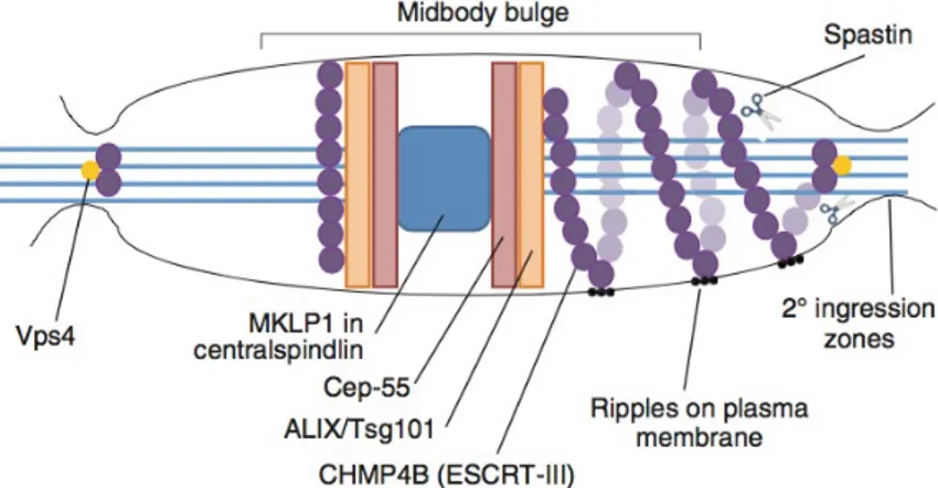
site of secondary ingression, shown here on the right of this schematic midbody. These rings also give rise to the appearance of ‘ripples’ on the plasma membrane. ESCRT-III can then recruit the microtubule-severing enzyme spastin, a microtubule severing AAA-ATPase. Indeed, spastin localizes at the midbody by binding the ESCRT subunits CHMP1B and IST1, and mediates microtubule scission at the end of the abscission process [73] (Figure 7).
Figure 7:The ESCRT complex in abscission
The centralspindlin component recruits Cep-55 to the midbody. Cep-55 in turn recruits I protein Tsg101 or ALIX. Tsg101 and the ESCRT-III subunit CHMP4B (purple spheres) are sequentially recruited into the center of the intercellular bridge. Later in cytokinesis, CHMP4B concentrates at the narrow secondary abscission zones, closely followed by Vps4 (yellow circles). ESCRT-III subunits extend towards these sites of cortical constriction forming filaments, which extend towards the site of secondary ingression. ESCRT-III can then recruit the microtubule-severing enzyme spastin.
1.6 U
BIQUITIN
-
DEPENDENT
CELL
CYCLE
TRANSITIONS
AND
THE
REGULATION
OF
CYTOKINESIS
A central role for ubiquitin lies in the turnover of cyclin proteins, which are the key elements driving the cell cycle by activating cell cycle dependent kinases (Cdks) (Glotzer et al., 1991). Besides cyclins a great number of other proteins need to be degraded during the cell cycle by the UPS (Pines, 2006). Modification of cyclins and other cell-cycle regulated proteins is carried out mostly by two classes of multi-subunit ubiquitin ligases. The most prominent is the anaphase-promoting complex/cyclosome (APC/C), which consists of at least 11 subunits that assemble on a scaffold (Apc2), a RING-finger protein (Apc11) and a subunit that is important for substrate recognition and extension of the poly-ubiquitin chain (Doc1/Apc10) (Peters, 2006). Substrate selection during the cell cycle requires the binding to one of two activating WD40-domain proteins known as Cdc20 and Cdh1 (Figure 5). Cdc20 exclusively binds to APC/C if phosphorylated by Cdk1, and the assembled APCCdc20 primarily recognizes proteins that harbor destruction boxes (a R/KXXL/I/M/V sequence). APCCdc20 is therefore active only until anaphase onset: at this point Cdk1 activity drops because its cyclin B activator is degraded and thus also phosphorylation of Cdc20 stops. Substrates
for APCCdc20 are proteins that need to be degraded in prometa- and metaphase (e.g. cyclin A, Nek2 kinase). In early M-phase and probably already in interphase (Di Fiore and Pines, 2007) APCCdc20 is kept inactive by binding to the inhibitor Emi1 (Machida and Dutta, 2007). Emi1 needs to be phosphorylated by Plk1 to be ubiquitylated by another E3 ligase complex, SCFßTrCP. However, the main inhibition of APCCdc20 takes place on the level of spindle assembly. APCCdc20 is active only if all kinetochores are attached to spindle microtubules. This scenario is also called spindle assembly checkpoint (Peters, 2006). At unattached kinetochores, the checkpoint proteins Mad2 and BubR1 build complexes with Cdc20 or APCCdc20 as long as unattached kinetochores are present. It has been suggested that the Mad2/Cdc20 and BubR1/Cdc20 complexes might sequester Cdc20 away from the APC/C and thereby keep it inactive (Peters, 2006). A similar mechanism involving the inhibitor Emi1 has recently been described (Ban et al., 2007). Emi1 binds the spindle-organizing NuMA/dynein-dynactin complex to anchor and inhibit the APC/C at spindle poles, probably limiting destruction of spindle-associated cyclin B.
When all kinetochores are finally attached, APCCdc20 can drive cell cycle progression by ubiquitylation of cyclin B leading to inactivation of Cdk1. Furthermore, APCCdc20 also promotes
that mediates sister chromatid separation. Due to the decrease in Cdk1 activity, the affinity of Cdc20 for the APC/C decreases and Cdh1, at anaphase onset, can bind to the APC/C as it is now hypo- phosphorylated, and Emi1, which also sequesters Cdh1 away from the APC/C, is now phosphorylated by Plk1 and degraded (Moshe et al., 2004; Hansen et al., 2004). The APCCdh1 modifies targets different from those recognized by APCCdc20, which include functional components of mitosis- and cytokinesis structures (kinetochores, spindle, contractile ring, midbody) to convert cells back into an interphase state. These comprise the spindle midzone protein PRC1 and the contractile ring regulators anillin (Zhao and Fang, 2005) and Ect2 (see above) (Pines, 2006). Furthermore, APCCdh1 drives degradation of mitotic regulators survivin, Aurora A and Plk1 (Castro et al., 2002; Lindon and Pines, 2004). Preventing Plk1 degradation at the end of mitosis interferes with the coordination of spindle relative to cleavage furrow position and, in consequence, results in cytokinesis failure (Lindon and Pines, 2004).
Another class of multi-subunit ubiquitin ligases with roles in mitosis are Skp1-Cullin-F- box (SCF) ligases. They are composed of a scaffolding cullin, a RING-finger protein (Rbx1), an adaptor protein (Skp1), and a substrate-interacting F-box protein. Approximately 70 F-box proteins are identified in humans so far allowing to accommodate for a wide range of substrates. In contrast
to the APC/C, SCF-ligases recognize phospho-degrons in target proteins and therefore the recognition of SCF-substrates does not depend on the modification of the ligase itself but on the modification of the substrate. As described above, the SCFßTrCP is crucial for cell cycle progression by degrading APC/C inhibitors like Emi1 (Moshe et al., 2004; Hansen et al., 2004). An SCF complex with the F-box protein Skp1 (SCFSkp1) has been found to localize on the midbody where it mediates ubiquitylation of a kinesin important for chromosome movement and midzone establishment, CENP-E (Liu et al., 2006).
Besides F-box proteins there are also proteins of the BTB-family (Bric-a- brac/Tramtrack/Broad complex) that can act as substrate recruiting factors in complex with cullin 3. Complexes of Cul3 with the BTB proteins KLHL9 and KLHL13 are required for chromosome alignment and midzone/midbody formation (Sumara et al., 2007). These complexes remove the CPC component Aurora B from chromosomes and allow transport to the midzone by mediating Aurora B ubiquitylation. As outlined above, exit from mitosis requires the degradation of cell cycle regulators by the ubiquitin proteasome system. Several observations allow to conclude that ubiquitin- dependent processes are also spatially linked to cytokinesis: Components of the UPS like the ubiquitin activating enzyme E1 and the proteasome itself are both
1995). Furthermore, the chromosomal passenger proteins survivin and Aurora B (Zhao et al., 2000; Sumara et al., 2007) as well as Plk1 (Lindon and Pines, 2004) are degraded most likely on midbody microtubules. Also the core APC/C subunit Doc1/Apc10 was found on the midbody (Kurasawa and Todokoro, 1999). In addition, proteasomal inhibition after anaphase onset revealed that cells fail to complete cytokinesis when ubiquitin-dependent degradation is shut down (Straight et al., 2003). Recent findings extend this view showing that a combination of Cdk1 and proteasomal inhibition can revert late cytokinetic stages to a pre-anaphase state (Potapova et al., 2006).
FIGURE 8: THE CELL CYCLE AND ITS REGULATORY PROTEINS DEGRADED BY THE APC/C AND THE UBIQUITIN PROTEASOME PATHWAY.
Cell cycle and the timing of APC/C activation and degradation of different substrate.
1.7 C
YTOKINESIS
FAILURE
Cytokinesis, the last step of cell division, proceeds throughout subsequent stages each owing to the previous one, that requires a complex interplay among a still enlarging number of regulatory and effectors components related to cytoskeleton, chromosome, cell cycle, lipid raft, vesicle, and membrane trafficking factors. Cytokinesis failure can arise through defects in any of the four stages in cytokinesis. Inhibition or excessive activation of different cytokinesis components can give rise to distinct phenotypes, including precocious ingression before the chromosomes have been separated, regression of the furrow giving rise to binucleated cells, or stabilization of the cytoplasmic bridge where daughter cells remain connected. High fidelity cytokinesis is crucial to ensure that both daughter cells inherit a diploid set of chromosomes. Cytokinesis failure and the resulting tetra- and poly-ploidization promote chromosomal and genomic instability, both hallmarks of cancer.
2. AIM
Cytokinesis is the critical final step of cell division. It is responsible for separation of the cytoplasm between daughter cells to complete mitosis. In animal cells, cytokinesis occurs through cortical remodeling orchestrated by the anaphase spindle and must be precisely controlled by many components and regulatory factors, in both establishing the spatial orientation of the cleavage plane and the timing of cleavage onset.
Cytokinesis failure leads to genomic instability and production of tetraploid cells, which may set the stage for the development of tumor cells.
CSA and CSB proteins have critical roles in a sub-pathway of nucleotide excision repair known as transcription-coupled repair (TCR). Although a defect in TCR pathway could potentially account for the enhanced photosensitivity of CS patients, other pathological features including neurological dysfunctions may not solely explained by a DNA repair defect and requires additional explanations [15]. Accordingly, in the last years, in our laboratories, it has been suggested that CSA and CSB proteins play multiple pleiotropic functions.
The aim of this work is to dissect the unknown extranuclear functions of CSA and CSB proteins in cytokinesis.
3. RESULTS
3.1 CSB
AND
CSA
LOCALIZATION
DURING
CELL
CYCLE
CSA and CSB proteins are known to be involved in the transcription-coupled DNA repair pathway and to have nuclear localization.
Although many of their functions could potentially be explained with their nuclear localization, other features, including their involvement in neurodegeneration, may not be solely explained by this way.
For this reason we asked ourself if the CSA and CSB localization may be not only restricted to the nuclei.
Searching for a new localization of the proteins we tested various antibodies that detect the endogenous proteins. By using various polyclonal antibodies directed against different epitopes of CSA and CSB proteins only a nuclear staining is detected.
Finally, using new rabbit polyclonal antibodies (GENETEX) directed against the internal region of CSA and CSB proteins, surprisingly we have identified for the first time a cytoplasmic proteins localization during the various stages of cell cycle.
Immunofluorescence (IF) staining for CSA and CSB (Figure 8, 9) during mitosis shows that in the early stages the cytoplasmic localization seems to be consequent of chromosomes condensation. However, from the onset of cleavage furrow the proteins take progressively a specific position.
In particular, while during metaphase CSA and CSB exit from the nucleus yet condensed, in anaphase, during cleavage furrow contraction, they are associated with the spindle midzone; then, in telophase, they accumulate at the midbody, where they have a characteristic ring-like arrangement embraced by Aurora B in the so called bulge-zone.
This specific localization let us hypothesize an active role of the proteins during cell division.
FIGURE 8: CSA LOCALIZATIONINCELLCYCLE
CS3BE-CSA-TY1-TET induced cells were fixed and stained with anti-Aim1 (green), anti-CSA (red) and Dapi (blue, DNA).
FIGURE 9: CSB LOCALIZATIONINCELLCYCLE
CS1AN-CSB-TY1-TET induced cells were fixed and stained with anti-Aim1 (green), anti-CSB (red) and Dapi (blue, DNA).
We confirmed the presence of CSA and CSB proteins at the midbody by employing both several human tumoral cell lines, MCF7, Hela and SK-N-BE (2c), and immortalized Cockayne fibroblast cell lines, CS1AN and CS3BE, rescue respectively for CSB and CSA.
Thus, to finally confirm this proteins localization, we utilized a protocol for the biochemical isolation and identification of proteins localized at the midbody (Skoop, 2004; Kuriyama and Ensrud, 1999; Mullins and McIntosh, 1982). Midbodies isolated and extracted from the various cell lines were analyzed by western blotting.
In particular, proliferating cell lines were enriched in telophase by nocodazole treatment, mitotic shake-off, and nocodazole wash-out to release replated cells from prometaphase. The midbody fraction was obtained after 80’ and compared by WB with equal volumes of total extracts from the same number of cells maintained in interphase or enriched in telophase. The transcription factor Sp1 and beta-actin, used as markers of nuclear and cytoplasmic contamination, were barely detectable in the purified midbody extracts, while there was the presence of AIM-1, indicating the quality of the preparations. In these conditions, both CSA and CSB, were enriched in the midbody, further confirming their presence in this subcellular compartment (Figure 10, 11).
FIGURE 10: CSA LOCALIZATIONINCELLCYCLE
Cell were fixed and stained with anti-Aim1 (green), anti-CSA (red) and Dapi (blue, DNA).
FIGURE 11: CSB LOCALIZATIONINCELLCYCLE
Cell were fixed and stained with anti-Aim1 (green), anti-CSA (red) and Dapi (blue, DNA).
3.2 CSA
AND
CSB
MUTATIONS
CAUSE
CYTOKINESIS
FAILURE
To better understand the functional roles of the CSA and CSB proteins during the final step of cell division process, the cytokinesis, we have utilized immortalized Cockayne fibroblast cell lines, CS1AN and CS3BE, respectively mutated for CSB and CSA genes.
Our experimental models (CS1AN-CSB-Ty1-TET and CS3BE-CSA-Ty1-TET) are inducible for the expression of functional proteins by adding Tetracycline, so reverting the phenotype after 4 h for CSA expression and 8 h for CSB.
There were not detectable staining at IF for CSA or CSB expression in not induced fibroblasts, while there were a correct localization of the proteins after TET treatment.
Compared to induced cells, mutated cells show an increased percentage of binucleate cells and aberrant mitosis, confirming the involvement of CSA and CSB in the cytokinesis.
Next, to elucidate which cytokinesis stage was compromised, we counted the percentage of early midbodies, II ingressions, signature of abscission, and long midbodies, indicative of cytokinesis failure. By counting emerged that CSA and CSB mutated cells show an increased number of long midbodies lacking of II ingression, thus
FIGURE 12: MULTINUCLEATEDANDABERRANTMITOSISIN COKAYNECELLS
Cell were fixed and stained with anti-alpha tubulin and anti-gamma tubulin (green), anti-actin and anti-gamma tubulin (red) and Dapi (blue, DNA).
Graphics show the number of cells in different phases of cell cycle and multinucleated, canonical and aberrant mitosis.
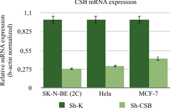
To evaluate the effect of CSB depletion in the cytokynesis, non-silencing control and CSB specific shRNA vectors (Santa Cruz Biotechnology) were used to create a CSB proficient pool of cell (sh-K) and a CSB-suppressed pool of cell (sh-CSB) respectively for different tumor cell lines: SK-N-BE (2c), Hela and MCF-7. Stably transduced cells were selected by puromycin (2µg/ml) resistance. Both RT-PCR and Western blot analyses (data not shown) showed a good suppression of CSB expression in sh-CSB cell line relative to scrambled vector transfected cells (Figure 13).
CSB mRNA expression Re la ti ve m RN A e xpre ss ion (b-a ct in norm al iz ed) 0 0,275 0,55 0,825 1,1 SK-N-BE (2C) Hela MCF-7 Sh-K Sh-CSB
FIGURE 13: CSB M-RNA EXPRESSION
Graphic of CSB suppression in SK-N-BE, Hela and MCF7 cells
We immunostained cells with antibodies against cytoscheletal proteins and we found that CSB-suppressed cells were multinucleated and showed a very high frequency of aberrant mitosis, characterized by the presence of spindle multipolarity, that we weren’t able to find in the control line. This phenotype was observed in all the CSB-suppressed tumor cells and was exacerbated, especially in SK-N-BE (2C), in respect to the one seen in Cockayne fibroblast, due to a deregulation of cell cycle check-points and apoptotic pathways, typical of tumoral cells.
Cytochalasin B treatment of SK-N-BE (2C) K reproduces the sh-CSB phenotype, inhibiting actin filament polymerization and the interaction of actin filaments, thus confirming cytokinesis failure. Finally, a time-lapse live-cell imaging performed using CellLight BacMam GFP-tubulin baculovirus vector, on sh-K and sh-CSB SK-N-BE (2C) cell line, confirmed our hypothesis of cytokinesis failure in interfered cells. In particular, the abscission clearly failed in Sh-CSB cells with re-fusion of daughter cells, confirming the observation made by IF and thus indicating that CSB depletion results in abscission failure.
Altogether, these data show that CSA and CSB depletion by siRNA induces accumulation of cytokinesis-dependent aberrations and abscission
defect suggesting that the proteins are required for completion of the late stage of cytokinesis (Figure 14).
FIGURE 14: MULTINUCLEATED AND ABERRANT MITOSIS IN SK-N-BE (2C) SH-CSB CELLS
Cell were fixed and stained with anti-alpha tubulin and anti-gamma tubulin (green), anti-actin and anti-gamma tubulin (red) and Dapi (blue, DNA).
Graphics show the number of cells in different phases of cell cycle and multinucleated, canonical and aberrant mitosis.
3.3 CSA
AND
CSB
MUTATIONS
CAUSE
DEFECT
IN
PRC-1
PROTEIN
LOCALIZATION
To investigate the mechanism of CSA and CSB protein in the abscission, we analyzed the spatio-temporal localization of a series of structural and functional proteins sequentially recruited during cytokinesis to assure proper cell division.
We analyzed:
1) proteins associated with midzone formation, such as Aurora B, PLK1 and PRC1;
2) proteins associated with midbody formation such as PLK1; 3) proteins associated with abscission events, such as CEP55, ALIX and Spastin.
In all cells analyzed at the early stages of cytokinesis, we observed that the localization patterns for all proteins examined are not affected during midzone formation and cleavage furrow ingression in the CSA and CSB mutated cells, indicating that the proteins are not mainly involved in the early events of cytokinesis.
In contrast, in the subsequent stage of midbody formation, PRC1(Figure 15) and PLK1 (Figure 16) proteins can be detected at the midbody but their distribution pattern is altered, becoming dispersed along the midbody microtubules and the midbodies appear elongated. These distributions are in contrast to their localizations at the flanking zone in the rescue cells.
Furthermore, by analyzing the proteins involved in abscission, in CSA and CSB mutated cells the critical abscission factors as ALIX and Spastin present several defects of organization localizing itself along the midbody (preliminary data).
Overall, these results indicate that CSA and CSB protein play a role at the terminal stage of cytokinesis by controlling the morphology of the midbody and by organizing the localization of a few critical regulators of the abscission.
FIGURE 15: PRC1 LOCALIZATION
Cell were fixed and stained with anti-AIM1 (green), anti-PRC1 (red) and Dapi (blue, DNA).
FIGURE 16:PLK1 LOCALIZATION
Cell were fixed and stained with anti-AIM1 (green), anti-PLK1 (red) and Dapi (blue, DNA).
4. DISCUSSION
Cockayne syndrome (CS) is a genetic disease inherited in an autosomal recessive pattern. CS patients are characterized by photosensitivity, severe growth retardation, cachectic dwarfism, feature of premature aging and progressive neurological abnormalities of the central nervous system including microcephaly, cerebellar atrophy and demyelinating peripheral neuropathy (2, 4). The average life span of children with this syndrome is about 12 years of age.
Two forms of CS have been described on the basis of age of onset of clinical symptoms. The classical type, Type I, becomes apparent after the child is about a year old. Type II, or severe Cockayne syndrome, instead becomes apparent at birth. There is currently no cure for CS and treatment is aimed at alleviating symptoms and improving the patient's quality of life.
CS patients have been assigned to two genetic complementation groups (CS-A and CS-B), whose corresponding genes (csa and csb) have been cloned and characterized (5, 6, 13). CSA and CSB proteins have critical roles in a sub pathway of nucleotide excision repair known as transcription-coupled repair (TCR) (21). Although a defect in TCR pathway could potentially account for the enhanced photosensitivity of CS patients, other pathological features
including neurological dysfunctions may not solely explained by a DNA repair defect and requires additional explanations (15). Accordingly, in the last years it has been suggested that CSA and CSB proteins play multiple pleiotropic functions. This notion is somewhat strengthened by the demonstrated involvement of CSB in basal transcription mediated by RNA polymerase I and II (16-22). More recently, we and others have demonstrated that CSB maintains the transcriptional programs promoting cell survival responses following exposure to cellular stressors such as UV, oxidative damage, inflammation and hypoxia (23-27). Therefore, beside DNA repair defects, abnormalities in the regulation of RNA pol I and II transcription activities of neural stem cells during the programs that govern either self-renewal or differentiation, might provide plausible explanations for many of the somatic features, including aspects of neurological symptoms associated with CS. It is not finished! Intriguingly, an unknown extranuclear role of CSA and CSB proteins is emerging. Our study reveals an unexpected subcellular localization and biological function of CSA and CSB proteins in cytokinesis.
We discovered, in several tumoral cells line and in Cockayne fibroblast, that CSA and CSB proteins have a different cytoplasmic localization during mitosis in cytoskeletal structures such as the centrosomes and the midbody, the intercellular microtubule bridge connecting two daughter cells at the end of cytokinesis. In
particular, CSA protein localizes at centrosomes during mitosis (data not show) and at midbody during cytokinesis, while CSB shows a cytoplasmic localization during mitosis, than moves along the cleavage furrow and finally localizes at midbody. Both proteins localize to the midbody in the so called bulge-zone.These observations suggest a potential role of CSA and CSB protein in the organization and functioning of cytoskeleton-mediated cellular activities encompassing polarity, motility and division.
Cytokinesis is a highly ordered process, requiring a complicated interplay between cytoskeletal, chromosomal, and cell cycle regulatory pathways. Cytokinesis failure is known to lead to genetically unstable states, such as tetra- and poly-ploidization, considered a critical step in tumourigenesis (109).
We discovered that depletion of CSB proteins in tumoral cells line and CSA and CSB mutation in Cockayne fibroblasts, results in cytokinesis failure.
In particular, we show the presence of syncytium-like cells, the increase of cells number at the midbody stage and abscission defects associated with the increase of aberrant mitosis and with the accumulation of binucleated cells.These signs are usually observed in cells with impaired abscission (110), that support the role of this proteins in this last stage of cell division.
enlarging number of regulatory and effectors components. During the last several years, multiple studies have considerably increased our understanding about cytokinesis and abscission. Although a large list of proteins participating in cytokinesis has been generated, it is still unclear how all of these proteins interact to drive cytokinesis and abscission.
To investigate the mechanism of CSA and CSB protein in the abscission, we analyzed the spatio-temporal localization of a series of structural and functional proteins sequentially recruited during cytokinesis to assure proper cell division.
In CSA and CSB mutated cells, cleavage furrow specification and ingression proceed normally, but the cell abscission fails to occur. We show, whit IF analysis, that localization of some proteins involved in specification/formation of the midzone, e.g. CPCs, PRC1 and PLK1, are unaffected in CSA and CSB mutated cells, indicating that the proteins are not mainly involved in the early events of cytokinesis.
In contrast, in the subsequent stage of midbody formation, PLK1 and PRC1 proteins and abscission factors as ALIX and Spastin, present several defects of organization localizing and the midbodies appear elongated, indicating abscission’s defects.
PLK1 and PRC1 proteins are closely associated in the regulation of midzone and midbody formation; the ubiquitination of both proteins it is required for the cytokinesis completion.
In particular, in metaphase, cyclin B–Cdk1 phosphorylates PRC1, which interferes with PRC1 binding to Plk1. Simultaneously, cyclin B-Cdk1 functions as priming kinase for Cdc25, which might contribute to Plk1 targeting to the centrosome. At the metaphase– anaphase transition, cyclin B is degraded and cyclin B–Cdk1 inactivated. Consequently, the inhibitory phosphorylation on PRC1 is lost. Plk1 then binds PRC1 and phosphorylates PRC1 on a distinct site (T602). This creates the docking site for stable Plk1 binding to PRC1 and enables PRC1 to target Plk1 to the central spindle.
How is the interaction between PRC1-2 and Plk1 terminated? Both Plk1 and PRC1 are substrates of the anaphase-promoting complex (APC) and the formation of a complex between PRC1 and Plk1 stimulates the ubiquitination of both proteins, and thereby limits the duration of their activity at the central spindle for the continuation of cytokinesis (111).
The CSA protein is a component of a CRL4 E3 ubiquitin ligase complex and CSB protein, whit a C-terminal UBD, is a substrate of this E3 ligase. In a previous work carried out in our laboratory, ubiquitination assays showed that CSA and CSB proteins interact with p53 and regulate its Mdm2-dependent ubiquitination, either in vivo or in vitro (112).
We speculate that in CSA and CSB mutated cells, PRC1 and PLK1 proteins are delocalized because of the lack of CSA protein that don’t permit the protein degradation.
Overall, these results indicate that CSA and CSB protein play a role at the terminal stage of cytokinesis by controlling the morphology of the midbody and organizing the localization of a few critical regulators of the abscission.
5. MATERIALS AND METODS
5.1 C
ELL
CULTURE
AND
SILENCING
MCF-7 were grown in DMEM medium supplemented with 10% Fetal Bovine Serum, 2mM L-Glutamine and 0,6 ug/ml of insulin from bovine pancreas. Hela were grown in DMEM medium supplemented with 10% Fetal Bovine Serum and 2mM L-Glutamine.
Cells in exponential growth phase were transduced with lentiviral shRNA particle (1 x 10⁵ infectious units of virus - Santa Cruz Biotechnology) expressing sh-RNA targeting CSB or sh-RNA non-targeting control. Puromicin selection (2ug/ml) is performed to achieve stable gene silencing.
CS1AN-CSB-Ty1-TET and CS3BE-CSA-Ty1-TET were grown in DMEM/F10 medium supplemented with 10% Fetal Bovine Serum and 2mM L-Glutamine.
SK-N-BE (2c) were grown in a MEM/DMEM F12 medium supplemented with 10% Fetal Bovine Serum and 2mM L-Glutamine. This lines are inducible for the expression of functional proteins by adding Tetracycline, so reverting the phenotype after 4 h for CSA expression and 8 h for CSB.
5.2 W
ESTERN
BLOT
ANALYSIS
Cells were lysed for 10 min on ice in RIPA buffer. The whole cell extracts were centrifuged at 13000 g for 5min and the supernatant was recovered. Protein concentration was determined by Bradford protein assay kit (BioRad, Hercules, CA, USA). Proteins (50 mg) were separated on polyacrylamide gradient gel (4–20%) electrophoresis and blotted onto PVDF membrane (Amersham, Pittsburgh, PA, USA) following the standard procedures. The membrane was incubated with TBST (20 mM Tris–HCl, pH 7.4, 137 mM NaCl; 0.2% Tween 20) buffer containing 5% non-fat dried milk for 60 min at RT and subsequently incubated with primary and horseradish peroxidase-conjugated secondary antibodies (Vector, Burlingame, CA, USA). The signal was detected using the enhanced chemiluminescence method following the manufacturer’s instructions (Amersham).
5.3 I
MMUNOFLUORESCENCE
M
ICROSCOPY
For immunofluorescence experiments, cells were seeded onto ibidi coverslips. Cells were fixed in ice-cold methanol or 2% formaldehyde, washed three times in phosphate buffered saline (PBS), permeabilized in 0.25% Triton X- 100 in PBS for 10 min., and then blocked in 0.2% Triton X-100, 3% bovine serum albumin
(BSA) in PBS for 30 min before the required primary Abs were applied. The following Abs were employed: anti- Aurora B Ab (BD-Bioscience), anti-Cep55 Ab (Santa Cruz), anti-PRC1 Ab (Santa Cruz), anti- MKLP1 Ab (Santa Cruz), anti-spastin Ab (Santa Cruz), anti PLK1 Ab (Santa Cruz), alpha-Tubulin (Sigma). Appropriate secondary ALEXA FLUOR were used. DNA was marked with DAPI in Vectalshied. Slides were analyzed with a confocal microscope system (Zeiss LSM 710).
5.4
R
ETROTRANSCRIPTION
AND
REAL
-
TIME
QUANTITATIVE
PCR
RNA was isolated using the NucleoSpin RNA II kit (Macherey-Nagel). cDNA synthesis was performed using the First Strand cDNA Synthesis kit (Fermentas). Real-time quantitative PCR was carried out with SYBR green master mixture (Promega) using Mx3005P Real-Time PCR system (Agilent). Results were normalized to β-actin. Primers sequences are available upon request.
5.5 M
IDBODY
I
SOLATION
AND
E
XTRACTION
MCF-7, SK-N-BE (2C), HeLa, CS1AN-CSB-Ty-TET and CS3BE-CSA-Ty-TET cells were enriched in telophase by treatment with nocodazole (0,1 ug/ml for 3-4 hr) followed by mitotic shake-off, nocodazole wash-out, and incubation of the collected cells for about 80 min to reach telophase stage. Midbodies were isolated as described by Kuriyama (Kuriyama et al., 1984) and extracted using an extraction buffer (50 mM Tris-HCl [pH 7.4], 600 mM NaCl, 0.1% SDS, 0.5% NP40, 1 mM DTT, 5 mM EDTA) supplemented with protease and phosphatase-inhibitor mix (Roche). Nonsynchronized interphase and telophase-enriched cells were obtained in the same manner and analyzed by WB together with midbody extracts.
5.6
L
IVE
C
ELL
I
MAGING
Cells seeded in 35 mm dishes (Ibidi) and transfected whit CellLight BacMan 2.0 GFP-tubulin. Incubate for 16 hours at 37°C for fluorescent protein expression. Slides were analyzed with a confocal microscope system (Zeiss LSM 710).
REFERENCES
1. Weidenheim KM, Dickson DW, Rapin I. Neuropathology of
Cockayne syndrome: evidence for impaired development, premature aging, and neurodegeneration. Mech Ageing Dev
2009; 130: 619–636.
2. Laugel V, Dalloz C, Durand M, Sauvanaud F, Kristensen U, Vincent MC et al. Mutation update for the CSB/ERCC6 and
CSA/ERCC8 genes involved in Cockayne syndrome. Hum
Mutat 2010; 31: 113–126.
3. Natale V.A Comprehensive description of the severity groups
in Cockayne syndrome. Am J Med Genet 2011; 155A: 1081–
1095.
4. Henning KA, Li L, Iyer N, McDaniel LD, Reagan MS, Legerski R et al. The Cockayne syndrome group A gene encodes a WD
repeat protein that interacts with CSB protein and a subunit of RNA polymerase II TFIIH. Cell 1995; 82: 555–564.
5. Troelstra C, van Gool A, de Wit J, Vermeulen W, Bootsma D, Hoeijmakers JH. ERCC6, a member of a subfamily of
preferential repair of active genes. Cell 1992; 71: 939–953.
6. Lehmann AR. Three complementation groups in Cockayne
syndrome. Mutat Res 1982; 106: 347–356.
7. Zhou, H.X., Wang, G., 2001. Predicted structures of two
proteins involved in human diseases. Cell Biochemistry and
Biophysics 35, 35–47.
8. Groisman, R., Polanowska, J., Kuraoka, I., Sawada, J., Saijo, M., Kisselev, A.F., Drapkin, R., Tanaka, K., Nakatani, Y., 2003.
Ubiquitin ligase activity in the DDB2 and CSA complexes, which are linked to xeroderma pigmentosum and Cockayne syn- drome respectively, is differentially regulated by the COP9 signalosome in response to UV irradiation. Cell 113,
357–367.
9. Citterio E1, Rademakers S, van der Horst GT, van Gool AJ, Hoeijmakers JH, Vermeulen W. Biochemical and biological
characterization of wild-type and ATPase-deficient Cockayne syndrome B repair protein. J Biol Chem. 1998 May
10. Troelstra C1, van Gool A, de Wit J, Vermeulen W, Bootsma D, Hoeijmakers JH. ERCC6, a member of a subfamily of
putative helicases, is involved in Cockayne's syndrome and preferential repair of active genes. Cell. 1992 Dec 11;71(6):
939-53.
11. Fousteri M1, Mullenders LH. Transcription-coupled
nucleotide excision repair in mammalian cells: molecular mechanisms and biological effects. Cell Res. 2008 Jan;18(1):
73-84. doi: 10.1038/cr.2008.6.
12. Mayne LV, Lehmann AR. Failure of RNA synthesis to recover
after UV irradiation: an early defect in cells from individuals with Cockayne’s syndrome and xeroderma pigmentosum. Cancer Res 1982; 42: 1473–1478.
13. Balajee AS, Proietti-De-Santis L, Brosh RM, Selzer R, Bohr VA. Role of the ATPase domain of the Cockayne syndrome
group B protein in UV induced apoptosis. Oncogene 2000;
19: 477–489.
14. Brooks PJ. Blinded by the UV light: how the focus on
transcription-coupled NER has distracted from understanding the mechanisms of Cockayne syndrome
15. Proietti-De-Santis L, Drane P, Egly JM. Cockayne syndrome B
protein regulates the transcriptional program after UV irradiation. EMBO J 2006; 25: 1915–1923.
16. Filippi S, Latini P, Frontini M, Palitti F, Egly JM, Proietti-De-Santis L. CSB protein is (a direct target of HIF-1 and) a
critical mediator of the hypoxic response. EMBO J 2008; 27:
2545–2556.
17. Frontini M, Proietti-De-Santis L. Cockayne syndrome B
protein (CSB): linking p53, HIF-1 and p300 to robustness, lifespan, cancer and cell fate decisions. Cell Cycle 2009; 8:
693–696.
18. Newman JC, Bailey AD, Fan HY, Pavelitz T, Weiner AM. An
abundant evolutionarily conserved CSB-PiggyBac fusion protein expressed in Cockayne syndrome. PLoS Genet 2008;
4: e1000031.
19. Velez-Cruz R, Egly JM. Cockayne syndrome group B (CSB)
protein: at the crossroads of transcriptional networks. Mech
20. F Ciaffardini, S Nicolai, M Caputo, G Canu, E Paccosi, M Costantino, M Frontini, AS Balajee and L Proietti-De-Santis
The cockayne syndrome B protein is essential for neuronal differentiation and neuritogenesis. Cell Death and Disease
(2014) 5, e 1268.
21. M. Frontini, L. Proietti-De-Santis Cockayne syndrome B
protein (CSB): linking p53, HIF-1 and p300 to robustness, lifespan, cancer and cell fate decisions. Cell Cycle 8 (2009)
693–696.
22. A.S. Balajee, L. Proietti-De-Santis, R.M. Brosh, R. Selzer, V.A. Bohr Role of the ATPase domain of the Cockayne syndrome
group B protein in UV induced apoptosis. Oncogene 19
(2000) 477–489.
23. Manuela Caputo, Mattia Frontini, Renier Velez-Cruz, Serena Nicolai, Giorgio Prantera, Luca Proietti-De-Santis. The CSB
repair factor is overexpressed in cancer cells, increases apoptotic resistance, and promotes tumor growth. Dna
Repair 12 (2013) 293-299.
and therapeutic targets in cancer. Cell Proliferation, 36: 131–
149. doi: 10.1046/j.1365-2184.2003.00266.
25. Kops GJ, Weaver BA, Cleveland DW. On the road to cancer:
aneuploidy and the mitotic checkpoint. Nat Rev Cancer. 2005
Oct; 5(10):773-85.
26. Fujiwara T, Bandi M, Nitta M, Ivanova EV, Bronson RT, Pellman D. Cytokinesis failure generating tetraploids
promotes tumorigenesis in p53-null cells. Nature. 2005 Oct
13;437(7061):1043-7.
27. Christine M. Caldwell, Rebecca A. Green, and Kenneth B. Kaplan APC mutations lead to cytokinetic failures in vitro
and tetraploid genotypes in Min mice. September 24, 2007 //
28. 1109-1120 The Rockefeller University Press, doi: 10.1083/jcb. 200703186.
29. Glotzer M. Animal cell cytokinesis. Annu Rev Cell Dev Biol. 2001;17:351-86.
30. Eggert US, Mitchison TJ, Field CM. Animal cytokinesis: from
parts list to mechanisms. Annu Rev Biochem (2006) 75: 543–
566.
31. Rappaport R. Cytokinesis in animal cells. Int Rev Cytol 1971; 31:169-213.
32. Rappaport R. Establishment of the mechanism of cytokinesis
in animal cells. Int Rev Cytol 1986;
105:245-281.
33. Danowski BA. Fibroblast contractility and actin
organization are stimulated by microtubule inhibitors. J Cell
Sci 1989; 93(Pt 2):255-266.
34. Ren XD, Kiosses WB, Schwartz MA. Regulation of the small