Childhood obesity: immune response and nutritional
approaches
Thea Magrone and Emilio Jirillo*
Department of Basic Medical Sciences, Neuroscience and Sensory Organs, University of Bari, Bari, Italy
Edited by:
Johan Garssen, Utrecht University, Netherlands
Reviewed by:
Edda Fiebiger, Children’s Hospital Boston and Harvard Medical School, USA
Margarida Castell, University of Barcelona, Spain
*Correspondence:
Emilio Jirillo, Department of Basic Medical Sciences, Neuroscience and Sensory Organs, University of Bari, Policlinico-Piazza G. Cesare, Bari 11-70124, Italy
e-mail: [email protected]
Childhood obesity is characterized by a low-grade inflammation status depending on the
multicellular release of cytokines, adipokines, and reactive oxygen species. In particular,
the imbalance between anti-inflammatory T regulatory cells and inflammatory T helper 17
cells seems to sustain such a phlogistic condition. Alterations of gut microbiota since
child-hood also contribute to the maintenance of inflammation. Therefore, besides preventive
measures and caloric restrictions, dietary intake of natural products endowed with
anti-oxidant and anti-inflammatory activities may represent a valid interventional approach for
preventing and/or attenuating the pathological consequences of obesity. In this regard,
the use of prebiotics, probiotics, polyphenols, polyunsaturated fatty acids, vitamins, and
melatonin in human clinical trials will be described.
Keywords: atherosclerosis, children, diabetes, immunity, microbiota, nutrition, obesity, oxidative stress
INTRODUCTION
Nowadays, childhood obesity has become an epidemic all over the
world. In terms of estimates, 200 million school-age children are
overweight/obese and 40–50 million of them are obese (1).
According to current literature, 70% of obese adolescents
become obese in adult life also in relation to parental obesity
(2–4). Of note, overweight and obesity seem to be irrespective
of the socio-economic conditions and income status, as reported
by Lobstein et al. (5), Popkin and Gordon-Larsen (6), and Wang
and Lobstein (7). However, in developed countries, social
inequal-ity represents a risk of developing obesinequal-ity (8). On the other
hand, genetic, epigenetic, environmental factors, and
inappropri-ate life styles (imbalanced diets and sedentary life) greatly
con-tribute to the development of obesity and related diseases, such
as insulin resistance, type 2 diabetes, and metabolic syndrome
(MetS) (9).
With regard to the etiology of childhood obesity (10), the
prenatal period should be considered as a critical period for its
development. Excessive or low calorie intake by the mother during
Abbreviations: AGEs, advanced glycation end products; AMP, antimicrobialpep-tide; BMI, body mass index; DCs, dendritic cells; DHA, docosahexaenoic acid; EGCG, epigallocatechin-3-gallate; EPA, eicosapentaenoic acid; GPCR, G-protein coupled receptor; IFN, interferon; Ig, immunoglobulin; IL, interleukin; LP, lamina propria; LPS, lipopolysaccharides; MCP, monocyte chemotactic protein; MedDiet, Mediterranean diet; MetS, metabolic syndrome; n-3 PUFAs, omega-3 polyun-saturated fatty acids; n-6 PUFAs, omega-6 polyunpolyun-saturated fatty acids; NADPH, nicotinamide adenine dinucleotide phosphate; NASH, non-alcoholic steatohepati-tis; NK, natural killer; NO, nitric oxide; PA, physical activity; PAI, plasminogen activator inhibitor; PKC, protein kinase C; PP, Peyer’s patches; PSA, polysaccha-ride A; RAGEs, advanced glycation end products receptors; ROS, reactive oxygen species; SCFAs, short chain fatty acids; SOD, superoxide dismutase; TGF, transform-ing growth factor; Th, T helper; ThF, T helper follicular; TLRs, toll-like receptors; TNF, tumor necrosis factor; Treg, T regulatory; VAT, visceral adipose tissue; VDR, vitamin D receptor; YHEI, youth healthy eating index.
the prenatal and perinatal period seems to predispose to obesity
in postnatal life stages (10). Children born from diabetic mothers
or mothers who smoked during pregnancy are more exposed to
the risk of obesity and type 2 diabetes (11). Also,
breastfeed-ing seems to reduce the risk of overweight/obesity. In addition,
the so-called “obesity rebound” at 5–7 years old seems to be very
critical for the development of obesity and risk of obesity in
adult-hood (10). Puberty is another critical period in life especially in
females, in terms of obesity risk since early menarche (<11 years)
in females predisposes to obesity in a more remarkable way than
later menarche (>14 years) (10). In these females, early menarche
leads to obesity in adolescence and in adulthood, while in 70% of
obese adolescent males weight tends to normalize at later ages in
comparison to 20% of obese adolescent females.
From a genetic point of view, humans have acquired the ability
to deposit fats and utilize them under a condition of starvation.
However, this characteristic turned into a disadvantage in
west-ern societies where the abundant consumption of food leads to an
increase of fat deposits. As far as nutritional factors are concerned
(12), evidence has been provided that obese children consume
more fat than non-obese children, thus leading to higher body
mass index (BMI). In addition, fats tend to deregulate control
mechanisms for body weight thanks to a greater amount of energy
per gram they provide. Fats are more palatable with a smaller
sati-ating effect in comparison to proteins and sugars and the actual
amount ingested. Another nutritional aspect in obese children and
adolescent is the irregular eating pattern with a lower frequency of
breakfast consumption.
Physical activity (PA) represents another important factor in
the development of obesity since reduced PA correlates with excess
body weight (12). Sedentary life due to playing video-games and
watching television affects basal expenditure. In particular,
watch-ing television also increases calorie intake for consumption of
foods which, in turn, are publicized by the same programs children
are watching. Finally, an association has been found between short
sleep and obesity in the sense that sleep duration is inversely
associated to BMI and waist circumference.
The excess of fat negatively influences health and/or
wellbe-ing in children. Data reported by Freedman and associates (13)
demonstrated that childhood obesity is associated to the risk of
cardiovascular events in adult life. In addition, Lauer and associates
(14) described some factors affecting the relationship between
childhood and adult cholesterol levels with special reference to
early development of atherosclerosis in children. Metabolic (high
levels of triacylglycerols, low levels of high density
lipoprotein-cholesterol) and clinical modifications (high blood pressure and
defective glucose metabolism) are part of the MetS, which
repre-sents one of the major complications of childhood obesity (15).
For instance, type 2 diabetes and cardiovascular disease represent
sequelae of MetS.
On these grounds, the aim of the present review is to describe
the profile of the immune response in obese children, taking into
consideration that obesity is an “inflammatory” disease.
Further-more, interventional studies aimed at preventing obesity and/or
attenuating the immune-inflammatory profile will be illustrated.
IMMUNITY IN CHILDHOOD OBESITY
Obesity is associated to a condition of systemic inflammation,
on the one hand (16), and to an impaired immunity, on the
other hand (17). For instance, obese individuals were more
sus-ceptible to the H1N1 influenza during the 2009 pandemia (18).
In obese individuals, visceral adipose tissue (VAT) is the major
source of pro-inflammatory cytokines, which, in turn, lead to
insulin resistance (16,
19). Experimentally, obese animals produce
higher amounts of adipose tissue-derived tumor necrosis factor
(TNF)-α in comparison to the lean counterpart (20), while in
obese humans Pradhan and associates (21) documented the
asso-ciation between C-reactive protein, interleukin (IL)-6, and the risk
of developing type 2 diabetes. Quite interestingly, macrophages
with an inflammatory phenotype have been detected in the
con-text of VAT of obese people (22,
23). They account for 40–60%
of VAT immune cells in obesity and differ from the
pheno-type of macrophages contained in lean adipose tissue (22,
23).
Obesity-associated macrophages in VAT are M1 “classically
acti-vated macrophages.” They secrete in large amounts TNF-α, IL-6,
IL-12, IL-1β, and monocyte chemotactic protein-1 (MCP-1), as
well as nitric oxide (NO) (24). On the contrary, in lean VAT
M2 macrophages are “alternatively activated macrophages,” which
secrete IL-10, IL-1 receptor antagonist, and arginase 1, thus
exert-ing anti-inflammatory activities. The contexert-ingent of obesity VAT
macrophages seems to be in part residential or, in alternative,
migrates into VAT from remote sites under the effect of MCP-1. M1
macrophages seem to favor insulin resistance and in murine
mod-els their deactivation protects against insulin resistance (25,
26).
Of note, VAT macrophages and adipocytes share common
func-tions in terms of pattern of cytokine released and insulin resistance
induction (27). It is worthwhile mentioning that, in obese mice,
neutrophils and mast cells also play an inflammatory role, while
the number of eosinophils is decreased (28). The above described
cellular profiles promote insulin resistance in murine obesity (28).
As far as adaptive immune responses are concerned in obese
mice, VAT contains higher numbers of T helper (Th) CD4+, T
cytotoxic CD8+ cells, and B cells. In particular, Th1 cells
pro-duce interferon (IFN)-γ in vitro (29). IFN-γ, in turn, polarizes M1
macrophages, also increasing release of TNF-α.
Conversely, obese mice lacking IFN-γ expression or T-cell
receptor
β-deficient mice are more protected with regard to
inflammatory cell infiltration of VAT (29,
30). T regulatory (Treg)
cells are decreased in both obese mice and humans (31,
32). In
murine VAT, depletion of Treg cells led to increased insulin
lev-els and reduced insulin receptor signaling (32). On the other
hand, expansion of Treg cell contingent in high-fat-diet-fed mice
and increased secretion of IL-10 led to a significant reduction
of blood glucose levels, insulin resistance, and glucose tolerance.
These data suggest the anti-inflammatory role exerted by Treg cells
via release of IL-10, which suppresses obesity-induced
inflamma-tion. Han and associates (33) have reported that insulin bears
receptors on Treg cells, thus decreasing IL-10 release by
acti-vating AKT/mammalian target of rapamycin signaling pathway.
These results suggest that high insulin levels in obesity play an
inflammatory role by impairing Treg cell-induced suppression.
Th17 cells are increased in obese mice and in humans, thus
leading to enhanced expression and release of IL-17 (34–36).
How-ever, evidence has been provided that
γδ T cells and neutrophils
can also produce IL-17 and, therefore, Th and Th17 cells are not
the only source of this cytokine (37,
38). According to Zúñiga
(37), these various sources of IL-17 may explain some
contradic-tory results obtained in IL-17 knockout mice which are overweight
and become obese as results of a high-fat diet compared to
con-trols. Despite these controversial results in mice, there is evidence
that obesity is associated to autoimmune diseases in both mice
and humans likely via a Th17-dependent mechanism (39). Finally,
as recently reported by Erbel and associates (40), IL-17A plays a
pathogenetic role in advanced murine and human atherosclerosis,
which is a complication of the obese status.
CD8+ cells accumulate into VAT in murine obesity and their
depletion led to reduction of macrophage infiltration and
amelio-ration of insulin sensitivity (41,
42). Adoptive transfer of CD8+
cells into CD8-deficient mice increased inflammatory cytokine
production in the context of VAT. Jiang and associates (43) have
documented that CD8+ cells in VAT are activated by IFN-γ
released by Th1 cells and, moreover, they express high levels of
the integrin CD11a, which promotes infiltration of CD8+ cells
into VAT.
B-cell infiltration into VAT seems to precede T cell and
macrophage homing into this tissue. According to Winer and
asso-ciates (44), B cells into VAT provoke insulin resistance, modulating
T cells and producing immunoglobulin (Ig)G, which account for
insulin resistance. Conversely, obese B null mice lack inflammatory
cytokines, produce high levels of IL-10, and are protected against
insulin resistance.
Major features of immune alterations in obese people are
indicated in Table 1.
THE IL-10/IL-17 RATIO AND TYPE OF DIET
In recent years, it has become clear that some lifestyle
fac-tors, including dietary habits, alcohol consumption, exercise, and
Table 1 | Alterations of innate and adaptive immunity in human obesity.
VAT tissue produces pro-inflammatory cytokines, which are responsible for insulin resistance (16,19)
The association between C-reactive protein, IL-6, and the risk of developing type 2 diabetes has been documented (21)
M1 macrophages with an inflammatory phenotype have been found in obese people VAT (22,23)
T regulatory cells are decreased in human obesity (31,32) Th17 cells are increased in obese humans (34–36)
CD8+ cells express high levels of the integrin CD11a, which promotes their infiltration of CD8+ into VAT (43)
B cells into VAT provoke insulin resistance, modulating T cells and producing immunoglobulin gG, which account for insulin resistance (44)
smoking play an important role in the control of both
post-prandial lipemia and inflammation. Advancing data suggest that
dietary anti-oxidants may influence both metabolic and
inflam-matory markers linked to low-grade systemic inflammation (45,
46). In general terms, low-grade inflammation seems to be
deter-mined by the ratio between IL-17 and IL-10. IL-17 has been
associated to the pathogenesis of multiple autoimmune diseases,
such as rheumatoid arthritis, multiple sclerosis, and
inflamma-tory bowel diseases (47). IL-17 also plays a crucial role in host
defense upon bacterial and fungal infections by recruiting
neu-trophils and producing antimicrobial peptides (AMPs) (48). Th17
cells release multiple molecules of IL-17 (A–F) (49) and IL-17F was
also found to be co-expressed in Th17 cells, thus contributing to
host defenses, inflammatory, allergic, and autoimmune functions
of Th17 cells (50).
Interleukin-10, a product of Treg cells (51) inhibits cytokine
production, particularly IFN-γ by T and natural killer (NK)
cells, and proliferation of T cells, performing primarily at the
level of antigen-presenting cells (52). IL-10 also inhibits other
monocyte/macrophage functions, including oxidative burst, NO,
pro-inflammatory cytokine production, and cytotoxicity (53).
The relationship between dietary habits and IL-10/IL-17 ratio
in children has been stressed out in a recent paper (Vitale et al.,
submitted). Schoolchildren with normal weight received healthy
eating recommendations and, then, BMI values, PA, and levels of
salivary IL-17 and IL-10, respectively, were followed-up at
enroll-ment, after 6 months and after 1 year (Vitale et al., submitted).
Results of this follow-up demonstrated that increase in BMI and
reduced PA was associated to a decrease in IL-10 and an increase
in IL-17 salivary levels in one group of children. In the other group
characterized by reduction in BMI and increase in PA, IL-10
sali-vary levels were higher than those detected in the case of IL-17.
Variations in BMI were oscillating within normal ranges.
With special reference to healthy food recommendations
pro-vided to children involved in this trial (Vitale et al., submitted),
they are in agreement with those elaborated by the American
Heart Association for children aged 2 years and older (54). The
suggested diet relies on fruits and vegetables, whole grains,
low-fat and non-low-fat dairy products, beans, fish, and lean meat. These
Table 2 | Type of diet and IL-10/IL-17 ratio.
Normal weight children who attended dietary recommendations and practiced PA exhibited a reduction of BMI and an increase in IL-10 salivary levels and a decrease in IL-17 salivary levels (Vitale et al., submitted) Normal weight children who did not attend dietary recommendations and did not practice PA exhibited an increase in BMI and in IL-17 salivary levels while IL-10 salivary levels were decreased (Vitale et al., submitted)
general recommendations associated to other recent dietary
guide-lines (55,
56) are primarily based on low intakes of saturated and
trans fat, cholesterol, added sugar and salt, energy intake, and
PA according to the Mediterranean diet (MedDiet) model (57,
58). In this framework, Knoops and associates (59) have shown
that in European men and women aged 70–90, adherence to a
Mediterranean-style diet was associated to a lower rate of all-cause
mortality. Taken together, the combination was associated to a
mortality rate of about one-third of those with none or only one
of these protective factors. In a large prospective survey involving
about 22,000 Greek adults, adherence to a Mediterranean-style
diet and death was associated to approximately 2/9 increment in
the MedDiet score with a 25% reduction in total mortality (60).
There is experimental evidence that a combination of diet
and exercise reduces adipose tissue derived-inflammation and
macrophage involvement (61,
62).
In the above mentioned study (Vitale et al., submitted),
increased levels of IL-17 in children with higher BMI seem to be
responsible for a low-grade inflammation attributable to the intake
of hypercaloric food [junk food; see also Ref. (59)] and reduced
PA. Instead, in children with lower BMI, elevated IL-10 levels
sup-port an anti-inflammatory status, likely dependent on the strict
adherence to healthy dietary recommendations and PA. In this
framework, it is worthwhile mentioning that post-prandial stress
is associated to a condition of low-grade inflammation (63) with a
massive increase of free radicals and pro-inflammatory cytokines
(64). In the long run, inflammatory mediators might cause
dam-age of intestinal barrier function, leading to endotoxin leakdam-age into
the portal blood (32). Increased endotoxin levels, in turn, might
provoke weight gain, insulin resistance, and a higher degree of
inflammation, a condition referred to as “metabolic endotoxemia,”
also associated to cardiovascular disease (65).
The major achievements related to the relationship between
dietary habits and IL-10/IL-17 ratio in normal weight children are
summarized in Table 2.
OXIDATIVE STRESS IN OBESITY
Obesity is associated to excessive production of reactive oxygen
species (ROS) from different sources such as mitochondrial
res-piratory chain and nicotinamide adenine dinucleotide phosphate
(NADPH) oxidase (66–69). This event leads to the development
of insulin resistance and MetS, thus deregulating adipokine and
pro-inflammatory cytokine release.
In obese mice with insulin resistance, an adipocyte fatty
acid-binding protein has been identified, which is modified by
4-hydroxynonenal, a lipid peroxidation-derived aldehyde (70). Thus,
adipose proteins, which play a role in cellular stress, lipotoxicity,
or insulin signaling are carbonylated as a result of the oxidative
process. Furthermore, in obese mice programed by early weaning,
a series of metabolic disturbances were detected such as visceral
adiposity, hypertension, dyslipidemia, hepatic steatosis, and high
concentrations of hepatic triglycerides (70). These alterations were
associated to plasmatic and hepatic oxidative stress supported
by elevated amounts of thiobarbituric acid-reactive substances
(markers of lipid peroxidation) and decreased activities of
super-oxide dismutase (SOD) and glutathione peroxidase. This
exper-imental model is an example of the link between obesity and
oxidative stress. In another study, evidence has been provided that
malondialdehyde (another biomarker of lipid peroxidation),
car-bonylated proteins, and SOD activity were increased in testicular
tissue and serum of obese rats (71).
In humans, studies by Keaney and associates (72) have clearly
demonstrated the association between oxidative stress and
obe-sity by monitoring urinary isoprostanes, which are a reliable
index of oxidative stress in vivo (73). Of note, weight loss due to
dietary changes and increased PA was able to significantly reduce
urinary levels of the isoprostane 8-epi-prostaglandin F2-α, thus
supporting the link between obesity and oxidative stress (74).
From a pathogenic point of view, evidence has been provided
that hydrogen peroxide-induced oxidative stress leads to the
dif-ferentiation of pre-adipocytes into adipocytes via transcription
factors such as CCAAT/enhancer-binding protein-β and
peroxi-some proliferator-activated receptors-γ (75). Another molecule,
protein kinase C (PKC), is involved in the adipocyte
differentia-tion (76). In fact, PKC deficiency increases fatty acid oxidadifferentia-tion and
reduces fat storage or its loss protects obese mice against hepatic
steatosis and insulin resistance (77,
78).
On the other hand, obesity can induce oxidative stress in
adipocytes via production of ROS by mitochondria which can
be increased in response to a high-fat diet (79,
80). In this
frame-work, it has been reported that mitoNEET present in the outer
mitochondrial membrane causes increased lipid storage, thus
augmenting the mass of murine adipose tissue and, ultimately,
leading to obesity (81). Quite interestingly, NOX-2 (the catalytic
core of NADPH oxidase) has been found to be over-expressed
in hypercholesterolemic and obese children (82). In these
chil-dren, the enhanced NOX-2-dependent oxidative stress and
reduc-tion of flow-mediated arterial dilareduc-tion indicated a condireduc-tion of
endothelial dysfunction. In addition, the increased oxidative stress
in obese people decreases the production of adiponectin, an
adipokine which inhibits plasminogen activator inhibitor
(PAI)-1, IL-6, and α (83). Adipocyte-induced increase of
TNF-α and PAI-1 is responsible for a prothrombogenic status and
insulin resistance in obese people (20,
84). Finally, inhibition of
adiponectin production increases insulin resistance and promotes
atherosclerosis (85,
86).
According to Furukawa and associates (83), adipocytes produce
ROS and the resulting oxidative stress is able to induce insulin
resistance in skeletal muscle and adipose tissue and decreased
secretion of insulin by pancreatic
β cells, thus, generating
ath-erosclerosis and hypertension. Furthermore, oxidative stress and
hyperglycemia generate advanced glycation end products (AGEs).
They bind to cellular receptors [advanced glycation end
prod-ucts receptors (RAGEs)], thus contributing to the condition of
low-grade inflammation in obesity (87). In addition, a deficit of
soluble RAGEs in obesity has been associated to low levels of
adiponectin and increased oxidative stress (88).
The effects of oxidative stress in human obesity are outlined in
Table 3.
On these grounds, in a recent report (89), salivary NO
concen-tration was determined in overweight/obese children, and, then,
compared to that of normal weight and underweight
counter-parts. In children, eating was assessed by considering the youth
healthy eating index (YHEI) (90) and compared to BMI,
activ-ity/inactivity, and salivary NO concentration. Data documented
that in overweight/obese individuals, BMI positively correlated to
YHEI scores, inactivity, and NO concentration, respectively, while
BMI inversely correlated to PA. Previous studies have
demon-strated the role of PA in the prevention of obesity (91,
92). In
addition, in the HELENA project (93), it has been demonstrated
that more physically active and leaner children undergo higher
energy intake than that observed in less active adolescents with
more fat-mass. The progressive increase in body weight results
from a daily cumulative effect of even a small caloric excess,
which may be related to the reduction of PA (94). Data
pre-viously discussed (89) as well as other epidemiological studies
confirm the inverse relationship between PA and BMI (95,
96).
The findings related to the increase in salivary NO
concentra-tion in overweight/obese children (89) are in accordance with a
series of reports (97–100) in which various other biomarkers were
monitored for assessing the inflammatory profile in obesity (101).
Particularly, NO participates to inflammation as a product of M1
macrophages, whose activities are depending on TNF-α, IL-1β,
and IL-6 release (102–104). In this direction, detection of salivary
NO in overweight/obese children may be interpreted as a
bio-marker of inflammation. However, other studies have found no
modifications of NO levels in obese children (105).
IMMUNE PROFILE IN CHILDHOOD ASTHMA AND OBESITY
Evidence has been provided that a correlation exists between
obe-sity and asthma in children (106,
107), which is often independent
from allergic sensitization, in accordance with same observation
in adults (108).
Asthma incidence in children may be ascribed to air pollution,
environmental tobacco smoke, infectious agents, and detrimental
dietary habits (109).
Table 3 | Oxidative stress and human obesity.
Oxidative stress decreases the release of adiponectin with an increase in TNF-α and PAI-1, thus leading to a prothrombogenic status and insulin resistance (83)
In obese children, the overexpression of NOX-2 and dependent oxidative stress suggests a condition of endothelial dysfunction (82)
Adipocytes produce ROS, thus leading to decreased secretion of insulin by pancreaticβ cells which is associated to atherosclerosis and
hypertension (83)
Generation of AGEs in response to oxidative stress and hyperglycemia contributes to low-grade inflammation and low levels of adiponectin in the presence of a deficit of soluble RAGEs (87,88)
Adipokines such as leptin, adiponectin, resistin, and visfatin
have been object of interest in childhood obesity and asthma
clin-ical manifestations. Leptin is a plasma protein involved in the
reg-ulation of food intake (110) and is able to exert pro-inflammatory
effects on dendritic cells (DCs), NK cells, T cells, B cells, and
Treg cells (111). Adiponectin exerts anti-inflammatory activities,
inhibiting IL-6 and TNF-α production (112). Resistin is expressed
in human macrophages, bone marrow, spleen, and peripheral
lym-phomonocytes and at low levels in adipose tissue (113). It is able
to activate NF-κB, thus promoting a pro-inflammatory cytokine
release. Visfatin, known as a pre-B-cell colony-enhancing factor,
is able to exert insulin-mimetic effect (114,
115). In addition,
vis-fatin has been shown to induce chemotaxis of phagocytes as well
as production of IL-1, TNF, IL-6 (116).
On these bases, Magrone and associates (117) have investigated
the immunological mechanisms involved in asthma and obesity
in terms of cytokine and adipokine release. Eighty children were
enrolled and were divided into four groups: asthmatic obese, obese,
asthmatic, and control children. For each group, BMI was
calcu-lated and asthma was defined following the Global Initiative for
Asthma (GINA) Guidelines (118). IL-2 and IFN-γ serum levels
were higher in asthmatic obese children than those of controls.
IL-4 serum levels were undetectable, while IL-13 serum levels
were not statically significant between groups. On the other hand,
IL-1β, IL-6, and IL-8 serum levels were increased in asthmatic
obese and obese children in comparison to asthmatic children and
controls.
With regard to adipokines, leptin serum levels were higher
in obese and asthmatic obese in comparison to asthmatic
chil-dren and controls. Conversely, a significant reduction of serum
adiponectin levels particularly in asthmatic obese children was
detected. In conclusion, one can hypothesize that increased levels
of leptin may account for the enhancement of Th1 responses (119),
while adiponectin reduction may be responsible for a
dimin-ished release of IL-10 by Treg cells (120), thus worsening the
pro-inflammatory status.
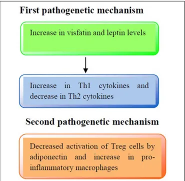
The immune profile in obese asthmatic children is illustrated
in Figure 1.
ROLE OF INTESTINAL MICROBIOTA IN OBESITY
Evidence has been provided that intestinal microbiota
composi-tion is altered in obese people, even including children, as recently
reviewed by Kabat and associates (121). Accordingly, intestinal
microbiota is active on both innate and adaptive immunity. With
regard to innate immunity, commensal bacterial products [e.g.,
lipopolysaccharides (LPS)] bind to Toll-like receptors (TLRs) and
Nod-like receptors present on gut immune and non-immune cells.
LPS, then, binds to TLR-4 on epithelial cells, soliciting release of the
AMP ASReg III-γ. This AMP is also produced by lamina propria
(LP) DCs via TLR-5 stimulation, which in turn, secrete IL-23, an
inducer of IL-22 producing Th17-type cells. The role of IL-22, in
this context is to amplify the release of other AMPs in the gut (121).
Furthermore, digestion of plant polysaccharides by microbiota
gives rise to short chain fatty acids (SCFAs), which, in turn, induce
release of IL-18 from intestinal epithelial cells via binding to the
G-protein coupled receptor (GPCR). Among SCFAs, acetate seems
to protect epithelial barrier function, mediating an anti-apoptotic
FIGURE 1 | Pathogenetic mechanisms involved in the immune profile in asthmatic obese children. First pathogenetic mechanisms: increased
production of visfatin and leptin serum levels may lead to an increased release of Th1 cytokines (IL-2, IFN-γ) and a decreased release of Th2 cytokines (IL-4, IL-13), respectively. Second pathogenetic mechanism: adiponectin reduces the activation of Treg cells and increases pro-inflammatory cytokines production (IL-1β, IL-6, and IL-8).
response. Taken together, all these gut microbiota-elicited activities
seem to be protective to the host.
With special reference to the adaptive immunity, microbiota
actively participates to intestinal IgA production via release of
B-cell activating factor, a proliferation-inducing ligand and
trans-forming growth factor (TGF)-β by intestinal epithelial cells and
DCs, thus leading to differentiation of B cells into IgA-producing
plasma cells. Also, follicular DCs promote differentiation of
IgA-producing plasma cells, as main producers of TGF-β in the Peyer’s
patches (PP). Moreover, interaction between Th17-like cells and
DCs favors IgA production within PP. Th17-like cells also promote
T-cell homing to the LP via soluble form of LTa3 lymphotoxin,
thus affecting T helper follicular (ThF) cell function. Also ATP,
generated by certain commensals, contributes to the induction of
Th17 cells which, in turn, differentiate into ThF cells, thus
pro-moting IgA production within PP. Polysaccharide A (PSA) from
Bacteroides fragilis, SCFAs, and TGF-
β induced by Clostridia (IV,
XIVa, XVII) promote Treg cell differentiation in the colon. PSA
acts on TLR-2 exposed on DCs, while SCFA operate via GPCR43
signaling (121). From these data, it is evident that
microbiota-induced IgA production and Treg cell differentiation in the gut
confers protection under healthy circumstances.
The altered microbiota in obesity subverts the protective
mech-anisms above illustrated. For instance, increase in segmented
fila-mentous bacteria induce release of serum amyloid A by epithelial
cells which acting upon DCs leads to differentiation and induction
of Th17 cells endowed with inflammatory activities (122,
123).
Furthermore, there is evidence that modifications of microbiota
composition in early life may increase the risk to developing
obe-sity in later life. For instance, children delivered by caesarian
section exhibit higher risk to become obese in adolescence when
compared to children born by vaginal delivery (124).
Further-more, breast-milk-fed children have less risk to become obese than
infant formula-fed children (125). Also, exposure of antibiotics in
early life may alter the composition of microbiota as observed
in mice which underwent changes in hepatic lipid and
choles-terol metabolism, thus leading to adiposity (126). In humans,
a correlation has been found between early-life antibiotic use
and obesity (127) and other studies have documented that
pres-ence/absence of specific microbiota components can modulate
immune response (128).
INTERVENTIONAL STUDIES IN CHILDHOOD OBESITY
Prevention seems to be the most appropriate strategy to
combat-ing obesity epidemic. Accordcombat-ing to a number of reports, there are
some major factors to be considered in the prevention of obesity
development (129–132). For instance, maternal factors encompass
monitoring of weight before conception and during pregnancy.
Breastfeeding may represent a favorable factor in terms of reduced
risk of obesity.
Dietary factors are based on limited consumption of
sugar-sweetened beverages and meals with servings of vegetable and
fruits, avoiding fast foods, and encouraging limited portions of
food.
Physical activity is based on levels of activity from moderate
to vigorous for one or more hours/day. Sedentary activity
(televi-sion, play station) should be limited to less than 2 h/day after age
two. Settings where food and PA can be influenced are represented
by schools and preschool institutions as well as after-school care
services. Built environment encompasses walking and cycling
net-works, parks, and recreation facilities. Home environment should
also be studied as a possible factor of obesity prevention but
the limitation is represented by the heterogeneity of homes and
possibility of access.
PREBIOTICS
By definition, prebiotics are non-digestible dietary fibers, which
are able to stimulate both growth and activity of gut bacteria. The
anti-obesogenic effects of prebiotics have mostly been evaluated
in experimental studies. In prebiotic-fed genetically obese mice
reduction of circulating endotoxins, pro-inflammatory cytokines
and intestinal permeability was reported (133). In rats with
steato-hepatitis induced by a high-fat diet, lactulose treatment reduced
liver inflammation and endotoxemia (134). Human clinical
tri-als based on the effects of prebiotics on obesity development
are very scanty. In infants receiving formula enriched in
pre-biotics (galacto-oligosaccharides and fructo-oligosaccharides at
9:1 ratio), an increase in Bifidobacteria was reported, thus
sug-gesting the possibility to influence adipocyte growth via
mod-ulation of microbiota composition (135). In patients with
non-alcoholic steatohepatitis (NASH), administration of
oligofruc-tose reduced serum aminotransferases and insulin levels (136).
Of note, in western countries NASH is very common in obese
children (137).
PROBIOTICS
Various strains of bacteria have been found in the gut of obese
and lean humans and, according to recent data, it seems that just
smaller modifications of intestinal commensals may account for
weight gain (138). On the other hand, in overweight adolescents,
the response to diet and PA was dependent on the microbiota
composition present before treatment (139). In this direction,
gut bacteria such as Ruminococcus bromii and Eubacterium rectale
were prevalent in individuals under a diet rich in resistant starch
who responded to a dietary weight loss program (140).
Further-more, Lactobacillus (L.) gasseri SBT2055 (LG2055) administration
to overweight subjects could lead to a significant reduction of
abdominal adiposity (141).
Just recently, fecal microbiota transplantation has been applied
to patients with inflammatory bowel disease and obese patients
(142). In particular, transfer of intestinal microbiota from lean
donors to obese recipients attenuated clinical manifestations of
MetS (143).
With special reference to pediatric obesity, the ratio between
Bacteroidetes and Firmicutes seems to play a role in weight gain
(144). Also, the size of Enterobacteriaceae, such as Desulfovibrio
and Akkermansia (A.) muciniphila were found to be related to
pediatric obesity (145). These last findings are also supported by
experiments in obese mice which underwent reduction of fat-mass
gain, endotoxemia, adipocyte-induced inflammation, and insulin
resistance following treatment with A. muciniphila (146).
Other clinical trials in pregnant mothers have documented that
administration of L. rhamnosus 4 weeks before expected delivery
up to 6 months after delivery could limit excessive weight gain
during the first 2 years of life but not between 2 and 4 years (147).
However, maternal supplementation with probiotics in the first
trimester of pregnancy did not modify prenatal and postnatal
growth rates (148,
149). In this framework, evidence has been
provided that milk from obese mothers is enriched in
Staphy-lococcus and Lactobacillus with lower counts of Bifidobacterium
when compared to that of normal weight women over the first
6 months of breastfeeding (150). The role of milk microbiota
on the development of neonatal microbiota needs to be further
investigated.
Synbiotics are a mixture of pro- and prebiotics which, when
ingested, are able to modulate gut microbiota and intestinal
immu-nity (151). In a recent clinical trial, Kelishadi and associates (152)
have administered overweight children with a synbiotic
(Protexin-London, England) composed by a combination of viable
Lacto-bacilli of human origin and fructo-oligosaccharides, as prebiotics.
Treated subjects exhibited a significant reduction in weight as
well as in TNF-α and IL-6 with an increase in adiponectin in
comparison to the placebo group. However, the modifications of
inflammatory markers were dependent on weight reduction.
FATTY ACIDS
Fatty acids exert important biological functions in the body as
a substrate for energy and the formation of membranes, also
acting as regulators of genetic expression (153). Excessive
con-sumption of saturated fatty acids or an altered ratio between
omega-3 polyunsaturated fatty acids (n-3 PUFAs) and n-6 PUFAs
leads to obesity, diabetes, neurodegenerative disease, and cancer
(154,
155). n-3 PUFAs and their derivatives, eicosapentaenoic
acid (EPA) and docosahexaenoic acid (DHA), are able to reduce
plasma triglyceride levels and body weight (156,
157).
Further-more, n-6 PUFAs promote excessive adipose tissue growth, while
n-3 PUFAs inhibit adipogenesis, while promoting storage and
accumulation of mature adipocytes (158–162). In terms of early
interventions at the level of maternal inflammation, studies in
transgenic Fat-1 mice have documented that increase in n-3/n-6
PUFA ratio diminished fetal-placental lipid exposure, thus
lim-iting adverse metabolic effects in adult offsprings (162). This
therapeutic model may be applied for preventive therapy in
obese pregnant women. However, as stated in a recent review
by Hauner and associates (163), results on the prevention of
childhood obesity obtained through modification of fatty
com-position during pregnancy and lactation are still contradictory
and inconsistent.
In a very recent paper (164), it has been reported that
con-sumption of n-3 PUFAs in obese adolescents along with dietary
restriction improved anthropometric parameters, while
decreas-ing plasma triglyceride levels. These effects correlated to a reduced
hypoxia in subcutaneous adipose tissue (164).
POLYPHENOLS
Polyphenols encompass flavonoids and non-flavonoids
(resvera-trol) compounds, which are widely distributed in the vegetal
king-dom. They are mostly contained in fruits, vegetables, and cereals,
and, therefore, contained in large amounts in MedDiet (165).
Polyphenols are endowed with oxidant and
anti-inflammatory activities and moderate consumption of red wine
has been shown to prevent cardiovascular disease according to
the French paradox (166–168). Experimentally, administration of
resveratrol from red grapes to obese rats reduced visceral obesity
and triglycerides and low-density lipoprotein plasma
concentra-tion, thus decreasing the risk of hypertension, dyslipidemia, and
steatosis (169). Furthermore, flavonoids hampered both
transcrip-tion factors and differentiatranscrip-tion of pre-adipocytes into mature
adipocytes (170). The in vitro demonstration that polyphenols
from fermented grape marc differentiate and activate peripheral
human Treg cells further supports the anti-inflammatory role of
these natural compounds (171). Just recently, evidence has been
provided that cocoa power supplementation ameliorated the
pro-inflammatory profile in high fat-fed obese mice (172). Same results
have been obtained with the administration of
epigallocatechin-3-gallate (EGCG) in high fat-fed mice (173). In obese women
admin-istration of green tea (EGCG) did not affect body weight, fat-mass,
energy, homeostasis, cardiometabolic risk factor, and liver
func-tion (174). Also, results by Li and associates (175) demonstrated
that green tea supplementation did not influence blood pressure
among overweight and obese adults. Conversely, in normal weight
obese syndrome subjects, regular consumption of dark
choco-late was useful in maintaining a good atherogenic profile for its
effects on HDL cholesterol, lipoprotein ratios, and inflammatory
markers (176).
In general terms, consumption of natural oxidants contained
in polyphenols with the diet may afford protection against
car-diovascular disease acting upon lipid profile, endothelial function,
and inflammatory mediators (177,
178).
MELATONIN
Melatonin is a pineal hormone endowed with anti-oxidant
proper-ties, thus, preventing nitro-oxidative stress mediated by
peroxyni-trites (179). Melatonin also exhibits anti-inflammatory activities
inhibiting ciclooxygenase-2 and inducible NO synthase and acting
upon transcriptional pathway involved in inflammation, such as
NF-κB, AP-1, Nrf2, as well as PI3K/Akt and MAPK kinase (180,
181). In view of its beneficial activities, melatonin has successfully
been used in rats with MetS diminishing insulin resistance, release
of TNF-α and IL-6 from adipocytes, low-density lipoprotein, and
very low-density lipoprotein plasma levels and body weight (182).
Evidence has been provided that melatonin can promote weight
loss in rodents via browning of white adipose tissue (183) and
this may represent a new approach to treat human obesity (184).
In fact, melatonin is a non-toxic compound widely distributed
in foodstuffs, such as olive oil, wine, coffee, tea, walnuts, and
grapes. Experimentally, a combination of resveratrol and
mela-tonin afforded protection in a model of myocardial infarction
(185). In this context, over the past few years, some clinical
tri-als have documented the beneficial effects of melatonin in patients
with MetS in terms of amelioration of blood pressure, lipid pattern,
and oxidative stress markers (186–188).
VITAMIN D
Vitamin D exerts anti-inflammatory activities, acting on DCs,
which in turn, induce activation of Treg cells. This vitamin
pos-sesses specific receptors, so-called Vitamin D receptors (VDR),
on gut epithelial and immune cells, while bacterial colonization
seems to affect distribution and expression of VDR (189). In
humans, vitamin D deficiency has been associated to asthma and
increased BMI (190–192). Furthermore, it has been hypothesized
that gut microbiota and vitamin D may be linked cofactors in the
pathogenesis of childhood asthma and obesity (193).
On these grounds, vitamin D may represent another possible
target of interventional studies in asthma-obesity but birth cohort
studies based on maternal and neonatal diet, gut microbiome,
immune response, and vitamin D-mediated immune regulation
are needed for asthma/obesity prevention.
The major nutritional attempts to prevent/attenuate childhood
obesity are illustrated in Table 4.
CONCLUSION
Nowadays, obesity is an epidemic in western and westernized
societies, thus representing one of the major consequences of
food-related disease (196). Besides appropriate dietary habits and
PA, an anti-inflammatory profile in response to food antigens
should be maintained throughout life span. For instance,
post-prandial low-grade inflammation is normally compensated by
dietary components, e.g., polyphenols, which activate gut Treg
cells (197).
A continuous intake of junk food since childhood may account
for the outcome of a systemic inflammation in overweight/obese
adults.
In interventional studies, another important aspect is
repre-sented by the identification of gut microbial components involved
in the development of obesity. In view of the diversity of human
intestinal microbiota (198), its variations among different obese
Table 4 | Some effects of natural products on obese humans.
Prebiotics, non-digestible dietary fibers, which are able to stimulate both growth and activity of gut bacteria (194), induce increase in Bifidobacteria in infants (135) and decrease in serum aminotransferases and insulin levels in NASH (136)
Probiotics, live bacteria, which when administered in adequate amounts confer a health benefit to the host (195). Administration of L. gasseri to overweight subjects reduced abdominal adiposity (141). Fecal microbiota transplantation attenuated clinical manifestation of MetS (143).
L. rhamnosus administration to pregnant mothers limited excessive weight gain during the first 2 years of life (147). Synbiotic administration to overweight children reduced weight gain and pro-inflammatory cytokine release (152)
n-3 Polyunsaturated fatty acids consumption in obese adolescents along with dietary restriction improved anthropometric parameters, while decreasing plasma triglyceride levels. These effects correlated to a reduced hypoxia in subcutaneous adipose tissue (164)
Polyphenols (flavonoids and non-flavonoids compounds) present in fruits vegetable and cereals exert anti-inflammatory and anti-oxidant activities (165)
Melatonin, a pineal hormone, has been shown to be protective in patients with MetS in terms of improvement of blood pressure, lipid profile, and oxidative biomarkers (186–188)
Vitamin D deficiency has been associated to asthma and increased BMI and, therefore, together with gut microbiota alterations may lead to childhood asthma and obesity outcome (193)
individuals should be investigated also in terms of interpersonal
microbiome differences (199). For instance, in the case of
treat-ment with probiotics generating a smaller effect size, the personal
microbiome effect which is very large may mask the more feeble
effects of treatment. Therefore, methods for the study of
micro-biome should be borrowed from the scientific areas and adjusted
for analyzing massive data as in the case of obese people.
In conclusion, more appropriate diets (e.g., MedDiet) or
sup-plements containing natural products (polyphenols, n-3 PUFA,
vitamins, synbiotics) are highly recommended to prevent or
atten-uate the noxious effects of obesity. In this last regard, in a very
recent review, Casas and associates (200) have stressed out the
immune protective effect of MedDiet, which may act on various
immune biomarkers, such as molecules involved in the stability of
atheromatous plaque.
ACKNOWLEDGMENTS
TM is a recipient of a contract in the context of the project
“Bioscience and Health (B&H)” (PONa3_00395). This paper
was in part supported by a grant from PON02_00186_2937475
(PRO.ALI.FUN.).
REFERENCES
1. International Association for the Study of Obesity. Obesity the Global Epidemic (2013). Available from: http://www.iaso.org/iotg/obesitytheglobalepidemic 2. Nicklas TA, Baranowski T, Cullen KW, Berenson G. Eating patterns, dietary
quality and obesity. J Am Coll Nutr (2001) 20:599–608. doi:10.1080/07315724. 2001.10719064
3. Parsons TJ, Power C, Logan S, Summerbell CD. Childhood predictors of adult obesity: a systematic review. Int J Obes Relat Metab Disord (1999)
23:S1–107.
4. Whitaker RC, Wright JA, Pepe MS, Seidel KD, Dietz WH. Predicting obesity in young adulthood from childhood and parental obesity. N Engl J Med (1997)
337:869–73. doi:10.1056/NEJM199709253371301
5. Lobstein T, Baur L, Uauy R; IASO International Obesity TaskForce. Obesity in children and young people: a crisis in public health. Obes Rev (2004) 5:4–104. doi:10.1111/j.1467-789X.2004.00133.x
6. Popkin BM, Gordon-Larsen P. The nutrition transition: worldwide obesity dynamics and their determinants. Int J Obes Relat Metab Disord (2004) 28:S2–9. doi:10.1038/sj.ijo.0802804
7. Wang Y, Lobstein T. Worldwide trends in childhood overweight and obesity. Int J Pediatr Obes (2006) 1:11–25. doi:10.1080/17477160600586747 8. Monteiro CA, Conde WL, Lu B, Popkin BM. Obesity and inequities in health
in the developing world. Int J Obes Relat Metab Disord (2004) 28:1181–6. doi:10.1038/sj.ijo.0802716
9. Aguilera CM, Olza J, Gil A. Genetic susceptibility to obesity and metabolic syndrome in childhood. Nutr Hosp (2013) 28:44–55. doi:10.3305/nh.2013.28. sup5.6917
10. Serra Majem L, Ribas Barba L, Aranceta Bartrina J, Pérez Rodrigo C, Saave-dra Santana P, Peña Quintana L. [Childhood and adolescent obesity in Spain. Results of the enKid study (1998-2000)]. Med Clin (Barc) (2003) 121:725–32. doi:10.1016/S0025-7753(03)74077-9
11. Flegal KM, Troiano RP, Pamuk ER, Kuczmarski RJ, Campbell SM. The influ-ence of smoking cessation on the prevalinflu-ence of overweight in the United States. N Engl J Med (1995) 333:1165–70. doi:10.1056/NEJM199511023331801 12. Bautista-Castaño I, Sangil-Monroy M, Serra-Majem L; Comité de Nutrición y
Obesidad Infantil de la Sociedad Española de Nutrición Comunitaria. [Knowl-edge and gaps on the role of nutrition and physical activity on the onset of childhood obesity]. Med Clin (Barc) (2004) 123:782–93.
13. Freedman DS, Dietz WH, Tang R, Mensah GA, Bond MG, Urbina EM, et al. The relation of obesity throughout life to carotid intima-media thickness in adulthood: the Bogalusa Heart Study. Int J Obes Relat Metab Disord (2004)
28:159–66. doi:10.1038/sj.ijo.0802515
14. Lauer RM, Lee J, Clarke WR. Factors affecting the relationship between child-hood and adult cholesterol levels: the Muscatine Study. Pediatrics (1988)
82:309–18.
15. Bremer AA, Mietus-Snyder M, Lustig RH. Toward a unifying hypothesis of metabolic syndrome. Pediatrics (2012) 129:557–70. doi:10.1542/peds.2011-2912
16. Shu CJ, Benoist C, Mathis D. The immune system’s involvement in obesity-driven type 2 diabetes. Semin Immunol (2012) 24:436–42. doi:10.1016/j.smim. 2012.12.001
17. Falagas ME, Kompoti M. Obesity and infection. Lancet Infect Dis (2006)
6:438–46. doi:10.1016/S1473-3099(06)70523-0
18. Milner JJ, Beck MA. The impact of obesity on the immune response to infec-tion. Proc Nutr Soc (2012) 71:298–306. doi:10.1017/S0029665112000158 19. Gregor MF, Hotamisligil GS. Inflammatory mechanisms in obesity. Annu Rev
Immunol (2011) 29:415–45. doi:10.1146/annurev-immunol-031210-101322 20. Hotamisligil GS, Shargill NS, Spiegelman BM. Adipose expression of tumor
necrosis factor-alpha: direct role in obesity-linked insulin resistance. Science (1993) 259:87–91. doi:10.1126/science.7678183
21. Pradhan AD, Manson JE, Rifai N, Buring JE, Ridker PM. C-reactive protein, interleukin 6, and risk of developing type 2 diabetes mellitus. JAMA (2001)
286:327–34. doi:10.1001/jama.286.3.327
22. Lumeng CN, Bodzin JL, Saltiel AR. Obesity induces a phenotypic switch in adipose tissue macrophage polarization. J Clin Invest (2007) 117:175–84. doi:10.1172/JCI29881
23. Lumeng CN, Deyoung SM, Bodzin JL, Saltiel AR. Increased inflammatory properties of adipose tissue macrophages recruited during diet-induced obe-sity. Diabetes (2007) 56:16–23. doi:10.2337/db06-1076
24. Osborn O, Olefsky JM. The cellular and signaling networks linking the immune system and metabolism in disease. Nat Med (2012) 18:363–74. doi:10.1038/nm.2627
25. Olefsky JM, Glass CK. Macrophages, inflammation, and insulin resistance. Annu Rev Physiol (2010) 72:219–46. doi:10.1146/annurev-physiol-021909-135846
26. Patsouris D, Li PP, Thapar D, Chapman J, Olefsky JM, Neels JG. Ablation of CD11c-positive cells normalizes insulin sensitivity in obese insulin resistant animals. Cell Metab (2008) 8:301–9. doi:10.1016/j.cmet.2008.08.015 27. Wellen KE, Hotamisligil GS. Inflammation, stress, and diabetes. J Clin Invest
(2005) 115:1111–9. doi:10.1172/JCI25102
28. Huh JY, Park YJ, Ham M, Kim JB. Crosstalk between adipocytes and immune cells in adipose tissue inflammation and metabolic dysregulation in obesity. Mol Cells (2014) 37:365–71. doi:10.14348/molcells.2014.0074
29. Rocha VZ, Folco EJ, Sukhova G, Shimizu K, Gotsman I, Vernon AH, et al. Interferon-gamma, a Th1 cytokine, regulates fat inflammation: a role for adaptive immunity in obesity. Circ Res (2008) 103:467–76. doi:10.1161/ CIRCRESAHA.108.177105
30. O’Rourke RW, White AE, Metcalf MD, Winters BR, Diggs BS, Zhu X. Systemic inflammation and insulin sensitivity in obese IFN-γ knockout mice. Metabo-lism (2012) 61:1152–61. doi:10.1016/j.metabol.2012.01.018
31. DeFuria J, Belkina AC, Jagannathan-Bogdan M, Snyder-Cappione J, Carr JD, Nersesova YR, et al. B cells promote inflammation in obesity and type 2 diabetes through regulation of T-cell function and an inflammatory cytokine profile. Proc Natl Acad Sci USA (2013) 110:5133–8. doi:10.1073/pnas.1215840110 32. Feuerer M, Herrero L, Cipolletta D, Naaz A, Wong J, Nayer A, et al. Lean, but
not obese, fat is enriched for a unique population of regulatory T cells that affect metabolic parameters. Nat Med (2009) 15:930–9. doi:10.1038/nm.2002 33. Han JM, Patterson SJ, Speck M, Ehses JA, Levings MK. Insulin inhibits IL-10-mediated regulatory T cell function: implications for obesity. J Immunol (2014)
192:623–9. doi:10.4049/jimmunol.1302181
34. Winer S, Paltser G, Chan Y, Tsui H, Engleman E, Winer D. Obesity pre-disposes to Th17 bias. Eur J Immunol (2009) 39:2629–35. doi:10.1002/eji. 200838893
35. Sumarac-Dumanovic M, Stevanovic D, Ljubic A, Jorga J, Simic M, Stamenkovic-Pejkovic D, et al. Increased activity of interleukin-23/interleukin-17 proinflammatory axis in obese women. Int J Obes (Lond) (2009) 33:151–6. doi:10.1038/ijo.2008.216
36. Jagannathan-Bogdan M, McDonnell ME, Shin H, Rehman Q, Hasturk H, Apovian CM, et al. Elevated proinflammatory cytokine production by a skewed T cell compartment requires monocytes and promotes inflammation in type 2 diabetes. J Immunol (2011) 186:1162–72. doi:10.4049/jimmunol. 1002615
37. Zúñiga LA, Shen WJ, Joyce-Shaikh B, Pyatnova EA, Richards AG, Thom C. IL-17 regulates adipogenesis, glucose homeostasis, and obesity. J Immunol (2010)
185:6947–59. doi:10.4049/jimmunol.1001269
38. Pini M, Fantuzzi G. Enhanced production of IL-17A during zymosan-induced peritonitis in obese mice. J Leukoc Biol (2010) 87:51–8. doi:10.1189/ jlb.0309188
39. Ahmed M, Gaffen SL. IL-17 in obesity and adipogenesis. Cytokine Growth Fac-tor Rev (2010) 21:449–53. doi:10.1016/j.cytogfr.2010.10.005
40. Erbel C, Akhavanpoor M, Okuyucu D, Wangler S, Dietz A, Zhao L, et al. IL-17A influences essential functions of the monocyte/macrophage lineage and is involved in advanced murine and human atherosclerosis. J Immunol (2014)
193:4344–55. doi:10.4049/jimmunol.1400181
41. Rausch ME, Weisberg S, Vardhana P, Tortoriello DV. Obesity in C57BL/6J mice is characterized by adipose tissue hypoxia and cytotoxic T-cell infiltration. Int J Obes (Lond) (2008) 32:451–63. doi:10.1038/sj.ijo.0803744
42. Nishimura S, Manabe I, Nagasaki M, Eto K, Yamashita H, Ohsugi M, et al. CD8+ effector T cells contribute to macrophage recruitment and adipose tissue inflammation in obesity. Nat Med (2009) 15:914–20. doi:10.1038/nm.1964 43. Jiang E, Perrard XD, Yang D, Khan IM, Perrard JL, Smith CW, et al.
Essen-tial role of CD11a in CD8+ T-cell accumulation and activation in adipose tissue. Arterioscler Thromb Vasc Biol (2014) 34:34–43. doi:10.1161/ATVBAHA. 113.302077
44. Winer DA, Winer S, Shen L, Wadia PP, Yantha J, Paltser G, et al. B cells promote insulin resistance through modulation of T cells and production of pathogenic IgG antibodies. Nat Med (2011) 17:610–7. doi:10.1038/nm.2353
45. Abeysekara P, Turchi R, O’Neil M. Obesity and children with special health-care needs: special considerations for a special population. Curr Opin Pediatr (2014) 26:508–15. doi:10.1097/MOP.0000000000000124
46. Del Chirico F, Vernocchi P, Dallapiccola B, Putignani L. Mediterranean diet and health: food effects on gut microbiota and disease control. Int J Mol Sci (2014)
15:11678–99. doi:10.3390/ijms150711678
47. Cao AT, Yao S, Stefka AT, Liu Z, Qin H, Liu H, et al. TLR4 regulates IFN-γ and IL-17 production by both thymic and induced Foxp3+ Tregs during intesti-nal inflammation. J Leukoc Biol (2014) 96:895–905. doi:10.1189/jlb.3A0114-056RR
48. Weber A, Zimmermann C, Kieseier BC, Hartung HP, Hofstetter HH. Bacteria and their cell wall components uniformly co-active IL-17 producing thymo-cytes. Clin Exp Immunol (2014) 178:504–15. doi:10.1111/cei.12414 49. Cosmi L, Liotta F, Maggi E, Romagnani S, Annunziato F. Th17 and non-classic
Th1 cells in chronic inflammatory disorders: two sides of the same coin. Int Arch Allergy Immunol (2014) 164:171–7. doi:10.1159/000363502
50. Zeng H, Chi H. The interplay between regulatory T cells and metabolism in immune regulation. Oncoimmunology (2013) 2:e26586. doi:10.4161/onci. 26586
51. Iyer SS, Cheng G. Role of interleukin 10 transcriptional regulation in inflam-mation and autoimmune disease. Crit Rev Immunol (2012) 32:23–63. doi:10. 1615/CritRevImmunol.v32.i1.30
52. Pereira S, Teixeira L, Aguilar E, Oliveira M, Savassi-Rocha A, Pelaez JN, et al. Modulation of adipose tissue inflammation by FOXP3+ Treg cell, IL-10, and TGF-β in metabolically healthy class III obese individuals. Nutrition (2014)
30:784–90. doi:10.1016/j.nut.2013.11.023
53. Cole TJ, Lobstein T. Extended international (IOTF) body mass index cut-offs for thinness, overweight and obesity. Pediatr Obes (2012) 7:284–94. doi:10.1111/j.2047-6310.2012.00064.x
54. O’Keefe JH, Gheewala NM, O’Keefe JO. Dietary strategies for improving post-prandial glucose, lipids, inflammation, and cardiovascular health. J Am Coll Cardiol (2008) 51:249–55. doi:10.1016/j.jacc.2007.10.016
55. Holt EM, Steffen LM, Moran A, Basu S, Steinberger J, Ross JA, et al. Fruit and vegetable consumption and its relation to markers of inflammation and oxidative stress in adolescents. J Am Diet Assoc (2009) 109(3):414–21. doi:10.1016/j.jada.2008.11.036
56. Casas R, Sacanella E, Estruch R. The immune protective effect of the Mediterranean diet against chronic low-grade inflammatory diseases. Endocr Metab Immune Disord Drug Targets (2014) 14:245–54. doi:10.2174/ 1871530314666140922153350
57. Bonaccio M, Di Castelnuovo A, Bonanni A, Costanzo S, De Lucia F, Per-sichillo M, et al. Decline of the Mediterranean diet at a time of economic crisis. Results from the Moli-Sani study. Nutr Metab Cardiovasc Dis (2014)
24:853–60. doi:10.1016/j.numecd.2014.02.014
58. Lim GB. Risk factors: mechanistic insights into the cardiovascular benefits of a Mediterranean diet. Nat Rev Cardiol (2014) 11:433. doi:10.1038/nrcardio. 2014.92
59. Knoops KT, de Groot LC, Kromhout D, Perrin AE, Moreiras-Varela O, Menotti A, et al. Mediterranean diet, lifestyle factors, and 10-year mortality in elderly European men and women: the HALE project. JAMA (2004) 292:1433–9. doi:10.1001/jama.292.12.1433
60. Vieira VJ, Valentine RJ, Wilund KR, Antao N, Baynard T, Woods JA. Effects of exercise and low-fat diet on adipose tissue inflammation and metabolic com-plications in obese mice. Am J Physiol Endocrinol Metab (2009) 296:E1164–71. doi:10.1152/ajpendo.00054.2009
61. Gleeson M, Bishop NC, Stensel DJ, Lindley MR, Mastana SS, Nimmo MA. The anti-inflammatory effects of exercise: mechanisms and implications for the prevention and treatment of disease. Nat Rev Immunol (2011) 11:607–11. doi:10.1038/nri3041
62. Miglio C, Peluso I, Raguzzini A, Villaño DV, Cesqui E, Catasta G, et al. Antiox-idant and antiinflammatory response following high-fat meal consumption in overweight subjects. Eur J Nutr (2012) 52:1107–14. doi:10.1007/s00394-012-0420-7
63. Calder PC, Ahluwalia N, Brouns F, Buetler T, Clement K, Cunningham K, et al. Dietary factors and low-grade inflammation in relation to overweight and obesity. Br J Nutr (2011) 106:S5–78. doi:10.1017/S0007114511005460 64. Korhonen R, Kosonen O, Hamalainen M, Moilanen E. Nitric oxide-releasing
compounds inhibit the production of interleukin-2, -4 and -10 in acti-vated human lymphocytes. Basic Clin Pharmacol Toxicol (2008) 103:322–8. doi:10.1111/j.1742-7843.2008.00275.x
65. Cani PD, Bibiloni R, Knauf C, Waget A, Neyrinck AM, Delzenne NM, et al. Changes in gut microbiota control metabolic endotoxemia-induced inflam-mation in high-fat diet-induced obesity and diabetes in mice. Diabetes (2008)