1130-0108/2020/112/6/474-476 • REVISTA ESPAÑOLA DE ENFERMEDADES DIGESTIVAS
© Copyright 2020. SEPD y © ARÁN EDICIONES, S.L. REV ESP ENFERM DIG 2020:112(6):474-476 DOI: 10.17235/reed.2020.6123/2018 Piccini B, Ulivelli M, Amato MP, Bartalini S, Falcini M, Giannini M, Magna-ni E, Massacesi L, Repice AM, Vascotto M, Grosso S. Association of celiac disease in patients with multiple sclerosis in Tuscany. Rev Esp Enferm Dig 2020;112(6):474-476
DOI: 10.17235/reed.2020.6123/2018
Association of celiac disease in patients with multiple sclerosis in Tuscany
ORIGINAL PAPERS
Barbara Piccini
1, Monica Ulivelli
2, Maria Pia Amato
3, Sabina Bartalini
4, Mario Falcini
5, Marta Giannini
6,
Eliana Magnani
7, Luca Massacesi
8, Anna Maria Repice
7, Marina Vascotto
9and Salvatore Grosso
101Tuscany Regional Centre of Pediatric Diabetes. Pediatric Department. Meyer Children’s Hospital. Florence, Italy. 2Neurology and
Neurophysiopathology Department. Center for Multiple Sclerosis. University of Siena. Siena, Italy. 3Department of NEUROFARBA. Section
Neurosciences. University of Florence, Florence. Italy. Institute Don Gnocchi. Florence, Italy. 4Neurology and Neurophysiopathology Department.
Center for Multiple Sclerosis. University Hospital Santa Maria alle Scotte. Siena, Italy. 5Neurology Department. Santo Stefano Hospital. Prato, Italy. 6Department of NEUROFARBA. Section Neurosciences. University of Florence. Florence, Italy. 7Division Neurology 2. Careggi University Hospital.
University of Florence. Florence, Italy. 8Department of Neurosciences. Careggi University Hospital. University of Florence. Florence, Italy. 9Clinical
Pediatrics. Department of Pediatrics. University Hospital Santa Maria alle Scotte. Siena, Italy. 10Clinical Pediatrics. Department of Pediatrics.
University of Siena. Siena, Italy
Received: 26/12/2018 · Accepted: 18/09/2019
Correspondence: Barbara Piccini. Tuscany Regional Centre of Pediatric Diabetes. Pediatric Department. “A. Meyer” University Children’s Hospital. Viale Pieraccini, 24 I-50139 Florence, Italy. e-mail: [email protected]
ABSTRACT
Background and study purpose: to describe the
comor-bidity of celiac disease among a large cohort of multiple sclerosis patients in Tuscany.
Methods: the association of celiac disease among
multi-ple sclerosis adult patients (n=2050) was retrospectively evaluated.
Results: 13 patients were diagnosed with celiac disease,
the female:male ratio was 3.3:1 and the median age at diag-nosis was 34.2 years (SD 13). Seventy-seven per cent of subjects complained about gastrointestinal symptoms. IgA anti- transglutaminase was positive in 85 % of cases and there was 70 % of villous atrophy.
Conclusions: the frequency of celiac disease among
multi-ple sclerosis patients examined was lower than in the gen-eral population (0.6 % vs 1 %) (p = 0.65).
Keywords: Celiac disease. Multiple sclerosis. Epidemiology.
Comorbidity.
INTRODUCTION
Multiple sclerosis (MS) is an autoimmune inflammatory demyelinating disease of the central nervous system (CNS).
Multiple sclerosis-like diseases and a headache associated with brain white-matter lesions (WMLs), which are docu-mented by magnetic resonance imaging (MRI), have been reported in celiac disease (CD) patients (1). No conclusive data support the hypothesis that there is an increased prev-alence of MS in patients with CD and of CD in patients with MS. The possible relationship between CD and MS was first recorded in 1965, when the pathogenesis of CD was not completely understood (2). MS and CD share common aspects of a dysregulation of the immune system and they are both inflammatory diseases due to T cell-mediated immunity. The aim of this study was to describe the asso-ciation of CD among a large cohort of MS patients in Tus-cany, compared to the prevalence of CD among the general population.
METHODS
The frequency of CD was retrospectively evaluated among a cohort of 2050 adult patients with MS followed-up during 2016 that were included in the database of the Centers for the diagnosis and treatment of MS in Firenze, Prato and Siena. MS cases were diagnosed between 1985 and 2015, based on the McDonald criteria and subsequent revisions (3). Patients who presented with gastrointestinal or extra-intes-tinal symptoms evocative of CD and/or with iron or vitamin
Funding/support: This research did not receive any specific grant from funding agen-cies in the public, commercial, or not-for-profit sectors.
Author´s contributions: PB, UM and GS wrote the manuscript, researched data and contributed to the discussion. BS, FM, AMP, GM, ME, RAM, ML and VM researched data. UM and ML contributed to the discussion and edited the manuscript. PB is the guarantor of this work and therefore, had full access to all the data in the study and takes responsibility for the integrity of the data and the accuracy of the data analysis.
Association of celiac disease in patients with multiple sclerosis in Tuscany
REV ESP ENFERM DIG 2020:112(6):474-476 DOI: 10.17235/reed.2020.6123/2018
475
deficiency, underwent serologic tests for CD, including tis-sue transglutaminase IgA antibodies (tTG-IgA), anti-endo-mysium IgA antibodies (EMA) and/or antibodies against deaminated gliadin peptides (DGP), together with dosage of total IgA levels. An esophagogastroduodenoscopy (EGDS) with small intestine biopsies was performed in subjects with a positive serology, in order to confirm CD diagnosis. The diagnosis was made according to the revised criteria of the European Society for Pediatric Gastroenterology Hepa-tology and Nutrition (ESPGHAN) modified in 1990 (4) and after 2012, according to the new ESPGHAN guidelines (5). A CD prevalence of 1:100 was considered in the general population, based on regional differences in Europe where the prevalence varies from 0.3 % in Germany to 2.4 % in Finland (6). Statistical significance was calculated using 2 x 2 contingency tables and the χ2 test and corrected with the
Fisher exact test when sample sizes were small. Values of p < 0.05 were considered as significant.
RESULTS
CD was diagnosed in 13/2050 patients attending 4 MS cen-ters in 3 hospitals in Tuscany region, including 3 males with a median age at CD diagnosis 34.2 years (SD 13) (Table 1). The median age at MS diagnosis was 28.8 years (SD 10.8) and 77 % of patients were female (F:M 3.3:1). Most patients
were self-sufficient and in the classification of MS subtypes, the relapsing-remitting form was the most common (61 % of cases). Thirty-one per cent of patients were first diag-nosed with CD and they started a gluten free diet (GFD) before being diagnosed with MS, 54 % of patients were diagnosed with CD after MS and 15 % of patients were diagnosed with CD and MS simultaneously. Seventy-seven per cent of the subjects complained about gastrointestinal symptoms. tTG-IgA levels were measured in all patients and tested positive in 85 % of cases. Patient 11 was neg-ative for tTG IgA, EMA and DGP and was treated ab initio with monoclonal antibodies. The negative tests results were in contrast to the presence of histologically docu-mented atrophic lesions (Marsh-Oberhuber 3C) (7) and with gastrointestinal symptoms consistent with CD, even though HLA-DQ2/DQ8 were negative. Patient 13 complained about abdominal pain that disappeared after gluten elimination, despite negative CD antibodies. This factor, a positive fam-ily history of CD and the presence of HLA-DQ2 homozy-gosity suggested a potential CD. Duodenal biopsies were performed in 12/13 patients (one patient refused). 70 % of subjects had a histological pattern indicative of CD with vil-lous atrophy (Marsh-Oberhuber 3 A, B or C). Patient 10 had mild histological lesions (Marsh 1), positive IgA-tTG and DGP antibodies, gastrointestinal symptoms and positive DQ2, indicating a potential CD. Patient 4 presented with a normal duodenal histology but moderate chronic gastritis,
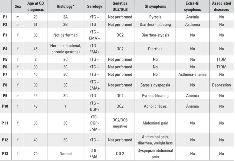
Table 1. Clinical data, serology, histology and haplotype among 13 patients with MS/CD Sex Age at CD
diagnosis Histology* Serology
Genetics DQ2/DQ8 GI symptoms Extra-GI symptoms Associated diseases
P1 m 29 3A tTG + Not performed Pyrosis Anemia No
P2 m 51 3B tTG + Not performed Diarrhea - bloating Asthenia No
P3 f 30 Not performed tTG + EMA + DQ2 Diarrhea-stypsis No No P4 f 46 Normal (duodenal, chronic gastritis) tTG + EMA+ DQ2 Diarrhea No No P5 f 2 3C tTG + Not performed No No T1DM P6 f 30 3C tTG + Not performed No No T1DM
P7 f 40 3C tTG + Not performed No Asthenia-anemia No
P8 f 30 3C tTG +
EMA+ Not performed Stypsis dyspepsia No Depression
P9 m 46 3C tTG + DQ2 Pyrosis bloating Anemia No
P10 f 43 1 tTG +
DGP+ DQ2 Acholic feces Anemia No
P 11 f 38 3C
tTG- DGP- EMA
-DQ2/DQ8
negative Abdominal pain No No
P12 f 40 3C tTG + Not performed Abdominal pain,
diarrhea, weight loss No No
P13 f 20 Normal tTG
-EMA - DQ 2
Dyspepsia abdominal
pain No No
*Modified Marsh-Oberhuber classification (7).
B. Piccini et al.
REV ESP ENFERM DIG 2020:112(6):474-476 DOI: 10.17235/reed.2020.6123/2018
476
positive IgA-tTG and EMA, a permissive haplotype (DQ2 positive) and gastrointestinal symptoms such as diarrhea. Thus, CD was suspected. Patient 13 had potential CD, as mentioned previously. DQ2 and DQ8 were investigated in 46 % of cases. Predisposing HLA-DQ2 and HLA-DQ8 were negative in just one patient (patient 11). All patients with permissive haplotype showed DQ2.
Only patient 11 repeated EGDS after 10 months of GFD and the atrophic lesions had reverted from Marsh-Ober-huber type 3C to type 1. This patient presented with neg-ative serology without a predisposing haplotype and a third endoscopy was not performed. In conclusion, a CD diagnosis was made in 13/2050 patients with MS, with a prevalence of 0.6 % in the cohort of patients considered. There were no differences between CD prevalence among MS patients and the general population (p 0.65).
DISCUSSION
The present study does not support an increased frequency of CD among MS patients. Moreover, the data surround-ing clinical presentation of CD among MS patients mainly indicate an atypical presentation with non-specific gastro-intestinal symptoms and extragastro-intestinal manifestations. In this study, gastrointestinal symptoms were present in 77 % of patients and 38 % of subjects had diarrhea as the main symptom.
Since 1966, a variety of neurological disorders have been reported in association with CD (8). The mechanisms impli-cated in the pathogenesis of neurological disorders asso-ciated with CD are not completely understood, due to the fact that only some subjects develop CNS symptoms and the extreme variability of these symptoms. TG6 represents a useful marker of the neurological manifestations in CD patients (9). MS and CD are inflammatory T-cell-mediat-ed autoimmune diseases. However, the evidence is insuf-ficient to support an increased frequency of CD among MS patients and conversely of MS among CD patients, despite these shared etiological aspects. Nicoletti et al. (10) evalu-ated the presence of CD relevalu-ated antibodies in 217 patients with MS and 200 healthy controls and there was only one control with CD specific antibodies. On the other hand, Rodrigo et al. (11) reported an increased prevalence of biopsy-proven CD in 8 out of 72 MS patients (11.1 %). Lud-vigsson et al. (12) evaluated the comorbidity of CD-neuro-degenerative/neuroinflammatory diseases using data from the Swedish national register. CD was positively associat-ed with polyneuropathy but not with MS. The strength of the present study is the large sample size, the expertise on immunological diseases of highly specialized centers in a third level hospital and the multidisciplinary approach. Some limitations must be highlighted: (i) only patients with evocative symptoms underwent serological screening and it is possible that CD was under-diagnosed due to the fact that cases with paucisymptomatic or silent CD were missed;
(ii) HLA-DQ2 or HLA-DQ8 molecules were investigated in only 46 % of CD-MS patients; and (iii) it should be taken into account that the positivity of serological markers for CD in these patients could be influenced by MS therapies. In conclusion, this study highlights the usefulness to imple-ment a case-finding strategy in order to address the issue of comorbidity CD-MS. HLA-DQ2 and HLA-DQ8 typing would allow the identification of patients with MS who are at risk and should be periodically screened for CD. This is likely due to the strong negative predictive value of DQ2-DQ8 aplotype and the different HLA class II susceptibility to MS (DQB1*0602 or DR2). On the other hand, CD should be retained as a red flag for a particular diagnostic caution in patients with non-specific MS symptoms and/or WMLs.
REFERENCES
1. Hadjivassiliou M, Grünewald RA, Lawden M, et al. Headache and CNS white matter abnormalities associated with gluten sensitivity. Neurology 2001;56:385-88. DOI: 10.1212/WNL.56.3.385
2. Shatin R. Gluten and multiple sclerosis. Br Med J 1965;1:1433-4. DOI: 10.1136/bmj.1.5447.1433-d
3. Polman CH, Reingold SC, Banwell B, et al. Diagnostic criteria for mul-tiple sclerosis: 2010 revisions to the Mc Donald criteria. Ann Neurol 2011;69:292-302. DOI: 10.1002/ana.22366
4. Revised criteria for diagnosis of coeliac disease. Report of working group of European Society of Pediatric Gastroenterology and Nutrition. Arch Dis Child 1990;65:909-11. DOI: 10.1136/adc.65.8.909
5. Husby S, Koletzko S, Korponay-Szabó IR, et al; ESPGHAN Working Group on Coeliac Disease Diagnosis; ESPGHAN Gastroenterology Committee; Eu-ropean Society for Pediatric Gastroenterology, Hepatology, and Nutrition. European Society for Pediatric Gastroenterology, Hepatology, and Nutri-tion guidelines for the diagnosis of coeliac disease. J Pediatr Gastroente-rol Nutr 2012;54:136-60. DOI: 10.1097/MPG.0b013e31821a23d0 6. Mustalahti K, Catassi C, Reunanen A, et al. The prevalence of celiac
disea-se in Europe: results of a centralized, International mass screening project. Ann Med 2010;42:587-95. DOI: 10.3109/07853890.2010.505931 7. Oberhuber G, Granditsch G, Vogelsang H. The histopatology of celiac
disea-se: time for a standardized report scheme for pathologists. Eur J Gastroen-terol Hepatol 1999;11:1185-94. DOI: 10.1097/00042737-199910000-00019 8. Cooke WT, Smith WT. Neurological disorders associated with adult coeliac
disease. Brain 1966;89:683-722. DOI: 10.1093/brain/89.4.683
9. Hadjivassiliou M, Rao DG, Grìnewald RA, et al. Neurological Dysfunction in Coeliac Disease and Non-Coeliac Gluten Sensitivity. Am J Gastroenterol 2016;111:561-7. DOI: 10.1038/ajg.2015.434
10. Nicoletti A, Patti F, Lo Fermo S, et al. Frequency of celiac disease is not increased among multiple sclerosis patients. Mult Scler 2008;14:698-700. DOI: 10.1177/1352458507087268
11. Rodrigo L, Hernández-Lahoz C, Fuentes D, et al. Prevalence of celiac di-sease in multiple sclerosis. BMC Neurol 2011;11:31. DOI: 10.1186/1471-2377-11-31
12. Ludvigsson JF, Olsson T, Ekbom A, et al. A population-based study of celiac disease, neurodegenerative and neuroinflammatory diseases. Aliment Pharmacol Ther 2007;25:1317-27. DOI: 10.1111/j.1365-2036.2007.03329.x