CHAPTER 4
Deficient DNA Mismatch Repair in Lynch
Syndrome-Associated Colorectal Adenomas
This chapter has been published under the title:
“Deficient DNA Mismatch Repair is Common in Lynch Syndrome-Associated
Colorectal Adenomas”, Pino MS, Mino-Kenudson M, Wildemore BM,
Ganguly A, Batten J, Sperduti I, Iafrate AJ, Chung DC; J Mol Diagn 2009
May;11(3):238-47.
Aim of the Study
Lynch syndrome (LS), also known as hereditary nonpolyposis colorectal cancer (HNPCC), is an autosomal dominant disorder that accounts for 2-5% of all colorectal cancers. LS is characterized by high penetrance, early-onset colorectal and endometrial cancer and an increased risk of certain extra-colonic cancers, including tumors of the stomach, small bowel, ovary, biliary tract, renal pelvis, and ureter. Germline mutations of DNA mismatch repair (MMR) genes, most commonly hMLH1, hMSH2, or less frequently
hMSH6 and hPMS2, underlie the majority of cases of LS. Mutations in these genes impair
the function of MMR proteins, which normally recognize and repair mismatched nucleotides and insertion/deletion loops caused by slippage of DNA polymerase. In tumors that develop due to defective DNA mismatch repair, repetitive DNA sequences known as microsatellites tend to undergo a high level of genetic alteration, resulting in microsatellite instability (MSI). MSI can be identified in greater than 90% of colorectalcancers that arise in individuals with LS, whereas in sporadic colorectal cancer it occurs in 15% of cases, typically from silencing of the hMLH1 gene by promoter hypermethylation. Microsatellite analysis has therefore been proposed as a useful diagnostic tool to screen for LS. In addition, immunohistochemical (IHC) analysis for the 4 MMR proteins is a complementary approach that can pinpoint the specific gene most likely to be mutated.
The diagnosis of LS can be difficult to make because family history information is often incomplete and there is no characteristic clinical phenotype such as diffuse polyposis. Nevertheless, its early recognition is essential to identify patients at high-risk who will require intensive cancer surveillance. In LS, carcinogenesis proceeds through the
adenoma-carcinoma sequence, albeit at a more rapid pace. A significant patient survival advantage and reduction in the incidence of colorectal tumors has been observed following colonoscopic screening and polypectomy [1]. Although the number of polyps in Lynch patients appears to be similar to the general population, the polyps are more likely to occur at a younger age, have a predilection for the proximal colon, be larger, display villous features or high-grade dysplasia, and most importantly, grow rapidly and progress to invasive cancer in less than 3 years [2-4].
The recognition of hereditary colon cancer syndromes and LS, in particular, is increasing in the population. Many individuals with suspected LS now undergo routine colonoscopic screening with polypectomy. In such a scenario, there is no colon cancer tissue available for MSI and IHC testing. The present study was undertaken to test the hypothesis that MSI testing and IHC analysis in pre-cancerous colorectal adenomas instead of colorectal cancers may be an alternative approach to screen for LS.
Patients and Methods
Patients
Carriers of a known germline mismatch repair gene mutation referred to the Gastrointestinal Cancer Genetics Clinic at the Massachusetts General Hospital (MGH) were identified. Those who had polyps endoscopically removed during surveillance examinations between January 1997 and December 2007 were selected for further analysis. Whenever available, hyperplastic polyps were also collected. Genetic counseling and testing were performed as part of routine clinical care. Sequencing of the hMLH1, hMSH2 and hMSH6 genes was performed in CLIA-certified laboratories as part of routine clinical care and included sequencing of all exons and exon-intron boundaries as well as gene rearrangement analysis. All mutations were determined to be deleterious with the exception of a variant of uncertain significance in the hMLH1 gene (H718P). Endoscopy and pathology reports from all patients enrolled were reviewed, and the location, size, and histopathological features of all polyps were recorded. The cecum, ascending, and transverse colon were regarded as the proximal or right colon, while the descending, sigmoid and rectum were referred to as the distal or left colon.
Analysis of MSI
MSI assays were performed on microdissected DNA, extracted using the Puregene DNA Purification Kit (Gentra Systems, Minneapolis, MN), from paraffin-embedded tissue blocks. Primer sets comprised the 5 reference panel markers recommended by the National Cancer Institute, with 5’ phosphoramidite fluorescent labeling of forward primers as follows: BAT-25 (NED), BAT-26 (6-FAM), D5S346 (VIC), D17S250 (6-FAM), and
D2S123 (VIC) [5].The primer sequences for D2S123 were
5’-AACATTGCTGGAAGTTCTGG-3’ (forward) and
5’-GTGTCTTGACTTTCCACCTATGGGACTG-3’ (reverse). Primer sequences for the remaining loci were identical to those previously described except that a 5’ GTGTCTT sequence was added to each reverse primer to facilitate non-template adenylation of the 3’ end of the forward strand. Polymerase chain reaction (PCR) amplifications were performed in an Eppendorf Mastercycler Gradient (Eppendorf, Hamburg, Germany). PCR was conducted in a total volume of 20 µl containing 1X Platinum Taq PCR buffer, 200 µM dNTPs, 2.0 mM MgCl2, 0.4 µM primers, 1.0 U of Platinum Taq polymerase (Invitrogen, Carlsbad, CA) with 40 ng of genomic DNA, using the following conditions: initial denaturation at 94oC for 5 min, followed by 38 cycles of denaturation at 94°C for 30 seconds, annealing at either 50°C or 55°C for 30 seconds, and primer extension at 72°C for 30 seconds. The final extension step was carried out at 72°C for 10 minutes. PCR products were pooled and fractionated by size using an Applied Biosystems 3730 DNA Analyzer (Applied Biosystems, Foster City, CA) and microsatellite status was analyzed using GeneMapper software. Only cases showing unequivocally distinct additional peaks or shifts in adenoma DNA in comparison to normal DNA were recorded and classified as MSI. The microsatellite status of each sample was determined based on the percentage of unstable loci. The status was defined as MSI-high (MSI-H) when 2 or more of 5 markers displayed instability and as MSI-low (MSI-L) when 1 marker exhibited instability. A sample was classified as microsatellite stable (MSS) when no MSI was found.
Immunohistochemistry
Staining of MMR proteins was performed using the BenchMark XT automated tissue staining system (Ventana Medical Systems, Inc., Tucson, AZ) using validated protocols. Briefly, slides were deparaffinized and endogenous peroxidase activity was blocked by incubation with 3% H2O2. Heat-induced antigen retrieval was performed using the Ventana CC1 mild reagent (Ventana Medical Systems), a combination of ethylenediaminetetraacetic acid (EDTA) and boric acid in Tris buffer, and the process was carried out for 30-60 minutes. After treatment with 10% normal goat serum to block nonspecific protein binding, prediluted primary mouse monoclonal antibodies against MLH1 (CellMarque, Rocklin, CA), MSH2 (Ventana Medical Systems), or MSH6 (B.D. Transduction Laboratories, Franklin, NJ, 1:50 dilution) were applied, followed by incubation with HRP-conjugated multimer antibody reagent (Igs; Ventana Medical Systems). The antigen-antibody reaction was visualized using diaminobenzidine as chromogen (UltraView, Ventana Medical Systems). Finally the slides were lightly counterstained with hematoxylin. Normal colonic crypt epithelium adjacent to the adenoma and lymphoid/stromal cells served as internal positive controls for staining. Adenomas that exhibited complete absence of nuclear staining in which adjacent normal mucosa or stromal/lymphoid cells showed intact nuclear staining were scored “negative” for expression of that protein.
Statistical Analysis
Continuous variables are expressed as median and range and categorical variables as absolute values or rates. Differences with respect to categorical covariates were evaluated using the χ2 test (overall or for trend) or Fisher’s exact test on appropriate cross-tabulations.
P values <0.05 were considered statistically significant. For the calculation, a statistical package (SPSS Inc, Chicago, IL, USA) was used.
Results
Clinicopathological Features of LS-Associated Colorectal Adenomas
Fifteen patients (10 males and 5 females) with a germline mutation in the hMLH1,
hMSH2 or hMSH6 gene and at least one adenoma removed during a surveillance
colonoscopy were identified (Table 1). These 15 patients comprised 15 different kindreds. The median age at first polypectomy was 49 years (range 39-68 years). Forty-four polyps were detected during 31 colonoscopies over a 10-years period from 1997 to 2007, resulting in a mean of 2.9 polyps per patient (range 1-15) and 1.46 polyps per exam (range 1-4). Thirty adenomas were from 6 carriers of an hMLH1 germline mutation, 11 adenomas were from 7 subjects with an hMSH2 germline mutation, and 3 adenomas were from two patients with an hMSH6 germline mutation. During this time period, 6 patients developed one adenoma, 4 patients had two adenomas, and 5 patients were found to have three or more adenomas. Twelve patients (5 with an hMLH1 germline mutation, 5 with an hMSH2 germline mutation, and 2 with an hMSH6 germline mutation) developed at least one LS-associated carcinoma (13 colorectal, 2 endometrial, 2 duodenal, 1 bladder and 1 gastric carcinoma), and four were diagnosed with 2 different primary tumors. In 2 of the 10 patients with a diagnosis of colorectal cancer, adenomatous polyps were diagnosed synchronously with a right-sided colon cancer. In the majority of cases, the adenomas were identified during surveillance colonoscopy.
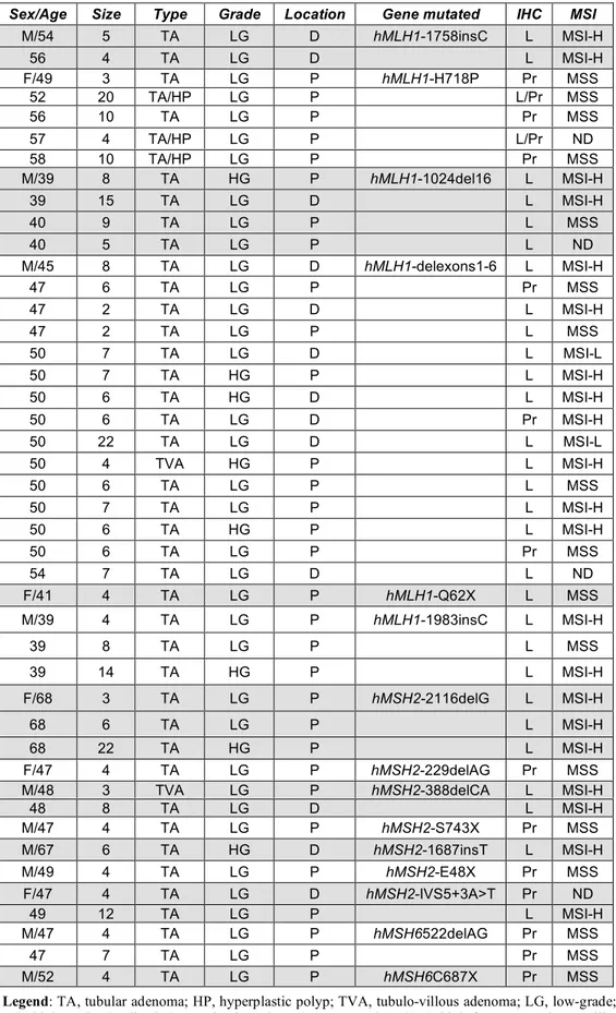
Table 1. Clinicopathological findings of adenomas from Lynch syndrome patients.
Legend: TA, tubular adenoma; HP, hyperplastic polyp; TVA, tubulo-villous adenoma; LG, low-grade;
HG, high-grade; D, distal; P, proximal; L, loss; Pr, preserved; MSI-H, high frequency microsatellite instability; MSI-L, low frequency microsatellite instability; MSS, microsatellite stable; ND, not done.
Sex/Age Size Type Grade Location Gene mutated IHC MSI
M/54 5 TA LG D hMLH1-1758insC L MSI-H 56 4 TA LG D L MSI-H F/49 3 TA LG P hMLH1-H718P Pr MSS 52 20 TA/HP LG P L/Pr MSS 56 10 TA LG P Pr MSS 57 4 TA/HP LG P L/Pr ND 58 10 TA/HP LG P Pr MSS M/39 8 TA HG P hMLH1-1024del16 L MSI-H 39 15 TA LG D L MSI-H 40 9 TA LG P L MSS 40 5 TA LG P L ND M/45 8 TA LG D hMLH1-delexons1-6 L MSI-H 47 6 TA LG P Pr MSS 47 2 TA LG D L MSI-H 47 2 TA LG P L MSS 50 7 TA LG D L MSI-L 50 7 TA HG P L MSI-H 50 6 TA HG D L MSI-H 50 6 TA LG D Pr MSI-H 50 22 TA LG D L MSI-L 50 4 TVA HG P L MSI-H 50 6 TA LG P L MSS 50 7 TA LG P L MSI-H 50 6 TA HG P L MSI-H 50 6 TA LG P Pr MSS 54 7 TA LG D L ND F/41 4 TA LG P hMLH1-Q62X L MSS M/39 4 TA LG P hMLH1-1983insC L MSI-H 39 8 TA LG P L MSS 39 14 TA HG P L MSI-H F/68 3 TA LG P hMSH2-2116delG L MSI-H 68 6 TA LG P L MSI-H 68 22 TA HG P L MSI-H F/47 4 TA LG P hMSH2-229delAG Pr MSS M/48 3 TVA LG P hMSH2-388delCA L MSI-H
48 8 TA LG D L MSI-H M/47 4 TA LG P hMSH2-S743X Pr MSS M/67 6 TA HG D hMSH2-1687insT L MSI-H M/49 4 TA LG P hMSH2-E48X Pr MSS F/47 4 TA LG D hMSH2-IVS5+3A>T Pr ND 49 12 TA LG P L MSI-H M/47 4 TA LG P hMSH6522delAG Pr MSS 47 7 TA LG P Pr MSS M/52 4 TA LG P hMSH6C687X Pr MSS
The sites and pathological features of the 44 polyps are summarized in Table 2.
Table 2. Pathological features of Lynch syndrome-associated adenomas.
Variables Adenomas (n=44) Location Proximal colon 31 (70%) Distal colon 13 (30%) Histologic Type Tubular adenoma 39 (89%) Villous adenoma 2 ( 4%) Mixed adenomatous/hyperplastic polyp 3 ( 7%)
Size (mm) <5 16 (36%) 5-10 22 (50%) >10 6 (14%) Dysplasia Low-grade 36 (82%) High-grade 8 (18%)
These Lynch syndrome-associated adenomas were more commonly identified in the right colon, with 31 out of the 44 (70%) adenomas located proximal to the splenic flexure. Histopathological review confirmed the diagnosis of 39 tubular and 2 villous adenomas, and there were 3 mixed polyps that exhibited features of both tubular adenoma and hyperplastic polyp. The adenomas displayed a wide range of size with a mean of 7.2 mm (range, 2-22 mm). Sixteen of the 44 (36%) adenomas were smaller than 5 mm, 22 (50%) were between 5 and 10 mm, and 6 (14%) were larger than 10 mm. The majority of adenomas (82%) exhibited only low-grade dysplasia, but high-grade dysplasia was identified in 8 of the 44 adenomas. Of these eight, 6 were located in the right colon with a median size of 9.1 mm (range 4-22 mm).
MSI Status in Adenomatous Polyps
Sufficient tissue was available for assessment of MSI in 40 of the 44 adenomas. MSI was detected in 23 of the 40 adenomas (58%). A high level of MSI (MSI-H) was found in 21 adenomas. This included 14 and 7 adenomas from carriers of an hMLH1 or
hMSH2 germline mutation, respectively. A low level of MSI (MSI-L) was detected in 2
adenomas from one patient with an hMLH1 germline mutation. The remaining 17 adenomas (42%), including all adenomas from patients with an hMSH6 germline mutation, were classified as MSS. When the data were analyzed per subject, the sensitivity of MSI analysis for the detection of defective mismatch repair in Lynch syndrome-associated adenomas was 53%. Using MSI testing, eight out of fifteen patients (4 with an hMLH1 and 4 with an hMSH2 gene mutation) were correctly identified as carriers of an MMR gene mutation. Table 3 shows the association between MSI status and the clinicopathological features of the adenomas.
Table 3. Relationship between MSI status and clinicopathological features of adenomas
n MSI-H/MSI-L MSS P Value Sex Male 30 19 11 Female 10 4 6 0.20 Age <50 20 9 11 ≥50 20 14 6 0.11 Size (mm) <5 14 6 8 5-10 20 12 8 >10 6 5 1 0.23 Location Proximal colon 29 12 17 Distal colon 11 11 0 0.001 Dysplasia Low grade 32 15 17 High grade 8 8 0 0.007
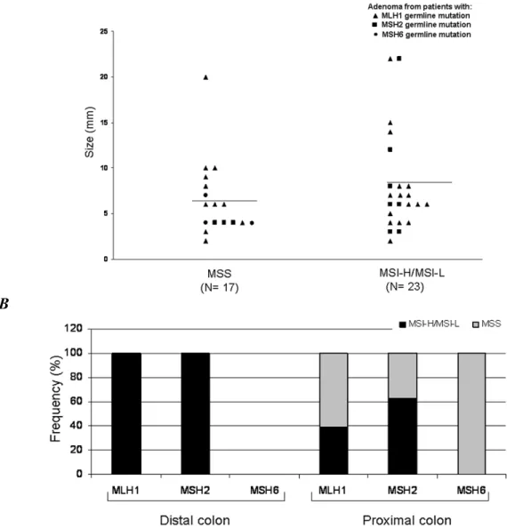
Although microsatellite instability was detected in 65% of the adenomas ≥ 5 mm compared with 43% of the adenomas < 5 mm, this difference was not statistically significant. Figure 1A depicts this distribution of the 40 adenomas by size and MSI status. Interestingly, whereas only 41% of the 29 proximally located adenomas showed MSI, all of the 11 distally located adenomas were MSI-H (P=0.0008). Figure 1B illustrates the distribution of the 40 adenomas by location and MSI status. Finally, all eight adenomas with high-grade dysplasia exhibited MSI (P=0.006).
A
B
Figure 1. A, Distribution of the 40 LS-associated adenomas by size and MSI status. The mean size is indicated by the horizontal bar. B, Distribution of the 40 LS-associated adenomas by location in the colon, MSI status, and genotype.
Immunohistochemical Analysis
Immunohistochemical staining was performed on all 44 adenomas. Loss of MMR protein immunostaining, defined as complete absence of nuclear staining within the adenoma, was detected in 31 of the 44 (70%) cases. Of these 31 adenomas, 24 were from patients with a germline hMLH1 mutation and 7 from patients with a germline hMSH2 mutation. Staining was preserved in the 3 adenomas from two patients with an hMSH6 mutation. Among the 30 adenomas from carriers of an hMLH1 mutation, loss of MLH1 staining was observed in 24 (80%) adenomas. The median size of these adenomas was 7.6 mm (range 2-22 mm), and the majority (15/24; 63%) were located proximal to the splenic flexure. Among the six adenomas from patients with a germline hMLH1 mutation with preserved MLH1 staining, all but one (83%) were located in the right colon with a median size of 6.8 mm (range 3-10 mm). Loss of MSH2 staining was identified in 64% (7/11) of the adenomas from carriers of an hMSH2 mutation. The median size of these adenomas was 8.6 mm (range 3-22 mm), and 5 out of 7 were located in the right colon. The four adenomas with preserved MSH2 staining were from 4 different patients, and 3 were located in the right colon, all with a size of 4 mm. IHC demonstrated a sensitivity of 67% when the analysis was performed on a per patient basis. Immunostaining for defective mismatch repair proteins in Lynch syndrome-associated adenomas identified the gene involved in ten out of fifteen patients (all 6 carriers of an hMLH1 mutation and 4 out of 7 carriers of an
hMSH2 mutation).
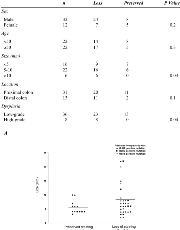
As shown in Table 4, no statistically significant correlation was found between sex, age or location with loss of immunostaining. However, adenomas located distally more frequently displayed loss of MMR protein staining. Among the 16 adenomas smaller than 5 mm, 9 (56%) lost MMR protein expression compared with 16 of 22 (73%) adenomas
between 5 and 10 mm and all 6 adenomas larger than 10 mm (P=0.04) (Figure 2A). Twenty of the 31 (65%) proximally located adenomas lost staining compared to 11 of the 13 (85%) distal adenomas, but this difference did not reach statistical significance.
Table 4. Relationship between IHC and clinicopathological features of adenomas.
n Loss Preserved P Value
Sex Male 32 24 8 Female 12 7 5 0.2 Age <50 22 14 8 ≥50 22 17 5 0.3 Size (mm) <5 16 9 7 5-10 22 16 6 >10 6 6 0 0.04 Location Proximal colon 31 20 11 Distal colon 13 11 2 0.1 Dysplasia Low-grade 36 23 13 High-grade 8 8 0 0.04 A
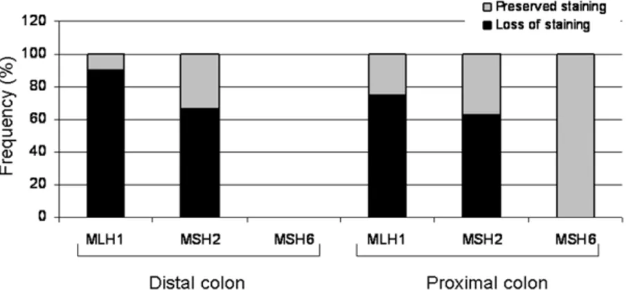
In Figure 2B, the distribution of all 44 adenomas by location and IHC results.
Finally, loss of staining was observed in all eight adenomas with high-grade dysplasia (P=0.04) (Figure 2C). Representative patterns are shown in Figure 2C.
B
Figure 2. B, Distribution of the 44 LS-associated adenomas by location in the colon, IHC results, and genotype
C
Correlation of MSI and IHC Testing in Adenomas
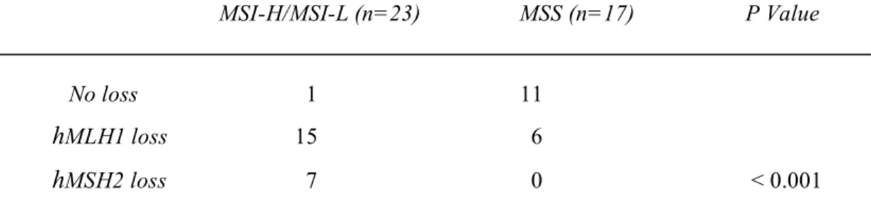
There were 40 adenomas in which both MSI and IHC testing were performed. In these 40 cases, negative protein expression by IHC was detected in 22 of the 23 adenomas that exhibited MSI. Specifically, 15/23 (65%) showed loss of MLH1 nuclear staining and 7/23 (30%) exhibited loss of MSH2 staining. The only adenoma with abnormal MSI but normal IHC was a 6 mm sigmoid adenoma derived from a carrier of an hMLH1 germline mutation. Among the 17 adenomas that did not exhibit MSI, 11 (65%) showed intact staining of the MMR protein tested and six, from four different carriers of an hMLH1 germline mutation, had loss of expression of the MLH1 protein. All 7 adenomas from carriers of an hMSH2 mutation in which IHC demonstrated loss of MSH2 protein staining also exhibited MSI. Whereas no abnormalities in IHC or MSI testing were observed in the adenomas from hMSH6 gene mutation carriers, IHC and MSI testing correctly identified
hMLH1 gene mutation carriers in 100% and 67% of cases, respectively, and hMSH2 gene
mutation carriers in 57% of cases. Collectively, an abnormal MSI result, IHC result, or either an abnormal MSI or IHC result correlated with the presence of a germline mutation in 58% (23/40), 70% (28/40) and 73% (29/40) of cases, respectively. This correlation between microsatellite status and expression of MMR proteins is shown in Table 5.
Table 5. Correlation of MSI status with IHC in Lynch syndrome-associated adenomas.
MSI-H/MSI-L (n=23) MSS (n=17) P Value
No loss 1 11
hMLH1 loss 15 6
MSI and IHC Testing in Hyperplastic Polyps
In our cohort of Lynch syndrome patients undergoing surveillance colonoscopy, 5 hyperplastic polyps were identified. MSI and IHC were performed on these polyps (Table 6). Three polyps from the same patient that carried an hMLH1 germline mutation were mixed polyps showing components of both hyperplastic polyp and tubular adenoma. The median size of these three polyps was 11.3 mm and all were located proximal to the splenic flexure. Microdissection was performed to separate these 2 components, and the results of the adenomatous components of these polyps were described above. In these three mixed polyps, the hyperplastic epithelium revealed preserved MLH1 staining and no MSI was observed in the two polyps in which MSI testing was performed. Two pure hyperplastic polyps were also removed from two carriers of an hMSH2 germline mutation. The median size of these polyps was 18.5 mm; one was located in the sigmoid and the other in the transverse colon. Staining for MSH2 by IHC was preserved in both cases, and both were MSS. Representative immunohistochemical images are shown in Figure 3.
Table 6. Clinicopathological findings in mixed and hyperplastic polyps.
Sex/Age Size Type Location Mutated gene IHC MSI
F/52 20 TA/HP P hMLH1-H718P L/Pr MSS 57 4 TA/HP P L/Pr ND 58 10 TA/HP P Pr MSS F/68 24 HP D hMSH2-2116delG Pr MSS M/48 13 HP P hMSH2-E48X Pr MSS Figure 3.
Discussion
The goal of this study was to determine the frequency of microsatellite instability and loss of immunostaining for MMR proteins in pre-cancerous adenomas of genetically defined LS patients. As the recognition of LS and its associated risk of other extra-colonic malignancies increases, a larger number of at-risk and pre-symptomatic individuals are presenting for genetic counseling. In such cases, adenomas may be the only tissue available, and analysis of these polyps for deficient mismatch repair may be a suitable alternative to testing of colon cancer samples. It is important to note that microsatellite instability and loss of MMR proteins by IHC are uncommon among adenomas in the general population. In a large series of 378 patients with adenomatous polyps, only six (1.6%) had at least one adenoma with MSI, and five of these six patients were indeed carriers of a germline MMR gene mutation [6]. We have demonstrated that a combined approach of MSI and IHC testing in adenomas is 73% sensitive in the detection of LS.
The particular strength of this study is that it represents the largest analysis of adenomas from LS patients who have been unambiguously defined by a germline mutation. Prior reports have sought to address the role of MSI and IHC testing in adenomas, but have defined LS more broadly (fulfillment of either genetic or clinical (Amsterdam) criteria). Unfortunately, the correlation between the Amsterdam criteria and a germline mutation is imperfect, and many families who fulfill the Amsterdam criteria may indeed carry an alternate diagnosis, such as Syndrome X [7]. A germline mutation thus serves as the most precise way to diagnose LS. Our analysis was restricted to polyps from individuals who
carried a defined mismatch repair gene mutation and therefore represents the largest group of true LS patients analyzed.
Iino et al. examined 30 adenomas from 24 patients with LS [8]. However, only 10 of these patients had a defined germline mutation in hMLH1 or hMSH2. Fifty-three percent of all adenomas displayed high levels of MSI. Among the 15 adenomas from the 10 patients with a known germline mutation in hMLH1 or hMSH2, loss of MLH1 or MSH2 protein by IHC was seen in all cases. DeJong analyzed 31 adenomas from 22 carriers of a germline hMLH1 or hMSH2 mutation, and loss of staining by IHC was observed in 65% of adenomas [9]. No MSI analysis was performed. Finally, Halvarsson included 32 adenomas from 23 patients with a known germline mutation in hMLH1 or hMSH2, and an abnormal IHC result was observed in 66% of polyps [10]. No analysis of MSH6 was performed in any of these studies. If adenomas from patients with an hMSH6 mutation are excluded from our analysis, then the sensitivity of a combined approach with MSI and IHC rises to 78% in our study.
Our findings suggest that mutations in hMSH6 are less likely to produce abnormal MSI or IHC results in colorectal adenomas. This is consistent with the attenuated phenotype sometimes observed in hMSH6 kindreds and the greater likelihood that cancers in patients with germline hMSH6 mutations display an MSI-L as opposed to an MSI-H phenotype [11]. Two of these proximally located MSH6 adenomas developed synchronously greater than 5 cm away from a colorectal cancer located in the transverse colon, and absence of MSH6 nuclear staining and MSI was observed in this cancer. Although some have observed similar patterns between synchronous adenomas and cancers in LS, our results are consistent with a previous study suggesting that LS-associated
adenomas developing at a distance of more than 5 cm away from a carcinoma do not show MSI and express all MMR proteins [12,13]. We cannot rule out the possibility that these particular adenomas were “sporadic” and did not arise in conjunction with the underlying
hMSH6 mutation. In agreement with previous observations, our data seem to suggest that
the MMR phenotype may not be necessary for adenoma formation in patients with LS [4,13-15]. Although some adenomas in these patients may arise as a consequence of dysfunctional DNA mismatch repair, others may be initiated as sporadic tumors that do not display microsatellite instability. Some colorectal cancers in carriers of an MMR gene mutation may indeed exhibit preserved IHC staining and MSS [16].
The combination of histological and molecular analyses is a powerful tool in the assessment of MMR, even in pre-cancerous lesions. Interestingly, the concordance between MSI and IHC testing in our cohort was good but not perfect. IHC was more sensitive than MSI analysis. Six adenomas, all from carriers of an hMLH1 germline mutation, exhibited loss of protein expression but no evidence of microsatellite instability. This finding suggests a MMR protein deficiency that has not yet manifested microsatellite instability. It is not inconceivable that loss of MMR protein may occur before the development of MSI, which may require multiple rounds of cell division before the appearance of this phenotype. Alternatively, these results may be due to contamination of DNA from normal stromal or inflammatory cells. Relying on IHC alone would have missed one case, a 6 mm polyp located in the left colon from a carrier of an hMLH1 gene mutation. Since multiple polyps were analyzed from this patient, a possible explanation for the preserved IHC staining may be differences in the second mutational hit, leading in some cases to detection of a non-functional or truncated MMR protein. From a technical perspective, these discordant results
could also be explained by the undefined binding site of the commonly used MLH1 antibody (a murine immunoglobulin G monoclonal antibody raised against full-length MLH1 protein) [17-19].
We found the highest rates of MSI and loss of immunostaining in large (≥ 5 mm) and high-grade dysplastic adenomas. Microsatellite instability and immunohistochemical loss of staining was detected in 65% and 79% of the larger adenomas, respectively. Only the latter relationship between loss of staining by IHC and polyp size was statistically significant. In our series, all high-grade dysplastic adenomas showed MSI (P=0.006) in association with loss of staining (P=0.04). In spite of the right-sided predominance of our Lynch-associated adenomas, we surprisingly detected MSI and loss of MMR protein staining more frequently in distally located adenomas. All the distal adenomas showed MSI compared with 41% (12/29) of the proximal (P=0.0008) polyps. Loss of staining was also reported in 85% (11/13) of the adenomas distal to the splenic flexure. These findings suggest that in spite of the right-sided predilection for Lynch-associated cancers, distally located adenomas should be considered to have equal malignant potential.
IHC and MSI testing correctly identified hMLH1 gene mutation carriers in 100% and 67% of cases, respectively, whereas in hMSH2 gene mutation carriers, loss of protein immunostaining and microsatellite instability were detected in 57% of cases. No abnormalities in IHC and MSI testing were observed in adenomas from the 2 patients with an hMSH6 gene mutation. Overall, it appears that carriers of an hMSH2 or hMSH6 germline mutation are less likely to show loss of staining and/or microsatellite instability; only four of the nine carriers exhibited abnormal MSI and IHC whereas all carriers of an hMLH1
germline mutation exhibited abnormal MSI and IHC. However, it should be noted that all adenomas with preserved staining and stability of the microsatellites were small (median size 4 mm) and did not exhibit high-grade dysplasia.
Our series also suggests that carriers of an hMLH1 germline mutation may develop more adenomas than carriers of an hMSH2 or hMSH6 mutation. However, this is likely a reflection of our smaller cohort, as a recent analysis of 695 patients with a proven germline MMR mutation did not identify significant differences in the prevalence of adenomas among individuals with hMLH1, hMSH2, or hMSH6 mutations [20].
An area of particular interest is whether MMR defects have a role in the development of hyperplastic and serrated polyps and whether these polyps indeed have malignant potential. Traditionally, hyperplastic polyps have been regarded as benign lesions with no potential for neoplastic progression. However, the recent findings of mutations in BRAF as well as microsatellite instability in certain hyperplastic/serrated polyps suggest that this paradigm may need re-evaluation and that these lesions may indeed be precursors of sporadic MSI-H colorectal cancers [21-27]. It is unknown whether hyperplastic polyps that arise in the context of LS may also be pre-cancerous. In the present study we analyzed 5 hyperplastic polyps and found stability of the microsatellites as well as preserved immunostaining in all cases. Interestingly, several of the polyps analyzed were mixed adenomatous/hyperplastic polyps, and abnormal MSI and IHC were confined to the adenomatous compartment. These results, although limited by the sample size, suggest that it is unlikely that hyperplastic polyps play a significant role in the pathogenesis of
microsatellite unstable tumors in subjects with a germline MMR gene mutation [28,29]. None of the polyps in the present study was classified as a serrated adenoma.
In conclusion, molecular analysis of colorectal adenomas may have a role in the workup of suspected LS. The combination of both MSI analysis and IHC staining for MMR proteins detected DNA repair deficiency in 73% of the Lynch-associated adenomas, and this included adenomas smaller than 5 mm. Thus, in the workup of patients suspected to have a germline MMR gene mutation, it is reasonable to begin with MSI and IHC analyses of adenomas, and our data suggest that IHC testing alone is nearly as sensitive as a combined approach. Adenoma size does not appear to be consistently correlated with a positive test result, so small adenomas should not be excluded from analysis. Positive results can be utilized to direct germline genetic testing. However, a negative MSI or IHC test result in an adenoma should be interpreted cautiously and cannot be used to formally exclude the diagnosis of LS if other clinical features suggest the diagnosis. This is particularly true for hMSH6 mutation carriers. Nevertheless, this approach would expand the diagnostic testing options in cases with suspected LS and increase the opportunities to recognize the syndrome before the development of invasive cancer.
References
1. Järvinen H, Aarnio M, Mustonen H, et al. Controlled 15-year trial on screening for colorectal cancer in families with hereditary nonpolyposis colorectal cancer.
Gastroenterology 118, 829-834 (2000).
2. Jass J, Stewart S, Stewart J, et al. Hereditary non-polyposis colorectal cancer: Morphologies, genes and mutations. Mutat Res 310, 125-133 (1994).
3. Lindgren G, Liljegren A, Jaramillo E, et al. Adenoma prevalence and cancer risk in familial non-polyposis colorectal cancer. Gut 50, 228-234 (2002).
4. Rijcken F, Hollema H, Kleibeuker J. Proximal adenomas in hereditary non-polyposis colorectal cancer are prone to rapid malignant transformation. Gut 50, 382-386 (2002).
5. Boland CR, Thibodeau SN, Hamilton SR, et al. A national cancer institute workshop on microsatellite instability for cancer detection and familial predisposition: Development of international criteria for the determination of microsatellite instability in colorectal cancer. Cancer Res 58, 5248-5257 (1998). 6. Loukola A, Salovaara R, Kristo P, et al. Microsatellite instability in adenomas as a
marker for hereditary nonpolyposis colorectal cancer. Am J Pathol 155, 1849-1853 (1999).
7. Lindor N, Petersen G, Hadley D, et al. Recommendations for the care of individuals with an inherited predisposition to lynch syndrome: A systematic review. JAMA 296, 1507-1517 (2006).
8. Iino H, Simms L, Young J, et al. DNA microsatellite instability and mismatch repair protein loss in adenomas presenting in hereditary non-polyposis colorectal cancer. Gut 47, 37-42 (2000).
9. De Jong A, Morreau H, Van Puijenbroek M, et al. The role of mismatch repair gene defects in the development of adenomas in patients with HNPCC. Gastroenterology 126, 42-48 (2004).
10. Halvarsson B, Lindblom A, Johansson L, et al. Loss of mismatch repair protein immunostaining in colorectal adenomas from patients with hereditary nonpolyposis colorectal cancer. Mod Pathol 18, 1095-1101 (2005).
11. Berends M, Wu Y, Sijmons R, et al. Molecular and clinical characteristics of MSH6 variants: An analysis of 25 index carriers of a germline variant. Am J Hum Genet 70, 26-37 (2002).
12. Müller A, Beckmann C, Westphal G, et al. Prevalence of the mismatch-repair-deficient phenotype in colonic adenomas arising in HNPCC patients: Results of a 5-year follow-up study. Int J Colorectal Dis 21, 632-641 (2006).
13. Shia J, Klimstra D, Nafa K, et al. Value of immunohistochemical detection of DNA mismatch repair proteins in predicting germline mutation in hereditary colorectal neoplasms. Am J Surg Pathol 29, 96-104 (2005).
14. Giuffre G, Müller A, Brodegger T, et al. Microsatellite analysis of hereditary nonpolyposis colorectal cancer-associated colorectal adenomas by laser-assisted microdissection: Correlation with mismatch repair protein expression provides new insights in early steps of tumorigenesis. J Mol Diagn 7, 160-170 (2005).
15. Leach F, Nicolaides N, Papadopoulos N, et al. Mutations of a MUTs homolog in hereditary nonpolyposis colorectal cancer. Cell 75, 1215-1225 (1993).
16. Fujiwara T, Stolker J, Watanabe T, et al. Accumulated clonal genetic alterations in familial and sporadic colorectal carcinomas with widespread instability in microsatellite sequences. Am J Pathol 153, 1063-1078 (1998).
17. Raevaara T, Vaccaro C, Abdel-Rahman W, et al. Pathogenicity of the hereditary colorectal cancer mutation hMLH1 del616 linked to shortage of the functional protein. Gastroenterology 125, 501-509 (2003).
18. Wahlberg S, Schmeits J, Thomas G, et al. Evaluation of microsatellite instability and immunohistochemistry for the prediction of germ-line MSH2 and MLH1 mutations in hereditary nonpolyposis colon cancer families. Cancer Res 62, 3485-3492 (2002).
19. Salahshor S, Koelble K, Rubio C, et al. Microsatellite instability and hMLH1 and hMSH2 expression analysis in familial and sporadic colorectal cancer. Lab Invest 81, 535-541 (2001).
20. Liljegren A, Barker G, Elliott F, et al. Prevalence of adenomas and hyperplastic polyps in mismatch repair mutation carriers among CAPP2 participants: Report by the colorectal adenoma/carcinoma prevention programme 2. J Clin Oncol 26, 3434-3439. (2008).
21. Otori K, Oda Y, Sugiyama K, et al. High frequency of k-ras mutations in human colorectal hyperplastic polyps. Gut 40, 660-663 (1997).
22. Jen J, Powell S, Papadopoulos N, et al. Molecular determinants of dysplasia in colorectal lesions. Cancer Res 54, 5523-5526 (1994).
23. Konishi M, Kikuchi-Yanoshita R, Tanaka K, et al. Molecular nature of colon tumors in hereditary nonpolyposis colon cancer, familial polyposis, and sporadic colon cancer. Gastroenterology 111, 307-317 (1996).
24. Konishi K, Yamochi T, Makino R, et al. Molecular differences between sporadic serrated and conventional colorectal adenomas. Clin Cancer Res 10, 3082-3090 (2004).
25. Jass J, Biden K, Cummings M, et al. Characterisation of a subtype of colorectal cancer combining features of the suppressor and mild mutator pathways. J Clin
Pathol 52, 455-460 (1999).
26. Chan T, Zhao W, Leung S, et al. Braf and kras mutations in colorectal hyperplastic polyps and serrated adenomas. Cancer Res 63, 4878-4881 (2003).
27. Iino H, Jass J, Simms L, et al. DNA microsatellite instability in hyperplastic polyps, serrated adenomas, and mixed polyps: A mild mutator pathway for colorectal cancer? J Clin Pathol 52, 5-9 (1999).
28. Jass J, Cottier D, Pokos V, et al. Mixed epithelial polyps in association with hereditary non-polyposis colorectal cancer providing an alternative pathway of cancer histogenesis. Pathology 29, 28-33 (1997).
29. Rijcken F, Van Der Sluis T, Hollema H, et al. Hyperplastic polyps in hereditary nonpolyposis colorectal cancer. Am J Gastroenterol 98, 2306-2311 (2003).