Alma Mater Studiorum
Alma Mater Studiorum –– Università di Bologna
Università di Bologna
DOTTORATO DI RICERCA IN
Biologia Cellulare e Molecolare
Ciclo XXVI
Settore Concorsuale di afferenza: 05/E1 Settore Scientifico disciplinare: Bio/12
TITOLO TESI
CELL DEATH MECHANISMS TRIGGERED BY MONOCLONAL ANTIBODIES AGAINST CD99 IN EWING SARCOMA: CROSS-TALK BETWEEN MDM2, IGF-1R AND RAS/MAPK
Presentata da: Terracciano Mario
Coordinatore Dottorato Relatore
Chiar.mo Prof. Vincenzo Scarlato Chiar.ma Prof.ssa Michela Rugolo
Correlatore Chiar.ma Prof.ssa Katia Scotlandi
INDEX
INTRODUCTION………...2 1. BONE SARCOMAS………...2 2. EWING SARCOMA………...3 2.1 Classification……….32.2 Epidemiology and risk factors………...3
2.3 Localization and histopathology………...4
2.4 Course and therapies……….5
2.5 Biology of Ewing sarcoma………6
3. CD99 ANTIGEN……….9
3.1 MIC2 gene and CD99 protein………...9
3.2 CD99 expression in normal and tumoral tissues……….10
3.3 CD99 function……….11
4. CELL DEATH MECHANISMS………...12
4.1 Apoptosis and autophagy: the main types of programmed cell death…….14
4.2 Macropinocytosis: an endocytic pathway leading to cell death…………..17
5. IGF SYSTEM………19
5.1 IGF-1 receptor (IGF-1R) and its molecular signaling……….20
5.2 Ras proteins: structure and function………22
5.3 IGF system and cell death: a “paradoxical” role of Ras-MAPK pathway………...23
PRELIMINARY DATA AND AIM OF THE STUDY………..24
MATERIALS AND METHODS………28
RESULTS………34
1. IGF-1R/MDM2 relationship in ES………34
3. CD99 internalization after its recruitment by 0662 mAb………..36
4. CD99 stability………38
5. Role of lysosomes and endosomes in CD99 internalization process…………40
6. CD99 internalizes both in a clathrin- and caveolin-1-dependent manner………...42
7. CD99 and IGF-1R interaction upon 0662 treatment……….44
8. IGF-1R is not degraded by 0662 mAb treatments……….45
9. Ras-MAPK signaling is activated in response to treatment………..48
10. Cytoplasm vacuolization during 0662 mAb-induced cell death: autophagy hypothesis………51
11. Ras has an active role in the observed cell death……….53
12. Mesenchymal stem cells are resistant to 0662 mAb-induced cell death………..55
13. EWS/FLI1 favors the delivering of the CD99-induced cell death message……….57
DISCUSSION………..59
INTRODUCTION
1. BONE SARCOMAS
Bone sarcomas represent an heterogeneous class of malignant tumors of different connective tissues like bones, muscles and cartilage. Global incidence is around 5 new cases/100000 inhabitants/year, in particular 3000 new cases/year are reported in Great Britain and Italy and about 12000 new cases in USA. This incidence is considered 1/5-1/6 of all sarcomas, with osteosarcomas the most frequent (0.2 new cases/100000 inhabitants/year) followed by the Ewing family tumors with an incidence considered one-half in comparison to osteosarcomas. In the past years the great heterogeneity of sarcomas made diagnoses complicated and targeted therapeutic regimes very difficult to be performed; recently, the development of much more accurated metodologies allowed to get a molecular profile – based classification that divides sarcomas into two big groups: the first one, which includes alveolar rhabdomyosarcoma, synovial sarcoma and Ewing Sarcoma, is characterized by a simple cariotype with rare chromosomal rearrangements; the second one, comprising osteosarcoma and chondrosarcoma, is associated to a complex cariotype with numerous genetic alterations. These alterations involve oncosuppressor genes, like p53 and Rb, which can lead to aneuploidy or chromosomal aberrations of different nature, while the ones with simple cariotype are usually characterized by specific chromosomal traslocations able to generate chimeric proteins with deregulated activity. Frequently the mutated genes code for transcription factors or signal molecules involved in proliferation mechanisms like growth factors and/or their
receptors and, following mutations, they show crucial alterations of regulatory or functional motives like DNA binding domains (1).
2. EWING SARCOMA
2.1 Classification
Ewing sarcoma (ES) is a very rare and aggressive bone tumor of mesenchymal origin (2) described for the first time by James Ewing as a “diffuse endothelioma of bone” and later as an “endothelial myeloma of bone”. Because of its highly undifferentiated small round cell phenotype, for many years it has been considered mainly an histologic diagnosis of exclusion. Nowadays, based on the identification of a common genetic lesion, a similar clinical behavior and response to treatment, the World Health Organization considers this type of tumor, Askin tumors, peripheral primitive neuroectodermal tumor (PNET) and extraosseous ES belonging to the Ewing sarcoma family tumors (ESFT)(3).
2.2 Epidemiology and risk factors
ES is the second most common bone tumor after osteosarcoma, it usually strikes children and young adults with a peak of incidence between 10 and 20 years of age(4) and with a slight prevalence of males than females (1,5:1). Moreover, for unknown reasons, Ewing tumors occur most often in whites (either non-Hispanic or Hispanic), they are less common among Asian Americans and are extremely rare among African Americans. Despite the plethora of data reporting neuroectodermic and stomach cancers in families of patients with ES(5), there are no evidences highlighting an association between this type of tumor and congenital or family syndrome, while it seems to arise as a second tumor in patients previously subjected to radiotherapy, although with a lesser extent if compared to osteosarcoma. Lifestyle-related risk
factors such as body weight, physical activity, diet, and tobacco use play a major role in many adult cancers, but they usually take many years to influence cancer risk, and they are not thought to be critical in childhood cancers, including Ewing tumors, for whom risk factors are still obscure.
2.3 Localization and histopathology
The tumor onset may affect flat, short or long bones and it is very rare in the skull. The most common localization in the appendicular skeleton is the femur, followed by the tibia, humerus, fibula and forearm bones, while in the trunk the pelvis is the most frequent site of occurrence, followed by vertebrae and sacrum, scapula, ribs and clavicle.
Microscopically this cancer is characterized by sheets of small rounded cells closely packed, without matrix and rich in glycogen: the cytoplasm is scarce, pale, granular and with poorly defined limits; nuclei, on the other hand, present a distinct nuclear membrane and they represent the bigger part of the cell.
Figure 2. Histology of Ewing sarcoma/PNET (4)
2.4 Course and therapies
This type of sarcoma usually displays an aggressive growth with a multiple metastatic spread to the lungs, skeleton, lymph nodes and brain, a feature that used to make the long-term survival less than 10% before the employment of chemotherapy. Nowadays, thanks to multimodal treatments, this percentage increased, making a 5-years survival possible without any recurrence in almost 60% of cases; nevertheless, if metastases are present at the time of diagnosis (20% of cases), the survival possibilities decrease dramatically to 20%. ES is sensitive to chemotherapy and radiotherapy, but surgery plays a fundamental role in treatment of the primary tumor, since it represents the best way to control the neoplasia locally and, in some cases, it allows to avoid radiotherapy. The preferred therapeutic approch includes chemotherapy given pre- and post-operatively, surgery (whenever feasible) and radiotherapy (together with surgery or as an alternative). The most effective drugs are cyclophosphamide, ifosfamide, adriamycin, vincristin, actinomycin D and etoposide;
multicentric clinical studies demonstrated a higher efficacy for the association of 4 drugs (vincristin, cyclophosphamide, adriamycin and actinomycin D) if compared to an association of 3 drugs or to the utilization of the single drugs alone. Patients with metastases at the time of diagnosis treated with the same therapeutic regimen utilized for patients having a localized tumor showed a worst prognosis. For these cases, promising results have been reached using a much more aggressive chemotherapy, with increased doses and the time between cycles of treatment reduced, followed by a bone marrow transplantation with a reinfusion of peripheral stem cells. Bone sarcomas are high grade tumors with a devastating tendency to metastatize; introduction of multidrug chemotherapy improved prognosis but the price is long-lasting treatments with very high toxicity and side effects. In addition, despite the plethora of recently developed new drugs, patients with metastasis and recurrent diseases still have a very poor prognosis due to the lack of effective novel therapies against these types of tumors. Further improvements obviously require advances in the biological and molecular understanding of these cancers.
2.5 Biology of Ewing sarcoma
Routinely, for ES diagnosis, in addition to the utilization of histochemical and immunohistochemical reactions to test different biological markers expression (Table 1), it is common the employment of electronic microscopy, cytogenetics and molecular biology techniques to identify the two main markers of Ewing sarcoma: the membrane glycoprotein p30/32MIC2, also known as CD99, and the chromosomal traslocation responsible for the generation of a chimeric protein, EWS-Ets, obtained as a result from the fusion of N-terminal portion of EWS protein to the C-terminal portion of a member of Ets family of trascription factors (2).
MARKERS IMMUNOHISTOCHEMISTRY VIM + CD99 + Caveolina-1 + Fli1 + CAM 5.2 +/- S100 +/- LAC - TdT -
Table 1. Markers routinely used with immunohistochemistry for ESFT diagnosis.
EWS gene is a member of TET transcription factors family, characterized by the presence of an 87 amminoacids region (RRM/RNP-CS) able to promote the RNA binding, and a glutammine-rich N-terminal portion involved in the fusion process to EST genes in ESFT tumors. EWS protein function is still poorly defined, even if it seems to be involved in trascription mechanisms since it was seen to be associated to RNA-polimerase subunits and products. Protein members of Ets family are transcription factors whose role can be to activate or downregulate the transcription, depending on their interaction with DNA and/or other regulatory proteins. DNA binding domain of Ets proteins is part of the chimeric product EWS-Ets, causing its aberrant trascriptional activity.
The traslocation t(11;22)(q24;q12) is present in almost 90% of ES cases; it causes the formation of EWS-FLI1 chimeric product, which is the main patognomonic alteration of ES, and it is characterized by binding of exon 7 of EWS gene to exon 5 (fusion type II) or 6 (fusion type I) of FLI1 gene (Figure 3). The chromosomal traslocation t(21;22)(q22;q12) responsible of EWS-ERG transcript generation is present in around
5% of the cases, while other chromosomal traslocations are much more rare (Table 2).
Figure 3. Different types of EWS/Fli-1 chimera (6)
TRASLOCATION GENES FREQUENCY
t(11;22)(q24;q12) EWS-FLI1 90-95% t(21;22)(q22;q12) EWS-ERG 5-10% t(2;22)(q33;q12) EWS-FEV Rare t(7;22)(q22;q12) EWS-ETV1 Rare t(17;22)(q12;q12) EWS-E1AF Rare t(2;16) FUS-FEV Rare t(21;16) FUS-ERG Rare
Table 2. Genetic rearrangements typically involved in ESFT
EWS-FLI1 protein activity is necessary not only for cell transformation, as demonstrated by its forced expression in immortalized murine NIH3T3 fibroblasts (7), but also for the maintenance of the disease: silencing EWS-FLI1 leads to a
decrease in tumoral growth in vivo and in vitro(8, 9). Besides, numerous genes involved in the regulation of proliferation and tumorigenicity are also EWS-FLI1 specific targets: E2-C, among these, regulates stromelysin, a metallo-protease involved in migration and invasion processes, and C-MYC, a transcription factor known to have a role in proliferation and apoptosis. However, effects of EWS-FLI1 expression are strongly dependent on cellular background; critical factors in the permissive background are largely unknown, but may include the presence of an intact IGF pathway as well as of CD99, both of them very attractive from a therapeutic point of view since the first one is known to be upregulated through an autocrine activation in most of ES cases, and the second one is a membrane glycoprotein highly expressed in this type of tumor, a feature that makes it also a good diagnostic marker for ES.
3. CD99 ANTIGEN
3.1 MIC2 gene and CD99 protein
The membrane glycoprotein CD99 has been described for the first time in 1979 as an antigen particularly expressed in Acute Limphoblastic Leukemia cells, being recognised by 12E7 monoclonal antibody (10).
It is coded by MIC2, a 50 kb gene mapping in the pseudo-autosomic region of both sexual chromosomes, X (Xp22.33-Xpter) and Y (Yp11-Ypter). Like all genes mapping in this region, MIC2 is characterized by interesting properties: the locus present on X chromosome does not undergo lyonization, allowing an equal distribution of MIC2 in males and females; besides, it does not follow the rules of sex-linked inheritance, and it segregates like autosomic alleles (11).
Particularly, MIC2 codes for two protein isoforms, resulting from an alternative splicing of the primary transcript, known as CD99 type I or wild type and CD99 type
II. Comparative gene sequence analyses revealed that cDNA of CD99 type II has a 18bp insert between the exon 8 and the exon 9 which is not present in the other isoform. Albeit its larger size, it codes for a truncated form of protein, since the 18bp insert introduces an in-frame stop codon responsible for the production of a 160 amminoacids instead of 185. Moreover, biochemical studies showed that CD99 undergoes post-translational modifications, like O-glycosylation, causing a reduction in protein molecular weight from 32 kDa to 28 kDa and 18 kDa (12).
To date a CD99 crystal structure is still missing because of its heavy glycosylation, but the use of cDNA sequence analyses gave us some indications: CD99 structure is made by an O-glycosylated extracellular domain of 100 amminoacids at the N-terminal portion, a putative hydrofobic transmembrane domain of 25 residues and a cytosolic C-terminal portion of 38 amminoacids, making possible the immunolocalization of the protein on the cellular surface (13-15). In order to have more information regarding the CD99 structure, circular dichroism and NMR spectroscopy analyses have been performed; these studies showed that the cytoplasmic portion of CD99 is characterized by the presence of an hairpin anchored to the plasma membrane with two flexible loops; for this reason, CD99 doesn’t seem to have a regular secondary structure. It has been postulated that CD99 could gain a rigid conformation only when associated to other partners (16).
2.2 CD99 expression in normal and tumoral tissues
Immunohystochemical studies revealed a ubiquitous distribution of CD99 antigen, showing a high expression level in particular in ependymal cells of brain and bone marrow, in pancreatic islet, in Sertoli and Leyding cells, and in endothelial cells of blood vessels(17). The CD99 function in these cellular contexts is basically unknown, on the contrary, its role in hematopoietic system is well documented.
CD99 is differently expressed according to the cell type and differentiation stage (18-20). Particularly, CD34+ progenitor cells of bone marrow hematopoietic tissue
display high levels of CD99 antigen, which tend to decrease progressively with the increasing of differentiation and maturation stage towards type B lymphocyte and granulocyte lineage. Regarding CD99 expression in T lymphocyte, it results highly expressed in immature thymocytes but its levels decrease proceeding throughout differentiation processes (18, 21).
CD99 resulted expressed also in numerous tumors like acute lymphoblastic leukemia (10), embrional rhabdomyosarcoma (17, 22, 23), synovial sarcoma (24), mesenchymal condrosarcoma (25, 26) and ependymal tumors (27). However, Ewing sarcoma and peripheral neuroectodermic tumors represent the the higher CD99-expressing neoplasms, making the receptor a reliable diagnostic marker for the cited tumors, together with the specific chromosomal traslocations(17, 23).
2.3 CD99 function
Because of its still unknown ligand and the absence of a crystal structure, CD99 functions in phisiological conditions are barely described. Moreover, the existence of two different isoforms makes its role even harder to define. Although present on the X chromosome, the CD99 gene does not undergo X inactivation, indicating that the molecule should be involved in crucial biological processes. Indeed, CD99 was proved to play a role in monocyte diapedesis, T cell adhesion, apoptosis and differentiation, HLA exposure and intracellular trafficking (14, 28-31). In pathology, CD99 is present on the cell surface of ES, acute lymphoblastic leukemia (ALL), thymic tumours, synovial sarcoma, haemangiopericytoma, meningioma and malignant glioma, in which it results in higher levels of invasiveness and lower rates of survival compared to normal brain (23, 24, 32-34). On the contrary, a lower expression of CD99 antigen has been found in Osteosarcoma, a cellular context where it plays an oncosuppressor role(35, 36). In ES, high levels of CD99 are maintained by EWS-FLI1 (37-41), the oncogenic driver of ES (2), and expression of CD99 is required to create the permissive conditions for EWS-FLI1 transformation
(40): CD99 knock-down, infact, leads ES cells to a terminal neural differentiated phenotype with neurite outgrowth, increased expression of β-III tubulin and neural differentiation markers; moreover, it reduces their ability to form tumors and bone metastases when xenografted into immunodeficient mice and diminishes their tumorigenic characteristics in vitro. On the other hand, triggering of CD99 with some, but not all, murine antibodies was found to deliver a massive and rapid cell-death message both in leukemic and ES cells (42-44) and significantly inhibits tumor growth and metastasis formation (45). All together these findings point out a potential immunotherapy employment of CD99, since it is easily accessible, it is expressed in virtually all cases (46) and it has a role in the pathogenesis of the tumor.
4. CELL DEATH MECHANISMS
Cell death can be classified according to its morphological appearance (which may be apoptotic, necrotic, autophagic or associated with mitosis), enzymological criteria (with and without the involvement of nucleases or of distinct classes of proteases, such as caspases, calpains, cathepsins and transglutaminases), functional aspects (programmed or accidental, physiological or pathological) or immunological characteristics (immunogenic or non-immunogenic)(47). The field of cell death research, anyway, has continued its expansion, significant progress has been made and new putative cell death modalities have been described since 2005, when the Nomenclature Committee on Cell Death (NCCD) formulated the guidelines for the definition of the different type of cell deaths (48).
Dying cells are engaged in a process that is reversible until a first irreversible phase or ‘point-of-no-return’ is trespassed. It has been proposed that this step could be represented by massive caspase activation (49), loss of ΔΨm (50), complete
phosphatidylserine (PS) residues that emit ‘eat me’ signals for normal neighboring cells. However, these parameters are also frequently present in the context of non-lethal processes and differentiation pathways, making the concept of “point-of-no-return” hard to be specifically defined.
In the absence of a clearly defined biochemical event that can be considered as the point-of-no-return, the NCCD proposes that a cell should be considered dead when any one of the following molecular or morphological criteria is met: (1) the cell has lost the integrity of its plasma membrane, as defined by the incorporation of vital dyes (e.g., PI) in vitro; (2) the cell, including its nucleus, has undergone complete fragmentation into discrete bodies (which are frequently referred to as ‘apoptotic bodies’); and/or (3) its corpse (or its fragments) has been engulfed by an adjacent cell in vivo (Table 3.)(52).
4.1 Apoptosis and autophagy: the main types of programmed cell death
Generally, a cell demise is “programmed” whether is genetically controlled and apoptosis and macroautophagy (here referred as autophagy) are universally known as the two fundamental types of programmed cell death.
The term “apoptosis” was coined in 1972 (53), it is a Greek word literally meaning “leaves falling from the tree” and represents a set of genetic and biochemical pathways essential for development and maintenance of pluricellular organisms. In particular, it plays an important role in the organogenesis during embrio development, and is involved in regulating cellular homeostasis in adult.
Alterations of the apoptotic machinery can lead to severe tissue damages and pathologies like cancer, autoimmune diseases, cronic diseases like diabetes and neurodegenerations.This type of death classically occurs through rounding-up of the cell, retraction of pseudopodes, pyknosis (reduction of cellular volume), chromatin condensation, nuclear fragmentation, plasma membrane blebbing (but maintenance of its integrity until the final stages of the process) (52). Cytoplasm shrinkage and the plasma membrane blebbing represent the final steps of the entire mechanism, ending with the formation of vesicles called “apoptotic corps” which, thanks to the presence of molecules like phosphatitilserine on their surface, are recognized by specialized phagocytes responsible for their degradation.
Caspases play a fundamental role in the apoptotic process, since their activation mark the “point of no return”(54). They represent a cystein-protease family necessary for the execution of both pathways of programmed cell death. The first one, called “the intrinsic pathway”, is basicly controlled by an interplay between proapoptotic and antiapoptotic members of Bcl2 family with the involvement of mithocondria; initiators of the pathway include increased intracellular reactive oxygen species, radiation-induced DNA damage, exposition to chemotherapeutic agents and the deprivation of growth factors. These initiators ultimately lead to increased mitochondrial permeability, thereby promoting the release of proapoptotic proteins (cytochrome c) from the intermitochondrial membrane space into the cytosol (55,
56). Another of these proteins, diablo homologue (SMAC/DIABLO), antagonizes cytosolic inhibitors of proapoptosis proteins, thus allowing the activation of caspases and hence progression to apoptosis. Activated caspase 9 mobilize caspases 3, 6, and 7, proteases that herald demolition of the cell by cleaving numerous proteins and activating DNases. The extrinsic pathway exploits the binding of members of the tumor necrosis factor (TNF) super-family to cell-surface “death receptors,” members of the TNF receptor family. Ligation of these receptors initiates the formation of the multiprotein death-inducing signaling complex. Aggregation of this complex causes conformational changes in its components that trigger the catalytic activity of caspase 8, a central mediator of apoptosis, and the activation of the same effector caspases of the intrinsic pathway (Figura 4).
Autophagy is the process by which cells recycle their own redundant or damaged organelles and macromolecular components(58, 59). It is an adaptive response to sublethal stress, such as nutrient deprivation, that supplies the cell with metabolites it can use for fuel. Nevertheless, if prolonged, can compromise the cell viability and become an alternative cell death mechanism, leading the cell to self-digestion.
It is morphologically defined (especially by transmission electron microscopy) as a type of cell death that occurs in the absence of chromatin condensation but accompanied by massive autophagic vacuolization of the cytoplasm. In contrast to apoptotic cells (whose clearance is ensured by engulfment and lysosomal degradation), cells that die with an autophagic morphology have little or no association with phagocytes(60, 61).
This type of programmed cell death is characterized by the sequestration of cytoplasmic material within autophagosomes for bulk degradation by lysosomes. Autophagosomes, by definition, are two-membraned and contain degenerating cytoplasmic organelles or cytosol(62, 63). which allows them to be distinguished by transmission electron microscopy from other types of vesicles such as lysosomes or apoptotic blebs.
The autophagic process and machinery are complex, the former consists of five basic phases: initiation, elongation, closure, maturation, and degradation (Figure 5). The core machinery is constituted by different proteins and each of them has a relevant role within the stages of the process: the ATG1/unc-51-like kinase (ULK) complex, including ATG13 and the scaffold FIP200, and a second complex comprised of Vps34, a class III phosphatidylinositol-3-kinase (PI3K), and ATG6/Beclin1 (64) both regulate the initiation phases of autophagy. A third subgroup regulates later steps of autophagy, autophagosome elongation, and ex- pansion, and is characterized by two ubiquitin-like proteins: LC3 (also known as ATG8/microtubule-associated protein 1 light chain 3) and ATG12 (65). LC3 becomes inserted into the inner and outer membranes of the autophagosome and has been used extensively to monitor autophagy. ATG12 is conjugated to ATG5 by the ATG7 and ATG10 proteins and
this ATG5– ATG12 conjugate interacts with ATG16L, promoting LC3 lipidation(66, 67). Autophagosomes, then, fuse with lysosomes, generating autolysosomes, in which both the autophagosome inner membrane and its luminal content are degraded by acidic lysosomal hydrolases. This catabolic process marks the completion of the autophagic pathway.
Figure 5. Proteins of the core machinery and autophagic process (68)
4.2 Macropinocytosis: an endocytic pathway leading to cell death
Apoptosis is the best characterized form of programmed cell death. However, during the last years nonapoptotic forms of cell death have been studied and recognized as playing significant roles during embryonic development, neurodegeneration, and cancer regression (69). In these cases, loss of cell viability may occur in a manner that is independent of caspase activation. In 1999, Chi et al.(70) reported that ectopic expression of activated Ras GTPase, which normally serves to stimulate cell proliferation, can trigger nonapoptotic cell death in glioblastoma and gastric carcinoma. This was described as autophagic death because the cells developed numerous cytoplasmic vacuoles. Later, in 2008, Overmeyer et al.(71) determined that
the large vacuoles observed in glioblastoma cells were derived from macropinosomes, terming this process “Methuosis” (from the Greek word methuo, which means “drink to intoxication”).
Macropinocytosis is a regulated form of endocytosis that mediates the uptake of solute molecules, nutrients and antigens in a clathrin-independent manner. It begins with an actin-mediated membrane ruffling of the plasma membrane, then some of the lamellipodia on the cell surface fold back onto themselves and fuse with the basal membrane creating large, irregular shaped vesicles termed macropinosomes which undergo a maturation process that is slightly different basing on the cellular context (Figure 6).
Figure 6. Pathway of Macropinocytosis (72)
This endocytic process has a physiological relevance: being associated with actin-dependent ruffling of the plasma membrane, this pathway is implicated in cell motility and, therefore, in tumor progression and metastasis; its role is also documented in the chemotactic response of neutrophils (73), while in the immune system, macropinocytosis is means by which cells internalize antigens to be then processed and loaded onto MHC molecules(74, 75). Although considerable progress has been made in understanding the cell biology of macropinocytosis, there remain
many unanswered questions: how do the different regulators of macropinocytosis regulate the entire process? Which events are associated with the formation and the maturation of macropinocytosis?
A key difference between clathrin-dependent endocytosis and macropinocytosis is that the latter requires actin cytoskeleton reorganisation. This was seen not only in Dictyoselium discoideum (76), but also in mouse macrophages (77), therefore, it does not surprise that components of signal transduction pathways linking receptor stimulation to actin cytoskeleton remodelling are also implicated as regulators of macropinocytosis. One such candidate is the Ras oncogene.
5. IGF SYSTEM
The role of growth factors in the pathogenesis of neoplasms is well documented, a lot of data in literature report a connection between tumors, growth factors and their own receptors. Among them, IGF system, being involved in cellular growth and proliferation because of its relevance in cell cycle progression from G1 to S phase, has been investigated by many research groups all around the world for years, disclosing its importance in developing and maintenance of different types of cancers. It is composed by several elements: two ligands IGF-1 and IGF-2; three membrane receptors IGF-1R, IGF-2R and IR; moreover, there are six proteins able to bind IGF ligands with a high affinity, IGFBinding Proteins (IGFBP1, 2, 3, 4, 5, -6), in order to modulate their bioavailability and preventing their degradation .
5.1 IGF-1 receptor (IGF-1R) and its molecular signaling
IGF-1R is a tetramer of 2 alpha and 2 beta chains linked by disulfide bonds; the alpha chains contribute to the formation of the ligand-binding domain, while the beta chain carries the kinase domain (78). Ligand binding (IGF-1 with a high affinity and IGF-2 with a lower affinity) activates the receptor kinase, leading to receptor autophosphorylation on tyrosines residues (Tyr-1165, Tyr-1161 and Tyr-1166) (79) and consequent phosphorylation of multiple substrates, that function as signaling adaptor proteins: the insulin-receptor substrates (IRSs proteins), Shc and 14-3-3 proteins. The IRS1 and Shc signaling pathways represent the main signal trasmission routes regulated by IGF-1R, leading, respectively, to the induction of the PI3K-AKT/PKB pathway and the Ras-MAPK pathway (Figure 7). The recruitment of IRSs proteins on IGF-1R causes the phosphorylation of specific residues which act as docking sites for proteins SH2-containing domains, like PI3-K (phosphoinositide-3-kinase). In particular, they bind PI3-K regulatory subunit, p85, favoring the traslocation of the catalitic subunit p110 on the plasma membrane, where, in turn, phosphorylates the phosphatidylinositole-(4,5)-biphosphate (PIP2) to phosphatidylinositole-(4,5)-triphosphate (PIP3). This event leads, ultimately, to the recruitment of B kinase protein (AKT) on the plasma membrane, with its subsequent conformational change and activation. AKT activation causes the induction of different proteins whose effect are the increase in protein synthesis mediated by mTOR on one hand, and on the other hand the apoptosis inhibition through the inhibitory phosphorylation of pro-apoptotic factors such as Bad. Shc activation stimulates, on the contrary, the Ras-MAPK pathway: Ras is a small membrane GTPase protein considered inactive if bound to GDP; following the receptor autophosphorylation, Sos factor (Son-of-sevenless) mediates the Ras activation catalizing its binding with GTP. The association between Sos and its substrate is controlled by the adaptor protein Grb2, which is able to bind specific phosphotyrosines on the receptor and, in turn, is activated by interacting with Shc(80). Once in the active form, Ras translocates to the plasma membrane and
induces the activation of the serine/treonine kinase Raf; the latter is able to positively modulate the phosphorylation of a series of tyrosine-kinase proteins belonging to the MAP kinase family (Mitogen Activate Protein Kinase). All these events cause the Erk1/2 activation (Extracellular signal-regulated kinase), through the double phosphorylation borne by tyrosines and treonines residues. Erk, then, may phosphorylates different targets, both at the membrane or at the cytoplasmic level, besides, is also able to translocate into the nucleus and regulate the expression of specific transcription factors, such as Fos and Elk1.
5.2 Ras proteins: structure and function
Mammals encode three ras genes, H-ras, K-ras, and N-ras, whose protein product is present in every cell type and tissue although the expression pattern varies depending on the organ and the stage of development (82).
The product encoded by the ras genes is a protein of 188 amino acids (189 in KRasB) of approximately 21 kDa. The three isoforms show an homology in their first 164 amino acids, while they differ completely in the so called “heterogeneous region”, a segment of the carboxy-terminal region made by residues 165-185 (83). Ras biological activity resides in what has been termed the effector domain (amino acids 32 – 40) by means of which it binds to the different effector molecules (84).
Ras proteins exert their function switching from an inactive state, in which they are bound to guanine dinucleotides (GDPs), to an active state, bound to guanine trinucleotides (GTPs). This mechanism is tightly regulated by two types of regulatory proteins: guanine nucleotide exchange factors (GEFs), able to catalyze the GDP/GTP exchange, and GTPase- activating proteins (GAPs), which enhance the intrinsic capacity of Ras to hydrolyze GTP into GDP, thereby returning Ras to the inactive state (83, 85). Structural differences between the active and inactive states are apparently restricted to the switch I (residues 30 – 38) and switch II (residues 60 – 76) regions (86). The switch I region comprises the main binding site for effector molecules and GAPs, while the switch II region is involved in part in the interaction with GEF regions(84, 87, 88).
5.3 IGF system and cell death: a “paradoxical” role of Ras-MAPK pathway
Despite the foregoing, an increasing number of evidence demonstrate also a “paradoxical” role for IGF-1R and its effectors Ras-MAPK. In particular IGF-1R, among others, has been shown to be responsible of a nonapoptotic form of cell death in 293T cells called “paraptosis”, associated with a massive cytoplasmic vacuolization(90, 91); moreover, IGF-1R and IR have been found implicated in apoptotic mechanisms in primary cultured MEFs and in brown preadipocytes wheter the two receptors were both present and unoccupied (92). Despite its well described role in tumorigenesis as a pro-survival/proliferation factor, increasing evidence indicates that RAS is able to switch cell fate toward death in specific cellular contexts, such as in normal primary cells and in cancer cells under certain conditions(89, 93). Its activation can induce non-apoptotic cell death in neuroblastoma through a classical autophagic process (94, 95), while in glioblastoma RAS-regulated death occurs through Rac-dependent pathways hyperstimulating macropinocytosis (71). Depending on the duration, the magnitude and its subcellular localization, ERK activation controls various cell responses, such as proliferation, migration and differentiation but also death (96). A constitutive activation of ERK by active Raf (97), and IGF-I receptor(91) has been demonstrated to induce a form of cell death correlated with extensive cell rounding and the formation of cytoplasmic macrovacuoles, a morphology not related to an apoptotic cell death but that resembles rather an autophagic-like death or paraptosis. Moreover, depending on the cell type and the nature of the injury, ERK activation has been found associated both with the intrinsic apoptotic pathway(98-100) and with the extrinsic one (101-103). Notably, ERK promotes p53 stability and activity, a tumor suppressor involved also in apoptotic processes, by phosphorylating its serine 15 residue (104-106), and by inhibiting MDM2 phosphorylation on its serine 166 residue, which is associated with its ubiquitin ligase activity toward p53(107).
Preliminary data and aim of the study
Preliminary data demonstrate that CD99 triggering by monoclonal antibodies leads to a massive and rapid death of Ewing sarcoma cell lines in vitro and in vivo, through a non canonical caspase-independent mechanism (44). The employment of the anti-CD99 0662 antibody promotes, indeed, the phosphatidylserine exposure on the outer leaflet of the plasma membrane, a typical marker of the apoptotic process; nevertheless, the cell death occurs without caspases involvement or PARP clivage (Figure 9).
Figure 9. CD99-induced death occurs in a caspase-independent manner. (a). FACS analysis of Annexin V/PI in different ES cell lines following 0662 mAb treatments in presence or not of the caspase inhibitor zVAD-fmk. (b) Western blot analysis of caspase/PARP clivage in the ES cell line 6647(44).
The observed cell death mechanism is promoted by a reactivation of p53 through the phosphorylation on its serine 15 residue, an event that makes this trascription factor able to traslocate into the nucleus, thus inducing the expression of some downstream effectors such as Bax and p21, the first one a pro-apoptotic factor and the second one a cell cycle inhibitor (Figure 10).
Figure 10. p53 phosphorylation and induction of its effectors p21 and Bax after 0662 mAb treatments in 6647 and LAP35 ES cell lines.
The fundamental role of p53 has been confirmed with loss of function experiments: exploiting siRNA oligonucleotides, p53 has been transiently knocked-down in 6647 and LAP35 cell lines, causing a reduction of the entire death mechanism if compared to the parental untransfected or scramble cell line, further suggesting that CD99-induced death is p53-dependent (Figure 11).
Figure 11. p53 partecipates actively to the CD99-induced cell death. In the upper panels Annexin V-PI analysis is shown, expressed as percentage of live (black), apoptotic (dark grey) or necrotic (light grey), in scramble or p53-silenced 6647 or LAP35 cells before and after treatment (30 min) with 5µg/mL 0662 MAb.
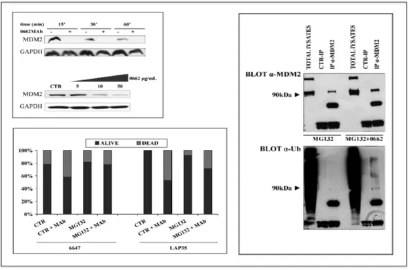
Normally, in non-transformed cellular contexts, p53 expression levels are regulated by MDM2, an E3 ubiquitin ligase which antagonizes the tumor suppressor by sequestering it into the cytoplasm, favouring, then, its ubiquitination and subsequent degradation by the proteasome. MDM2 is frequently overexpressed in ES (108), and our preliminary data demonstrate that its protein levels decrease in a time- and dose-dependent manner following 0662 mAb treatments, a reduction caused by a self-ubiquitination and proteasome degradation (Figure 12), leading, as a result, to the p53 reactivation previously described.
Figure 12. MDM2 expression in ES cell lines. Upper-left panel: whole cell lysates from untreated (-) or MAb treated (+) LAP35 cells and whole cell lysates from 6647 cells, untreated or treated with increasing concentration of 0662 MAb; right panel: Western blot evaluation of MDM2 (upper panel) and ubiquitin (lower panel) on total cell lysates, anti-MDM2 or anti-IgG immunoprecipitates from LAP35 cells treated with MG132 alone or plus 0662 MAb. Lower-left panel: Histogram depicting 6647 (left) and LAP35 (right) percentage of viable cells (vital cell counting) before and after a16 h treatment with 10µg/mL 0662 MAb, alone or in combination with MG132.
Besides p53, many studies report that MDM2 serves as a ubiquitin ligase also for the IGF-1R(109-111), thereby causing its degradation by the proteasome. On these bases, the present PhD project aims to:
• Evaluate IGF-1R modulations in response to 0662 mAb treatments in ES cells; • Identify IGF-1R effectors positively or negatively regulated, and figure out
what is the significance of this modulation;
• Dissect the mechanism by means of which CD99 starts its signalling and assess its fate when triggered by mAbs ;
• Investigate the effects of anti-CD99 mAb treatments in normal cells, in order to rule out possible concerns about toxicity related to the employment of these antibodies.
MATERIALS & METHODS
Cell lines and primary cell coltures.
SK-ES-1, SK-N-MC and RD-ES were obtained from the American Type Culture Collection (Rockville, MD, USA). TC71 and 6647 ES cell lines were a generous gift from TJ. Triche (Children Hospital, Los Angeles, CA, USA); Peripheral neuroectodermal tumor cell line LAP-35 was previously established and characterized at the Istituti Ortopedici Rizzoli, Bologna, Italy. The 6647 variant 6647/CD99low, was previously obtained in our laboratory after a long and continous
exposure to 0662 mAb as previously described (40). The stably expressing CD99 U2/CD99wt57 was previously established in our laboratory (35). Murine C3H10T1/2 cells transfected with EWS/FLI-1(C3H10T1/2 EF) were provided by F. Lecanda (University of Navarra, Pamplona, Spain). ES cell lines have been tested for mycoplasma contamination (Mycoplasma detection Set, Takara Bio Inc.) and have been authenticated by STR PCR analysis. Cells were cultured in IMDM (Gibco), supplemented with 100 units/ml penicillin, 100 µg/ml streptomycin and 10% inactivated fetal bovine serum (FBS) (Lonza). Bone marrow-derived human mesenchimal stem cells were obtained from healthy donors or patients with benign bone lesions. After washings, cells were plated in α-MEM (Gibco), supplemented with 100 units/ml penicillin, 100 µg/ml streptomycin and 20% inactivated fetal bovine serum (FBS) (Lonza). Primary hMSC cell cultures were characterized for mesenchymal markers as well as for their differentiative ability and used within 6 in vitro passages.
Lentiviral infections, transient and stable transfections
All stable transfections were performed using Calcium Phosphate transfection Kit (Invitrogen): LAP35 cells were transfected with pCMV or pCMV-MDM2 and maintained in selective medium containing neomycin [250 µg/ml]; 6647 was transfected with pBABEpuro-HRasN17, and cells expressing the mutant form of Ras were selected with puromycin [500 ng/ml]; U2/CD99 wt57 were derived from the osteosarcoma cell line U2-OS transfected with pcDNA3-CD99 and maintained in selective medium containing neomycin [500 µg/ml]; C3H10T1/2 EF cells were transfected with pcDNA3-CD99 and cultured in medium containing neomycin [500 µg/ml]; 6647 cells were transiently transfected with pcDNA3/His-Ub by using Lipofectamine 2000 (Invitrogen), 48 hours later treatments and nichel-beads pull down were performed; pLKO.1 shAtg7-E8 (TET-ON inducible) and pGIPZ sh SCR (constitutive) were transfected on HEK293T cells to generate lentiviral supernatant to infect ES cell lines. Genetically-modified cells were employed for 0662-mAb treatment and subsequent apoptosis assays, cell count or western blot analysis.
0662 mAb treatments
The anti-CD99 0662 monoclonal antibody (mAb) was produced in the Unite´ INSERM 343, Hospital de l’Archet, Nice, France, and cauterized during the Human Leukocyte Differentiation Agents International Workshop in 1989 and 1993 (Gelin C et al., 1995). Treatments with 0662 mAb were either performed on adherent cells or in suspension. Particularly, cells were washed in PBS, and detached from substrate with trypsin-EDTA 0.5%, counted and seeded at final concentration of 5X10⁶/mL and incubated in IMDM 10% FBS with or w/o 0662 mAb. After the indicated time-points, control and treated samples were washed in PBS and employed for Annexin-V/propidium iodide (PI) evaluation (Mebcyto apoptosis Kit, MBL) by flow cytometry, trypan blue cell count, cell viability analysis (MTT assay kit, Roche) or for protein/RNA extraction. To evaluate the proteasome- and lysosome-mediated degradation, treatments have been performed with 0662 alone or together with
MG132 [20 µM] and lysosome inhibitor [100 µM], both alone or combined; lysosome inhibitor was composed by leupeptin and pepstatin. Unless differently specified, all treatments have been carried out with 0662 [3 µg/ml]. Morphological alterations were analyzed by electron microscopy. LC3-II positive autophagosomes were detected by immunogold staining on ultrathin section by electron microscopy.
Electron microscopy analysis.
6647 or LAP35 cells treated with 10 µg/mL of 0662 mAb for 30 min were fixed with 2.5 % glutaraldehyde in 0.1 M cacodilate buffer, pH 7.4, postfixed with 1% OsO4 ,
dehydrated to absolute ethanol and embedded in Epon. For immunogold labeling LAP35 cells untreated or exposed to anti-CD99 0662 mAb (10 µg/mL) for 3h were fixed with 4 % paraformaldehyde and immunolabeled with anti-LC3B primary antibody; a goat anti-rabbit IgG conjugated with 5 nm colloidal gold particles was used as secondary antibody (GE Healthcare Biosciences, USA). Signal was amplified with Silver Enhancer kit (GE healthcare Biosciences). After immunolabeling cell monolayers were fixed with 1 % glutaraldehyde in 0.1 M Na-phosphate buffer, postfixed with 0.5% OsO4 , dehydrated to absolute ethanol and embedded in Epon.
Ultrathin sections were stained with uranyl acetate and lead citrate before electron microscopy observation with a Zeiss EM109 electron microscope.
Immunofluorescence staining.
Cells were seeded on poly-D-lysine-coated coverslips (BD Biosciences) and after 24h stimulated with anti-CD99 mAb. The stained preparations were observed with a confocal microscopy Nikon A1R and/or with a fluorescence microscopy Nikon Eclipse 90i or 300 (Nikon); the fluorescence was visualized with a digital imaging system using an inverted epifluorescence microscope (Nikon Eclipse 300) with 63X/1.4 oil objective (Nikon Eclipse Ti-U, Nikon, Japan) and Omega Filter pinkel Set XF66-1 triband (Omega Optical Inc., Brattleboro,VT, USA). Images were captured with a back-illuminated Photometrics Cascade CCD camera system (Roper
Scientific, Tucson, AZ, USA) with 500ms of exposure time and elaborated with Metamorph acquisition/analysis software (Universal Imaging Corp., Downingtown, PA, USA). After washing in PBS, cells were fixed in 4% paraformaldeyde and permeabilized in TritonX-100 0.15%-PBS, blocked in 4% BSA and incubated with specific antibodies for immunofluorescence analysis: anti-CD99 primary antibody (Abcam, clone 12E7) + anti-mouse PE; anti-Lamp-1 polyclonal antibody (Santa Cruz Biotechnologies) + rabbit FITC (Dako); Lamp-1PE (BD Pharmigen); anti-Rab4 polyclonal antibody (Thermo Scientific) + anti-rabbit FITC(Dako); anti-Rab11 (Abcam) + anti-rabbit FITC(Dako); anti-clathrin polyclona antibody (Cell Signaling) + anti-rabbit FITC(Dako); anti-caveolin-1 (BD-transduction labs) + anti-rabbit FITC(Dako); anti-IGF-1R (Santa Cruz Biotechnologies, clone C20) + anti-rabbit FITC(Dako); IGF1RPE (Pharmigen); Rab5 (Thermo Scientific) + goat anti-rabbit Dy Light 350 (Thermo scientific). Nuclei were counterstained with Hoechst 33342.
Western blotting and immunoprecipitation.
Cells were lysed with Phospho-protein extraction Buffer (Upstate) supplemented with protease-phosphatase cocktail inhibitor (Sigma). Proteins of interest were detected with specific antibodies: p53 (DO-1) monoclonal antibody (SEROTEC), anti-ser15-P p53 polyclonal antibody (Cell signaling Technologies), anti-p21 (H-164) polyclonal antibody (Santa Cruz Biotechnology), anti-Ub (P4D1) monoclonal antibody (Santa Cruz Biotechnology), anti-mdm2 (OP-46) monoclonal antibody (Calbiochem) and anti-gapdh (FL-335) polyclonal antibody (Santa Cruz Biotechnology), anti-pan RAS (Ab-1) monoclonal antibody (Oncogene), anti-Fli1 (C-19) rabbit polyclonal antibody (Santa Cruz Biotechnology), anti-ATG7 (2631) polyclonal antibody (Cell signaling Technologies), anti IGF-1R (C-20) polyclonal antibody (Santa Cruz Biotechnology), anti - ERK (Cell signaling), anti - thr202-tyr204-pERK (Covance). For immunoprecipitation, 500µg or 900 µg of cell lysates were incubated for 16 hours with Protein G-Plus agarose beads (Calbiochem) in the
presence of 2µg anti-CD99 (abcam) or 3 µg anti-IGF-1R antibody (Calbiochem). Immunoprecipitates and 40 µg total lysates were then resolved on a 10% Tris-HCl gel and immunoblotted with specific antibodies.
ELISA assay.
To determine the CD99 internalization, ELISA assays were performed in 6647 cell line: 70000 cells/time-point treatment were seeded in multi 24 plates and mAb treated after 24h for the indicated times; cells were then starved for 3 hours and washed in TBS 1X, subsequently they were fixed in 3.7% TBS-paraformaldeyde and blocked in 1% TBS-BSA. After the incubation with the anti-mouse alkaline phosphatase-conjugated secondary antibody, the relative substrate (BIO-RAD Cat# 172-1063) was added to the cells. The reaction was stopped by adding NaOH 0.4 N, then absorbance was read at 405 nm.
Nichel-beads pull down.
Co-treatments with 0662 [3 µg/ml] and MG132 [20 µM] + lysosome inhibitor [100 µM] have been performed in 6647 cell lines, then cells were lysed in Buffer A (HNTG+MG132/leu/pep + ”complete” cocktail phosphatase inhibitors (Sigma) +PMSF+Na3VO4); in parallel, nichel beads (Qiagen) have been washed 3 times in PBS and one time in Buffer A, then resuspended in Buffer A and added to each lysate previously solubilized in Buffer A (500 µg). After an over-night rotate incubation at 4°C samples have been washed 3 times in Buffer B (HNTG+Inhibitors+Imidazole 20 mM pH 8), then Buffer C (HNTG+Inhibitors+Imidazole 250 mM) was added to the pellets; the suspensions have been vortexed and spinned, then resuspended in Buffer C + laemli buffer, boiled and resolved on a 10% Tris-HCl gel.
Flow cytometry analysis.
The expression of CD99 was analyzed by indirect immunofluorescence and flow cytometry analysis (FACS Calibur, Becton Dickinson): cells were counted and a
pellet of 500000 cells was obtained; then, they have been resuspended using the anti-CD99 O13 monoclonal antibody (mAb) (Covance), 1: 80 + Goat anti-Mouse IgG (H+L) FITC (diluizione 1:100) (Thermo Fisher Scientific Inc). Both incubations were stopped by adding serum-free medium and, as a final step, cells have been resuspended in PBS-diluited ethidium bromide [1µg/ml], before proceeding to FACS analysis.
RNA extraction and Real Time PCR.
RNAs were extracted using TRIZOL reagent (Life Tecnologies, cat# 15596018). Pre-designed TaqMan probes were chosen for IGF-1R (Taqman assay # Hs00181385_m1) and for GAPDH endogenous control (Taqman assay # Hs99999905_m1) (Applied Biosystems). Samples were analyzed using an ABI Prism 7900 Sequence Detection System (Applied Biosystems, Foster City, CA), according to manufacturer's instructions. Expression levels of target genes were normalized to that of glyceraldehyde 3-phosphate dehydrogenase (GAPDH), and the relative quantification analysis was performed on the basis of the ΔΔCT method. cDNA from untreated cells was used as calibrator for the comparative analysis.
Plasmids
pLKO.1 shAtg7-E8 (TET-ON inducible) and pGIPZ sh SCR (constitutive) were kindly provided by Bruno Calabretta, Philadelphia, USA; pCMV or pCMV-MDM2 were kindly provided by Dr Fabiola Moretti, Rome; pBABE-HRasN17 was kindly provided by Dr Kurosaki, Moriguchi, Japan; pcDNA3/His-Ub was kindly provided by Dr Andrea Morrione, Philadelphia,PA.
Statistics
Differences among means were analyzed using 2-tailed Student’s t test. Fisher’s exact test was used for frequency data. A P value less than 0.05 was considered significant.
RESULTS
1. IGF-1R/MDM2 relationship in ES
In order to confirm the well documented role of MDM2 as a negative regulator of IGF-1R also in the ES cellular context, we took advantage of a Ewing cell line stably expressing WT-MDM2 (LAP35-MDM2) or carrying the pCMV empty vector (LAP35-E.V.) to evaluate IGF-1R protein expression. As shown by the western blot analysis in Figure 13, LAP35 cells overexpressing MDM2 not only present a marked p53 downregulation, but they are characterized also by decreased levels of IGF-1R expression.
Figure 13. MDM2 downregulates IGF-1R in ES.
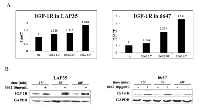
2. IGF1R modulations following 0662 mAb treatments
Once highlighted the inverse correlation between the ubiquitin ligase and IGF1R, and since preliminary data demonstrated the MDM2 downregulation in response to 0662 mAb treatments, we proceeded to evaluate IGF-1R modulations following CD99 triggering by 0662 mAb, both at mRNA and protein level. Real Time PCR and Western blot analyses performed, respectively, using cDNAs and lysates from LAP35 and 6647 cell lines previously treated with 0662 mAb for different times,
revealed a marked induction of the receptor at every time point of the treatment (Figure 14).
Figure 14. IGF-1R induction following CD99 engagement by 0662 mAb in 6647 and LAP-35. Upper panels: Graphs showing results of Real Time PCR; lower panels: western blot analysis confirming RT PCR data. All treatments have been carried out for 15-30-60 minutes with 0662 10 µg/ml.
Concomitantly, flow cytometry analysis of IGF-1R carried out on a panel of 0662 mAb-treated ES cells resulted in a decrease of the receptor expression on the cell surface if compared to the respective untreated control; same phenomenon has been observed in a ES cell line obtained by continous and long exposure to the 0662 mAb,
6647CD99low: despite the IGF-1R induction, together with ERK phosphorylation and
MDM2 downregulation detected by western blot, the expression level of the receptor on the plasma membrane was significantly lower than that registered in 6647 (Figure 15).
Figure 15. Flow cytometry analysis showing the IGF-1R decreased levels on the surface of ES
cells. Western blot analysis of IGF-1R, p-ERK and MDM2 basal expression in 6647CD99low are
shown in the lower-right panel.
3. CD99 internalization after its recruitment by 0662 mAb
One of the issues not mentioned in literature and totally unknown was the CD99 fate following treatments; to get more insight, we explored the internalization hypothesis by using ELISA and immunofluorescence (IF) assays. Taking advantage of U2/CD99wt57, an osteosarcoma cell line derived from U2-OS and stably transfected with CD99 (Figure 16,left panel), we treated cells for 1h and 3h with 0662 mAb, then we proceeded to the receptor immunostaining: untreated U2/CD99wt57 showed
CD99 widespread homogeneously throughout the entire cell surface; after 1 hour of treatment it began to form aggregates right beneath the plasma membrane, moving into the cytoplasm after 3 hours (Figure 16).
Figure 16. On the left, CD99 internalization in U2/CD99wt57 treated for 1h and 3h with 0662 3µg/ml. In the right panel FACS analysis of CD99 expression in 6647, U2-OS and U2/CD99wt57.
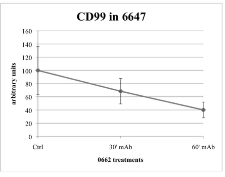
In parallel, in order to have a quantitative evaluation of CD99 internalization, we carried out some ELISA assays in 6647 mAb-treated, concluding that it internalizes into the cell in a time-dependent manner (Figure 17).
Figure 17. Time-dependent CD99 internalization in 6647 treated for 30’ and 60’ with 0662 3µg/ml.
4. CD99 stability
Once determined that CD99 traslocates into the cytoplasm, it remained to be elucidated its stability. We set up a time-course treatment in 6647 and U2/CD99wt57 and, as depicted in Figure 18, we found out that CD99 protein levels decreased after 2 hours of treatment in 6647 and after 4 hours in U2/CD99wt57. The next step has been to figure out which mechanism could cause the observed CD99 reduction: to this purpose, we transiently transfected 6647 with a plasmid expressing a 6xHis-tagged ubiquitin (His-Ub), then I treated cells with 0662 mAb and proteasome/lysosome inhibitors at the same time and I subsequently performed a nichel beads pull-down to assess wheter CD99 could be ubiquitinated as a consequence of the treatment. Anti-CD99 western blotting represented in Figure 18 confirmed that Anti-CD99 is poly-ubiquitinated, suggesting that its degradation could be proteasome-mediated.
Figure 18. CD99 stability upon 0662 mAb treatments. (A) Western blots and respective densitometric analyses showing CD99 reduction in 6647 and U2/CD99wt57. (B) Nichel beads pull-down on lysates from 6647 transiently expressing a 6x His-tagged ubiquitin and treated with 0662 mAb and proteasome + lysosome inhibitors.
5. Role of lysosomes and endosomes in CD99 internalization
process
In order to better define the mechanism by means of which CD99 protein amounts underwent the observed reduction, we explored, in parallel, two possible ways: the proteasome- and the lysosome-mediated degradation. 6647 have been treated with 0662 alone or in combination with MG132 (proteasome inhibitor), with lysosome inhibitor or both; based on the results shown in Figure 19, CD99 protein expression decreased following 2 hours of 0662 treatment alone, while the employment of the inhibitors visibly blocked the mAb-induced reduction of CD99. In particular, densitometric analysis emphasizes the CD99 rescue by the lysosome inhibitor, which seems to be predominant compared to that mediated by MG132.
For this reason, I evaluated by confocal microscopy a colocalization between CD99 and Lamp-1, the latter considered the main marker of lysosomes, in U2/CD99wt57 0662-treated for 1 hour, 3 hours and 6 hours in presence of both MG132 and lysosome inhibitor. Normally the lysosome degradation is a very late event which becomes to be clearly visible after many hours upon a stimulus, in this case the two molecules colocalized after only 1 hour, increasing considerably their interaction in the other time points, concomitantly with the formation of enormous aggregates (Figura 20).
Figure 20. IF (left panel) and confocal microscopy (right panel) images of CD99/Lamp-1 colocalization upon 1h, 3h and 6h of 0662 treatment in combination with MG132 and lysosome inhibitor. The interaction is depicted as yellow dots, which become bigger in the last two time-points because of the employment of the inhibitors that favour the CD99 accumulation.
As a further confirm of its degradation, CD99 was not found to colocalize with Rab11, a late endosome marker implicated in recycling mechanisms, after 30 and 90 minutes of 0662 treatment. Notably, the internalization process resulted to be endosome-mediated, since CD99 was found associated to Rab4, an early endosome marker involved also in the biogenesis of endosomes, particularly after 30 minutes of
treatment (Figura 21).
Figure 21. Evaluation of CD99/Rab11 (upper panel) and CD99/Rab4 (lower panel) colocalizations by IF assays upon 30 and 90 minutes of 0662 treatment. The interaction (yellow areas) is detected for CD99/Rab4, but not for CD99/Rab11.
6. CD99 internalizes both in a clathrin- and caveolin-1-dependent
manner
After having demostrated that CD99 undergoes an internalization process leading, as a final step, to its degradation by lysosomes, we examined the beginning of the entire process, in order to identify the endocytosis route mainly involved. The latter can occur through two different pathways: the dependent and the independent endocytic pathways; the first one requires the formation of clathrin-coated vescicles, while the second one takes place through the formation of the caveolae, invaginations of plasma membrane whose main component is caveolin-1.
We performed IF assays in U2/CD99wt57 0662-treated for 10, 30 and 60 minutes to test the eventual colocalization between CD99 and clathrin or caveolin-1, and, surprisingly, we found out that both pathways partecipated in the endocytosis process (Figura 22).
Figure 22. Evaluation of CD99/Clathrin (A) and CD99/Caveolin-1 (B) colocalizations upon 10, 30 and 60 minutes of 0662 treatment in U2/CD99wt57. On the right, confocal microscopy analyses confirm the results from IF assays depicted on the left.
7. CD99 and IGF-1R interaction upon 0662 treatment
Previously we reported an increase of IGF-1R in ES cell lines both at protein and mRNA level following treatments; notably, FACS analysis highlighted a decrease of its expression on the cell surface, suggesting that the receptor could possibly undergo an internalization process. Based on these results and considering the documented mAb-induced internalization of CD99, we explored the hypothesis of an interaction between the two receptors, which could likely share the same fate. Once again we carried out combined treatments using 0662 and MG132 + lysosome inhibitors for 1 hour, 3 hours and 6 hours and we evaluated the CD99/IGF-1R co-localization by IF assays in U2/CD99wt57: both receptors resulted homogeneously widespread throughout the cell surface in the untreated control cell line, while after 1 hour of treatment CD99 is visible in the form of aggregates beneath the cell surface, even if there is no trace of interaction with IGF-1R; on the contrary, the co-localization is evident into the cytoplasm after 3 hours of treatment, and it is still slightly present in the last time point (Figure 23).
Figure 23. CD99/IGF-1R double staining upon 1, 3 and 6 hours of 0662 and MG132+lysosome inhibitor treatments.
To further confirm this result, we performed an immunoprecipitation (IP) anti-IGF-1R using lysates from 6647 previously treated with 0662 for 2 hours; anti-CD99 western blot analysis showed a marked interaction between the two molecules following treatment, corroborating the initial hypothesis (Figura 24).
Figure 24. Western blotting α-CD99 showing the CD99/IGF-1R strong interaction after IP α-IGF-1R on lysates of 6647 previously treated for 2 hours with 0662 mAb.
8. IGF-1R is not degraded by 0662 mAb treatments
Once determined the IGF-1R internalization and its interaction with CD99 upon treatments, we then proceeded to investigate the observed process in details. In order to prove that the complex IGF-1R/CD99 could be internalized inside the endosomes, we performed a triple staining to detect the two receptors and Rab5, an early endosome marker involved in the biogenesis of endosomes. Confocal microscopy analysis on U2/CD99wt57 mAb-treated for 30, 60 and 90 minutes revealed that not only IGF-1R and CD99 formed a complex, confirming previous results, but each of them colocalized also with Rab5 (Figure 25).
Figure 25. CD99/IGF-1R/Rab5 complex (merge) detected by confocal microscopy analysis in U2/CD99wt57 treated for 30, 60 and 90 minutes with 0662 [3 µg/ml].
The next step has been to evaluate wheter IGF-1R could share the same fate of CD99 and undergo a lysosome-mediated degradation. We set up the same time-course treatment used to investigate the CD99/Lamp-1 co-localization in U2/CD99wt57, employing the combination of 0662 and MG132 + lysosome inhibitors, but in this case IGF-1R showed a different sorting, since none of the treatments caused the interaction with Lamp-1 observed for CD99 (Figure 26).
Figure 26. IGF-1R does not undergo lysosome-mediated degradation. Treatments were carried out with 0662 [3 µg/ml] and MG132 [100 µM] + lysosome inhibitor [20 µM] (leupeptin + pepstatin).
Conversely, confocal microscopy images depicted in Figure 27 show that IGF-1R is recycled on the cell surface, since it was found to colocalize with Rab 11, particularly after 60 and 90 minutes of treatment.
Figure 27. IGF-1R recycling on the cell surface.
9. Ras-MAPK signaling is activated in response to treatment
Considered the up-regulation of IGF-1R in protein and mRNA, and since it is not degraded following its internalization, but recycled on the surface, we investigated the modulation of Ras-MAPK pathway, one of the two main pathways associated to IGF-1R activation. As shown in Figure 25, 0662 mAb treatments in 6647 and LAP35 cell lines caused a strong Ras induction detected by western blotting and by IF assays, together with ERK phosphorylation and its traslocation to the nucleus.
Figure 25. Ras-MAPK up-regulation in ES cells. (A) Western blotting depicting Ras and pERK induction following treatments; in the lower panel the cytosol/nucleus separation shows the ERK traslocation into the nucleus after its mAb-induced phosphorylation. (B) Ras activation detected in 6647 by IF assay.
A plethora of data in literature point out to a “non conventional” localization of Ras proteins; all Ras isoforms, infact, have been found to signal from a variety of intracellular membranes, including endosomes (112, 113). For this reason, we evaluated an eventual Ras interaction with CD99 upon or concomitantly with its induction by 0662 treatment. IF images in Figure 26 prove that the two molecules resulted physically linked in U2/CD99wt57 after 1, 3 and 6 hours of mAb exposure; this co-localization has been detected also by Ras western blotting after anti-CD99 immunoprecipitation (IP), as a first instance, and anti-IGF-1R IP as a second
instance: both circumstances supported the supposed interaction (Figura 26).
Figure 26. CD99/Ras interaction in ES and osteosarcoma cells.(A) IF images of 0662-treated U2/CD99wt57;(B) Anti-Ras western blotting after IP anti-CD99 (right) and IP anti-IGF-1R (left). In the first case the IP has been performed using 500 µg of lysates, while in the second case 900 µg have been used.