Featured Article
Synergistic Cytotoxicity and Pharmacogenetics of Gemcitabine and
Pemetrexed Combination in Pancreatic Cancer Cell Lines
Elisa Giovannetti, Valentina Mey,
Romano Danesi, Irene Mosca,
and Mario Del Tacca
Division of Pharmacology and Chemotherapy, Department of Oncology, Transplants and Advanced Technologies in Medicine, Pisa, Italy
Abstract
Purpose: Gemcitabine is an inhibitor of ribonucleotide reductase (RR) and DNA synthesis and is an effective agent in the treatment of pancreas cancer. The present study investigates whether the multitargeted antifolate pem-etrexed would be synergistic with gemcitabine against MIA PaCa-2, PANC-1, and Capan-1 pancreatic cancer cell lines. Experimental Design: Cells were treated with gemcitab-ine and pemetrexed, and the type of drug interaction was assessed using the combination index. Cytotoxicity of gem-citabine was examined with inhibitors of (a) deoxycytidine kinase (dCK), which activates gemcitabine by phosphoryla-tion, and (b) 5ⴕ-nucleotidase (drug dephosphorylation) and cytidine deaminase (drug deamination), the main inactivat-ing enzymes. The effects of gemcitabine and pemetrexed on cell cycle were analyzed by flow cytometry, and apoptosis was examined by fluorescence microscopy. Finally, quanti-tative, real-time PCR was used to study the pharmacogenet-ics of the drug combination.
Results: Synergistic cytotoxicity and enhancement of apoptosis was demonstrated, mostly with the sequence pemetrexed3gemcitabine. Pemetrexed increased cells in S phase, the most sensitive to gemcitabine, and a positive correlation was found between the expression ratio of dCK:RR and gemcitabine sensitivity. Indeed, pemetrexed significantly enhanced dCK gene expression (ⴙ227.9, ⴙ86.0, andⴙ135.5% in MIA PaCa-2, PANC-1, and Capan-1 cells, respectively), and the crucial role of this enzyme was con-firmed by impairment of gemcitabine cytotoxicity after dCK saturation with 2ⴕ-deoxycytidine.
Conclusions: These data demonstrate that the gemcit-abine and pemetrexed combination displays schedule-dependent synergistic cytotoxic activity, favorably modu-lates cell cycle, induces apoptosis, and enhances dCK expression in pancreatic cancer cells.
Introduction
In the last decade, the availability of several new active drugs has improved the efficacy of combination regimens and substantially increased the response rate of refractory tumors, including pancreatic cancer (1, 2). Antimetabolites are widely used in combination regimens because of their activity and generally manageable toxicities; however, several preclinical studies have shown schedule-dependent drug interaction (3). For example, gemcitabine (2⬘,2⬘-difluoro-2⬘-deoxycytidine) is syn-ergistic in vitro with cisplatin in non-small cell lung cancer cell lines and this interaction is most pronounced when gemcitabine precedes this drug (4, 5). Similar results were obtained using PANC-1 and BxPc3 pancreatic cancer cells for which the most synergistic schedule is gemcitabine followed by cisplatin (6), whereas additive to slightly synergistic or antagonistic effects are observed with gemcitabine followed by topotecan, pacli-taxel, and docetaxel in various non-small cell lung cancer cell lines (7–9).
Gemcitabine has a broad spectrum of anticancer activity against solid tumors, and it is an effective agent in the treatment of pancreatic cancer (10). Gemcitabine requires intracellular phosphorylation to its active metabolites, 2⬘,2⬘-difluoro-dCDP and 2⬘,2⬘-difluoro-dCTP, which, respectively, inhibits ribonu-cleotide reductase (RR) and is incorporated into the DNA, leading to chain termination (11). The rate-limiting step of drug activation is catalyzed by deoxycytidine kinase (dCK), which phosphorylates gemcitabine, whereas 5⬘-nucleotidase and cyti-dine deaminase inactivate gemcitabine by dephosphorylation and deamination, respectively (12). Pemetrexed is a new anti-metabolite that inhibits thymidylate synthase, dihydrofolate re-ductase, and glycinamide ribonucleotide formyltransferase (13), thereby depleting nucleotide pools and blocking DNA synthesis (14, 15). Because of these effects on nucleotide biosynthesis, pemetrexed has the potential to sensitize cells to the cytotoxic activity of gemcitabine. Indeed, the accumulation of cells in the S phase caused by pemetrexed may enhance both gemcitabine incorporation into the DNA and apoptosis (15, 16). Moreover, dCTP depletion and glycinamide ribonucleotide formyltrans-ferase inhibition by pemetrexed may up-regulate dCK, a key enzyme of the nucleotide salvage pathway (14).
For these reasons, the present study was performed in pancreatic cancer cell lines to characterize, from a pharmaco-logical and pharmacogenetic point of view, the combination of pemetrexed and gemcitabine and to provide the experimental basis for potential clinical application of this combination. Received 11/3/04; revised 12/31/03; accepted 1/16/04.
Grant support: R. Danesi was supported by unrestricted research grants from the Ministero dell’Istruzione, Universita` e Ricerca (MIUR, Rome, Italy). M. Del Tacca was supported by unrestricted research grants from Eli-Lilly (Florence, Italy).
The costs of publication of this article were defrayed in part by the payment of page charges. This article must therefore be hereby marked
advertisement in accordance with 18 U.S.C. Section 1734 solely to
indicate this fact.
Requests for reprints: Mario Del Tacca, Division of Pharmacology and Chemotherapy, Department of Oncology, Transplants and Ad-vanced Technologies in Medicine, 55, Via Roma, 56126 Pisa, Italy. Phone: 39-050-830148; Fax: 39-050-562020; E-mail: m.deltacca@med. unipi.it.
Materials and Methods
Drugs and Chemicals. Gemcitabine and pemetrexed were generous gifts from Eli Lilly (Indianapolis, IN). Drugs were dissolved in sterile distilled water and diluted in culture medium immediately before use. RPMI and DMEM media, fetal bovine serum, horse serum,L-glutamine (2 mM), penicillin (50 IU/ml), and streptomycin (50g/ml) were from Life Technol-ogies, Inc. (Gaithersburg, MD). All other chemicals were from Sigma (St. Louis, MO).
Cell Cultures. MIA PaCa-2 (American Type Culture Collection, Manassas, VA), PANC-1 and Capan-1 cell lines (generous gift of Prof. S. Pedrazzoli and Dr. P. Fogar, Univer-sity of Padova, Padua, Italy), were grown in DMEM with 10% fetal bovine serum and 2.5% horse serum (MIA PaCa-2), DMEM with 10% fetal bovine serum (PANC-1), and RPMI with 20% fetal bovine serum (Capan-1), glutamine, and peni-cillin-streptomycin. Cells were cultivated in 75-cm2
flasks (Costar, Cambridge, MA), at 37°C in 5% CO2and 95% air, and were harvested with EDTA when they were in logarithmic growth.
Assay of Cytotoxicity. Cells were plated in 6-well sterile plastic plates (Costar) at 105
cells/well and were allowed to attach for 24 h. Cells were treated with (a) gemcitabine (0.001– 100g/ml) for 1 h; (b) pemetrexed (0.001–100 g/ml) for 24 h; (c) gemcitabine for 1 h followed by a 24-h washout in drug-free medium and then pemetrexed for 24 h; (d) the reverse sequence of point (c) above, i.e., cells were treated with a 24-h washout in drug-free medium followed by treatment with gemcitabine for 1 h. After drug treatments were completed, cells were grown for an additional 24 h in drug-free medium, and cytotoxicity was expressed as the percentage of cells surviving relative to un-treated cultures; the 50% inhibitory concentration of cell growth (IC50) was calculated by non-linear least squares curve fitting. Drug interaction between gemcitabine and pemetrexed was assessed, at a concentration ratio of 1:1, using the combination index (CI; Ref. 17), where CI⬍1, CI ⫽ 1, and CI ⬎1 indicate synergistic, additive, and antagonistic effects, respectively. On the basis of the isobologram analysis for mutually exclusive effects, the CI value was calculated as follows:
CI⫽(D)1 (Dx)1⫹
(D)2 (Dx)2
where (Dx)1and (Dx)2are the concentrations of pemetrexed and gemcitabine, respectively, required to inhibit cell growth by 50%, and (D)1and (D)2are the drug concentrations in combi-nation treatments that also inhibit cell growth by 50% (isoef-fective as compared with the single drugs). Data analysis was performed by the Calcusyn software (Biosoft, Oxford, United Kingdom).
Effect of Inhibition of Gemcitabine Metabolism on Cy-totoxicity. Cells were plated in 6-well plates as described above and were treated with gemcitabine (0.1 ng/ml to 10 g/ml) for 24 h, alone or in combination with 2⬘-deoxycytidine (natural substrate of dCK), tetrahydrouridine (cytidine deami-nase competitive inhibitor), and diethylpyrocarbonate (5 ⬘-nucle-otidase noncompetitive inhibitor), at 10 M to inhibit drug activation by phosphorylation (dCK), as well as drug inactiva-tion by dephosphorylainactiva-tion (5⬘-nucleotidase) or deamination
(cytidine deaminase), respectively. IC50was calculated as de-scribed above.
Cell Cycle Analysis. Cells were plated at 1 ⫻ 106 in 100-mm plastic dishes (Costar) and were allowed to attach for 24 h. After treatments with gemcitabine (1 h) and pemetrexed (24 h) alone at their IC50levels, followed by a 24-h washout, cells were harvested with trypsin-EDTA and were washed twice with PBS. DNA was stained with a solution containing pro-pidium iodide (25g/ml), RNase (1 mg/ml), and NP40 (0.1%); and samples were kept on ice for 30 min. Cytofluorimetry was performed using a FACScan (Becton Dickinson, San Jose, CA), and data analysis was carried out with CELLQuest software, and cell cycle distribution was determined using Modfit software (Verity Software, Topsham, ME).
Analysis of Apoptosis. Cells were treated with gemcitab-ine and pemetrexed and their combinations at their IC50levels, as described in “Assay of Cytotoxicity,” and, at the end of the incu-bation, were washed twice with PBS and fixed in 4% buffered paraformaldehyde for 15 min. Cells were resuspended and incu-bated for an additional 15 min in a solution containing 8g/ml bisbenzimide HCl (18). Cells were spotted on glass slides and were examined by fluorescence microscopy (Leica, Wetzlar, Germany). A total of 200 cells from randomly chosen microscopic fields were counted, and the percentage of cells displaying chromatin conden-sation and nuclear fragmentation relative to the total number of counted cells (apoptotic index) was calculated.
PCR Analysis of dCK and RR. Total RNA was extracted from cells treated as described above in “Cell Cycle Analysis,” using the TRI REAGENT LS. RNA was dissolved in RNase-free water containing 10 mmol/liter DTT and 200 units/ml RNase inhibitor, and measured at260 nm. Oneg of RNA was reverse transcribed at 37°C for 1 h in a 100-l reaction volume containing 0.8 mMdNTPs, 200 units of moloney murine leuke-mia virus reverse transcriptase, 40 units of RNase inhibitor, and 0.05 g/ml random primers. The cDNA was amplified by quantitative real-time PCR with the Applied Biosystems 7900HT sequence detection system (Applied Biosystems, Fos-ter City, CA). quantitative real-time PCR reactions were per-formed in triplicate using 5l of cDNA, 12.5 l of TaqMan Universal PCR Master Mix, 2.5 l of probe, and 2.5 l of forward and reverse primers in a final volume of 25l. Samples were amplified using the following thermal profile: an initial incubation at 50°C for 5 min, to prevent the reamplification of carryover-PCR products by AmpErase uracil-N-glycosylase, followed by incubation at 95°C for 10 min, to suppress AmpEr-ase uracil-N-glycosylAmpEr-ase activity and to denature the DNA, 40 cycles of denaturation at 95°C for 15 s followed by annealing and extension at 60° for 1 min.
Forward (F) and reverse (R) primers and probe (P) were designed with Primer Express 2.0 (Applied Biosystems) on the basis of dCK gene sequence obtained from the GenBank: 5 ⬘-TTC CTG AAC CTG TTG CCA GAT-3⬘(F); 5⬘-GAG ACA TTG TAA GTT CCT CAA ATT CAT C-3⬘(R), and 5⬘-TGC AAT GTT CAA AGT ACT CA-3⬘ (P). Primers and probes for the regulatory (RRM1) and catalytic (RRM2) subunits of RR were from Applied Biosystems Assay on-Demand Gene expres-sion products Hs00168784 and Hs0035724. Amplifications were normalized to glyceraldehyde 3-phosphate dehydrogenase (GAPDH), and quantitation of gene expression was performed
using the⌬⌬CTcalculation, where CTis the threshold cycle; the amount of target gene, normalized to GAPDH and relative to the calibrator (untreated control cells), is given as 2⫺⌬⌬CT (19).
Optimal primer concentration, i.e., associated with minimum SDs between CTvalues, was 300 nM, as preliminarily assessed in PCR reactions with all of the combinations of forward and reverse primers. A validation experiment was performed to demonstrate that the efficiencies of the target (dCK, RRM1, and RRM2) and reference (GAPDH) gene amplifications were ap-proximately equal, using a standard curve method with several dilutions of the cDNA sample from untreated control cells. An
ideal slope should be⫺3.32; the values of the slopes of cDNA calibrator relative to CTwere⫺3.06 for dCK, ⫺3.12 for RRM1, ⫺3.24 for RRM2, and ⫺3.32 for GAPDH (Fig. 1). Therefore, PCR efficiencies were 92.2, 94.0, 97.6, and 100%, respectively, for dCK, RRM1, RRM2, and GAPDH.
Statistical Analysis. All of the experiments were per-formed in triplicate and were repeated at least three times. Data were expressed as mean values⫾ SE and were analyzed by Student’s t test or ANOVA followed by the Tukey’s multiple comparison; the level of significance was P⬍ 0.05.
Results
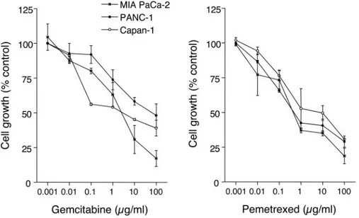
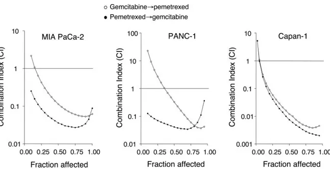
Cytotoxicity of Gemcitabine and Pemetrexed. A dose-dependent inhibition of cell growth was observed with gemcitabine and pemetrexed (Fig. 2), with IC50s of 2.90⫾ 0.34 and 1.58 ⫾ 0.40g/ml (MIA PaCa-2), 42.21 ⫾ 5.74 and 2.49 ⫾ 0.29 g/ml (PANC-1), and 4.75⫾ 1.07 and 7.33 ⫾ 1.93 g/ml (Capan-1), respectively. The sequential exposure of cell lines to pemetrexed followed by gemcitabine reduced the IC50s of gemcitabine to 36.10 ⫾ 1.31, 21.50 ⫾ 2.50, and 94.0 ⫾ 5.62 ng/ml in MIA PaCa-2, Capan-1, and PANC-1, respectively, whereas the IC50s of gemcitabine resulting from the reverse sequence were 123.70⫾ 1.45, 352.28⫾ 43.87, and 748.00 ⫾ 64.32 ng/ml in MIA PaCa-2, Capan-1, and PANC-1 cells, respectively. The calculation of the CI showed synergism at effect levels ⬎30% (fraction of cells affected by the treatments) for both schedules in the three cell lines (Fig. 3), but the degree of synergism obtained with the pemetrexed3gemcitabine sequence was considerably greater than that observed with the reverse schedule (Fig. 3).
Modulation of Drug Metabolism and Gemcitabine Cy-totoxicity. A key role for dCK activity on sensitivity to gem-citabine of the three pancreatic cancer cell lines was demon-strated. Indeed, after treatment with gemcitabine for 24 h, there was a modest increase in cytotoxicity by inhibition of 5 ⬘-nucleotidase and cytidine deaminase, whereas a 10-fold increase in IC50, indicating suppression of cytotoxicity, was observed in all cell lines with simultaneous exposure to 2⬘-deoxycytidine and gemcitabine (Table 1).
Fig. 1 Deoxycytidine kinase (dCK), RRM1 (regulatory subunit of
ri-bonucleotide reductase), RRM2 (catalytic subunit of riri-bonucleotide re-ductase), and glyceraldehyde 3-phosphate dehydrogenase (GAPDH) standard curves for validation of quantitative real-time PCR (QRT-PCR) method. The following equations apply to the target gene amplification:
y⫽ ⫺3.06x ⫹ 30.82, R2⫽ 0.96 (dCK); y ⫽ ⫺3.12x ⫹ 28.32, R2⫽ 0.99
(RRM1); y ⫽ ⫺3.24x ⫹ 28.96, R2⫽ 0.99 (RRM2); y ⫽ ⫺3.32x ⫹
23.96, R2⫽ 0.99 (GAPDH). y, C
T; x, logarithm of cDNA (mg).
Fig. 2 Inhibitory effect of gemcitabine
and pemetrexed on cell proliferation in pancreatic cancer cells. Points, mean val-ues obtained from three independent ex-periments; bars,⫾SE.
Cell Cycle Effects of Gemcitabine and Pemetrexed. Both pemetrexed and gemcitabine were able to affect cell cycle distribution of pancreatic cancer cells (Fig. 4). In particular, the percentage of cells in S phase significantly
increased (P ⬍ 0.05), after treatment with pemetrexed for 24 h, from 15.3 to 46.6% (MIA PaCa-2), from 10.6 to 80.1% (PANC-1), and from 31.1 to 63.2% (Capan-1). The same effect on cell cycle was observed after a 1-h treatment with gemcitabine in MIA PaCa-2 (⫹34.0%) and PANC-1 cells (⫹18.7%), whereas a modest increase was detected in Capan-1 cells (⫹5.3%; Table 2).
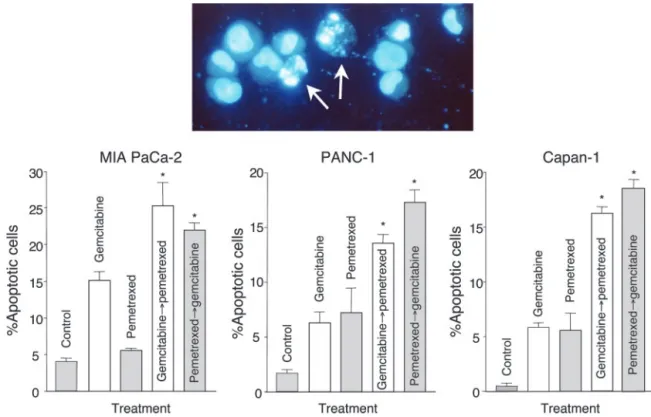
Induction of Apoptosis by Gemcitabine and Pem-etrexed. Cells exposed to pemetrexed, gemcitabine, and their combination presented typical apoptotic morphology with cell shrinkage, nuclear condensation and fragmentation, and rupture of cells into debris (Fig. 5). Only 5– 6% of apoptotic cells were observed after pemetrexed treatment, and a higher percentage (6 –15%) was found after gemcitabine exposure in all cell lines, but the pemetrexed3gemcitabine sequential exposure signifi-cantly increased apoptotic index up to 22.0 ⫾ 1.4%, 17.3 ⫾
Fig. 4 Histograms of DNA content of MIA PaCa-2 cells demonstrating the accumulation of cells in S phase in a representative experiment as a
consequence of treatment with gemcitabine and pemetrexed. FL2-A, fluorescence measured by FL2-A channel.
Table 1 Effects of deoxycytidine (dCyd) diethylpyrocarbonate (DEPC) and tetrahydrouridine (THU) on gemcitabine cytotoxicity
after 24 h of continuous exposure
IC50 a(ng/ml)
Gemcitabine ⫹dCyd ⫹DEPC ⫹THU
MIA PaCa-2
12.29⫾ 3.89 86.11 ⫾ 6.34 10.15 ⫾ 2.21 7.54 ⫾ 1.31 PANC-1 53.43⫾ 6.24 503.97 ⫾ 23.40 28.03 ⫾ 2.07 10.52 ⫾ 0.12 Capan-1 29.90⫾ 0.66 272.53 ⫾ 62.24 13.71 ⫾ 0.40 9.40 ⫾ 0.70
aMean values⫾ SE of at least three independent experiments.
Fig. 3 Combination index (CI) plots of pemetrexed-gemcitabine combinations in MIA PaCa-2, PANC-1, and Capan-1 cells. The most pronounced
2.6%, and 19.4⫾ 1.5% in MIA PaCa-2, PANC-1, and Capan-1 cells, respectively (Fig. 5).
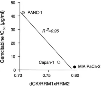
Inducible dCK Gene Expression and Correlation with Cytotoxicity. The expression of dCK was increased by pem-etrexed up to⫹227.9, ⫹86.0, and ⫹135.5% and by gemcitabine up to 8.5, 153.1, and 55.3% in MIA PaCa-2, PANC-1, and Capan-1 cells, respectively (Fig. 6), as also demonstrated by the shift to the left of the amplification plot (Fig. 6). Because the expression of dCK and RR are thought to be involved in gem-citabine chemosensitivity, the dCK/RRM1⫻RRM2 expression ratio was calculated and a correlation (R2⫽ 0.95) was demon-strated with gemcitabine sensitivity in this panel of cells, the cell line with higher dCK/RRM1⫻RRM2 value (MIA PaCa-2) being the most chemosensitive (Fig. 7). Moreover, after
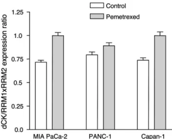
pemetrexed treatment, there was a marked increase in dCK expression and a modest up-regulation in RRM1 and RRM2 levels; therefore, the ratio dCK/RRM1⫻RRM2 mRNA expres-sion increased by 39.3, 12.0, and 12.3% in MIA PaCa-2, PANC-1, and Capan-1 cells respectively, potentially facilitating gemcitabine activity (Fig. 8).
Discussion
The aim of the present study was to investigate the cyto-toxic activity of gemcitabine and pemetrexed in combination and to define the optimal schedule and the cellular mechanism involved in drug interaction against human pancreatic cancer cells.
In preclinical studies, the combination of pemetrexed and gemcitabine yielded conflicting results on various colorectal cancer cell lines. A recent study showed a synergistic cytotox-icity of gemcitabine followed by pemetrexed in HCT-8 cells (20), and similar results were obtained in LoVo, WiDr, and LRWZ cells, in which a higher synergistic interaction was observed with gemcitabine followed by pemetrexed, and the reverse sequence caused an additive-synergistic effect (21). On the contrary, a previous study demonstrated that the schedule-dependent synergism was maximal when pemetrexed preceded gemcitabine in HT29 cells (16). In agreement with these find-ings, in vitro experimental data obtained in this study indicate that pemetrexed and gemcitabine interacted synergistically against MIA PaCa-2, PANC-1, and Capan-1 cells, and the highest chemotherapeutic activity was observed with the se-quence pemetrexed3gemcitabine.
Recent studies have shown the importance of modulating
Fig. 5 Induction of apoptosis by gemcitabine, pemetrexed, and their combinations. Inset panel, morphological appearance of apoptotic cells. Columns, mean values obtained from three independent experiments; bars, SE;ⴱ, statistically significantly different from controls (P ⬍ 0.05).
Table 2 Effect of gemcitabine and pemetrexed on cell cycle of pancreatic cancer cell linesa
Treatment G1(%) a
S phase
(%) G2(%)
MIA PaCa-2 Control 77.01 15.30 7.69 Gemcitabine 46.36 49.29 4.35 Pemetrexed 30.12 46.63 23.25 PANC-1 Control 88.25 10.55 1.20 Gemcitabine 66.98 29.29 3.73 Pemetrexed 17.21 80.13 2.66 Capan-1 Control 50.73 31.13 18.14 Gemcitabine 54.19 36.40 9.41 Pemetrexed 31.10 63.21 5.69
aMean percentage values of total number of cells examined in
the cell cycle to exploit the effect of drug combinations (8, 22). In the present study, flow cytometry demonstrated that both pemetrexed and gemcitabine caused an accumulation of cells in S phase, as a result of the inhibition of DNA synthesis. This
finding is in agreement with previous data on increased propor-tion of MIA PaCa-2 cells in S phase after gemcitabine treatment (23), and on the accumulation of CCRF-CEM and HT29 cells in S phase after 12–24 h of exposure to pemetrexed (15, 16). Because gemcitabine is a S-phase-specific drug, the increase in its activity in the schedule pemetrexed3gemcitabine may be the result of a modulation of cell cycle potentially facilitating 2 ⬘,2⬘-difluoro-dCTP incorporation in DNA.
Deregulation of apoptosis machinery might explain the resistance of cancer cells to chemotherapeutic agents, and a recent study showed that the up-regulation of the phosphatidy-linositol-3⬘-kinase-AKT cell survival pathway correlated with impairment of gemcitabine-induced apoptosis and antitumor activity in human pancreatic adenocarcinoma PK1 and PK8 cells (24). Therefore, not only impaired drug uptake, but also the loss of ability to undergo apoptosis may be involved in gemcit-abine resistance (25). Thus, the development of drug combina-tions that increase apoptosis represents an important approach for the rational design of treatment schedules. In particular, the combination with pemetrexed may improve the therapeutic ac-tivity of gemcitabine by increasing the activation of the apo-ptotic pathway. In the present in vitro study MIA PaCa-2, PANC-1, and Capan-1 cells were exposed to the gemcitabine and pemetrexed in combination at their IC50 levels, and a significant enhancement of apoptosis in treated cells was ob-served when compared with control cells. A similar observation has been reported in the colon cancer cell line WiDr, with which
Fig. 7 Correlation of the deoxycytidine kinase dCK RRM1 (regulatory
subunit of ribonucleotide reductase)⫻RRM2 (catalytic subunit of ribo-nucleotide reductase) expression ratio with gemcitabine chemosensitiv-ity (IC50after 1-h treatment) in MIA PaCa-2, PANC-1 and Capan-1
cells. Points, mean values obtained from three independent experiments.
Fig. 6 Left panel, Representative plot of deoxycytidine kinase (dCK) and glyceraldehyde 3-phosphate dehydrogenase (GAPDH) amplification in
MIA PaCa-2 cells showing the up-regulation of dCK expression by pemetrexed in comparison with control cells. Right panel, dCK expression in MIA PaCa-2, PANC-1, and Capan-1 pancreatic cancer cells. Columns, mean values obtained from two independent experiments; bars,⫾SE; ⴱ, statistically significantly different from control cells (P⬍ 0.05).
only 1% of apoptotic cells were observed after gemcitabine treatment, whereas a higher percentage was found after gemcit-abine–pemetrexed combination (21).
Several observations have suggested that dCK, a key en-zyme of the nucleoside salvage pathway, is a limiting factor for the antitumor effect of gemcitabine because its activity is often decreased in cells resistant to nucleoside analogs, and the sen-sitivity to these drugs could be restored by transfection with a wild-type dCK (26, 27). Recent studies also showed a clear correlation between dCK expression and gemcitabine sensitivity in human tumor xenografts (28); in agreement with these find-ings, the crucial role of dCK was confirmed in the present work by the 10-fold increase in the IC50 in gemcitabine with 2 ⬘-deoxycytidine in MIA PaCa-2, PANC-1, and Capan-1 cells. Because pemetrexed inhibits various enzymes, including glyci-namide ribonucleotide formyltransferase, which catalyzes the first reaction in de novo purine biosynthesis (13, 29), treatment with this drug could increase the expression of dCK as a com-pensatory mechanism. The present study confirmed this hypoth-esis and suggested the potential predictive value of the dCK/ RRM1⫻RRM2 expression ratio with respect to chemosensitivity to gemcitabine. Therefore, the up-regulation of dCK by pem-etrexed, without a parallel increase in RR expression, and the favorable modulation of cell cycle may be considered the most important mechanisms underlying the synergistic interaction with gemcitabine. Two potential applications of these findings may be thus envisaged: (a) integration of pemetrexed in gem-citabine combination regimens to increase drug activity and restore chemosensitivity in tumor cells with dCK down-regula-tion; and (b) application of pharmacogenetic profiling to assess the potential tumor cell sensitivity to gemcitabine.
Finally, an increase in dCK expression was also observed after gemcitabine exposure in PANC-1 and Capan-1 cells, and this result is in agreement with previous studies demonstrating that the salvage pathway initiated by dCK accounted for the
majority of nucleotide synthesis for DNA repair (30), particu-larly after treatment with antimetabolites (31).
In conclusion, this study characterizes the synergistic effect between gemcitabine and pemetrexed against in vitro models of pancreatic cancer and the potential mechanisms involved, and provides the experimental basis for the rational development of this combination for the treatment of this malignancy.
References
1. Oettle H, Riess H. Gemcitabine in combination with 5-fluorouracil with or without folinic acid in the treatment of pancreatic cancer. Cancer (Phila) 2002;95:912–22.
2. Philip PA. Gemcitabine and platinum combinations in pancreatic cancer. Cancer (Phila) 2002;95:908 –11.
3. Peters GJ, van der Wilt CL, van Moorsel CJ, Kroep JR, Bergman AM, Ackland SP. Basis for effective combination cancer chemotherapy with antimetabolites. Pharmacol Ther 2000;87:227–53.
4. van Moorsel CJ, Pinedo HM, Veerman G, et al. Mechanism of synergism between cisplatin and gemcitabine in ovarian and non-small-cell lung cancer non-small-cell lines. Br J Cancer 1999;80:981–90.
5. Edelman MJ, Quam H, Mullins B. Interactions of gemcitabine, carboplatin and paclitaxel in molecularly defined non-small-cell lung cancer cell lines. Cancer Chemother Pharmacol 2001;48:141– 4. 6. Symon Z, Davis M, McGinn CJ, Zalupski MM, Lawrence TS. Concurrent chemoradiotherapy with gemcitabine and cisplatin for pan-creatic cancer: from the laboratory to the clinic. Int J Radiat Oncol Biol Phys 2002;53:140 –5.
7. Tolis C, Peters GJ, Ferreira CG, Pinedo HM, Giaccone G. Cell cycle disturbances and apoptosis induced by topotecan and gemcitabine on human lung cancer cell lines. Eur J Cancer 1999;35:796 – 807. 8. Theodossiou C, Cook JA, Fisher J, et al. Interaction of gemcitabine with paclitaxel and cisplatin in human tumor cell lines. Int J Oncol 1998;12:825–32.
9. Zoli W, Ricotti L, Dal Susino M, et al. Docetaxel and gemcitabine activity in NSCLC cell lines and in primary cultures from human lung cancer. Br J Cancer 1999;81:609 –15.
10. Noble S, Goa K. Gemcitabine, a review of its pharmacology and clinical potential in non-small cell lung cancer and pancreatic cancer. Drugs 1997;54:447–72.
11. Gandhi V, Mineishi S, Huang P. Cytotoxicity, metabolism and mechanism of action of 2⬘,2⬘-difluoro-deoxyguanosine in the Chinese hamster ovary cells. Cancer Res 1995;55:1517–24.
12. Bergman AM, Pinedo HM, Peters GJ. Determinants of resistance to 2⬘,2⬘-difluorodeoxycytidine (gemcitabine). Drug Resist Upd 2002;5:19–33. 13. Shih C, Chen VJ, Gossett LS, et al. LY231514, a pyrrolo[2,3-d]pyrimidine-based antifolate that inhibits multiple folate-requiring en-zymes. Cancer Res 1997;57:1116 –23.
14. Chen VJ, Bowley JR, Andis SL, et al. Cellular pharmacology of MTA: a correlation of MTA-induced cellular toxicity and in vitro enzyme inhibition with its effects on intracellular folate and nucle-oside triphosphate pools in CCRF-CEM cells. Semin Oncol 1999; 26:48 –54.
15. Tonkinson JL, Marder P, Andis SL, et al. Cell cycle effects of antifolate antimetabolites implications for cytotoxicity and cytostasis. Cancer Chemother Pharmacol 1997;39:521–31.
16. Tonkinson JL, Worzalla JF, Teng CH, Mendelsohn LG. Cell cycle modulation by a multitargeted antifolate, LY231514, increases the cy-totoxicity and antitumor activity of gemcitabine in HT29 colon carci-noma. Cancer Res 1999;59:3671– 6.
17. Chou TC, Motzer R, Tong Y, Bosl G. Computerized quantitation of synergism and antagonism of Taxol, topotecan, and cisplatin against human teratocarcinoma cell growth: a rational approach to clinical protocol design. J Natl Cancer Inst (Bethesda) 1994;86:1517–24.
Fig. 8 Enhancement of deoxycytidine kinase dCK RRM1 (regulatory
subunit of ribonucleotide reductase)⫻RRM2 (catalytic subunit of ribo-nucleotide reductase) expression ratio after pemetrexed treatment in MIA PaCa-2, PANC-1, and Capan-1 cells. Columns, mean values ob-tained from three independent experiments; bars, SE.
18. Hsueh CT, Chiu CF, Kelsen DP, Schwartz GK. Selective inhibition of cyclooxygenase-2 enhances mitomycin-C-induced apoptosis. Cancer Chemother Pharmacol 2000;45:389 –96.
19. Mocellin S, Rossi CR, Pilati P, Nitti D, Marincola FM. Quantitative real-time PCR: a powerful ally in cancer research. Trends Mol Med 2003;9:189 –95.
20. Adjei AA, Erlichman C, Sloan AJ, et al. Phase I and pharmacologic study of sequences of gemcitabine and the multitarget antifolate agent in patients with advanced solid tumors. J Clin Oncol 2000;8:1748 –57. 21. Tesei A, Ricotti L, De Paola F, Amadori D, Frassinetti GL, Zoli W. In vitro schedule-dependent interactions between the multitarget antifo-late LY231514 and gemcitabine in human colon adenocarcinoma cell lines. Clin Cancer Res 2002;8:233–9.
22. Zoli W, Ricotti L, Barzanti F, et al. Schedule-dependent interaction of doxorubicin, paclitaxel and gemcitabine in human breast cancer cell lines. Int J Cancer 1999;80:413– 6.
23. Yip-Schneider MT, Sweeney CJ, Jung SH, Crowell PL, Marshall MS. Cell cycle effects of nonsteroidal anti-inflammatory drugs and enhanced growth inhibition in combination with gemcitabine in pancre-atic carcinoma cells. J Pharmacol Exp Ther 2001;298:976 – 85. 24. Ng SSW, Tsao MS, Chow S, Hedley DW. Inhibition of phosphati-dylinositide 3-kinase enhances gemcitabine-induced apoptosis in human pancreatic cancer cells. Cancer Res 2000;60:5451–5.
25. Mackey JR, Mani RS, Selnerm M, et al. Functional nucleoside transporters are required for gemcitabine influx and manifestations of toxicity in cancer cell lines. Cancer Res 1998;58:4349 –57.
26. Gregoire V, Rosier JF, De Bast M, et al. Role of deoxycytidine kinase (DCK) activity in gemcitabine’s radioenhancement in mice and human cell lines in vitro. Radiother Oncol 2002;63:329 –38.
27. Hapke DM, Stegmann AP, Mitchell BS. Retroviral transfer of deoxycytidine kinase into tumor cell lines enhances nucleoside toxicity. Cancer Res 1996;56:2343–7.
28. Kroep JR, Loves WJ, van der Wilt CL, et al. Pretreatment deoxy-cytidine kinase levels predict in vivo gemcitabine sensitivity. Mol Cancer Ther 2002;1:371– 6.
29. Chong LK, Tattersall MH. 5,10-Dideazatetrahydrofolic acid re-duces toxicity and deoxyadenosine triphosphate pool, expansion in cultured L1210 cells treated with inhibitors of thymidylate synthase. Biochem Pharmacol 1995;49:819 –27.
30. Iwasaki H, Huang P, Keating MJ, Plunkett W. Differential incor-poration of ara-C, gemcitabine, and fludarabine into replicating and repairing DNA in proliferating human leukemia cells. Blood 1997;90: 270 – 8.
31. Sasvari-Szekely M, Csapo Z, Spasokoukotskaja T, Eriksson S, Staub M. Activation of deoxycytidine kinase during inhibition of DNA synthesis in human lymphocytes. Adv Exp Med Biol 1998;431:519 –23.