Pharmacognosy Magazine
Phcog.Net - Bringing Medicinal Plant Researchers Together
Publication of Pharmacognosy Network Worldwide
www.phcog.com
ISSN : 0973-1296
July-September
2016 | Volume 12 | Issue 4
7
CAB Abstracts, Caspur, Chemical Abstracts, CNKI (China National Knowledge Infrastructure), CSA databases, DOAJ, EBSCO Publishing’s Electronic Databases, Excerpta Medica / EMBASE, Genamics JournalSeek, Google Scholar, Health & Wellness Research Center, Health Reference Center Academic, Hinari, Index Copernicus, Indian Science Abstracts, Journal Citation Reports, National Science Library, OpenJGate, PrimoCentral, ProQuest, PubMed, Pubmed Central, Science Citation Index Expanded, Scimago Journal Ranking, SCOLOAR, SCOPUS, SIIC databases, Summon by Serial Solutions, Ulrich’s International Periodical Directory and Web of Science.
Pharmacognosy Magazine • V olume 12 • Issue 46 • April-June 2016 • Suuplement 2 Pages S102-S292 •
Included
Impact Factor
®for 201
5: 0.831
ABSTRACT
Background: Thymelaea microphylla Coss. et Dur. (Thymelaeaceae) (TM)
is a rare medicinal plant endemic to Algeria. Leaves decoction is used in folk medicine for anticancer, anti‑inflammatory, and antidiabetic properties.
Objective: Herein, the antioxidant and anti‑inflammatory properties of
different extracts from leaves and flowers of Algerian TM were evaluated.
Materials and Methods: The study was carried out by in vitro cell‑free
assays (antioxidant/radical properties), ex vivo experiments (inhibition of prostaglandin E2 and thromboxane B2 release in human whole blood) and in vitro experiments on cell systems (cytotoxicity on peripheral blood mononuclear cells, and protective effects on human vein endothelial cells exposed to TNF‑α). Results: The acetone TM extract showed significant antioxidant properties and excellent anti‑inflammatory and cyclooxygenase‑inhibitory activity, together with lack of toxicity on normal human blood cells; furthermore, it was able to protect endothelial cells against dysfunction induced by TNF‑α, as shown by decrease in cell death, e‑selectin expression and leukocyte adhesion. Conclusion: On these bases, TM leaves and flowers appear to be a good source of bioactive compounds with significant antioxidant and antiinflammatory capability, and potentially effective in prevention and treatment of pathological conditions related to oxidative stress and inflammation, such as endothelial dysfunction.
Key words: Cyclooxygenase‑2, inflammation, medicinal plant, oxidative
stress, radical scavenger activity, Thymelaea microphylla, tumor necrosis factor‑α
SUMMARY
• Thymelaea microphylla leaves and flowers are a good source of bioactive
compounds with significant antioxidant/free radical scavenger and anti‑ inflammatory activity.
• The acetone extract from leaves and flowers of Algerian Thymelaea
microphylla possesses excellent cyclooxygenase‑inhibitory activity.
• This extract is able to protect against endothelial dysfunction, an early event in development of atherosclerosis and vascular diseases.
Abbreviations used: TM: Thymelaea microphylla; BCB: Beta‑carotene
bleaching; AcE: Acetone extract; PGE2: Prostaglandin E2; TxB2: Thromboxane B2; FL: Fluorescein; Cat: Catechin; DPPH: 2,2‑diphenyl‑1‑picrylhydrazyl; ABTS: 2,2′‑azinobis‑(3‑ethyl‑benzothiazolin‑6‑sulfonic acid)+; Que: Quercetin; ORAC: Oxygen radical absorbance capacity; AAPH: 2,2’‑azobis (2‑methylpropionamidine) dihydrochloride; PMS/NADH: Phenazine methosulfate/nicotinamide adenine dinucleotide; HUVECs: Human umbilical
vein endothelial cells. Correspondence: Dr. Antonio Speciale,
Department of Drug Sciences and Health Products, University of Messina, Viale Annunziata,
98168 Messina, Italy. E‑mail: [email protected] DOI: 10.4103/0973‑1296.186345
INTRODUCTION
Thymelaea is a genus (Thymelaeaceae family) comprising about 30 species of evergreen shrubs under the flowering plant family Thymelaeaceae. Thymelaea microphylla (TM) Coss. et Dur. is a medicinal species endemic to Algeria and very common in the arid and desert pastures.[1] The leaves decoction is used in folk medicine to treat abscess, skin diseases, and abdominal pain and for anticancer, anti‑inflammatory, and antidiabetic properties.[2,3] Another species of Thymelaea genus (Thymelaea hirsuta Endl.) is traditionally used for its several and well‑documented antioxidant, anti‑inflammatory, and anticancer properties.[4‑7] On the contrary, only a few data dealing with the antibacterial and antioxidant activity of the extracts of TM, [3,8,9] as well as on its chemical composition, are available.[10,11]
The present work aims to investigate, using in vitro cell‑free chemical assays, ex vivo experiments, and cell culture‑based tests, the antioxidant/ free radical scavenger and anti‑inflammatory properties of several
extracts (obtained by water, ethanol, acetone, and hexane) from leaves and flowers of Algerian TM.
For this purpose, these extracts, characterized for their total content in flavonoids and flavonols, were tested in a battery of in vitro chemical assays differing in the mechanisms involved, the chemical environment used, and the stressor applied (Folin–Ciocalteu assay; bleaching of This is an open access article distributed under the terms of the Creative Commons Attribution‑NonCommercial‑ShareAlike 3.0 License, which allows others to remix, tweak, and build upon the work non‑commercially, as long as the author is credited and the new creations are licensed under the identical terms.
For reprints contact: [email protected]
www.phcog.com | www.phcog.net
Access this article online Website: www.phcog.com
Quick Response Code:
Cite this article as: Dehimi K, Speciale A, Saija A, Dahamna S, Raciti R, Cimino F, et al. Antioxidant and anti‑inflammatory properties of Algerian Thymelaea
microphylla coss. and dur. extracts. Phcog Mag 2016;12:203-10.
Antioxidant and Anti‑inflammatory Properties of Algerian
Thymelaea microphylla Coss. and Dur. Extracts
Khadidja Dehimi, Antonio Speciale
1, Antonina Saija
1, Saliha Dahamna, Roberto Raciti
1, Francesco Cimino
1,
Mariateresa Cristani
1Department of Biology and Animal Physiology, Faculty of Nature Sciences and Life, Laboratory of Phytotherapy Applied to Chronic Diseases, University Setif, 19000 Setif, Algeria, 1Department of Drug Sciences and Health Products, University of Messina, Viale Annunziata, 98168 Messina, Italy
KHADIDJA DEHIMI, et al.: Biological Effects of Thymelaea microphylla Extracts
204 Pharmacognosy Magazine, Vol 12, Issue 47, Jul-Sep, 2016
the stable 1,1‑diphenyl‑2‑picrylhydrazyl radical; 2,2’‑azino‑bis(3‑ ethylbenzthiazoline‑6‑sulfonic acid) (ABTS) radical scavenging activity; beta‑carotene bleaching [BCB] test; scavenging activity against superoxide anion; prevention of heating‑induced albumin denaturation). On the basis of the results of these experiments and also taking into account the evidence of cytotoxicity shown by these extracts on in vitro cultured peripheral blood mononuclear cells (PBMCs), the TM acetone extract (AcE) was investigated about its capability to inhibit cyclooxygenase (COX) activity, and consequently prostaglandin E2 (PGE2) and thromboxane B2 (TxB2) release, in whole blood, and to protect vessel endothelial cells against the damage induced by the proinflammatory cytokine tumor necrosis factor‑α (TNF‑α), in terms of cell viability, E‑selectin expression, leukocyte adhesion, and cell‑reduced glutathione (GSH) content.
MATERIALS AND METHODS
Chemicals
Folin–Ciocalteu phenol reagent, analysis grade methanol, high‑performance liquid chromatography (HPLC) grade methanol, fluorescein (FL) sodium nitrite, and hydrochloride acid were purchased from Carlo Erba (Milan, Italy). HPLC grade water, acetonitrile, aluminum chloride anhydrous and potassium peroxodisulfate were purchased from VWR International (Radnor, Pennsylvania, United States). All other reagents, if not specified, were purchased from Sigma‑Aldrich (Milan, Italy).
Plant extract preparation
The plants of TM were collected in September 2011 from the region of M’sila in East Algeria. The identification of the plant was based on the work of Quezel and Santa[12] and validated by botanists in the Department of Botany and Ecology at the University of Setif (Algeria). The plant was air‑dried in shade for 10 days at room temperature. Leaves and flowers were then separated from stems and used for extraction.
Four extracts (aqueous, ethanol, acetone, and hexane) were prepared from dried leaves and flowers of TM. Briefly, drug samples were powdered by a mill and to obtain the aqueous extract, 10 g of the sample was mixed with 100 ml of distilled water, left for 30 min at 70°C, and then left for 2 days under occasional stirring. To obtain ethanol, acetone, and hexane extracts, 10 g of the sample were mixed with 100 ml of each solvent for 48 under stirring and on the dark. Finally, the mixtures were filtered and evaporated to dryness under vacuum using a Rotavapor. The yields of the extractions, expressed as percentage, were 10.44%, 4.46%, 0.9%, and 1% for the aqueous, ethanolic, acetonic, and hexanic extracts, respectively. For the chemical and biochemical assays, the extracts were redissolved in the appropriate volume of dimethyl sulfoxide (DMSO).
Phytochemical screening
Extracts were screened for the presence of phytoconstituents (tannins, saponins, flavonoids, steroids, alkaloids, terpenoids, cardiac glycosides, and quinones) using the standard procedure described by Ganatra et al.[13] and Doughari.[14]
Total flavonoid and flavonol content
The total content of flavonoids and flavonols in TM extracts was determined by the method described by Tomaino et al.[15]
For the flavonoid content, aliquots (50 µl) of the solution containing the extracts to be tested (or catechin [Cat] used as standard) were diluted with distilled water to a final volume of 0.5 ml, and 30 µl of 5% NaNO2 was added. After 5 min, the mixture with 60 µl of 10% AlCl3 was added,
consequently after 6 min, with 200 µl of 1 M NaOH and 210 µl of distilled water. Absorbance was recorded at 510 nm using a Shimadzu UV‑1601 spectrophotometer.
For the total content of flavonols, aliquots (125 µl) of the solutions containing the extracts to be tested (or quercetin [Que] used as standard) were mixed with 125 µl of AlCl3 (2 mg/ml) and 750 µl of sodium acetate (50 mg/ml). The absorbance at 440 nm was detected after 2.5 h. Total flavonoid and flavonol contents were expressed as µg of Cat equivalents (eq) and µg of Que eq per mg of dried extract, respectively. All determinations were carried out in duplicate and repeated at least 3 times. Results are expressed as means ± standard deviation (SD) from three experiments.
In vitro biochemical assays
Folin–Ciocalteu colorimetric method
The antioxidant capacity (expressed as total phenols content) of the extracts was determined using the Folin–Ciocalteu reagent.[16] Fifty microliters of the solutions containing different concentrations of the extracts to be tested, or of the vehicle alone (DMSO), were added to 450 µl of deionized water, 500 µl of Folin–Ciocalteu reagent, and 500 µl of 10% aqueous sodium carbonate solution; samples were then maintained at room temperature for 1 h. Absorbance was measured at 750 nm (UV‑Vis Spectrophotometer, Shimadzu, Japan) against the blank containing 50 µl of the same solvent used to dissolve the extracts. Total phenol content was expressed as µg of gallic acid eq per mg of extract, using calibration curve prepared with gallic acid standard solutions. Each determination was carried out in triplicate. Results are expressed as means ± SD from three experiments.
2,2‑diphenyl‑1‑picrylhydrazyl test
The free radical‑scavenging capacity of extracts was determined by the 2,2‑diphenyl‑1‑picrylhydrazyl (DPPH) assay,[17] a method based on the reduction of the stable radical DPPH. The reagent mixture consisted of 1.5 ml of 100 mM DPPH in methanol, to which 37.5 µl of solutions containing various concentrations of the extracts to be tested, or of the vehicle alone (DMSO), were added; an equal volume of the solvent employed to dissolve the extracts was added to control tubes. After 20 min of incubation at room temperature, the absorbance was recorded at 517 nm in a UV‑Vis spectrophotometer. Each determination was carried out in triplicate.
2,2′‑azinobis‑(3‑ethyl‑benzothiazolin‑6‑sulfonic acid) assay
This method determines the capacity of the TM extracts to quench the stable 2,2’‑azino‑bis(3‑ethylbenzthiazoline‑6‑sulfonic acid) radical (ABTS·+). In our experiments,[18] the ABTS·+ radical cation was produced by the oxidation of 1.7 mM ABTS with potassium persulfate (4.3 mM final concentration) in water. The mixture was allowed to stand in the dark at room temperature for 12–16 h before use, and then the ABTS·+ solution was diluted with phosphate buffered saline (PBS) at pH 7.4 to give an absorbance of 0.7 ± 0.02 at 734 nm. One hundred microliters of a solution containing different concentrations of TM extracts to be tested or of the vehicle alone (DMSO) was added to 2 ml of the ABTS·+ solution, and the absorbance was recorded at 734 nm in a UV‑Vis spectrophotometer after allowing the reaction to stand for 6 min in the dark at room temperature. Each determination was carried out in triplicate.
Oxygen radical absorbance capacity assay
The oxygen radical absorbance capacity (ORAC) assay was performed as described by Dávalos et al.[19] and adapted in our laboratory.[20] This assay is based on the effect of peroxyl radicals generated from the thermal decomposition of 2,2’‑azobis
(2‑methylpropionamidine) dihydrochloride (AAPH) on the signal intensity from the fluorescent probe FL in the presence of an oxygen radical absorbing substance. The reaction was carried out in 75 mM phosphate buffer (pH 7.4), and the final reaction mixture was 2 ml. An aliquot of the solution containing different concentrations of TM extracts to be tested (200 µl), or the same volume of the vehicle alone (DMSO), and 1.2 ml of FL solution (70 nM, final concentration) was placed in a cuvette. The mixture was preincubated for 15 min at 37°C in the spectrofluorometer. Freshly prepared AAPH solution (600 µl; 12 mM, final concentration) was rapidly added. Fluorescence was recorded every minute for 80 min at 37°C; the fluorescence conditions were excitation at 485 nm and emission at 520 nm. A blank containing FL and AAPH but only the solvent employed to dissolve the extracts, and different calibration solutions using Trolox (1–7.5 µM, final concentration) as reference antioxidant, was also carried out in each assay. All the reaction mixtures were prepared in triplicate, and at least three independent assays were performed for each sample. The area under the curve (AUC) was calculated for each sample by integrating the relative fluorescence curve. The net AUC was calculated by subtracting the AUC of the blank. The final ORAC values were determined by linear regression equation of Trolox concentrations and expressed as mmol Trolox eq/g dried extract. Results are expressed as means ± SD from three experiments.
Beta‑carotene bleaching assay
The BCB potential of TM extracts was determined following the method of Martorana et al.[21] To prepare a stock solution of a β‑carotene‑linoleic acid mixture, 1 mg of β‑carotene was dissolved in 10 ml of chloroform (HPLC grade), and then 5 ml of this solution were added to 40 µl of linoleic acid and 400 µl of Tween 40. Chloroform was removed using a rotary evaporator at 40°C for 5 min, and then 100 ml of distilled water were slowly added to the residue to form an emulsion. Five milliliters of the emulsion was added to 200 µl of a solution containing the extracts to be studied at different concentrations; the same volume of the solvent alone (DMSO) was used in control samples. The absorbance was immediately measured (t = 0 min) at 470 nm against a blank, consisting of an emulsion without β‑carotene. Then, the samples were placed in a water bath at 50°C, and the oxidation of the emulsion was monitored by measuring absorbance at 470 nm 120 min after the beginning of the reaction. The percentage of inhibition respect to the control was calculated as follows:
% of inhibition = ([At − Ct]/[C0 − Ct])/100
where At and Ct are the absorbances measured for the sample to be tested and the control one, respectively, at t = 120 min, and C0 is the absorbance value for the control measured at t = 0 min. Each determination was carried out in triplicate.
Scavenging activity against the superoxide anion (superoxide
dismutase assay)
The scavenging capability against the superoxide anion of TM extracts was measured by the method described by Tomaino et al.[15] and Tuttolomondo et al.[16] with little modifications. The nonenzymatic system phenazine methosulfate/nicotinamide adenine dinucleotide (PMS/ NADH) generates O2−, which reduces nitroblue tetrazolium (NBT) to a purple formazan. Twenty microliters of each extract solution, tested at different concentrations, or the vehicle alone was added to 1.5 ml of the reaction mixture containing 500 µl of NADH (73 µM), 500 µl of NBT (50 µM), and 500 µl of PMS (15 µM); all reagents were prepared in Tris‑HCl (16 mM, pH 8). After incubation for 2 min at room temperature, the absorbance was measured at 560 nm to determine the concentration of formed formazan. Each determination was carried out in triplicate.
Inhibition of heat‑induced albumin denaturation
We measured the capability of TM extracts to inhibit heat‑induced albumin denaturation.[22] The reaction mixture consisted of 100 µL of solution of each extract to be tested, or the same volume of the vehicle alone (DMSO), 1.9 ml of phosphate buffer 0.2 M (pH 7.4) and 1 ml of bovine serum albumin solution (1%). The mixture was incubated at 37°C for 20 min and then heated to 51°C for other 20 min. After cooling, the turbidity of the samples was measured by reading the absorbance at 660 nm. Each determination was carried out in triplicate.
Ex vivo experiments
Release of prostaglandin E
2and thromboxane B
2in whole blood
To characterize the anti‑inflammatory properties of the TM water and AcE, we used a human whole blood assay to assess its ability to act as in vitro selective inhibitors of COX‑1 or COX‑2 pathways, by decreasing release of TxB2 and PGE2, respectively.[23,24]
Peripheral blood samples were drawn from healthy volunteers who had taken no anti‑inflammatory drug during the last 2 weeks before the study. Informed consent was obtained from each subject.
To evaluate changes in PGE2 release, 1 mL of peripheral blood aliquots containing 10 IU of sodium heparin was incubated in the presence of lipopolysaccharide (LPS; 10 µg/mL) for 24 h at 37°C. The contribution of platelet prostaglandin endoperoxide synthase‑1 was suppressed by adding aspirin (10 µg/mL) at time 0. Then, plasma was separated by centrifugation (10 min at 1600 × g) and kept at − 30°C until assayed for the content of PGE2, which is an index of the COX activity of blood monocyte prostaglandin endoperoxide synthase‑2.
To evaluate changes in TxB2 release, 1 mL of whole blood aliquots was immediately transferred into glass tubes and allowed to clot at 37°C for 1 h. Serum was separated by centrifugation (10 min at 1600 × g) and kept at − 30°C until assayed for TxB2, which is an index of endogenously formed thrombin‑stimulated COX activity of platelet prostaglandin endoperoxide synthase‑1. Plasma PGE2 and serum TxB2 were measured by enzyme immunoassay (using kits purchased from Cayman chemical, USA).
To evaluate the effects of the extract on PGE2 and TxB2 in whole blood, TM extracts were dissolved in DMSO and 2 µl of these solutions was put into test tubes to give a final concentration in whole blood ranging between 320 and 5 µg/mL. The experiments were then carried out as described above. As reference drugs, the nonsteroid anti‑inflammatory drugs, indomethacin as non‑selective COX inhibitor and nimesulide as selective COX‑2 inhibitor, were used. Each determination was performed in triplicate; the results were expressed as percentage decrease with respect to control values and were reported as mean ± SD of three experiments.
In vitro experiments on cell systems
Toxicity on peripheral blood mononuclear cells
PBMCs (pool of five donors) have been isolated from whole blood with Histopaque‑1077 (Sigma‑Aldrich), according to the manufacturer’s instructions, suspended in RPMI‑1640 and immediately used.[25] One milliliter of PBMC suspension (1 × 10⁶ cells/mL) was exposed to TM extracts (dissolved in DMSO) and incubated at 37°C for 24 h. Cell cultures exposed only to the same volume of the vehicle (DMSO; 0.1% final concentration) were used as controls. Changes in viability of PBMCs exposed to plant extracts were evaluated using the trypan blue exclusion assay.[26] Briefly, 10 µl of cell suspension was mixed with 30 µl of trypan blue isotonic solution (0.4% w/v) and loaded into a hemocytometer for both live and dead cell counting.
KHADIDJA DEHIMI, et al.: Biological Effects of Thymelaea microphylla Extracts
206 Pharmacognosy Magazine, Vol 12, Issue 47, Jul-Sep, 2016
Protective effects on human umbilical vein
endothelial cells exposed to tumor necrosis
factor-α
Cell cultures
Human umbilical vein endothelial cells (HUVECs) were isolated from freshly obtained human umbilical cords by collagenase digestion of the interior of the umbilical vein and were cultured in medium 199, supplemented with 20% fetal bovine serum, 1% L‑glutamine, 20 mM HEPES, 100 units/mL penicillin/streptomycin, 50 mg/mL endothelial cell growth factor, and 10 µg/mL heparin, in gelatin‑pretreated flasks. Cells were maintained in a humidified atmosphere containing 5% CO2 in an incubator at 37°C. Cells used in this study were from the second to fourth passage. The final concentration of DMSO in the culture medium during different treatments was <0.1% (v/v). The subconfluent cells were treated for 24 h in serum‑free medium with various doses of TM AcE (20–40 µg/ml) whereas control cells were treated with 0.1% DMSO only. After this incubation time, cells were washed with PBS under sterile conditions and then incubated for 2 h with serum‑free medium containing various doses of recombinant human TNF‑α (20 ng/mL) as previously reported (Speciale et al. 2010, 2013). Control cells were not exposed to TNF‑α. At the end of the exposure time, cells were immediately processed and/or preserved at − 80°C until analysis as expected for each test. The cytotoxic effect of TNF‑α on HUVECs pretreated or not with TM AcE was evaluated using trypan blue exclusion assay.
Leukocyte adhesion
Mononuclear cells have been isolated from human whole blood with Histopaque‑1077, following the procedure recommended by the manufacturer. Briefly, heparinized venous blood from healthy donors was centrifuged over Histopaque‑1077; the mononuclear cell layer was collected, washed twice with DPBS, suspended in medium 199, and immediately used. Two flasks with subconfluent HUVECs were treated with various doses of AcE (20–40 µg/ml) and were incubated for 24 h at 37°C in a humid 5% CO2 atmosphere incubator. Two controls were treated with the medium containing only 0.1% DMSO. After this pretreatment, cells were washed with DPBS under sterile conditions and then co‑cultured with leukocytes (3 × 10⁶ leukocytes/flask) and TNF‑α 20 ng/mL for 2 h at 37°C with gentle shaking. Cells not exposed to TNF‑α were used as controls. After this incubation time, medium was removed and cells were washed with DPBS. Co‑cultures were visualized under an inverted microscope and photographed using a digital camera. Four areas for each flask were selected and used to count the number of adherent leukocytes. Increase in leukocyte adhesion upon stimulation of HUVECs with TNF‑α was calculated in relation to the basal adhesion of leukocytes to unstimulated HUVECs, which was set to 1.
Quantitative reverse transcription-polymerase
chain reaction
Total cellular RNA was isolated according to the TRIzol protocol as previously described.[27,28] The quality of the RNA was tested in 1% formaldehyde‑agarose gel stained with ethidium bromide (EtBr) and spectrophotometrically quantified. After reverse transcription with oligo(dT)15 primers polymerase chain reaction (PCR) was performed for identification of E‑selectin mRNA levels. Glyceraldehyde‑3‑phosphate dehydrogenase (GAPDH) was used as housekeeping gene for normalization. Gene expression was assessed by real‑time PCR (Applied Biosystem 7300 Real‑Time PCR System, Monza, Italy) coupled with the Sybr green JumpStart Taq ReadyMix kit. The specific primers set for the target genes were as follows: GAPDH, forward, 5’‑GGC TCT
CCA GAA CAT CAT CCC TGC‑3’, reverse, 5’‑GGG TGT CGC TGT TGA AGT CAG AGG‑3’; E‑selectin, forward, 5’‑CTG CCA AGT GGT AAA ATG TTC AAG‑3’, reverse, 5’‑TTG GAC TCA GTG GGA GCT TCA‑3’. Cycling conditions were 40 cycles of 94°C denaturation (15 s), 60°C annealing and extension (1 min). A final dissociation stage was run to generate a melting curve for verification of amplification product specificity. Each sample was assayed at least 3 times from the same RNA. Data were collected and processed with SDS 1.3.1 software (Applied Biosystems, Monza, Italy) and given as threshold cycle (Ct). The fold increase compared with the control cells not treated and not exposed to TNF‑α mRNA expression was determined using the 2−ΔΔCt method. Primer efficiencies for the test gene were comparable to those for GAPDH (reference gene).
Cell content of glutathione
Intracellular GSH concentration was detected by HPLC (Shimadzu, Milan, Italy) coupled with a fluorescent detector (Hewlett‑Packard 1046A) as described elsewhere.[28,29] Quantization was performed by comparing derivative peak area to known standards. Results were normalized with protein content determined with the Bradford assay.
Statistical analysis
The Litchfield and Wilcoxon test was used to express the results as mean scavenging concentrations (SC50 for DPPH and ABTS assays) or mean inhibition concentrations (IC50 for superoxide dismutase [SOD] assay, BCP assay, protein denaturation inhibition, PGE2, and TxB2 release inhibition) and 95% confidence limits (95% CL). Statistics were carried out using PHARM/PCS Version 4 (MCS Consulting) software.
The results obtained in experiments on cell cultures are expressed as means ± SD from three experiments and were statistically analyzed by the ANOVA test, followed by Tukey’s HSD, using the statistical software ezANOVA (http://www.sph.sc.edu/comd/rorden/ezanova/home.html). Differences in groups and treatments were considered statistically significant for P < 0.05.
RESULTS AND DISCUSSION
Phytochemical profile and antioxidant properties
The phytochemical characterization evidenced the presence of tannins in all extracts; flavonoids and flavonols in all extracts, especially in the acetone extract [Table 1]; saponins only in the aqueous extract; steroids in ethanolic and acetonic extracts; terpenoids in all the extracts except the ethanolic one; cardiac glycosides in hexanic and acetonic extracts; quinones in aqueous and acetonic extracts. No presence of alkaloids was observed in all the extracts examined. Furthermore, preliminary studies, carried out by means of HPLC with UV/VIS detection, confirmed the presence, only in the aqueous extract, of discrete amounts (expressed as mean ± relative SD of three different analyses) of phenolic acids (in particular: gallic acid 2.30 ± 1.79 µg/mg extract, ferulic acid 1.71 ± 3.08 µg/mg extract, p‑coumaric acid 2.32 ± 0.56 µg/mg extract, caffeic acid 2.812 ± 0.346 µg/mg extract), as well as the presence of luteolin and kaempferol in acetone and ethanol extracts (luteolin: 4.12 ± 1.48 and 6.61 ± 1.48 µg/mg extract, respectively; kaempferol: 1.96 ± 4.65 and 2.19 ± 4.73, respectively). These data are in agreement with those previously reported by other authors[10,11] who evidenced the presence of flavonoids and phenolic acids together with monoterpene glucosides, phenylpropanoid glucosides, ionol glucosides, and lignans in hydroalcoholic extracts from TM aerial parts.Since the antioxidant activity measured by an individual assay can reflect only the chemical reactivity under the specific conditions applied in that assay, we carried out a battery of four different simple redox‑based assays
differing in the mechanisms involved and the chemical environment used: Folin–Ciocalteu assay, bleaching of the stable radical DPPH, ABTS assay, ORAC assay, BCB test.
Among these assays, ORAC is a hydrogen atom transfer (HAT) reaction based assay while total phenols assay by Folin–Ciocalteu reagent is a single electron transfer (ET) reaction based assay; furthermore, we used the ABTS and DPPH assays which are usually classified as ET reactions although the two indicator radicals employed may be neutralized either by direct reduction via ETs or by radical quenching via H atom transfer. However, ORAC‑ and Folin–Ciocalteu‑based assays are not suitable for measuring lipid‑soluble antioxidants; conversely, the strong point of ABTS is that it can be applied for both water‑ and lipid‑soluble antioxidants while DPPH is soluble only in organic solvents. Unlike all the other assays, the BCB test is performed in a heterogeneous system; in fact, the presence of two phases, one hydrophilic and one hydrophobic, might limit the antioxidant power of compounds unable to reach the organic phase (where lipoperoxidation occurs) and/or to interact with lipid micelles/biomembranes. The BCB test is a proton‑transfer based assay, given that the scavenging of the lipo‑peroxyl radicals by an antioxidant is thought to be a proton‑transfer based reaction, and thus it allows us to evaluate the capacity of a compound to act as a chain‑breaking antioxidant in the process of lipid peroxidation. The pro‑oxidant agent used in this assay is heating, the same that is used in the albumin denaturation assay. The heat‑induced albumin denaturation, as well as the superoxide anion scavenging assay, was carried out to investigate the anti‑inflammatory capability of the TM extracts under the study. In fact, the superoxide anion O2•- is the major reactive oxygen species and plays a pivotal role in inflammation; the enzyme SOD neutralizes O2•- by transforming it into hydrogen peroxide, thereby preventing the formation of highly aggressive compounds such as peroxynitrite and hydroxyl radical. On the other hand, protein denaturation (a process in which proteins lose their tertiary structure and secondary structure, and consequently their function, by application of external stressors or compounds, such as strong acids or bases, concentrated inorganic salts, organic solvents or heat) is a well‑documented cause of inflammatory diseases.[30]
The full results are summarized in Table 1 and in Table 2. All tested extracts showed a good antioxidant/free radical scavenger activity. In the ET‑based assays, the potency order was water > acetone > ethanol >> hexane while in the HAT‑based assay (ORAC), the potency order was water > ethanol > acetone >> hexane. These findings demonstrate that all the extracts of TM have a similar chemical profile as concerns the presence of antioxidants/radical scavengers acting by both HAT and ET. On the other hand, the acetone and hexane extracts are very likely richer, in comparison with the water and ethanol extracts, of antioxidants able to rich the lipophilic phase; in fact, in the BCB assay, the potency order was acetone > hexane > ethanol >> water (IC50 values ranging between 0.16 and 1.29 mg/ml). Similarly, in the protein denaturation assay, IC50 values ranged respectively between 1.807 and
11.54 mg/ml and the potency order was acetone > hexane > ethanol >> water. Interestingly, further, in the SOD mimetic assay (where IC50 values ranged respectively between 0.147 and 1.115 mg/ml), the AcE appeared to be endowed with the highest activity (acetone > hexane > ethanol >> water) [Table 2].
Taking into account all these findings, the TM AcE is that showing the best anti‑inflammatory activity together with good antioxidant/radical scavenger properties while the TM water extract has the best antioxidant activity but seems lacking anti‑inflammatory properties.
Citotoxicity on blood mononuclear cells
To evaluate the cytotoxic effects of TM extracts, we used human fresh‑isolated PBMCs because these cells can be easily collected and cultured and the results obtained may be of great significance as biomarkers of toxic responses to chemicals.[25] Cell viability of PBMCs exposed to different doses of TM extract (20, 40, 80 µg/ml) was evaluated by the trypan blue exclusion assay. As reported in Figure 1, after 24 h of cell exposure to TM extracts, only the TM ethanolic extract, at all tested doses, and also the water extract, at the highest dose tested, induced a significant cell death in a dose‑dependent manner. On the contrary, the other two TM extracts did not affect cell viability.
Anti-inflammatory activity of extracts
The two COX isoforms COX‑1 and COX‑2 have an important role in inflammation and tumorigenesis. We assessed the ability of the TM AcE and water extract to act as selective inhibitors of COX pathways, by decreasing release of TxB2 and PGE2 in whole blood. In fact, constitutive COX‑1 and inducible COX‑2 convert arachidonic acid released from the cell membrane in response to stimulation to PGE2, which is then further metabolized by other enzymes to various prostaglandins, prostacyclins, and thromboxanes.
The TM AcE showed a good capability to inhibit release of both TxB2 and PGE2. However, also, at the highest dose tested (320 µg/ml), the inhibition in TxB2 release was not more than 50%. Conversely, it appeared more strongly inhibit PGE2 allowing us to calculate an IC50 15.12 µg/
Figure 1: Cell viability, evaluated in the trypan blue assay, of human blood mononuclear cells exposed for 24 h to three doses of Thymelaea
microphylla extracts. Cultures treated with vehicle alone were used as
controls. Data represent percentage of viable cells (mean percentage) calculated from the number of viable cells in treated samples versus control/that was considered as 100%). Each point represents mean ± standard deviation of three experiments. *P < 0.05 versus vehicle treated cells
Table 1: Content of total flavonoids and flavonols, and of gallic acid equivalents (as calculated in the Folin assay) determined in different TM extracts. Results are expressed as mean±S.D. of three determinations
Water Ethanol Acetone Hexane
*GaE (µg/mg) 60.45±1.70 37.26±1.21 47.59±4.55 16.56±0.54 Total flavonoids (µg §Cat eq/mg) 41.05±13.10 60.45±11.11 137.56±12.85 31.72±1.07 Total flavonols (µg #Que eq/mg) 15.78±1.13 43.15±2.57 94.14±8.92 31.32±4.97
*GaE: Gallic acid equivalents. §Cat eq: Catechin equivalents. #Que eq: Quercetin
KHADIDJA DEHIMI, et al.: Biological Effects of Thymelaea microphylla Extracts
208 Pharmacognosy Magazine, Vol 12, Issue 47, Jul-Sep, 2016
ml (95% CL: 12.84–17.79 µg/ml). Thus, the TM AcE could be able to inhibit the COX pathways. However, unlike tests performed on purified enzymes that only show a purely drug–enzyme interaction, it should be considered that other interactions can exist in an ex vivo test where the inhibitory potency is measured at a cellular level. In particular, the rate of
PGE2 production is related to COX‑2 expression elicited by LPS. Several natural drugs are able to modulate cell signaling pathways and gene expression; thus, the effect of TM AcE could be due, partially at least, to a downregulation of COX‑2 expression.
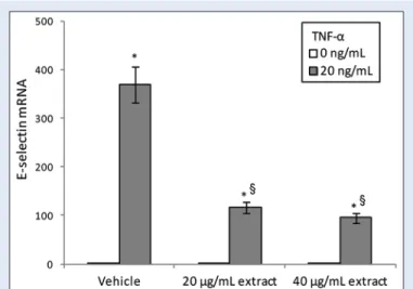
Figure 4: Effect of Thymelaea microphylla acetone extract on tumor necrosis factor-α induced mRNA expression of the adhesion molecule E-selectin, in human umbilical vein endothelial cells. Cells were pretreated with Thymelaea microphylla extract (20–40 µg/ml) for 24 h and then exposed for 2 h to tumor necrosis factor-α (20 ng/ml). Cultures treated with the vehicle alone (dimethyl sulfoxide 0.1%) were used as controls. Values are expressed as 2−ΔΔCt normalized to control and reported as mean
± standard deviation of three experiments. *P < 0.05 versus respective tumor necrosis factor-α 0 ng/ml; §P < 0.05 versus vehicle exposed to tumor necrosis factor-α 20 ng/ml
Figure 3: Cell viability in human umbilical vein endothelial cells pretreated with Thymelaea microphylla acetone extract (20–40 µg/ml) for 24 h and then exposed for 2 h to tumor necrosis factor-α (20 ng/ ml). Cultures treated with the vehicle alone (dimethyl sulfoxide 0.1%) were used as controls. Data represent percentage of viable cells (mean percentage) calculated from the number of viable cells in treated samples versus control untreated and unexposed. *P < 0.05 versus respective tumor necrosis factor-α 0 ng/ml; §P < 0.05 versus vehicle exposed to tumor necrosis factor-α 20 ng/ml
Figure 2: Effect of Thymelaea microphylla acetone extract on release of (a) thromboxane B2 (in comparison with that of indomethacin, Indo) and of (b) prostaglandin E2 (in comparison with that of nimesulide, NIM) in human whole blood. Data are expressed as mean ± standard deviation of three independent experiments
a b
Table 2: Antioxidant activity/free radical scavenger of T. microphylla extracts measured by means of different in vitro tests. Results are expressed as: mean±SD of 3 experiments for ORAC values; 50% scavenging concentration (SC50) and 95% confidence limits (CL) in the DPPH and ABTS assays; 50% mean inhibition
concentration (IC50) and 95% confidence limits (CL) in the SOD, protein denaturation and BCB assay. §TE: Trolox equivalents
Extracts DPPH SC50mg/ml ABTS SC50 mg/ml ORAC mmol §TE/g dw BCB IC50 mg/ml Protein denaturation IC50 mg/ml SOD IC50 mg/ml Water 3.54 (2.76‑4.54) 0.39 (0.04‑3.76) 1,98±0.07 1.29 (0.53‑3.11) 1.12 (0.89‑1.39) 11.54 (9.06‑14.69) Ethanol 8.67 (6.96‑10.80) 1.02 (0.78‑1.33) 1.39±0.05 0.53 (0.42‑0.68) 0.22 (0.16‑0.28) 7.24 (4.37‑11.99) Acetone 5.98 (5.05‑7.09) 0.67 (0.55‑0.81) 1,09±0.01 0.16 (0.10‑0.28) 0.15 (0.10‑0.21) 1.81 (1.18‑2.77) Hexane 47.97 (33.76‑68.17) 7.98 (4.93‑12.91) 0.22±0.03 0.25 (0.17‑0.37) 0.24 (0.18‑0.56) 3.71 (2.91‑4.72)
Conversely, the TM water extracts appeared unable, under these experimental conditions, to inhibit TxB2 release at all dose tested and showed only a weak capability to inhibit PGE2 release, being the IC50 value 348.45 µg/ml (95% CL 262.96–562.16 µg/ml) [Figure 2].
Protective effect on tumor necrosis
factor-α-induced endothelial dysfunction
Since the TM AcE was the only extract showing good chemical profile, significant antioxidant properties, and excellent anti‑inflammatory and COX‑inhibitory activity, together with lack of toxicity on normal human blood cells, only these TM extracts were tested about its protective effect on TNF‑α‑induced endothelial dysfunction.
In fact, oxidative stress and inflammation are considered among the prominent pathways of vascular endothelial dysfunction in many pathophysiological conditions. The proinflammatory cytokine TNF‑α is often present in chronic inflammatory diseases, exerting a prominent effect on the expression of proinflammatory genes in endothelial cells. This effect takes place predominantly through activation of intracellular signaling pathways involving NF‑κB. In endothelial cells, NF‑κB is involved in the
regulation of the expression of several genes, including those encoding E‑selectin and COX‑2. Many studies have demonstrated that the increased adhesion of circulating monocytes to the injured endothelial layer is a critical early event in the development of atherogenesis. In fact, endothelial cells recruit monocytes by selectively expressing various cell surface adhesion molecules such as E‑selectin. Proinflammatory cytokines, such as TNF‑α, can induce, through its ability to promote intracellular ROS formation and activation of the redox‑sensitive transcription factor NF‑κB, the expression and release of chemotactic factors and additional cytokines that can each further contribute to inflammation.
Hence, in this study, we studied the capability of TM AcE to alter the TNF‑α‑changes on HUVECs which are a model system widely used to identify the effects of and targets for deleterious vascular risk factors. At this aim, cells were pretreated with the extract (20–40 µg/ml) and then exposed for 2 h to TNF‑α 20 ng/mL.
Our results indicate a clear cytotoxic effect of TNF‑α. Cell pretreatment with TM AcE showed a dose‑dependent protective effect against TNF‑α‑induced cell death. Furthermore, TM AcE alone was unable to affect cell viability at the tested doses [Figure 3].
Real‑time PCR was used to evaluate E‑selectin gene expression in endothelial cells as representative genes involved in cell adhesion. According to the data reported in the literature, cell exposure to TNF‑α significantly induced the surface expression of E‑selectin [Figure 4]. TNF‑α‑induced upregulation of RNA expression of this adhesion molecule was significantly suppressed by pretreatment with TM AcE [Figure 4]. TM AcE per se, without any kind of stimulus, had no effects on the basal expression of this gene.
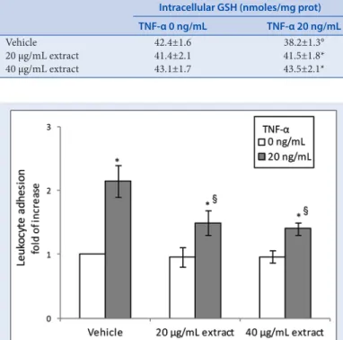
Adhesion molecules are responsible for leukocyte adhesion to vascular endothelium, so promoting their migration into subendothelial space, an early event in atherogenesis. To confirm the inhibitory activity of TM AcE on endothelial activation induced by TNF‑α, we investigated isolated leukocytes co‑cultured with HUVECs. Figure 5 shows that the number of leukocytes adhered to the endothelial cells exposed to TNF‑α was higher than that observed in controls, but it appeared to be reduced by pretreatment with TM AcE. These results confirm the inhibitory activity of TM AcE on TNF‑α‑induced endothelial activation.
To further investigate the protective effect of TM AcE against cytotoxic effects of TNF‑α on HUVECs, we measured intracellular content of GSH. In fact, cellular thiol redox status is critical for a variety of biological process including transcriptional activation of various genes and regulation of cell proliferation, inflammation, and apoptosis. Thiols, particularly GSH, are also critical for cellular antioxidant defenses, including protecting the cells from oxidant injury and inflammation. In our present experiments, we found that GSH levels were significantly decreased in HUVECs exposed to TNF‑α. As shown in Table 3, the pretreatment with the TM AcE was able to prevent, in a concentration‑dependent way, the depletion in GSH cell content following exposure to TNF‑α.
These findings contribute to confirm the hypothesis that several plant‑derived polyphenols can suppress TNF‑α activated inflammatory pathways.[31] In particular, both luteolin and kaempferol, two flavonoids recovered in this drug, have been previously demonstrated to protect endothelial cells against cytokine‑induced proinflammatory status.[32,33]
CONCLUSIONS
This is the first time that leaves and flowers from TM are identified as a good source of bioactive compounds possessing significant antioxidant and anti‑inflammatory properties, potentially effective in prevention and treatment of pathological conditions in which oxidative stress and inflammation play a significant role. In fact, in our experiments, TM
Figure 5: Leukocyte adhesion to human umbilical vein endothelial cells pretreated with Thymelaea microphylla acetone extract (20–40 µg/ml) for 24 h and then exposed to tumor necrosis factor-α (20 ng/ml) for 2 h. A flask containing the co-culture and not exposed to tumor necrosis factor-α was used as control. Increase in leukocyte adhesion upon stimulation of human umbilical vein endothelial cells with tumor necrosis factor-α was calculated in relation to the basal adhesion of leukocytes to nonstimulated human umbilical vein endothelial cells (that was set to 1). Data are reported as mean ± standard deviation of three experiments. *P < 0.05 versus respective tumor necrosis factor-α 0 ng/ml; §P < 0.05 versus vehicle exposed to tumor necrosis factor-α 20 ng/ml
Table 3: Changes in intracellular GSH levels in HUVECs pretreated for 24 h with T. microphylla acetone extract (20-40 µg/ml) and then for 2 h with TNF-α (20 µg/ml). Cultures treated with the vehicle alone (DMSO 0.1%) were used as controls. Data are expressed as mean±SD of the three independent experiments. °P<0.05 vs respective TNF-α 0 ng/ml; *P<0.05 vs vehicle exposed to TNF-α 20 ng/ml
Intracellular GSH (nmoles/mg prot) TNF‑α 0 ng/mL TNF‑α 20 ng/mL
Vehicle 42.4±1.6 38.2±1.3° 20 µg/mL extract 41.4±2.1 41.5±1.8* 40 µg/mL extract 43.1±1.7 43.5±2.1*
KHADIDJA DEHIMI, et al.: Biological Effects of Thymelaea microphylla Extracts
210 Pharmacognosy Magazine, Vol 12, Issue 47, Jul-Sep, 2016
appears able to decrease TxB2 and PGE2 release, very likely by an inhibition of COX activity, and further experiments could demonstrate a possible inhibitory effect of TM extracts selectively on the COX1 and COX2 pathways. Furthermore, due very likely to the mentioned bioproperties, the TM AcE is able to protect against endothelial dysfunction, which is an early event in development of atherosclerosis and vascular diseases. Further studies are warranted to better understand both the chemical composition of this drug and the cellular mechanisms involved in its pharmacological activity.
Financial support and sponsorship
This research was partially supported by the Erasmus Mundus Programme of the European Union – EMMAG.
Conflicts of interest
There are no conflicts of interest.REFERENCES
1. Galicia‑Herbada D. Origin and diversification of Thymelaea (Thymelaeaceae): Inferences from a phylogenetic study based on ITS (rDNA) sequences. Plant Syst Evol 2006;257:159‑87. 2. Benhammou N, Bekkara FA, Constard JM. Antioxidant activity of methanolic and water
extracts from Marrubium deserti (De Noë) and Thymelaea microphylla from Algerian Sahara. Adv Food Sci 2009;31:194‑201.
3. Boukef MK. Traditional medicine and pharmacopoeia. Plants in Tunisian traditional medicine. Ed. ACCT, Paris; 1986.
4. Miyamae Y, Villareal MO, Abdrabbah MB, Isoda H, Shigemori H. Hirseins A and B, daphnane diterpenoids from Thymelaea hirsuta that inhibit melanogenesis in B16 melanoma cells. J Nat Prod 2009;72:938‑41.
5. Akrout A, Gonzalez LA, El Jani H, Madrid PC. Antioxidant and antitumor activities of Artemisia
campestris and Thymelaea hirsuta from Southern Tunisia. Food Chem Toxicol 2011;49:342‑7.
6. Trigui M, Hsouna AB, Tounsi S, Jaoua S. Chemical composition and evaluation of antioxidant and antimicrobial activities of Tunisian Thymelaea hirsuta with special reference to its mode of action. Ind Crops Prod 2013;41:150‑7.
7. Amari NO, Bouzouina M, Berkani A, Lotmani B. Phytochemical screening and antioxidant capacity of the aerial parts of Thymelaea hirsuta L. Asian Pac J Trop Dis 2014;4:104‑9. 8. Djeridane A, Yousfi M, Brunel JM, Stocker P. Isolation and characterization of a new steroid
derivative as a powerful antioxidant from Cleome arabica in screening the in vitro antioxidant capacity of 18 Algerian medicinal plants. Food Chem Toxicol 2010;48:2599‑606.
9. Ladjel S, Gherraf N, Zellagui A, Brahim L, Hameurlaine S. Phytochemical and antibacterial screening of some Algerian Saharan medicinal plants. Plant Sci Feed 2011;1:179‑82. 10. Mekhelfi T, Kerbab K, Guella G, Zaiter L, Benatache S, Benayache F. Antioxidant and
antimicrobial activities of an endemic desert species Thymelea microphylla Coss. et Dur. Pharm Lett 2014;6:152‑6.
11. Kerbab K, Mekhelfi T, Zaiter L, Benayache S, Benayache F, Picerno P, et al. Chemical composition and antioxidant activity of a polar extract of Thymelaea microphylla Coss. et Dur. Nat Prod Res 2015;29:671‑5.
12. Quezel P, Santa S. New Flora of Algeria and the southern desert regions. Paris: National Centre Editions of Scientific Research;1962‑63. p. 475‑6.
13. Ganatra SH, Durge SP, Patil SU. Preliminary phytochemicals investigation and TLC analysis of
Ficus racemosa leaves. J Chem Pharm Res 2012;4:2380‑4.
14. Doughari JS. Phytochemicals: Extraction Methods, Basic Structures and Mode of Action as Potential Chemotherapeutic Agents, Phytochemicals A Global Perspective of Their Role in Nutrition and Health. Rijeka, Croatia:InTech Publisher; 2012. p. 2‑15.
15. Tomaino A, Martorana M, Arcoraci T, Monteleone D, Giovinazzo C, Saija A. Antioxidant activity and phenolic profile of pistachio (Pistacia vera L. variety Bronte) seeds and skins. Biochimie 2010;92:1115‑22.
16. Tuttolomondo T, La Bella S, Licata M, Virga G, Leto C, Saija A, et al. Biomolecular characterization of wild sicilian oregano: Phytochemical screening of essential oils and extracts, and evaluation of their antioxidant activities. Chem Biodivers 2013;10:411‑33. 17. Spagna G, Pifferi PG, Rangoni C, Mattivi F, Nicolini G, Palmonari R. The stabilization of
white wines by adsorption of phenolic compounds on chitin and chitosan. Food Res Int 1996;29:241‑8.
18. Morabito G, Trombetta D, Singh Brajendra K, Prasad Ashok K, Parmar Virinder S, Naccari C,
et al. Antioxidant properties of 4‑methylcoumarins in in vitro cell‑free systems. Biochimie
2010;92:1101‑7.
19. Dávalos A, Gómez‑Cordovés C, Bartolomé B. Extending applicability of the oxygen radical absorbance capacity (ORAC‑fluorescein) assay. J Agric Food Chem 2004;52:48‑54. 20. Boussahel S, Speciale A, Dahamna S, Amar Y, Bonaccorsi I, Cacciola F, et al. Flavonoid profile,
antioxidant and cytotoxic activity of different extracts from Algerian Rhamnus alaternus L. Bark. Pharmacogn Mag 2015;11 Suppl 1:S102‑9.
21. Martorana M, Arcoraci T, Rizza L, Cristani M, Bonina FP, Saija A, et al. In vitro antioxidant and in vivo photoprotective effect of pistachio (Pistacia vera L. variety Bronte) seed and skin extracts. Fitoterapia 2013;85:41‑8.
22. Leelaprakash G, Mohan Dass S. In vitro anti‑inflammatory activity of methanol extract of
Enicostemma axillare. Int J Drug Dev Res 2011;3:189‑96.
23. Trombetta D, Giofrè SV, Tomaino A, Raciti R, Saija A, Cristani M, et al. Selective COX‑2 inhibitory properties of dihydrostilbenes from liquorice leaves – in vitro assays and structure/ activity relationship study. Nat Prod Commun 2014;9:1761‑4.
24. Siracusa L, Saija A, Cristani M, Cimino F, D’Arrigo M, Trombetta D, et al. Phytocomplexes from liquorice (Glycyrrhiza glabra L.) leaves – chemical characterization and evaluation of their antioxidant, anti‑genotoxic and anti‑inflammatory activity. Fitoterapia 2011;82:546‑56. 25. Cimino F, Speciale A, Siracusa L, Naccari C, Saija A, Mancari F, et al. Cytotoxic effects induced
in vitro by organic extracts from urban air particulate matter in human leukocytes. Drug Chem
Toxicol 2014;37:32‑9.
26. Cimino F, Speciale A, Anwar S, Canali R, Ricciardi E, Virgili F, et al. Anthocyanins protect human endothelial cells from mild hyperoxia damage through modulation of Nrf2 pathway. Genes Nutr 2013;8:391‑9.
27. Speciale A, Canali R, Chirafisi J, Saija A, Virgili F, Cimino F. Cyanidin‑3‑O‑glucoside protection against TNF‑α‑induced endothelial dysfunction: Involvement of nuclear factor‑κB signaling. J Agric Food Chem 2010;58:12048‑54.
28. Speciale A, Anwar S, Ricciardi E, Chirafisi J, Saija A, Cimino F. Cellular adaptive response to glutathione depletion modulates endothelial dysfunction triggered by TNF‑α. Toxicol Lett 2011;207:291‑7.
29. Speciale A, Anwar S, Canali R, Chirafisi J, Saija A, Virgili F, et al. Cyanidin‑3‑O‑glucoside counters the response to TNF‑alpha of endothelial cells by activating Nrf2 pathway. Mol Nutr Food Res 2013;57:1979‑87.
30. Puglia C, Santagati NA, Bonina F, Trombetta D, Cristani M, Speciale A, et al. Protective effect of Mediterranean fish oil extracts on heat‑induced denaturation of albumin. J Pharm Pharmacol 2006;58:1411‑3.
31. Gupta SC, Tyagi AK, Deshmukh‑Taskar P, Hinojosa M, Prasad S, Aggarwal BB. Downregulation of tumor necrosis factor and other proinflammatory biomarkers by polyphenols. Arch Biochem Biophys 2014;559:91‑9.
32. Crespo I, García‑Mediavilla MV, Gutiérrez B, Sánchez‑Campos S, Tuñón MJ,
González‑Gallego J. A comparison of the effects of kaempferol and quercetin on cytokine‑induced pro‑inflammatory status of cultured human endothelial cells. Br J Nutr 2008;100:968‑76.
33. Jia Z, Nallasamy P, Liu D, Shah H, Li JZ, Chitrakar R, et al. Luteolin protects against vascular inflammation in mice and TNF‑α‑induced monocyte adhesion to endothelial cells via suppressing IKBα/NF‑κB signaling pathway. J Nutr Biochem 2015;26:293‑302.
ABOUT AUTHOR
Dr. Antonio Speciale, is a Researcher at University of Messina, Italy.
Graduated in Pharmaceutical Chemistry and Technology, he obtained his PhD degree in Toxicology in 2009 from University of Messina, and is a Specialist in Farmacognosy since 2013. He is author of more than 30 peer‑ reviewed papers in international scientific journals and of more than 40 publications in national and international conferences.