Obstructive sleep apnea in sarcoidosis and impact of cpap
treatment on fatigue
Pier-Valerio Mari
1,2, Giuliana Pasciuto
1,2, Matteo Siciliano
1,2, Jacopo Simonetti
1,2, Federico Ballacci
2,
Francesco Macagno
1,2, Bruno Iovene
1, Filippo Martone
3, Giuseppe Maria Corbo
1,2, Luca Richeldi
1,21 Fondazione Policlinico Universitario A. Gemelli, IRCCS, Rome (Italy); 2 Università Cattolica del Sacro Cuore; 3 Amici Contro La Sarcoidosi
Italia ONLUS
AbstrAct. Rationale: An increased incidence of Obstructive Sleep Apnea (OSA) in sarcoidosis has been de-scribed in small sample size studies. Fatigue is common in sarcoidosis and OSA could be a relevant, treatable comorbidity. To date, the effect of Continuous Positive Airway Pressure (CPAP) on fatigue has never been assessed. Objectives: To investigate the prevalence of OSA in sarcoidosis, fatigue status and daytime sleepiness in patients of our center. To explore the effect of CPAP in fatigue and daytime sleepiness after 3 months using validated questionnaires. Method: Single group, one center, open-label prospective cohort study. Measurements and main result: We enrolled 68 patients and OSA was diagnosed in 60 (88.2%): 25 (36.8%) were mild while 35 (51.5%) were moderate-to-severe. 38 (55.9%) patients received CPAP but only 20 (30.9%) were compliant at 3-month evaluation. Questionnaires demonstrated fatigue in 34 (50%) and daytime sleepiness in 21 (30.9%). In multivariate regression analysis, Scadding stage and FAS behave as predictors of Apnea-Hypopnea Index (AHI) severity while sleepiness and steroids weren’t associated. FAS score (ΔFAS = 6.3; p = 0.001) and ESS score (ΔESS =
2.8; p = 0.005) improved after three months of CPAP. Conclusions: OSA is highly prevalent in patients affected by sarcoidosis. ESS questionnaire is not reliable for OSA screening and other pre-test probability tool should be evaluated in further studies. CPAP leads to a significative reduction of fatigue and daytime sleepiness at three-month. Further studies are needed to confirm the high prevalence of OSA in sarcoidosis and the positive role of CPAP in fatigue. (Sarcoidosis Vasc Diffuse Lung Dis 2020; 37 (2): 169-178)
Key words: Sarcoidosis; Sleep Apnea; CPAP
SARCOIDOSIS VASCULITIS AND DIFFUSE LUNG DISEASES 2020; 37 (2);169-179 DOI: 10.36141/svdld.v37i2.9169 © Mattioli 1885
Received: 14 January 2020
Accepted after revision: 18 March 2020 Correspondence: Mari Pier-Valerio, MD (ORC-ID: 0000-0002-3307-217X)
Fondazione Policlinico Universitario A. Gemelli, Rome, Italy Largo Agostino Gemelli, 8
Rome (IT) 00168
Introduction
Sarcoidosis is a granulomatous disease charac-terized by a great heterogeneity in clinical presen-tation, in the disease course and therapeutic results. Pathogenesis is still unclear; exaggerated immune re-sponse to unidentified antigens is thought to be the key pathogenic mechanism1. Sarcoidosis is a
world-wide disease but prevalence is higher in the north-ern European and African-American populations2.
Involvement of lung parenchyma and thoracic lym-phadenopathies are very common features3.
An increased incidence of Sleep-disordered Breathing (SDB) and Obstructive Sleep Apnea (OSA) has been observed in patients affected by sar-coidosis though the pathogenesis is still unclear4.
OSA is a disorder of upper airways collapse dur-ing sleep time leaddur-ing to oxygen desaturation, sleep fragmentation and hypercapnia5. Moreover, once the
obstruction has taken place, an arousal from sleep is induced by a respiratory effort allowing hyperventi-lation to occur6. Thus, arousals prevent asphyxia
dur-ing upper airway obstructions but can lead to cardio-vascular activation and contribute to adverse events of sleep apneas. Such condition is common, and re-cent studies suggest a significant prevalence of mod-erate-to-severe disordered breathing in high-income countries7 of 49.7% in men and 23.4% in women8.
Obstructive Sleep Apnea (OSA) is defined by an Apnea-Hypopnea Index (AHI) more than five and the collapse leads to a fall of blood saturation dur-ing sleep linked to daytime sleepiness, and stroke9,10.
Patients with sleep apneas report excessive daytime sleepiness, non-restorative sleep and fatigue. Con-tinuous positive airway pressure (CPAP) is the treat-ment of choice since the first publication in 198111 in
moderate-to-severe OSA syndrome although mild diseases may be allocated to treatment.
The association between OSA and sarcoidosis is nowadays without a clear pathogenesis explanation. Upper airway collapsibility might play a key role in pathogenesis of OSA in Interstitial Lung Dis-eases. A restrictive pattern on pulmonary function test leads to an increased airway collapsibility due to the caudal traction on these structures12. Also, a
high Body Mass Index with deposition of fat around upper airways may enhance the collapsibility. More hypotheses have been proposed: sarcoid neuropathy, obesity due to steroids and upper airway resistance
secondary to airway disease4,13. A higher OSA
preva-lence was found in subjects with lupus pernio and increased AHI was reported among patients with lung parenchymal involvement14. In addition to the
OSA, sarcoidosis is frequently associated with fa-tigue, a state of physical and mental weariness15 as
a consequence of inflammatory mediators16. Fatigue
is common with 50-70% prevalence in patients af-fected by sarcoidosis17. Regardless of its underlying
cause, fatigue has a great impact on the life of pa-tients affected with sarcoidosis and questionnaires such as the Fatigue Assessment Scale (FAS) have been validated18 and used in clinical practice in order
to assess the burden of such condition.
Up-to-date, poor data is available on the preva-lence of OSA in Sarcoidosis and no data is neither available about the effect of obstructive sleep apnea treatment with CPAP device on sarcoidosis nor the effect on the fatigue state assessed with FAS ques-tionnaire.
Therefore, we aimed to define the prevalence of OSA in patients from our Sarcoidosis Clinic and to explore the effects on fatigue due to the treatment of OSA with CPAP.
Methods
The SARCOIDOSAS study is a single group, open labeled prospective cohort study conducted in Fondazione Policlinico Universitario A. Gemelli, IRCCS, Roma (Italy). The trial was approved by the Institutional Board Review (ID 2455, approved on 05 April 2019; principal investigator: R. L) and have been registered on ClinicalTrials.gov (SARCOIDO-SAS number, NCT03926832, registered 10 April; principal investigator: M. PV). Patients affected by sarcoidosis older than 18 years to a maximum of 85 years were eligible in the Sarcoidosis Clinic of our institution: a reference, tertiary care center for rare interstitial lung diseases. All patient met ATS cri-teria1 for diagnosis and had a histology-proven
sar-coidosis. The exclusion criteria included: ongoing CPAP treatment or having a diagnosis of psychiatric disorders. We contacted (telephone / mail) the whole cohort of our Sarcoidosis Clinic (n = 122 patients) but only 84 (68.9%) gave back us a feedback after the contact. Contacted patients were addressed for a first eligibility evaluation and enrolled between April and May 2019. All participants provided informed
consent. We did not impose criteria to select pa-tients with higher probability of diagnosing sleep apnea syndrome. Furthermore, patients with OSA diagnosis19 were allocated to treatment according
to the latest American Academy of Sleep Medicine (AASM)20: Continuous Positive Airway Pressure
(CPAP) therapy was started in all those with a mod-erate-to-severe OSA and in those with mild OSA with a documented symptoms of excessive daytime sleepiness or history of insomnia or hypertension/ ischemic heart disease or history of stroke.
Baseline evaluation
After the initial screening of the entire cohort of our Sarcoidosis Clinic, a total of 84 subjects were eligible but 16 of them declined to participate in the study. Thus, 68 patients were recruited and performed a baseline evaluation collecting data about demo-graphic, Scadding stage on chest x-ray and corticos-teroid treatment. Lung function tests of the cohort were collected including the following parameters: Forced Vital Capacity (FVC), Total Lung Capacity (TLC) and Diffusing Capacity divided by the Al-veolar Volume (DLCO/Va). Moreover, fatigue status
and daytime sleepiness were assessed by completing the Fatigue Assessment Scale (FAS) and Epworth Sleepiness Scale (ESS) questionnaires. The FAS is a 10-item validated questionnaire to assess the fatigue in patients affected by sarcoidosis ranging from 10 to 50 points while the ESS determines the likelihood of falling asleep during various scenarios for a total score from 0 to 24. Fatigue was determined if FAS ≥ 22 and extreme fatigue status if FAS ≥ 35 points while daytime sleepiness was established if ESS > 10 points. Both FAS and ESS questionnaires were administered via face-to-face interview during the same baseline or follow up medical evaluation. Re-gardless of the result of the screening questionnaires, all patients scheduled an overnight home sleep study using a level III portable diagnostic device (Vital-Night Plus®, VitalAire®, Milan, Italy). The
follow-ing parameters were measured: airflow measured by thermistor and nasal pressure cannula, thoraco-abdominal respiratory bands, pulse oximetry and body position. Obstructive sleep apnea syndrome was identified when apnea/hypopnea index (AHI) was found more than five events per hour during the night sleep study. Sleep studies were automatically
analyzed and then rescored using a manual editing of the total recording time. OSA severity was de-termined using the rescored apnea/hypopnea index (AHI) according to the following grading score: mild (5 ≥ AHI > 15), moderate (15 ≥ AHI > 30), severe (AHI ≥ 30). All moderate-to-severe patients, along with those with mild OSA that have excessive daytime sleepiness or cardiovascular disease, were al-located to treatment. An autoset device was given with a pressure range between 8.0 and 15.0 cm H2O (AirSense 10 AutoSet®, Resmed®) in order to start a
CPAP treatment. Three-month follow-up
Patients that were allocated to treatment were evaluated after three months of CPAP therapy: fa-tigue status and daytime sleepiness were newly as-sessed and change from baseline to 90 days were collected. The minimal important difference (MID) in the change of FAS (ΔFAS) is defined as a
reduc-tion of at least 4 points or 10% of the baseline val-ue, while the MID in the change of ESS (ΔESS) is a
reduction of at least 2 points of the baseline value. Also, compliance to CPAP therapy was evaluated by reading the device report and threshold of “good” compliance was defined as device use for more than 4 hours per night and at least 70% of total number of nights.
Statistical analysis
We analyzed the OSA prevalence in our Sar-coidosis Clinic. The sample was described using de-scriptive statistics techniques. Quantitative variables were summarized using means ± standard deviation; categorical variables presented using (absolute and percentage) frequency tables. The strength of asso-ciations was assessed using Chi-Squared test (cat-egorical variables) or Pearson correlation coefficient (continuous variables). Apnea-Hypopnea Index po-tentially independent predictors were investigated post hoc by using multiple linear regression analysis. Statistical results were adjusted for potential con-founding variables. Correlation between baseline and follow up outcomes were assessed using a paired T-Test after being tested for normality. SAS version 9.4 or higher statistical software was used (SAS In-stitute Inc®, Cary, North Carolina, USA).
Results
Baseline characteristics
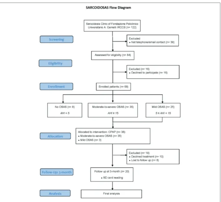
The flowchart diagram of the study is presented in Figure 1. We enrolled 68 patients and the cohort was predominantly female in 42 (61.8%). Mean age was 60.2 ± 10.0 SD along with a mean BMI of 27.9 ± 5.1 SD. 32 (50.0%) were former smokers with a mean pack/year of 8.8 ± 9.4 SD. Baseline lung func-tion tests did show FVC % 108.6 ± 17.6 SD, TLC % 98.2 ± 13.1 SD and DLCO % 91.2 ± 16. We also defined Sarcoidosis staging on the chest radiograph:
stage 0-I was identified in 28 (41.2%), stage II-III in 38 (55.9%) while stage IV in only 2 (2.9%). Cor-ticosteroid therapy was confirmed in 31 (45.6%) and 26 (38.2%) were treated for more than three months with steroids.
Fatigue and daytime sleepiness
Fatigue status and daytime sleepiness were found to be common in our cohort: the FAS ques-tionnaire assessed a mean FAS of 24.8 ± 9.7 SD, the fatigue status in 34 (50.0%) and an extreme fatigue status in 15 (22.1%), while the ESS questionnaire
did show a mean ESS of 7.9 ± 5.2 and the presence of daytime sleepiness in 21 (30.9%). Moreover, the baseline FAS score did show a negative correlation with male gender (coefficient B: -8.8; p = 0.0001) and correlate with ESS score at baseline (coefficient B: 0.74; p = 0.001).
Prevalence and severity of OSA
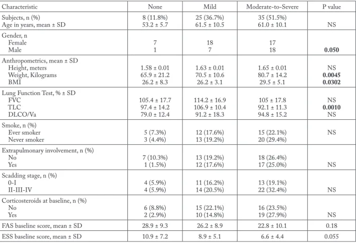
OSA was prevalent in the Sarcoidosis Clin-ic of our center and was diagnosed in 60 (88.2%): 25 (36.8%) had a mild grade and 35 (51.5%) were moderate-to-severe. CPAP was proposed according to AASM guidelines to all the moderate-to-severe patients (n = 35) and some of the mild group (n = 3 with history of ischemic heart disease) allocating a total of 38 (55.9%) patients to treatment. Table 1 de-scribes the demographic and clinical characteristics of the groups.
Association between AHI and Baseline evaluation AHI did show a negative, weak correlation with questionnaires results of FAS and ESS. The regres-sion plots in Figure 2 illustrate the negative correla-tion coefficient (ρ) when AHI was compared to FAS (ρ: -0.31, p = 0.009) and ESS (ρ: -0.32, p = 0.007) at baseline. Also, TLC% predicted seems to be nega-tively correlated to AHI (ρ: -0.47, p = 0.001) along with FVC% predicted (ρ: -0.34, p = 0.015), while a positive, moderate correlation was established with weight (ρ: 0.49, p = 0.001) and BMI (ρ: 0.41, p = 0.001). The post hoc regression analysis (Table 2) adjusted for multiple variables (male gender, BMI, ad-vanced Scadding stages, FAS/ESS at baseline, treat-ment with corticosteroids for more than 3 months at baseline) revealed some interesting insights. In this model, BMI, Scadding stage and FAS score behave as independent predictors of Apnea-Hypopnea Index
Table 1. Demographic and clinical characteristics by OSA severity
Characteristic None Mild Moderate-to-Severe P value
Subjects, n (%)
Age in years, mean ± SD 8 (11.8%)53.2 ± 5.7 61.5 ± 10.525 (36.7%) 35 (51.5%)61.0 ± 10.1 NS
Gender, n Female Male 71 187 1718 0.050 Anthropometrics, mean ± SD Height, meters Weight, Kilograms BMI 1.58 ± 0.01 65.9 ± 21.2 26.2 ± 8.3 1.63 ± 0.01 70.5 ± 10.6 26.2 ± 3.1 1.65 ± 0.01 80.7 ± 14.2 29.5 ± 5.1 NS 0.0045 0.0302 Lung Function Test, % ± SD
FVC TLC DLCO/Va 105.4 ± 17.7 97.4 ± 14.2 79.0 ± 12.4 114.2 ± 16.9 106.9 ± 10.4 91.2 ± 18.3 105 ± 17.8 92.1 ± 11.3 94.8 ± 15.2 NS 0.0010 NS Smoke, n (%) Ever smoker Never smoker 5 (7.3%)3 (4.4%) 12 (17.6%)13 (19.2%) 15 (22.1%)20 (29.4%) NS Extrapulmonary involvement, n (%) No Yes 7 (10.3%)1 (1.5%) 13 (19.2%)12 (17.6%) 18 (26.4%)17 (25.0%) NS Scadding stage, n (%) 0-I II-III-IV 4 (5.9%)4 (5.9%) 11 (16.2%)14 (20.5%) 13 (19.1%)22 (32.4%) NS Corticosteroids at baseline, n (%) No Yes 6 (8.8%)2 (2.9%) 15 (22.1%)10 (14.8%) 16 (23.5%)19 (27.9%) NS
FAS baseline score, mean ± SD 28.9 ± 9.3 26.2 ± 8.9 22.8 ± 10.1 0.18
ESS baseline score, mean ± SD 10.9 ± 7.2 8.9 ± 5.1 6.6 ± 4.4 0.055
BMI = Body Mass Index, FVC = Forced Vital Capacity, TLC = Total Lung Capacity, DLCO/Va = Diffusing Capacity / Alveolar Volume, FAS = Fatigue Assessment Scale, ESS = Epworth Sleepiness Scale, NS = Not significant
Fig. 2. Regression plot: FAS, ESS and AHI. FAS: Fatigue Assessment Scale, ESS: Epworth Sleepiness Scale, AHI: Apnea-Hypopnea Index, ρ: Pearson correlation coefficient.
Table 2. Regression analysis summary for independent predictors of AHI
Unadjusted Adjusted*
Variable B (95% CI) t P>|t| B (95% CI) t P>|t|
Male gender 8.0 (-0.2-16.3) 1.94 0.056 10.2 (-0.1-20.1) 2.02 0.050
BMI 1.3 (0.6-2.0) 3.55 0.001 1.4 (0.6-2.2) 3.54 0.001
Scadding stages II-III-IV 5.6 (-2.7-13.8) 1.35 0.183 6.3 (-1.5-17.3) 2.04 0.048
TLC -0.7 (-1-0.3) -3.65 0.001 -0.5 (-0.8-0.2) -2.83 0.007
FAS Baseline -0.5 (-0.9-0.1) -2.68 0.009 0.1 (-0.5-0.6) 0.11 0.916
ESS Baseline -1.0 (-1.8-0.3) -2.76 0.007 -1.1 (-1.9-0.2) -2.40 0.021
Steroid treatment > 3m 5.2 (-3.0-13.4) 1.27 0.208 0.1 (-8.7-9.2) 0.03 0.997
*Adjusted R-squared: 0.47
AHI = Apnea-Hypopnea Index, BMI = Body Mass Index, CI: 95% Confidence Interval of Coefficient (B), ESS = Epworth Sleepiness Scale, FAS = Fatigue Assessment Scale, “m” = months, TLC = Total Lung Capacity.
severity while other covariates such as ESS score and steroids at baseline seems to be not related to AHI prediction. These results confirmed the known role of BMI along with the male gender in predicting se-verity of AHI but also enlightened that radiological impairment in sarcoidosis is associated to the OSA severity. Furthermore, the therapy with corticoster-oids for more than 3 months in patients evaluated at baseline was not predictive of the AHI severity as described in the multiple regression analysis.
Three-month evaluation
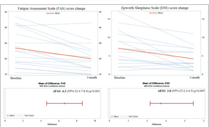
An evaluation after three months since the CPAP start was obtained and 20 (29.4%) patients were analyzed. The follow-up questionnaires (FAS90
and ESS90) investigate fatigue and daytime sleepiness
after CPAP treatment using the same items of the baseline questionnaires: FAS90 had a mean of 18.7 ±
8.6 SD while ESS90 was 4.0 ± 2.8 SD. Changes in
FAS and ESS questionnaires score from baseline to 90-day are shown in Figure 3 and confirmed an im-provement of fatigue and sleepiness: the change in FAS (ΔFAS) was 6.3, 95% CI 4.7-8.9, p = 0.001 and
the change in ESS (ΔESS) was 2.8, 95% CI 2.3-4.5,
p = 0.005. CPAP adherence tracking data were ana-lysed using the device’s SD card. Compliance to treatment was good in 13 (65.0%) with a mean AHI residual of 3.4 ± 5.1 SD and a percentage of used days > 4 hours of 65% ± 34.7 SD. The explorative analysis shows that only good adherence to CPAP treatment (Figure 4) reduced fatigue (7.5, 95% CI
Fig. 3. Changing in FAS and ESS questionnaires score from baseline to 3-month. FAS: Fatigue Assessment Scale, ESS: Epworth Sleepiness Scale, ΔFAS: Difference between baseline FAS and FAS at 3-month, ΔESS: Difference between baseline ESS and ESS at 3-month.
Fig. 4. Adherence to CPAP impact in FAS change at 3-month. FAS: Fatigue Assessment Scale. Compliance to CPAP therapy was evalu-ated by reading SD card at 3-month. Good compliance was defined as “device use for more than 4 hours per night and at least 70% of total number of nights”.
3.5-9.1; p = 0.0011) while poor compliance to CPAP therapy missed the statistical significance (4.4, 95% CI -1.0-9.9; p = 0.0960). Similarly, a statistical sig-nificative reduction in ESS score at 3-month was also demonstrated only in good compliance (2.92, 95% CI 1.1-4.7; p = 0.0041) when compared to poor one (3.14, 95% CI -0.1-6.3; p = 0.0519).
Discussion
The analysis of the cohort from our institu-tion demonstrate that OSA affects the majority of patients with sarcoidosis in our center and it must be taken into account during the clinical evaluation. The impressively high OSA prevalence might be ex-plained by an impaired upper airway stability. We found that BMI, lower lung volumes in pulmonary function tests (TLC) and parenchymal involvement (Scadding stage) are independent predictors of OSA in sarcoidosis. Thus, the caudal traction on the upper airways results in increased collapsibility during the sleep due to the deposition of fat around upper air-ways and the reduction of the lung volumes. Moreo-ver, advanced Scadding stages (II-to-IV) are inde-pendent predictors of higher AHI. The design of the study is not suitable to investigate if such significant association resulted from the inflammation media-tors of the disease’s activity or the concurrent steroid treatment. Considering that no patient received any other second-line treatment for sarcoidosis, the ad-justed multivariate analysis of the AHI did not pro-vide us with robust data to support the hypothesis of steroids as a possible risk factor or primum movens of sleep disorder because of the weight gain and BMI impact during treatment time. Notably, the small sample size and the limits might affect such inves-tigations and we think that a call for further stud-ies focusing on the relationship between steroids and OSA is justified.
Despite the high prevalence of OSA in patients affected by sarcoidosis, as far as we are aware, no data is available regarding the effect of OSA treatment with CPAP.
In this study, we report for the first time that CPAP treatment has a positive impact on reducing fatigue and sleepiness in sarcoidosis. Patients toler-ant to positive pressure at the 3-month goal for ad-herence assessment, did show that a good compli-ance can reduce fatigue and sleepiness. These results
are particularly encouraging because no exhaustive treatment demonstrate d efficacy for fatigue in sar-coidosis. Thus, the investigation and the treatment of comorbidities such as OSA, might represent a first step for treatment of fatigue status in sarcoido-sis. Moreover, CPAP therapy improved symptoms of sleepiness assessed with ESS questionnaire more than the minimal clinically important difference at 3-month. Considering that the definition of exces-sive daytime sleepiness was met at baseline only in a small proportion, we may agree the clinical value of such ESS reduction is slightly limited. Though, we investigated the effect of positive pressure on sleepiness assessed with ESS score not only because no previous data was available in literature but also with regard to future, controlled studies specifically designed to address the role of CPAP treatment in sarcoidosis. Ultimately, a significant drop-out rate or poor adherence should be taken into account in forthcoming studies with CPAP treatment and face to face appointments or wireless telemonitoring could be possible, feasible strategies in order to im-prove CPAP compliance.
Limitations
The study demonstrated limitations. We per-formed a single-center study and the described prev-alence of OSA may suffer from a selection bias that only a further, multi-center investigation may solve in order to generalize that Sleep Apnea is highly prevalent in sarcoidosis. A selection bias could also be implied in the higher FAS score at baseline in none-to-mild subjects when compared to the FAS score of moderate-to-severe cohort: FAS correlates with gender and the great proportion of females in none-to-mild cohort did play a role, resulting in the overestimation of the FAS. Also, the overnight sleep study was performed using a portable device for di-agnosis of OSA instead of supervised in-laborato-ry polysomnography. In order to limit the possible overestimations21, one polysomnologist performed
a manual editing of the total recording time of the whole sleep studies. We chose a single arm design without any control group when assessing the CPAP treatment effect on fatigue. Thus, the results must be considered as explorative findings and the effect of CPAP on fatigue raises a clinical question that should be addressed in future studies.
Conclusions
Obstructive Sleep Apnea Syndrome is prevalent in the Sarcoidosis Clinic of our institution and has a great impact on fatigue status in patients affected by sarcoidosis. A higher upper airway collapsibility could be implied in such important prevalence of OSA in sarcoidosis due to the lower lung volumes and excessive BMI. Advanced Scadding Stages with severe parenchymal involvment is also an inde-pendent predictor of AHI and forthcoming studies should investigate on the association between ster-oids and OSA in sarcoidosis. Treatment with CPAP successfully reduced fatigue and daytime sleepiness symptoms after three months of therapy in those with good adherence to positive pressure treatment. Contributions
Drs Mari PV, Pasciuto G and Richeldi L had full access to all of the data in the study and take responsi-bility for the integrity of the data and the accuracy of the statistical analysis. We also acknowledge the pa-tient’s society “Amici Contro La Sarcoidosi Italia ON-LUS” for the invaluable support. Concept and design: PVM, GP, LR, GMC, MS. Acquisition and collection of data: PVM, MS, JS, FB. Data analysis: PVM, LR, GMC. Manuscript drafting: PVM, LR, GP, GMC. Final approval of the manuscript: all authors.
Disclosure Statement
The design, management, analysis and reporting of the study are independent. We did not receive any funding.
Abbreviations: AHI, Apnea-Hypopnea Index; BMI, Body
Mass Index; CPAP, Continuous Positive Airway Pressure; CS, Corticosteroids; DLCO/Va, Diffusing Capacity divid-ed by the Alveolar Volume; ESS, Epworth Sleepiness Scale; FAS, Fatigue Assessment Scale; FVC, Forced Vital Capac-ity; MID, Minimum Important Difference; ODI, Oxygen Desaturation Index; OSA, Obstructive Sleep Apnea; SDB, Sleep-Disordered Breathing; TLC, Total Lung Capacity Disclosures. Drs Mari, Pasciuto, Siciliano, Si-monetti, Ballacci, Macagno, Corbo have nothing to disclose. Dr. Iovene reports personal fees from Me-narini, personal fees from Boehringer Ingelheim, outside the submitted work. Dr. Martone reports
non-financial support from Foundation for sarcoido-sis research, non-financial support from European Lung Foundation, outside the submitted work. Dr. Richeldi reports personal fees from Sanofi-Aventis, grants and personal fees from Roche, personal fees from ImmuneWorks, grants and personal fees from Boehringer Ingelheim, personal fees from Celgene, personal fees from Nitto, personal fees from Fibro-gen, personal fees from Promedior, personal fees from Bristol Myers Squibb, personal fees from DynaMed, personal fees from Pliant Therapeutics, outside the submitted work.
References
1. Hunninghake GW, Costabel U, Ando M, et al. Statement on sarcoido-sis. Am J Respir Crit Care Med. 1999;160(2):736–755. doi:10.1164/ ajrccm.160.2.ats4–99
2. Valeyre D, Prasse A, Nunes H, Uzunhan Y, Brillet P-Y, Müller-Quern-heim J. Sarcoidosis. Lancet (London, England). 2014;383(9923): 1155–1167. doi:10.1016/S0140-6736(13)60680-7
3. Iannuzzi MC, Rybicki BA, Teirstein AS. Sarcoidosis. N Engl J Med. 2007;357(21):2153–2165. doi:10.1056/NEJMra071714
4. Lal C, Medarov BI, Judson MA. Interrelationship between sleep-disordered breathing and sarcoidosis. Chest. 2015;148(4):1105–1114. doi:10.1378/chest.15–0584
5. Jordan AS, McSharry DG, Malhotra A. Adult obstructive sleep apnoea. Lancet (London, England). 2014;383(9918):736–747. doi:10.1016/S0140-6736(13)60734–5
6. Jordan AS, O’Donoghue FJ, Cori JM, Trinder J. Physiology of Arousal in Obstructive Sleep Apnea and Potential Impacts for Seda-tive Treatment. Am J Respir Crit Care Med. 2017;196(7):814–821. doi:10.1164/rccm.201612-2511PP
7. Sforza E, Chouchou F, Collet P, Pichot V, Barthélémy JC, Roche F. Sex differences in obstructive sleep apnoea in an elderly French population. Eur Respir J. 2011;37(5):1137–1143. doi:10.1183/09031936.00043210 8. Heinzer R, Vat S, Marques-Vidal P, et al. Prevalence of sleep-disordered breathing in the general population: the HypnoLaus study. Lancet Respir Med. 2015;3(4):310–318. doi:10.1016/S2213-2600(15)00043-0 9. Rosenzweig I, Glasser M, Polsek D, Leschziner GD, Williams
SCR, Morrell MJ. Sleep apnoea and the brain: a complex relation-ship. Lancet Respir Med. 2015;3(5):404–414. doi:10.1016/S2213-2600(15)00090-9
10. Ismail K, Roberts K, Manning P, Manley C, Hill NS. OSA and pulmonary hypertension: Time for a new look. Chest. 2015;147(3): 847–861. doi:10.1378/chest.14-0614
11. Sullivan CE, Issa FG, Berthon-Jones M, Eves L. Reversal of ob-structive sleep apnoea by continuous positive airway pressure applied through the nares. Lancet (London, England). 1981;1(8225):862– 865. http://www.ncbi.nlm.nih.gov/pubmed/6112294.
12. Troy LK, Corte TJ. Sleep disordered breathing in interstitial lung dis-ease: A review. World J Clin cases. 2014;2(12):828–834. doi:10.12998/ wjcc.v2.i12.828
13. Turner GA, Lower EE, Corser BC, Gunther KL, Baughman RP. Sleep apnea in sarcoidosis. Sarcoidosis, Vasc Diffus lung Dis Off J WASOG. 1997;14(1):61–64. http://www.ncbi.nlm.nih.gov/pub-med/9186990.
14. Bingol Z, Pihtili A, Gulbaran Z, Kiyan E. Relationship between pa-renchymal involvement and obstructive sleep apnea in subjects with sarcoidosis. Clin Respir J. 2015;9(1):14–21. doi:10.1111/crj.12098
15. Chervin RD. Sleepiness, fatigue, tiredness, and lack of energy in obstructive sleep apnea. Chest. 2000;118(2):372–379. doi:10.1378/ chest.118.2.372
16. Lower EE, Harman S, Baughman RP. Double-blind, randomized trial of dexmethylphenidate hydrochloride for the treatment of sarcoido-sis-associated fatigue. Chest. 2008;133(5):1189–1195. doi:10.1378/ chest.07-2952
17. Drent M, Lower EE, De Vries J. Sarcoidosis-associated fatigue. Eur Respir J. 2012;40(1):255–263. doi:10.1183/09031936.00002512 18. Hendriks C, Drent M, Elfferich M, De Vries J. The Fatigue
As-sessment Scale: quality and availability in sarcoidosis and other dis-eases. Curr Opin Pulm Med. 2018;24(5):495–503. doi:10.1097/ MCP.0000000000000496
19. Sateia MJ. International classification of sleep disorders-third edi-tion highlights and modificaedi-tions. Chest. 2014;146(5):1387–1394. doi:10.1378/chest.14–0970
20. Kapur VK, Auckley DH, Chowdhuri S, et al. Clinical Practice Guide-line for Diagnostic Testing for Adult Obstructive Sleep Apnea: An American Academy of Sleep Medicine Clinical Practice Guideline. J Clin Sleep Med. 2017;13(03):479–504. doi:10.5664/jcsm.6506 21. Zhao YY, Weng J, Mobley DR, et al. Effect of Manual Editing of
Total Recording Time: Implications for Home Sleep Apnea Testing. J Clin Sleep Med. 2017;13(01):121–126. doi:10.5664/jcsm.6404