A
A
l
l
m
m
a
a
M
M
a
a
t
t
e
e
r
r
S
S
t
t
u
u
d
d
i
i
o
o
r
r
u
u
m
m
–
–
U
U
n
n
i
i
v
v
e
e
r
r
s
s
i
i
t
t
à
à
d
d
i
i
B
B
o
o
l
l
o
o
g
g
n
n
a
a
DOTTORATO DI RICERCA
Joint International Ph.D Programme in Cognitive Neuroscience
Ciclo XXX
Settore Concorsuale: 11/E1
Settore Scientifico Disciplinare: M-PSI/02
Encoding of visual targets during 3D reaching movements
in human and non-human primates
Presentata da: Dott.ssa Valentina Piserchia
Coordinatore Dottorato
Supervisore
Prof.ssa Monica Rubini
Prof.ssa Patrizia Fattori
Abstract
Executing a movement towards a sensory target requires a transformation between reference frames. Neuropsychological and neurophysiological studies have provided evidence that the neural correlates of these transformations are reflected in the activity of the posterior parietal cortex (PPC). In addition, the PPC is also crucial for the encoding of reach target position in three dimensional (3D) space. This has been highlighted also by neuropsychological studies on patients with parietal cortex lesion (optic ataxia) which provided evidence of impaired movement control especially in the sagittal plane.
The aim of my thesis was to investigate how reaching for visual targets placed in 3D space, at various depths and directions, influences the coordinate frames and the kinematics in non-human and human primates. To this end, I conducted three studies, which provided new insights in this topic. The first study was conducted on non-human primates to find the predominant reference frame of cells in a specific reach related area of the PPC (area PEc) while reaching towards targets placed at different depths and directions; we tested whether PEc reaching cells displayed hand-centered and/or body- centered coding of reach targets. We found that the majority of PEc neurons encoded targets in a mixed body/hand-centered reference frame. Our findings highlight a role for area PEc as intermediate node between the visually dominated area V6A and the somatosensory dominated area PE. The second study was conducted on healthy human subjects to find the reference frame used while reaching towards targets placed at different depths and directions. Our results revealed reach error patterns based on both eye- and centered coordinate systems: in depth more biased towards a space-centered representation and in direction mixed between space- and eye-space-centered representation. These behavioral results, together with the previous work from my lab where both eye- and space-centered representations were found differently balanced across neurons, can suggest that what we have found here is the outcome, at behavioral level, of the neural discharges investigated in non-human primates. The third study was conducted on a patient with a parietal cortex lesion who showed optic ataxia (OA) symptoms. We wanted to verify which component of visuo-motor control was impaired, given that these patients show deficits in visuo-manual guidance especially when reaching to targets located in the periphery of the visual field. By manipulating gaze position and hand position of visual reaching targets, placed at different depth and directions, we investigated how reaching in peripheral and central viewing conditions
influenced the trajectories and reach errors of the patient and control subjects. Firstly, with our results, we suggest that the reaching inaccuracies observed, in particular in the configurations where the direction of gaze and reach differed, are due to a disruption of the online correction mechanism, which is able to adjust in healthy subjects the predefined motor plan, and secondly, that the PPC is involved in these automatic corrections.
Overall, the studies described in this thesis aim for a deeper understanding of how the brain represents objects in space and how action related regions of the dorso-medial visual stream are involved in higher level cognitive functions related to actions such that of coordinate frame transformations.
Table of Contents
1. Introduction ... 1
2. Current state of knowledge and aim ... 3
2.1 The posterior parietal cortex ... 3
2.1.1 General anatomy of the posterior parietal cortex ... 4
2.1.2 Area PEc ... 5
2.2 Reaching and reference frames: the role of posterior parietal areas ... 6
2.3 Coordinate systems for reaching in 3D space: the role of posterior parietal areas... 8
2.4 Kinematic studies of reference frames and depth and direction contribution to reaching ... 10
2.5 Effects of lesions of the posterior parietal cortex: optic ataxia ... 11
2.5.1 Optic ataxia: deficit in reaching towards targets located at different depths and directions ... 14
2.6 Aim of the projects ... 15
3. Mixed body/hand reference frame for reaching in 3D space in macaque parietal area PEc ... 18
3.1 Abstract ... 18
3.2 Introduction ... 18
3.3 Materials and Methods ... 22
3.3.1 Behavioral Paradigm: reaching in depth task ... 23
3.3.2 Data Analysis ... 24
3.3.3 Population Analysis of Reference Frames ... 25
3.4 Results ... 30
3.4.1 Population Analyses of Reference Frames ... 32
3.4.1.1 Euclidean Distance Analysis ... 32
3.4.1.3 Convergence of the different analyses ... 40
3.4.1.4 Correlation Analysis ... 43
3.5 Discussion ... 45
4. Multiple coordinate systems and motor strategies for reaching movements when eye and hand are dissociated in depth and direction ... 50
4.1 Abstract ... 50
4.2 Introduction ... 51
4.3 Materials and Methods ... 53
4.3.1 Participants and Ethics statement ... 53
4.3.2 Apparatus and Stimuli ... 53
4.3.3 Behavioral Paradigm ... 56
4.3.4 Data Analysis ... 57
4.4 Results ... 61
4.4.1 Analysis of reach error patterns ... 61
4.4.2 Analysis of movement variability ... 68
4.5 Discussion ... 72
4.5.1 Comparison with other studies of reaching in depth ... 76
4.5.2 Conclusion ... 77
5. Reaching in 3D space, effects of brain lesions in the posterior parietal cortex .... 79
5.1 Abstract ... 79
5.2 Introduction ... 80
5.3 Materials and Methods ... 82
5.3.1 Participants ... 82
5.3.2 Case history and neuropsychological assessment ... 83
5.3.3 Apparatus and Stimuli ... 84
5.3.4 Behavioral Paradigm ... 85
5.4 Results ... 89
5.4.1 Analysis of the trajectories ... 89
5.4.2 Analysis of reach errors ... 101
5.5 Discussion ... 108
5.5.1 Trajectory impairments ... 108
5.5.2 Reaching errors ... 109
6. General conclusions ... 111
List of figures
Figure 2-1 The two main pathways of visual input into the dorsal and ventral streams .. 4
Figure 2-2 Dorsal view of left hemisphere and medial view of right hemisphere showing the location and extent of medial PPC areas ... 5
Figure 2-3 Posterolateral view of a partially dissected macaque brain illustrating the location of area PEc ... 6
Figure 2-4 Schematic representation of how the brain computes the location of objects in a coordinate system relative to the observer ... 8
Figure 2-5 Reaching for a target in an exemplary patient with optic ataxia ... 12
Figure 2-6 Schematic representation of the experimental setup for movements towards different directions and depths ... 15
Figure 3-1 Brain location of area PEc and experimental setup. ... 21
Figure 3-2 Examples of neuronal modulation in PLAN and REACH epoch ... 31
Figure 3-3 Population analysis of the reference frames of PLAN and REACH activity 33 Figure 3-4 Results from separability analysis ... 35
Figure 3-5 Strength of modulation by target and hand signals ... 36
Figure 3-6 Results of the gradient analysis. ... 39
Figure 3-7 Convergence of the results of different analyses ... 42
Figure 3-8 Correlation analysis of the reference frames of PLAN and REACH activity for the population of PEc modulated cells ... 44
Figure 3-9 Functional gradient in medial PPC ... 49
Figure 4-1 Experimental setup ... 55
Figure 4-2 Gradient and vector analysis for real reach errors in direction in an exemplary participant ... 62
Figure 4-3 Gradient and vector analysis for real reach errors in depth ... 64
Figure 4-5 Correlation analysis... 68
Figure 4-6 Actual trajectories of the movement ... 69
Figure 4-7 Spatial variability analysis ... 71
Figure 5-1 Site of the lesion of the patient in the right parietal-occipital cortex ... 84
Figure 5-2 Experimental setup ... 87
Figure 5-3 Examples of hand trajectories ... 90
Figure 5-4 Deviation of the first and last half of the trajectories ... 92
Figure 5-5 Mean deviations of the first and last half of the trajectories towards targets placed at different depths ... 95
Figure 5-6 Mean deviations of the first and last half of the trajectories towards targets placed at different directions ... 97
Figure 5-7 Correlation of deviations from the ideal trajectory ... 100
Figure 5-8 Reach errors... 104
Figure 5-9 Linear regression of reach errors ... 106
Figure 5-10 Representation of the correlation of the reach errors and the deviations of the first and last half of the trajectory ... 107
1
1. Introduction
Reaching towards a visual target in space, even though in everyday life is an action that is performed without intense cognitive effort, requires many complex computational processes that integrate visual and proprioceptive information to program and execute reaching movements. The processes between the transduction of the sensory stimulus into biochemical inputs and the muscle contractions that are needed to move the limb include attention, decision-making, response selection, coordinate frame transformations. To execute reaching movements firstly visual information about the object location is mapped within the early stages of the visual cortex in a coordinate system based on eye position (retinocentric frame of reference). Then information about the object location is mapped within the motor cortex in a coordinate system based on hand and body position (hand- and body-centered frame of reference). Executing a movement towards a sensory target requires a transformation between reference frames (for a review see: Soechting and Flanders, 1992; Andersen et al., 1993 and Crawford and Guitton, 1997). Neuropsychological and neurophysiological studies have provided evidence that the neural correlates of these transformations are reflected in the activity of the posterior parietal cortex (PPC) (for a review see: Andersen and Buneo, 2002). In addition, the PPC is also crucial for the encoding of reach target position in three dimensional (3D) space. This has been highlighted by neuropsychological studies on patients with parietal cortex lesion (optic ataxia) which provided evidence of impaired movement control (Holmes, 1919; Brain, 1941; Karnath and Perenin, 2005) especially in the sagittal plane (Danckert et al., 2009).
The present thesis will examine the coordinate frames used during reaching for depth and directional targets and how reach related regions contribute to the planning and control of arm movements towards objects placed at different depths and direction in 3D space.
To this aim we conducted three studies: the first study was conducted on non- human primates to find the reference frame of cells in a specific reach related area of the PPC, area PEc, while reaching towards targets placed at different depths and directions; the second study was conducted on healthy human subjects to find the reference frame used while reaching towards targets placed at different depths and directions; the third study was conducted on a human subject with optic ataxia (OA) to verify which component of visuo-motor control is impaired and to functionally relate the impaired
2
behavior with the lesioned anatomical substrates. In particular, we studied the involvement of the PPC in eye-hand coordination and tested how depth and direction signals influence arm movements.
The thesis is organized in seven chapters: the first chapter and second chapter report a general introduction and state of knowledge of the topic of encoding of spatial information in the PPC during reaching movements, along with the aim of my research; the third, fourth and fifth chapters include three scientific articles: the first (already published: Piserchia V, Breveglieri R, Hadjidimitrakis K, Bertozzi F, Galletti C, Fattori P. Mixed body/hand reference frame for reaching in 3D space in macaque parietal
area PEc. Cerebral Cortex, 2017; 27:1976-1990) is based on electrophysiology data
from non-human primates, the second (already published: Bosco A, Piserchia V and Fattori P. Multiple coordinate systems and motor strategies for reaching movements
when eye and hand are dissociated in depth and direction. Frontiers in Human
Neuroscience, 2017; 11:323) and the third (in preparation: Piserchia V, Bosco A and Fattori P. Reaching in 3D space, effects of brain lesions in the posterior parietal cortex) are based on behavioral data from human subjects. The sixth chapter summarizes the results of my studies and draws a general conclusion. The seventh chapter includes all the references.
3
2. Current state of knowledge and aim
2.1 The posterior parietal cortex
When we interact with our surroundings, visual information in the brain follows two main pathways: the dorsal and ventral streams (Fig. 2-1). In the classical view, which has been slightly refined in recent years (for a review see: Binfofsky and Buxbaum, 2013; Galletti and Fattori, 2017) the ventral stream, which starts from the striate cortex and continues to the temporal cortex, is involved in perception of the incoming visual information (the so called “what” pathway) and the dorsal stream, which starts from the striate cortex and continues to the parietal cortex is involved in the guidance of action (the so called “where/how” pathway) (Ungerleider and Mishkin, 1982; Goodale and Milner, 1992; Milner and Goodale, 1995). Evidence for this proposal came from findings on neurological patients with lesions of one of the two pathways. Patients with bilateral lesions of the ventral stream showed the inability to identify objects or estimate their shapes and size, whereas they were able to execute correct grasping movements to the same objects (Goodale, Milner, Jakobson and Carey, 1991; Milner et al., 1991); instead it was shown that patients with lesions of the posterior parietal cortex (PPC), located along the dorsal stream, exhibited severe visuo-motor deficits showing large deviations of goal directed movements towards targets whereas they were able to identify correctly shape and size of the targets (Perenin and Vighetto, 1988; Milner et al., 2001).
The PPC, located along the dorsal stream, is a set of regions connected to visual cortices and premotor areas. “It can be considered as an intermediate stage in the process leading from vision to movement” (Battaglia-Mayer et al., 2006). PPC is crucial for the encoding of space and the control of visually guided movements. It includes several reach-sensitive cortical areas: for example area V6A (Galletti et al., 1999; Fattori et al., 2001; 2012; Galletti and Fattori, 2017), area PEc (Breveglieri et al., 2006, 2008; Bakola et al., 2010). Neurophysiological evidence support this division of competence among the ventral and dorsal stream. It has been shown that posterior parietal lesions impair the ability to reach for targets in space (Karnath and Perenin, 2005).
4
Figure 2-1. (Adapted from Milner and Goodale, 1995). The two main pathways of visual input
into the dorsal and ventral streams. The schematic drawing of this human brain illustrates the approximate routes of the cortico-cortical projections from the primary visual cortex to the posterior parietal and the inferotemporal cortex, respectively.
2.1.1 General anatomy of the posterior parietal cortex
The PPC is located between the somatosensory cortex in the postcentral gyrus and the visual cortex in the occipital lobe. Anatomically it is delineated by three sulci: the lateral sulcus (LuS) separates it from the temporal lobe, the central sulcus (CeS) from the frontal lobe, and the parieto-occipital sulcus (POS) from the occipital lobe. The PPC is composed of two lobules: the superior parietal lobule (SPL) and the inferior parietal lobule (IPL) separated by the intraparietal sulcus (IPS). The IPL in humans extends to the angular and supramarginal gyrus, the regions classified as Brodmann area 39 and 40, respectively. The SPL, on the medial part of the PPC, includes several reach selective areas (Fig. 2-2) as demonstrated by recent physiological and neuroanatomical studies in the macaque monkey (Ferraina et al., 1997; Snyder et al., 1997; Battaglia-Mayer et al., 2001; Fattori et al., 2001, 2005, Galletti and Fattori, 2017; McGuire and Sabes, 2011; Hwang et al., 2014; Hadjidimitrakis et al., 2014a, 2015): areas PE and PEc, located nearby on the exposed surface of SPL, area PGm located on the mesial surface of the hemisphere, area V6A, located in the parieto-occipital sulcus, and the functionally defined parietal reach region (PRR) which includes a number of
5
anatomically defined cortical areas, including MIP (Gail and Andersen, 2006), located in the medial bank of intraparietal sulcus (Andersen et al., 2014; Hwang et al., 2014).
Figure 2-2. (Adapted from Galletti and Fattori, 2017) Dorsal view of left hemisphere (left) and medial view of right hemisphere (right) showing the location and extent of medial PPC areas: purple V6A (Galletti et al., 1991); green: PEc (Pandya and Seltzer, 1982); orange: PE (Pandya and Seltzer, 1982); blue: MIP/PRR, medial intraparietal area/parietal reach region (Colby and Duhamel, 1991; Snyder et al., 1997); magenta: PGm (Pandya and Seltzer, 1982); Sulci are also shown: as, arcuate sulcus; cal, calcarine sulcus; cin, cingulate sulcus; cs, central sulcus; ips, intraparietal sulcus; lf, lateral fissure; ls, lunate sulcus; pos, parieto-occipital sulcus; ps, principal sulcus; sts, superior temporal sulcus. D, dorsal; P, posterior.
2.1.2 Area PEc
PEc is an area of the PPC, located in the posterior part of the SPL, that Brodmann ascribed to area 7 (Brodmann, 1909) and other authors later recognized as a distinct parietal cytoarchitectural pattern (Pandya and Seltzer, 1982; Luppino et al., 2005). It is a small portion of the cortex linking V6A posteriorly with PE anteriorly. It was defined on cytoarchitectural grounds by Pandya and Seltzer (1982). This area belongs to a network of areas in the SPL, the dorsomedial network, that integrate and process visual, somatosensory and motor information to program and control reaching arm movements (Fig. 2-3) (Snyder et al., 1997; Buneo et al., 2002; Galletti et al., 2003; Breveglieri et al., 2006; Bakola et al., 2010; McGuire and Sabes, 2011). Several physiological studies
6
on non-human primates found that PEc contains cells modulated by passive somatosensory stimuli (Breveglieri et al., 2006), cells sensitive to visual stimuli (Squatrito et al., 2001) as well as neurons sensitive to oculo-motor activity and arm reaching movements (Batista et al., 1999; Battaglia-Mayer et al., 2001; Ferraina et al., 2001). It has been suggested that PEc is a visuomotor area involved in creating and maintaining an internal representation of one’s own body (Breveglieri et al., 2006).
Figure 2-3. Posterolateral view of a partially dissected macaque brain (modified from Galletti et
al., 1996; Gamberini et al., 2009) illustrating the location of area PEc and location and extent of all the other areas that form the superior parietal cortex of the macaque brain: V6, V6Ad, V6Av, PE, MIP, PEip, VIP, PGm. PEc is located in the posterior part of the superior parietal lobule of macaque brain. This area belongs to a network of areas in the SPL that integrate and process visual, somatosensory and motor information like areas of the dorsocaudal part of the superior parietal lobule such as area PE and V6A. The main sulci are also shown: POs, parietal occipital sulcus; Cal, calcarine sulcus; Cin, cingulated sulcus; IOs, inferior-occipital sulcus; OTs, occipital-temporal sulcus; STs, superior temporal sulcus; Cs, central sulcus; ARs, superior arcuate sulcus; ARi, inferior arcuate sulcus; Ps, principal sulcus.
2.2 Reaching and reference frames: the role of posterior parietal areas
The PPC has been classically viewed as a sensory and motor structure; it receives visual information from the extra-striate areas and it is connected to the premotor and motor
7
cortices (for a review see: Battaglia-Mayer et al., 2006). However, recent work indicates that the PPC is involved in higher level cognitive functions related to actions (for a review see: Andersen and Buneo, 2002). Among these cognitive functions there is the planning for action: several and segregated intentions-related areas are involved in multisensory integration and coordinate transformations.
One of the main challenges for the central nervous system (CNS) is how to integrate input signals from different sensory modalities and transform them into motor commands. In order to interact with the world around us, the brain has to construct multiple representations of space. These representations are central for the efficient coordination of behaviors like reaching, walking etc. These maps of space are represented by the brain in a variety of reference frames. The reference frame can be described as a set of axes, reference position and reference orientations, with respect to which the brain represents the location, direction of objects in space. The choice of a particular reference frame impacts on the mathematical operations that the brain undertakes to generate the adequate motor output (Khan et al., 2008). There are two main classes of reference frames: egocentric and allocentric. The egocentric reference frame represents objects and location in a coordinate system relative to the observer (for example eye-centered, head-centered, arm-centered coordinates); allocentric reference frames represent the location in a coordinate system external to the observer: in enviromental (world-centered) coordinates, for example room-centered, and in coordinates centered on an object of interest for example object-centered coordinates (for a review see: Colby, 1998; Szczepanski and Saalmann, 2013; Filimon, 2015).
As far as the egocentric representations are concerned, to efficiently direct actions to where the target of interest is, firstly visual information about the object location is mapped within the early stages of the visual cortex in a coordinate system based on eye position. We refer to this as coding an object in a retinocentric frame of reference. To code reaching movements in space, information about the object location is mapped within the motor cortex in a coordinate system based on hand and body position. We refer to this as coding an object in a hand- and body-centered frame of reference. For example, when we want to reach for a cup (Fig. 2-4) the brain computes the location of the cup in a coordinate system based on several body parts like eye, head, hand etc. How does the brain translate the position of the cup from the coordinates of the retina into coordinates centered on the hand that executes the reaching? How is eye-hand coordination achieved? Executing a movement towards a sensory target requires a
8
transformation between reference frames (for a review see: Soechting and Flanders, 1992; Andersen et al., 1993; Crawford and Guitton, 1997). Several studies helped to elucidate the neural correlates of these transformations which are reflected in the activity of the posterior parietal cortex.
Figure 2-4. Schematic representation of how the brain computes the location of objects in a
coordinate system relative to the observer (egocentric representation). Blue vectors represent the location of the cup in an eye-centered frame of reference. The red vector represents the location of the cup in a head-centered frame of reference and the green vector in a hand-centered frame of reference.
2.3 Coordinate systems for reaching in three-dimensional space: the role of posterior parietal areas
The PPC plays an important role in the coding of three-dimensional space, in particular related to the depth and direction dimension.
Several physiological studies showed that the PPC encodes the direction and depth of reaching movements. Several areas of the PPC have been found to be implicated in the processing of distance in peripersonal space.Each area of the PPC of non-human primates studied, encodes the distance in a specific coordinate system: PRR/MIP, located in the medial bank of the intraparietal sulcus, encodes the location of reach targets in an eye-centered reference frame (Batista et al., 1999; Bhattacharyya et al., 2009); area PE, located in the rostral part of the SPL, in hand-centered frame
9
(Ferraina et al., 2009); area V6A, located in the caudal part of the superior parietal lobule (Galletti et al., 1999) in a body-centered and mixed-centered frame of reference (Hadjidimitrakis et al., 2014a). Hadjidimitrakis and colleagues, tested whether hand-centered coding of reach targets occurs in V6A. Reaching targets were presented at different distances and lateralities from the body and were reached from two initial hand positions located at different depths. The authors found that the majority of neurons encoded targets in a body-centered and mixed body- and hand-centered frame of reference while only a minority of cells encoded targets in a hand-centered frame of reference (Hadjidimitrakis et al., 2014a). Bhattacharayya and colleagues investigated how a reach target is represented in three dimensions in the posterior parietal reach region (PRR) studying the integration of disparity and vergence signals. The authors found that PPR/MIP encoded the location of reach targets in an eye-centered reference frame (Bhattacharyya et al., 2009); Ferraina and colleagues examined how PE integrates information about eye and hand position to encode target distance for reaching in depth. The authors found that PE neurons encoded depth in hand-centered frame of reference (Ferraina et al., 2009); Marzocchi and colleagues (2008), by comparing the neuronal discharges of the same cell during the execution of foveated and non-foveated reaching movements towards the same or different spatial locations, found that in V6A neurons had an eye-centered coding in addition to body- and mix-centered coding. Several of the previous cited works have studied only depth neglecting direction (Bhattacharyya et al., 2009; Ferraina et al., 2009), others have studied direction but not depth (Marzocchi et al., 2008). The first study that showed the neural correlates of both depth and direction in the medial PPC was that by Lacaquaniti and colleagues (1995). The authors tested both the effects of depth and direction on the neurons in area PE during arm reaching movements. The monkeys performed movements starting from one of three possible initial hand positions towards one of eight reach targets located in three-dimensional space. The authors found that the majority of neurons was influenced by the spatial location of the hand within a shoulder-centered reference frame with neurons encoding azimuth, elevation or reach amplitude individually. Lately a study by Hadjidimitrakis and colleagues (2014b) addressed the neural correlates of both depth and direction dimensions; the authors found that in V6A information about distance and direction was jointly encoded in many neurons supporting the existence of a common neural substrate for the encoding of target depth and direction in the PPC.
10
depth and direction dimension was present in area V6A during a three-dimensional reaching task with nine target locations where gaze position and target position were decoupled. The authors found a mixed eye- and spatial-centered encoding of target position with the eye-centered encoding and the spatial-centered encoding differently balanced within the same neuron: depth was encoded in an eye-centered reference frame whereas direction was encoded in a spatial-centered reference frames.
In addition, another recent paper showed that a large percentage of neurons in area PEc is involved in encoding both direction and depth during arm reaching movements (Hadjidimitrakis et al., 2015); though the reference frames displayed by PEc neurons during arm reaching movements has not been investigated so far and it is the topic of the present thesis (Chapter 3).
2.4Kinematic studies of reference frames and depth and direction contribution to reaching
Physiological and behavioral investigations have targeted reaching movements to highlight principles underlying the process of visuomotor transformation. To study how the brain computes locations in space, the role of direction during arm reaching/grasping movements has been investigated in previous psychophysical studies in healthy subjects. Several authors showed that the location of the target is encoded in an eye-centered reference frame (Henriques et al., 1998; Medendorp and Crawford, 2002). The first important study which demonstrated the use of a gaze-centered reference frame in humans was conducted by Henriques and colleagues (1998). Participants had to reach to the location of a remembered, visually presented target (LED). In different conditions, participants had to fixate their gaze on various locations relative to the target when it was presented or had to move their eyes to a certain location between target presentation and movement onset. Results revealed reaching errors that varied systematically with fixation location relative to the target. The authors concluded that the reach targets were encoded in a gaze-dependent frame of references. Of particular interest for our study was the work by Van Pelt and Medendorp (2008) since the authors studied the combined role of depth and direction on healthy subjects (Van Pelt and Medendorp, 2008). Their aim was to study the reference frames used by the subjects to encode reaching movements in depth and direction. Nine LEDs targets were presented in front of the subject on a horizontal plane, slightly below the eyes.
11
They had either the subject fixating central and touching all the possible nine target locations or the subject changing fixation (intervening saccade) to a second fixation light and touching to where the first fixation target previously appeared. Their results, both in depth and direction, showed that the location of the target was encoded in an eye-centered reference frame. Even though the eye-centered reference frame seems to play an important role, there are also other egocentric reference frames for coding locations of visual and proprioceptive targets (such as eye-centered, hand-centered, body-centered) (Beurze et al., 2006; Tramper and Medendorp, 2015; Mueller and Fiehler, 2016). Khan and colleagues (2007), varying target position and initial hand position in the direction dimension, found that multiple reference frames are used to execute reaching movements: reaching errors of all the subjects revealed an influence of target position in gaze-centered coordinates but also showed a shoulder-centered influence of target position. Gordon et al. (1994), in a task where depth and direction component were studied conjunctly, instead found that the pattern of errors during reaches revealed a hand-centered coding for the reach.
The analyses of kinematics in previous psychophysical experiments suggested that the spatial coordinate system used by the brain to represent target position changes depending on the task requirements (Graziano, 2001a). The topic of the present thesis is to study the effect of both depth and direction signals on the kinematic of movement and to analyze the patterns of errors to define the coordinate system in which the movements are executed: eye-centered, space-centered (Chapter 4).
2.5 Effects of lesions of the posterior parietal cortex: optic ataxia
The role of depth and direction during arm reaching/grasping movements has been investigated both in non-human and human primates. In particular, studies of posterior parietal cortex lesions in human subjects, have been of valuable importance in understanding more in general dorsal stream functions and in discovering the PPC functions. In fact, it has been shown that the accuracy for reaching to targets can be severely affected by lesions of the PPC, like those that are observed in patients with OA.
OA is a high level visuomotor deficit that cannot be attributed to a simple visual or motor deficit since patients can perform accurate reaches under some circumstances and not under others: patients typically misreach when guiding a limb in peripheral
12
space (Fig. 2-5) towards targets that are not foveated (non foveal OA) (Perenin and Vighetto, 1988; Karnath and Perenin, 2005) and most often make errors in the direction of their gaze (Buxbaum and Coslett, 1997); in a less usual form, called foveal OA, patients misreach targets even when they are directly foveated (Pisella et al., 2000). Performance is generally worse with the hand contralateral to the lesion and in the visual field contralateral to the lesion (Khan et al., 2007; Blangero et al., 2008). Nevertheless it has been found that memorized visual info improve grasping movements when the movement onset is postponed around 5 seconds after target presentation (Milner et al., 2001, 2003; Rossetti et al., 2005; Himmelbach and Karnath, 2005).
Figure 2-5. (Taken from Karnath and Perenin, 2005) Reaching for a target in a patient with
optic ataxia. The left brain-damaged patient showed gross and uncorrected misreaching for a target in peripheral vision (when he had to fixate the camera lens in front of him) (left picture) and normal reaching under foveal vision (when he had to orient eyes and head towards the object while reaching for it) (right picture). Ataxic reaches were performed most frequently with the contralesional hand in contralesional space.
Initially OA was described by a Hungarian physician Rezso Balint, and few years later by Holmes, as a component of one of the three visuospatial symptoms of the Balint’s syndrome (Balint, 1909; Holmes et al., 1919). The syndrome includes three main spatial deficits including OA; Balint described the other two symptoms as a psychic paralysis of gaze, probably corresponding to simultagnosia (the inability to perceive more than one object at a time), and as lateralized spatial disorder of attention,
13
probably corresponding to neglect. A few years later Holmes (1918) described a “visual disorientation syndrome” in soldiers who had a bilateral parietal damage, these patients had difficulties in judging the location and distance of an object. The syndrome today is also called Balint-Holmes syndrome, incorporating the reports of Balint and Holmes. More recent studies showed that OA can also occur as a distinct disorder in isolation, without the other symptoms associated with the Balint’s syndrome (Perenin and Vighetto, 1988). It can occur with unilateral (Perenin and Vighetto, 1988; Karnath and Perenin, 2005) or bilateral PPC lesion (Karnath and Perenin, 2005; Khan et al., 2005; Pisella et al., 2000, 2004). The sites of the lesion typically involve the parietal occipital junction (POJ), the superior parietal lobule (SPL) and areas around the intra parietal sulcus (IPS) (Karnath and Perenin, 2005; Martin, Karnath, Himmelbach, 2015).
Optic ataxia, as originally observed by Balint (1909), is modality specific: patients exhibit misreaching errors towards visual stimuli but not to auditory or tactile stimuli (Rossetti et al., 2003) and this suggests that OA could be the result of a deficit in coupling vision and action. Nevertheless different hypotheses about the causes of the misreachings observed in OA patients have been put forward by several authors according to their results.
The interpretation of OA as a visuomotor deficit came from patients which showed an interaction between reaching deficits in different visual fields (field effect) and reaching deficits with either hand (hand field effect) (Perenin and Vighetto, 1988). Recent studies provided other interpretations of this deficit (Pisella et al., 2000; Khan et al., 2013). Pisella and colleagues proposed that OA is an online control deficit; they found that patients fail to make corrections when the target is unexpectedly displaced (Pisella et al., 2000). In their experiment both healthy subjects and the patient were able to point correctly to a target when it was still, but when the target was unexpectedly displaced the healthy subjects were able to adjust while the patient could not. An alternative hypothesis was provided by other authors who interpreted OA as a coordinate frames transformation deficit (Jax et al, 2009; Khan et al., 2013). Several authors found that reaching errors are caused by the disruption of different reference frames according to the type of task employed: gaze-centered (Khan et al, 2005; Jax et al., 2009), head-centered (Frassinetti et al., 2007; Jax et al., 2009). Khan et al., (2007) proposed that OA is a result in deficit of sensorimotor integration, that is to say that reach errors observed in OA can be explained by deficits which involve sensorimotor
14
transformation and integration of hand and target position to form the movement vector within the PPC.
2.5.1 Optic ataxia: deficit in reaching towards targets located at different depths and directions
Several studies showed that patients with OA are able to recognize objects but not their spatial relationships more in depth than in direction (Brain, 1941; Holmes and Horrax, 1919; Perenin and Vighetto, 1988; Baylis and Baylis, 2001; Dankert et al., 2009; Cavina-Pratesi et al., 2010).
Baylis and Baylis (2001) were the first to examine the role of the PPC in depth/direction representation. Their results showed that the OA patient exhibited deficits in visually guided reaching movements towards targets located at different depths more than towards targets located at different directions, underlying that lesions to the SPL produce severe deficits in the depth component of visually guided arm movements.
Consistent with the findings by Baylis and Baylis (2001), also the study by Dankert and colleagues (2009) supported the role of PPC in encoding the location of reach targets. In their experimental set-up (Fig. 2-6), three targets were positioned one after the other in the sagittal plane in one condition and in the frontoparallel plane in another condition. The subjects were required to reach to the foveated targets in sequence. The hand starting position was placed either next to the body or far from it. The eyes were free to move. The authors found that the movements of the controls and the OA patient executed in the sagittal axis were more disordered than movements executed in the frontoparallel plane but the patient showed more deficits compared to the controls, as a consequence of a lesion in the right superior parietal cortex.
15
Figure 2-6. (Adapted from Dankert et al., 2009) Schematic representation of the experimental
setup for movements towards different directions (frontoparallel movements, left panel) and different depths (sagittal movements, right panel).
In line with the findings by Dankert and colleagues (2009), the study by Cavina-Pratesi and colleagues (2010) explored the role of depth during reaching performance. The authors investigated the performance of an OA patient, manipulating the position of the target either far from the starting hand position or close to it. The subject had to reach or grasp objects of different size placed close or far from the body while always fixating a central fixation cross. The errors in reaching were present only when reaching for objects presented at the far distance.
In this thesis I will examine the mechanism of hand reaching movements towards targets placed at different depth and directions using both a foveal and extrafoveal reaching paradigm in healthy human subjects (Chapter 4) and in a subject with OA symptoms (Chapter 5).
2.6 Aim of the projects
In this thesis, with a series of experiments, I aimed to examine how reaching behavior in depth and direction influences the coordinate frames and the kinematics in non-human and human primates.
16
The aim of the first experiment (Chapter 3) was to study the coordinate system displayed by cells in area PEc of non-human primates during arm reaching movements in 3D space.
A very recent paper showed that PEc is involved in encoding both direction and depth of reaching (Hadjidimitrakis et al., 2015), but the reference frames displayed by PEc neurons during reaching movements is still unknown. To study the above issues in area PEc, we used the same experimental paradigm employed by Hadjidimitrakis and colleagues (2014a) in nearby area V6A, where the arm movement started from different positions in depth, and tested whether PEc reaching cells displayed hand-centered and/or body-centered coding of reach targets. We found that the hand position influences the activity of PEc cells, but this effect is not strong enough to express a pure hand-centered reference frame. The majority of PEc neurons encodes targets in a mixed body/hand-centered reference frame. Our findings highlight a role for area PEc as intermediate node between the visually dominated area V6A and the somatosensory dominated area PE.
In human subjects we have performed two studies (Chapters 4 and 5): one on healthy human subjects and one on a subject with OA symptoms with the primary purpose to investigate the role of depth and direction during arm reaching movements when damage of the parietal lobe is present.
The aim of the second experiment (Chapter 4) was to study how the brain computes locations in space, gaining insight into the underlying reference frames utilized during pointing to targets that vary in depth and direction. The role of depth and the role of direction during arm reaching/grasping movements have been investigated in previous psychophysical studies in healthy subjects, and several authors showed that the location of the target is encoded in an eye-centered reference frame (Henriques et al., 1998; Medendorp and Crawford, 2002; Van Pelt and Medendorp, 2008). In particular, we wanted to elucidate whether the depth and the direction components of a reaching movement were encoded in eye- or space-centered reference frames. To this aim, we employed a memory-guided reaching task with different eye-target configurations in depth and direction. The task design maximizes natural reaching conditions where objects are reached on a horizontal surface and at a comfortable distance. We explored whether different coordinate systems and movement strategies are employed to encode reach direction and depth. Therefore, we compared reach errors patterns and trajectory variabilities for pairs of configurations that shared the same eye/target relative position
17
and those that shared the same absolute target position. We found a difference between reach errors in direction and depth, with the use of a more mixed reference frame for direction, whereas a stronger tendency towards more space-centered reference frame was found for reaches in depth.
The aim of the third experiment (Chapter 5) was to analyze trajectories and reaching errors to assess the reaching accuracy of a patient with OA in order to study the role of depth and direction on the trajectories of the reaching movement. Several studies, in fact, have shown that the accuracy for reaching to targets can be impaired by lesions of the PPC, like those that have patients who exhibit OA symptoms (Brain, 1941; Holmes and Horrax, 1919; Perenin and Vighetto, 1988; Dankert et al., 2009; Cavina-Pratesi et al., 2010). We employed a reaching tasks with nine target locations at different depth and directions, as already tested in healthy subjects by Bosco and colleagues (Bosco et al., 2017), in which, by manipulating gaze position and hand position (Dankert et al., 2009; Cavina-Pratesi et al., 2010) of visual reach targets we investigate how reaching in peripheral and central viewing conditions influence the trajectories and reach errors of the patient and controls subjects. We employed different configurations of gaze and hand relative position in depth and direction so to test all possible conditions of dissociation of visual target and reaching target, given that OA patients typically show impairments in reaching in peripheral viewing conditions (Buxbaum and Coslett, 1997).
18
3. Mixed body/hand reference frame for reaching in 3D space in
macaque parietal area PEc
A similar version of this manuscript has been published as:
Valentina Piserchia, Rossella Breveglieri, Kostas Hadjidimitrakis, Federica Bertozzi, Claudio Galletti, Patrizia Fattori. Mixed body/hand reference frame for reaching in 3D
space in macaque parietal area PEc. Cerebral Cortex, 2017; 27:1976-1990.
3.1 Abstract
The neural correlates of coordinate transformations from vision to action are expressed in the activity of posterior parietal cortex (PPC). It has been demonstrated that among the medialmost areas of the PPC, reaching targets are represented mainly in hand-centered coordinates in area PE, and in eye-hand-centered, body-hand-centered, and mixed body/hand-centered coordinates in area V6A. Here, we assessed whether neurons of area PEc, located between V6A and PE in the medial PPC, encode targets in body-centered, hand-body-centered, or mixed frame of reference during planning and execution of reaching. We studied 104 PEc cells in three Macaca fascicularis. The animals performed a reaching task towards foveated targets located at different depths and directions in darkness, starting with the hand from two positions located at different depth, one next to the trunk and the other far from it. We show that most PEc neurons encoded targets in a mixed body/hand-centered frame of reference. Although the effect of hand position was often rather strong, it was not as strong as reported previously in area PE. Our results suggest that area PEc represents an intermediate node in the gradual transformation from vision to action that takes place in the reaching network of the dorsomedial PPC.
3.2 Introduction
Reference frames for reaching is one of the most relevant topics of current neuroscience. Defining the reference frame displayed by neurons while a primate is performing, or even just preparing, a reach is of great importance to understand how our
19
brain encodes object location and processes spatial orientation strategies to interact with objects in the peripersonal space (see for reviews Andersen and Buneo, 2002; Crawford et al., 2011).
Many works performed in the field focused mainly on two portions of the primate cortex: the premotor areas of the frontal cortex and the areas of the posterior parietal cortex (PPC). In the dorsal premotor cortex, neural activity during reach planning is influenced by the location of reach targets relative to the arm and the eyes, either using reference frames centered on hand, eye, or both (Batista et al., 2007), or on the relative position between hand and eye (Pesaran et al., 2006). In the ventral premotor cortex, head- and limb-centered frames of reference are displayed (Graziano and Gross, 1998; Graziano, 1999, 2001a). In the PPC, many distinct subregions were extensively studied at this regard. Among them, there is the parietal reach region (PRR), a functionally defined region located in the medial bank of the intraparietal sulcus. In this region many studies were performed, and gave different contributions: PRR neurons encode object locations in eye-centered coordinates (Batista et al., 1999; Pesaran et al., 2006; Bhattacharyya et al., 2009), in mixed hand-eye reference frames (Chang et al., 2009), or in mixed eye/head reference frames (Mullette-Gillman et al., 2005, 2009). Area V6A, a visuomotor area located in the caudal part of the superior parietal lobule (SPL) (Galletti et al., 1999), has been extensively studied in the last ten years. V6A occupies the most anterior, medial part of Brodmann’s area 19 (Brodmann, 1909), but shows a typical parietal cytoarchitectural pattern (Luppino et al., 2005). When reaching targets were arranged in a frontal plane (Marzocchi et al., 2008; Bosco et al., 2014), V6A was reported to encode reach targets in eye-centered and in a combination of eye-centered and spatial reference frames. During reaches in depth, when body-centered versus hand centered coding was compared, V6A neurons showed mostly body-centered or mixed body/hand-centered reference frames, with a few neurons using hand-centered reference frames (Hadjidimitrakis et al., 2014a). Contrary to V6A, area PE (often referred to as area 5 or 5d, Pandya and Seltzer, 1982), located in the rostral part of the SPL, was reported to be strongly influenced by hand position during reaches in depth, and to represent reach targets mainly in a hand-centered frame of reference (Ferraina et al., 2009; Bremner and Andersen, 2012).
In between areas V6A and PE, there is another visuomotor area called PEc (see Fig. 3-1A). PEc occupies a small cortical region in the caudal aspect of SPL, that Brodmann ascribed to area 7 (Brodmann, 1909) and other authors later recognized as a
20
distinct parietal cytoarchitectural pattern (Pandya and Seltzer, 1982; Luppino et al., 2005). PEc belongs to the dorsomedial network of areas in the PPC that are involved in reaching and integrate visual, somatosensory and motor information to program and control arm movements (Snyder et al., 1997; Buneo et al., 2002; Galletti et al., 2003; Breveglieri et al., 2006; Breveglieri et al., 2008; Bakola et al., 2010; McGuire and Sabes, 2011). It has been also suggested that PEc is an area involved in creating and maintaining an internal representation of one’s own body (Breveglieri et al., 2006), and in navigation (Bakola et al., 2010). Finally, a very recent paper showed that PEc is involved in encoding both direction and depth of reaching (Hadjidimitrakis et al., 2015), but the reference frames displayed by PEc neurons during reaching movements is still unknown.
The aim of the present work was to study the coordinate system displayed by cells in area PEc during reaching movements in the 3D peripersonal space. We used the same experimental paradigm employed by Hadjidimitrakis and colleagues (2014a) in nearby area V6A, where the arm movement started from different positions in depth, and tested whether PEc reaching cells displayed hand-centered and/or body- centered coding of reach targets. We found that the hand position influences the activity of PEc cells, but this effect is not strong enough to express a pure hand-centered reference frame. The majority of PEc neurons encodes targets in a mixed body/hand-centered reference frame. Our findings highlight a role for area PEc as intermediate node between the visually dominated area V6A and the somatosensory dominated area PE.
21
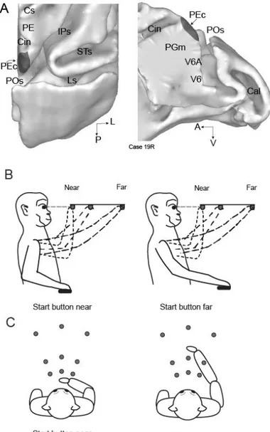
Figure 3-1. Brain location of area PEc and experimental setup. A) dorsal (left) and medial
(right) views of the surface-based reconstruction of the caudal half of the right macaque hemisphere. Dark gray areas show the extent and location of area PEc according to the cytoarchitectural pattern (see text). The location of areas PE, V6A, PGm, and V6 is also shown. Cin, Cingulate sulcus; Cal, calcarine fissure; IPs, intraparietal sulcus; Ls, lunate sulcus; POs, parieto-occipital sulcus; STs, superior temporal sulcus; A, anterior; V, ventral; L, lateral; P, posterior. B-C) Setup for the reaching in depth task (B, lateral view, and C, top view). The animals performed reaching movements towards one of the nine LEDs (grey dots) located at different depths and directions starting the movement either from an initial hand position located next to the body (left panel) or from an initial hand position located far from the body (right panel).
22
3.3 Materials and Methods
Three male macaque monkeys (Macaca fascicularis) weighing 3.9-4.4 kg were involved in the study.The animals were first trained to sit in a primate chair and interact with the experimenters. Then, a head restraint system and a recording chamber were surgically implanted under general anesthesia (sodium thiopenthal, 8 mg/kg*h, i.v.) following the procedures reported by Galletti et al. (1995). A full program of postoperative analgesia (ketorolac tromethamine, 1 mg/kg i.m. immediately after surgery, and 1.6 mg/kg i.m. on the following days) and antibiotic care (Ritardomicina, benzatinic benzylpenicillin + dihydrostreptomycin + streptomycin, 1–1.4 ml/10 kg every 5–6 days) followed surgery. Experiments were performed in accordance with national laws on care and use of laboratory animals and with the European Communities Council Directive of 22 September 2010 (2010/63/EU). All the experimental protocols were approved by the Bioethical Committee of the University of Bologna. During training and recording sessions, particular care was taken to avoid any behavioral and clinical sign of pain or distress.
Extracellular recording techniques and procedures to reconstruct microelectrode penetrations were similar to those described in other papers (Galletti et al., 1996; Breveglieri et al., 2006; Gamberini et al., 2011). Single cell activity was extracellularly recorded from the exposed surface of the posterior part of the superior parietal lobule. We performed multiple electrode penetrations using a 5-channel multi-electrode recording system (Thomas Recording). The electrode signals were amplified (at a gain of 10000) and filtered (bandpass between 0.5 and 5 kHz). Action potentials in each channel were isolated with a waveform discriminator (Multi Spike Detector; Alpha Omega Engineering) and were sampled at 100 kHz.
Histological reconstructions have been performed following the procedures detailed in a recent paper from our laboratory (Gamberini et al., 2011). Electrode tracks and the approximate location of each recording site were reconstructed on histological sections of the brain on the basis of electrolytic lesions and the coordinates of penetrations within recording chamber. The present work include only the neurons assigned to area PEc (Fig. 3-1A) following the cytoarchitectonic criteria according to Luppino et al. (2005) and to Pandya et al. (Pandya and Seltzer, 1982).
23
3.3.1 Behavioral Paradigm: reaching in depth task
Electrophysiological signals were collected while monkeys were performing a reaching task in darkness with the hand contralateral to the recording site. During the task, the monkeys maintained steady fixation of the reaching targets with their head restrained. The task was performed in two blocks that differed for the starting position of the hand: in both cases, the starting position was on the mid-sagittal plane at waist level, but in one block the hand started from a button placed 4 cm in front of monkey’s chest (‘near button’: left panels in Fig. 3-1B and 3-1C), in the other from a button located 14 cm farther from the near one (‘far button’: right panels in Fig. 3-1B and 3-1C). In each block, only one of the two buttons was available to press because the other was covered. For each neuron, the block sequence was random. Fixation and reaching targets were 9 Light Emitting Diodes (LEDs) positioned at eye level, at three different distances and directions (Fig. 3-1B and 3-1C). Three LEDs targets were placed at three isovergence angles: the nearest targets were located at 10 cm from the eyes (17.1°); the LEDs located at intermediate and far positions were at a depth of 15 cm (11.4°) and 25 cm (6.9°), respectively. At each isovergence angle, LEDs were positioned in three directions: one central, along the sagittal midline, and two lateral, at iso-version angles of −15° and +15°. Target positions were chosen in order to be within the peripersonal space.
The time sequence of the task was identical to the one used in a recent report (Hadjidimitrakis et al., 2014a): a trial began when the monkey pressed the button (far or near). After 1000 ms, 1 of the 9 LEDs lit up green and this cue instructed the monkey to fixate it, while maintaining the button pressed. Then, the monkey had to wait 1000– 2000 ms for a change in color of the fixation LED without performing any eye or arm movement. The color change was the go-signal for the animal to release the button and start an arm movement toward the foveated target. Then, the monkey held its hand on the target for 800–1200 ms, keeping the gaze fixed on the same LED. The switching off of the target cued the monkey to release the target and return to the button in order to receive reward. The presentation of stimuli and the animal’s performance were monitored using custom software written in Labview (National Instruments), as described previously (Kutz et al., 2005). Eye position signals were sampled with 2 cameras (1 for each eye) of an infrared oculometer system (ISCAN) at 100 Hz and were controlled by an electronic window (4 × 4°) centered on the fixation target. If the
24
monkey fixated outside this window, the trial was aborted. The task was performed in darkness, in blocks of 90 randomized trials, 10 for each LED target position. The background light was switched on for some minutes between blocks to avoid dark adaptation. At the start of each recording session, monkeys were required to perform a calibration task, following the details reported in Hadjidimitrakis et al. (2014a).
3.3.2 Data Analysis
Data were analyzed with the same approach used in Hadjidimitrakis et al. (2014a) and summarized hereafter. Neural activity was quantified and studied in two epochs: the PLAN epoch that corresponded to the last 500 ms before the go-signal, and the REACH epoch that started 200 ms before the arm movement onset and ended at the pressing of the LED target.
To check the stability of each recorded unit between the 2 blocks, we used the HOLD epoch as a reference. This epoch started with the pressing of LED target and ended with the switching off of the target. The activity in HOLD was assumed to be equal in the 2 blocks because visual, eye position, and arm somatosensory signals were identical. To check for this, we performed a t-test (two sided; Bonferroni’s correction, p =0.01/9= 0.001) for each cell, comparing the nine mean firing rates (1 mean per LED) of the HOLD epoch recorded in one block with the 9 mean firing rates of the HOLD epoch in the other block. Neurons having a significantly different activity in the HOLD epoch between the two blocks were excluded from the analysis. The threshold of statistical difference between the two blocks was in agreement with other criteria of isolation stability (visual inspection of the raster histograms and the distribution of the interspike intervals). A similar procedure has been employed in other studies of reaching activity (Chang et al., 2008; Hadjidimitrakis et al.,2014a). Considering the variability of the neural discharges, only cells tested in at least 7 trials per position and with a mean firing rate higher than 5 spikes/s for at least 1 target position were selected for further analysis (Kutz et al., 2003). Significant modulation of neural activity relative to different positions of the reach targets or to different initial hand positions was studied with a 2-way analysis of variance (ANOVA) performed for PLAN and REACH epochs (factor 1: target position, factor 2: initial hand position). Task-related cells in each epoch were defined as cells where factor 1 and/or factor 2 and/or the interaction factor 1 × 2 were significant (p < 0.05). Only these cells were further analyzed.
25
3.3.3 Population Analysis of Reference Frames
With the task configuration described above, we could study whether spatial target representation in PEc neurons is organized in body-centered or hand-centered coordinates. It is worth specifying that in the two task conditions the targets were located in the same spatial positions. Therefore, being both tasks foveal reaching, the targets remained in constant centered coordinates, so we cannot assess the eye-centered reference frames in the neurons we studied. It should also be considered that, in our experimental condition, the fact that the monkey fixated the target to be reached may lead to a potential confound between target position coding and eye position gain field. Moreover, given that the head of the animal was fixed, our experiment cannot distinguish body from head- or world-centered frames of reference. We will refer to this frame as “body-centered” coordinates, This terminology has been kept consistent with the one used for area V6A (Hadjidimitrakis et al., 2014a).
All the analyses here proposed have been performed following the approaches used for V6A in a recent paper of our lab (Hadjidimitrakis et al., 2014a), so to allow direct comparisons between the two areas. Several analyses have been used so to avoid that observed differences may be attributed to the different methods of analysis employed (Mullette-Gillman et al., 2009; Bremner and Andersen, 2012).
Euclidean Distance Analysis
At single cell level, to compare the similarity of firing rates in body and in hand-centered reference frames, we calculated in each cell the average activity of all the conditions that were equivalent in each frame of reference. Cells could have significantly different firing rates between condition pairs in both reference frames. To find which reference frame accounted more for the neural responses, we quantified the similarity between the mean firing rates in each frame by computing the normalized Euclidean distance (ED) between them (Batista et al., 2007)
26
The mean PLAN/REACH activity for targets n and m that were equivalent in a given reference frame were normalized between 0 and 1 and T corresponds to the targets number. The 95% confidence intervals (CIs) on the distance value were estimated using a bootstrap test. Synthetic response profiles were created by drawing N firing rates (with replacement) from the N repetitions of experimentally determined firing rates. Five-hundred iterations were performed, and CIs were estimated as the range that delimited 95% of the computed distances. These confidence intervals indicate the range within which distance metric would have fallen 95% of the time. Neurons falling outside one of these CI are sensitive to one reference frame, whereas neurons falling inside these 2 CIs are influenced by both reference frames. To compare the RFs of single cells in PLAN and REACH, we used their Euclidean distance values in each frame to calculate a single RF index (Fig. 3-3C). To compute the RF index, individual data points from Figure 3-3A, B were projected on the negative diagonal line. The RF index was equal to the distance of the projection point from the upper end of the negative diagonal line that had Euclidean coordinates 0 and 1. As a result, the RF index ranged from 0 to 1.414. Small index values (<0.5) indicate stronger effect of body-centered coordinates, whereas RF index values equal to 1 or higher indicate a prevalence of hand-centered coding.
Separability Analysis
To examine whether in single neurons target location was separable from starting hand position, we applied the singular value decomposition (SVD) analysis (Peña and Konishi, 2001; Pesaran et al., 2006; Bhattacharyya et al., 2009; Blohm and Crawford, 2009; Blohm, 2012; Hadjidimitrakis et al., 2014a). A 2D matrix M was constructed from the mean activity across target and hand conditions. This matrix was subsequently reconstructed to calculate the diagonal matrix S than contained the singular values. Responses were considered to be separable if the first singular value was significantly larger than the singular values obtained when trial conditions were randomized (randomization test, α = 0.05). More specifically, we randomly rearranged the data in each matrix 1000 times and subjected each “shuffled” matrix to SVD. The first singular values from each shuffled matrix were accumulated into a vector, which was then sorted in ascending order. This sorted vector (n = 1000) formed the reference distribution for determining statistical significance. If the first singular value obtained from the original unshuffled matrix was greater than 95% of the singular values in this distribution, the
27
responses were considered separable. The fractional energy (FE) of the first singular value was computed from the equation below (Mulliken et al., 2008; Hadjidimitrakis et al., 2014a):
Neural responses were classified as separable if the first singular value was significantly larger (p<0.05) compared with the first singular value calculated when conditions were randomized by permuting the rows and the columns of the initial 2D matrix (Randomization test, 1000 permutations) (Mulliken et al., 2008; Bhattacharyya et al., 2009).
Modulation Indexes
To measure the relative strength of neural modulations by target location in body- and hand-centered coordinates, we calculated two indexes in the same way used to quantify modulations in hand or body centered coordinates in area V6A (Hadjidimitrakis et al., 2014a), and to quantify the modulations of reaching activity by disparity and vergence angle in area PRR (Bhattacharyya et al., 2009). Index TB, referring to target in body coordinates, quantified the modulation between pairs of conditions where target position with respect to the body changed while movement vector was constant.
As we tested neurons in three lines of LEDs (see Fig. 3-1C and Fig. 3-3B), our experimental configuration allowed us to have three pairs of equal movement vectors for each neuron. The three indexes TB were subsequently averaged for each neuron to obtain a single index.
Index TH, referring to target in hand-centered coordinates, measured the strength of the gain modulation by hand position while target position remained the same.