A
A
l
l
m
m
a
a
M
M
a
a
t
t
e
e
r
r
S
S
t
t
u
u
d
d
i
i
o
o
r
r
u
u
m
m
–
–
U
U
n
n
i
i
v
v
e
e
r
r
s
s
i
i
t
t
à
à
d
d
i
i
B
B
o
o
l
l
o
o
g
g
n
n
a
a
DOTTORATO DI RICERCA IN
SCIENZE VETERINARIE
Ciclo XXVI
Settore Concorsuale di afferenza: 07/H3
Settore Scientifico disciplinare: VET/06
TITOLO TESI
Toxoplasma gondii in animals and the
environment
Presentata da: Maria Parigi
Coordinatore Dottorato
Relatore
Prof. Carlo Tamanini
Prof. Giovanni Poglayen
Correlatore
Dr. Frank Katzer
CONTENTS
History of the etiological agent
1
Life cycle and routes of infection
3
Characteristic of T. gondii population
12
Toxoplasmosis in animals
15
Toxoplasmosis in humans
36
Laboratory diagnostic
48
Development and application of an in-house indirect ELISA for
anti-T. gondii antibody detection in sheep sera collected in the
Forli-Cesena district
57
PCR detection methods of Toxoplasma gondii in raw and finished
drinking water samples from Scotland
77
1
HISTORY OF THE ETIOLOGICAL AGENT
It’s 1908 when Nicolle and Manceux, two scientists of the laboratory of Charles Nicolle at the Pasteur Institute in Tunis, found a protozoan in spleen, liver and blood of a North-African rodent, the gundi (Ctenodactylus gundi), which was being used for leishmaniasis research. Even if they initially believed the parasite to be a piroplasm and then a Leishmania, soon they realized that they had been discovered a new organism. They named the parasite Toxoplasma gondii based on its morphology and the original host: toxon= bow or arc, plasma= life and gondii may have resulted from a misspelling of the original host, the gundi (Nicolle and Manceaux 2009). In the same year, Splendore discovered the same parasite in a rabbit in Brazil and erroneously identified it as Leishmania (Splendore 2009).
For the next 30 years, T. gondii-like organisms were found in several other hosts, including humans.
In 1937 Sabin and Olitsky showed that Toxoplasma was an obligate intracellular parasite that could be passaged in laboratory animals by intracranial, subcutaneous and intraperitoneal inoculation of brain homogenates (Sabin and Olitsky 1937). In the same years, following a fatal case of T. gondii infection in a three-day old infant, it has been showed that Toxoplasma was a parasite capable of and associated with congenital transmission (Wolf et al. 1941). In the late 1940s and early 1950s Toxoplasma was shown to be involved in inflammatory diseases of the eye (Frenkel and Jacobs 1958).
In 1948 the developing of “The Sabin-Feldman dye test”, the first serological technique to detected specific Toxoplasma antibodies, made it possible to find out that a high proportion of the human and domestic animal population was infected (Sabin and Feldman 1948). Due to this surprising data, Toxoplasma changed from being considered a rare exotic infection into one of more common human parasitic infections, but it was still unknown the mechanism whereby adults were infected.
As the only parasites stages known at that time were tachyzoites and tissue cysts, numerous fruitless studies were performed to demonstrate the relation of Toxoplasma to Leishmania and to show the presence of an insect vector. It’s when Jacobs et al. (1960) proved that that bradyzoites in tissue cysts could survive exposure to acid and trypsin that it was first suggested that carnivorism could be a possible mechanism of infection. The role played by carnivorism was then confirmed in 1965 by Desmonts et al. in an epidemiological study in a tubercolosis hospital in Paris: it was observed that the incidence of T. gondii infection rose from 10% to 50% (and even to 100%) when raw or undercooked meat started to be given to patients for therapeutic purposes. However, the observation that Toxoplasma could be transmitted through carnivorism did not explain the widespread infection in herbivores or strict vegetarians; indeed, a study in Bombay, India, found the prevalence of T. gondii in strict vegetarians to be similar to that in non-vegetarians (Rawal 1959).
In 1965, Hutchinson first linked T. gondii infectivity with cat feaces. He fed tissue cysts to a cat infected with the nematode Toxocara cati, collected and floated faeces in a 33% zinc sulphate solution, stored this sample in tap water for 12 months to embryonate T. cati eggs and then fed these faeces to mice. The great discovery was that “something” in the faeces, that survived
2 for over a year in water, induced toxoplasmosis in mice (Hutchison 1965). As neither the tachyzoites nor bradyzoites could survive this treatment, it was clear that it should be a new form of T. gondii; initially it was proposed that the parasite might be protected within the egg of the cat nematode, T. cati, similar to the transmission of the flagellate Histomonas through Heterakis eggs (Hutchison 1967). The nematode egg theory of transmission was discarded when Toxoplasma infectivity was found in faeces of worm-free cats fed T. gondii (Frenkel et al. 1969; Sheffield and Melton 1970).
In 1970, almost simultaneously, scientific groups in the USA, UK, Germany and Netherlands resulted in the identification of the resistant form of Toxoplasma as a coccidian oocyst with typical coccidian asexual and sexual development occurring in the small intestine of the cat, recognized as the definitive host of the parasite (Hutchison et al. 1969; Dubey et al. 1970a; Hutchison et al. 1970; Overdulve 1970; Sheffield and Melton 1970; Weiland and Kuhn 1970; Witte and Piekarski 1970; Hutchison et al. 1971).
Thus, knowledge on the life cycle of T. gondii was completed more than 60 years after its first description and still remains the only known member of the genus Toxoplasma (Dubey 2008).
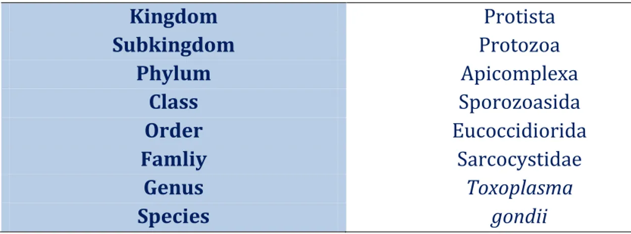
Table 1 Taxonomic classification of T. gondii
Kingdom
Protista
Subkingdom
Protozoa
Phylum
Apicomplexa
Class
Sporozoasida
Order
Eucoccidiorida
Famliy
Sarcocystidae
Genus
Toxoplasma
Species
gondii
3
LIFE CYCLE AND ROUTES OF INFECTION
Toxoplasma gondii is an obligate intracellular parasite that occurs in most areas of the world, capable of infecting virtually all warm-blooded species, including humans.
The life cycle of T. gondii is facultatively heteroxenous (intermediate host is not essential for the completion of the life cycle) and includes both sexual and asexual multiplication; the sexual phase takes place only felids, including the domestic cat, and makes them the definitive hosts of the parasite. A large range of vertebrates, including domestic and wild mammals, birds, and humans, are intermediate hosts.
Like other protozoa, its life cycle requires switching between distinct developmental stages. In the case of T. gondii, the stages are :
the rapidly replicating tachyzoite,
the slower growing bradyzoites within tissue cysts;
the sporozoites within oocysts.
The tachyzoite
The term “tachyzoite” (tachos=speed in Greek), coined by Frenkel, describes the stage of the parasite that rapidly multiply in any cell of the intermediate host and in nonintestinal epithelial cells of the definitive host; it has also been called the proliferative form and the endozoite (Frenkel 1973). It is the stage that Nicolle and Manceaux found in 1909 in the gundi (Dubey 2008). The presence of fast-replicating tachyzoites is an indication of an initial infection and an acute disease.
The tachyzoite is lunate, approximately 2 by 6 µm, with a pointed anterior (conoidal) end and a rounded posterior end. Ultrastructurally, the tachyzoite consists of various organelles, inclusion bodies and an outer covering called pellicle; the nucleus is usually situated toward the central area of the cell and contains clusters of chromatin and a centrally-located nucleolus. Although tachyzoites can move by gliding, flexing, undulating, and rotating, they do not have visible means of locomotion such as cilia, flagella, or pseudopodia.
Both sporozoites or bradyzoites differentiate into tachyzoites once that an intermediate or definitive host ingests a sporulated oocyst or a tissue cyst and this conversion takes place in the small intestinal lamina propria within 18 h post infection (Dubey 1998a).
Tachyzoites enter host cells by actively penetrating through the host cell plasmalemma or by phagocytosis. After entering the host cell, the tachyzoite becomes ovoid and is surrounded by a parasitophorous vacuole (PV), which appears to be derived from both the parasite and the host cell (Silva et al. 1982; Werk 1985; Bonhomme et al. 1992; Morisaki et al. 1995).
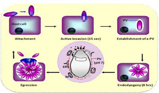
Tachyzoites multiply asexually within the host cell by repeated endodyogeny (Ferguson 2009) (Fig.1); this is a specialized form of reproduction in which the two progenies form within the parent parasite, consuming it (Sheffield and Melton 1968). Tachyzoites continue to
4 divide until the host cell can no longer support their growth and ruptures. In this way, the parasite can disseminate and replicate within the new host: it is reported that T. gondii organisms circulate in the blood only 1 h after infection (Chinchilla et al. 1993).
Fig. 1 T.gondii endodyogeny (http://www.charite.de/sysbio/research/toxoplasma/)
In the blood flow, the majority of parasites exist intracellularly, inside infected leukocytes which contribute to the dissemination of the parasite from the general circulation into the peripheral tissue by a “Trojan horse” mechanism (Unno et al. 2008). This parasitemia in primarly infected pregnant hosts (both animal and humans) may results in the invasion of the placenta and eventually of the foetus, if some tachyzoites cross the placenta (Tenter et al. 2000).
Tachyzoites are rapidly killed outside the host; they are considered sensitive to proteolytic enzymes and they are usually destroyed by gastric digestion (Powell et al., 2001). However, a study showed that tachyzoites may occasionally survive for a short period of time (up to 2 h) in acid pepsin solutions, and that oral application of high doses of tachyzoites may cause an infection in mice and cats (Dubey 1998b). It has also been suggested that tachyzoites may enter the host by penetration of mucosal tissue and thereby gain access to the host's circulation or lymphatic system before reaching the stomach (Riemann et al. 1975; Sacks et al. 1982).
Bradyzoites and tissue cysts
The term “bradyzoite” (brady= slow in Greek) was also coined by Frenkel (1973) to describe the organism multiplying slowly within a tissue cyst in the intermediate host; they are also called cystozoites.
Tachyzoites differentiate into bradyzoites around 10-14 days post infection. It is still not completely clear what is the molecular environment that regulates development from tachyzoites to bradyzoites; it is likely that differentiation into bradyzoites during in vivo infection is multifactorial with contributions from host immunity (Lyons et al. 2002), . Data
5 from in vitro studies suggest that the bradyzoite differentiation program is genetically programmed after the brief period of rapid division as tachyzoites (Skariah et al. 2010). Bradyzoites differ structurally only slightly from tachyzoites: they are more slender, with a nucleus situated toward the posterior end and they contain several amylopectin granules which stain red with PAS (Dubey et al. 1998a). Similar to tachyzoites, bradyzoites are approximately 7 by 1.5 µm in size (Mehlhorn and Frenkel 1980) and divide by endodyogeny (Ferguson and Hutchison 1987).
In the past, the methods used to distinguish tachyzoites from bradyzoites were the resistance of bradyzoites to acid-pepsin digestion and bioassay in cats. Cats fed tachyzoites may become infected and may shed oocysts with a long pre-patent period (>15 days), whereas cats fed bradyzoites shed oocysts with a short (<10 days) pre-patent period. Dubey (199.b) suggests that these cannot be the sole criterion to distinguish between the two stages and that, to accurately determine the cyst-like structures, the identification of molecular markers is required; in fact, stage conversion is associated with the expression of stage-specific surface antigens (Lyons et al. 2002). Several stage-specific surface antigens (SAG) have been identified and many of these fall into two distinct families: SAG1 and SAG2. The SAG1 family includes SAG3, bradyzoitespecific recombinant (BSR) 4 SAG-related sequences (SRS) 1–4 proteins, SAG5, SAG5.1 and SAG5.2. Whereas SAG1 and SRS1–SRS3 are present only on tachyzoites, BSR4 is present only on bradyzoites and SAG3 is present on both stages. This family of proteins is likely to play a role in the attachment process before parasite invasion, as proven for SAG1 and SAG3. The SAG2 family comprises four related proteins designated SAG2A (previously SAG2) and SAG2B–SAG2D. Whereas SAG2A and SAG2B are expressed by tachyzoites exclusively, SAG2C and SAG2D are expressed by bradyzoites exclusively (Lyons et al. 2002).
The conversion to bradyzoites takes place within the parasitophorous vacuole (PV), which converts itself into a cyst. In this way, tissue cysts remain intracellular with an elastic and thin wall (<0.5µm), composed either of host cell and parasite materials (Ferguson and Hutchison 1987). Their size is dependent on cyst age and the type of host cell parasitized; young tissue cysts may be as small as 5 mm in diameter and contain only two bradyzoites, while older ones may contain hundreds of organisms.
Although tissue cysts may develop in virtually any cell type in vitro, in infected animals they are more prevalent in the neural and muscular tissues, including the brain, eyes, and skeletal and cardiac muscles; they can be also detected in visceral organs as lungs, liver, and kidneys (Dubey 1998a). The ones in the brain are often spheroidal and rarely reach a diameter of 70 µm, whereas intramuscular cysts are elongated and may be 100 µm long.
Tissue cysts are the terminal life-cycle stage in the intermediate host and their finding represent the establishment of a chronic infection; the species of intermediate host may also affect the ability of tachyzoites to convert to bradyzoites and to persist as tissue cysts. In some intermediate host species, such as sheep and goats, they may persist for the life of the host. The mechanism of this persistence is unknown. However, many investigators believe that
6 tissue cysts break down periodically, with bradyzoites transforming to tachyzoites that reinvade host cells and again transform to bradyzoites within new tissue cysts. This reactivation can results in clinical symptoms and is a serious and life-threatening condition in immune-compromised individuals (Skariah et al. 2010).
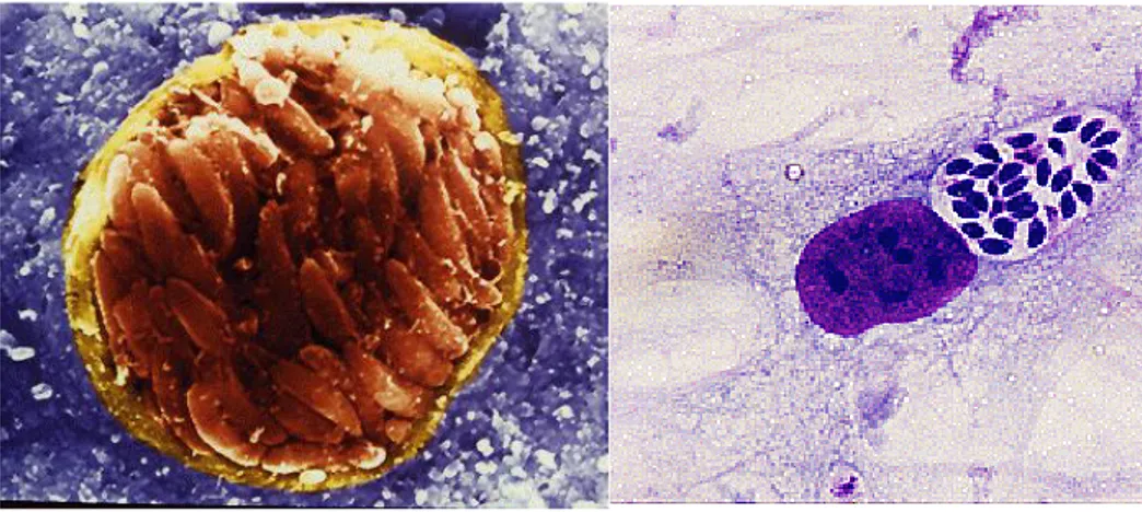
Fig. 2 On the left: tissue cyst with many bradyzoites within the brain of an infected mouse (scanning electron micrograph (http://cmgm.stanford.edu/micro/boothroyd/test.html). On the right: tachyzoites in a human foreskin fibroblast (HFF) grown in culture (Giemsa stain)
(http://cmgm.stanford.edu/micro/boothroyd/test.html).
Enteroepithelial asexual and sexual stages
The sexual development of T. gondii, also called the coccidian stage, takes part only in the gut of the definitive hosts, the felines, when an uninfected cat ingests one of the three infectious stages of the parasite, tachyzoites, bradyzoites or sporozoites. Felidae excrete T. gondii oocysts in faeces 3 to 10 days after ingesting bradyzoites, ≥ 18 days after ingesting of sporulated oocysts and ≥ 13 days after ingesting tachyzoites. Under experimental conditions, it has been demonstrated that the bradyzoite induced cycle in cats is the most efficient since nearly all experimentally infected cats fed tissue cysts shed oocysts, whereas < 30 % of cats fed tachyzoites or oocysts shed oocysts (Dubey 1998a).
Only the bradyzoite induced cycle in cats has been studied in detail (Dubey and Frenkel 1972). After the ingestion of tissue cysts, contained in a prey animal or in meat, the cyst wall is dissolved by gastrical or intestinal proteolytic enzymes and the bradyzoites released in the gut’s lumen; once they penetrate the epithelial cells of the small intestine, they start to replicate asexually by a process called Toxoplasma endopolygeny. This development represents an asexual proliferative phase with repeated nuclear divisions followed by the formation of between 8-20 daughters’ cells within the mother cell (Ferguson 2009). After five asexual stages of this multiplication, the mature merozoites are released, invade new enterocytes and undergo a repeated cycle of asexual development or differentiate into a sexual stage, either macrogametocyte (female gamete) or microgametocyte (male gamete). The factors involved in deciding the fate of a merozoite are unknown. The initial asexual cycles are required to increase parasite density for two reasons: firstly because the male and female gametes have to be able to find each other; secondly, since each macrogamete only produces a single oocyst, there requires being literally millions of merozoites available to
7 develop into macrogametes to produce the large number (millions) of oocysts seen in the faeces (Ferguson 2009).
The sexual development starts 2 days after tissue cysts are ingested by the cat (Dubey et al. 1998a) and gametes are found throughout the small intestine, more commonly in the ileum, above the nucleus of the host epithelial cell, near the tips of the villi. The female gamete or macrogametocyte, is sub spherical and has two very important functions. Firstly, it has to synthesize and store all the nutritional requirements to allow sporulation in the external environment and sustain the viability of the sporozoites over long periods (in excess of 1 year); secondly, it has to synthesize the specific components necessary to form the oocyst wall (Ferguson 2009). Microgametes are elongated, consist mainly of nuclear material and present two long, free flagella projected posteriorly. Microgametes use their flagella to swim, to penetrate and fertilize mature macrogametes in order to form zygotes; after fertilization, an oocyst wall is formed around the parasite.
Finally infected epithelial cells rupture discharging unsporulated oocysts into the intestinal lumen of the cat (Dubey et al. 1998a) which will eventually shed them into the environment or cat litter box.
Sporozoites
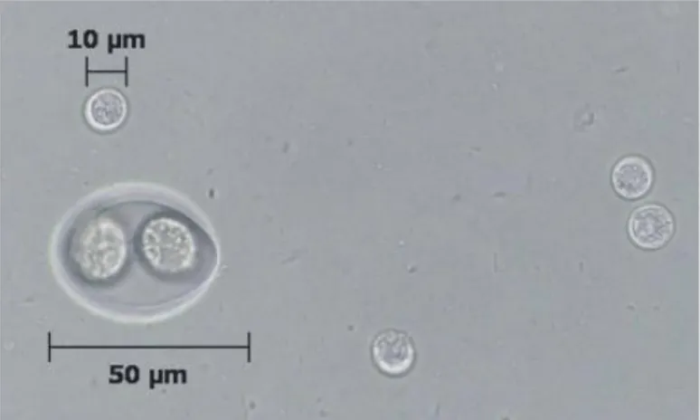
Unsporulated oocysts (zygote) are subspherical to spherical with a diameter of 10 by 12 µm. The cytoplasm of the unsporulated oocyst has a large nucleus with amorphous nucleoplasm and a distinct nucleolus (Fig. 3).
Sporulation occurs outside the cat within 1 to 5 days of excretion depending on oxygenation and temperature. Firstly, the nucleus divides twice and two spherical sporoblasts are formed, each with two nuclei. As the sporulation continues, the sporoblasts elongate and sporocysts are formed, each nucleus divides into two and four sporozoites are formed in each sporocyst; a prominent residual body is left after sporozoites are formed and it is enclosed in a single-unit membrane (Ferguson et al. 1979). Sporulated oocysts are subspherical to ellipsoidal and are 11 by 13 µm in diameter. Each oocyst contains two ellipsoidal sporocysts that measure 6 by 8 µm; sporozoites are 2 by 6 to 8 µm in size with a subterminal nucleus and, ultrastructurally, they are similar to tachyzoites.
Cats can shed upward of 360 million oocysts in their feces in a single day and the shedding period lasts for a median of 8 days, although it may be as long as 3 weeks (Dabritz and Conrad 2010). Sporulated oocysts can survive for long periods in moist soil, under most ordinary environmental conditions, and in water (Dubey and Beattie 1988).
8
Fig. 3 Multiple oocysts and a single Isospora oocyst (arrow): comparison of the relative size of Isospora
felis to the Toxoplasma oocysts
9
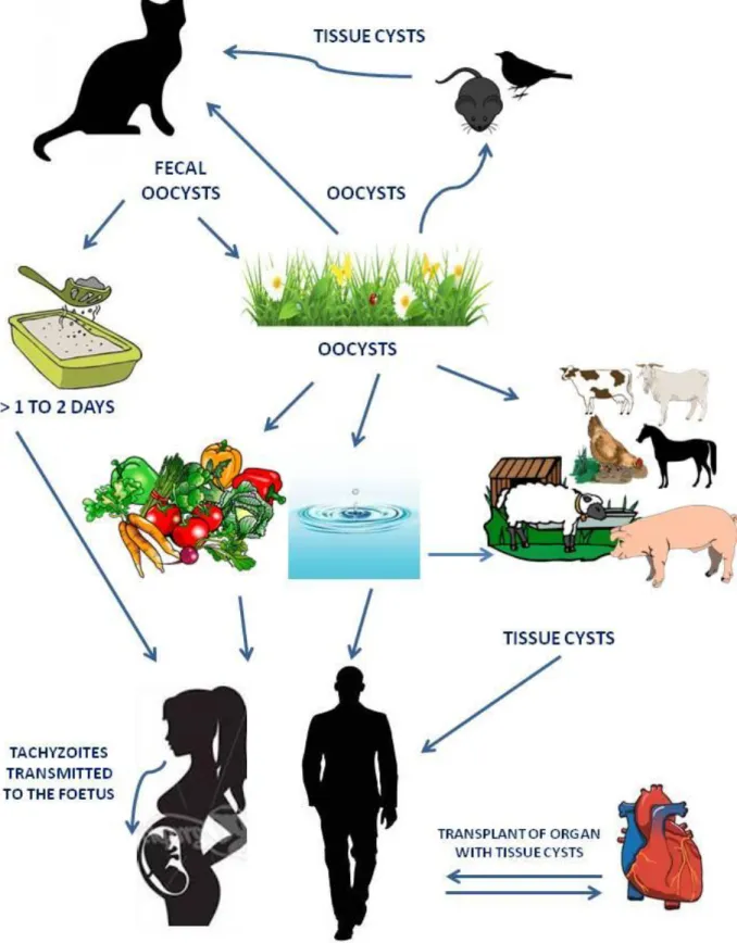
ROUTES OF TRANSMISSION OF T. gondii
Tachyzoites, bradyzoites contained in tissue cysts, and sporozoites inside of sporulated oocysts are the three infectious stages in the life cycle of T. gondii. All the stages are infectious for both intermediate and definitive hosts which may acquire T. gondii infection mainly via one of the following routes:
1. horizontally by oral ingestion of infectious oocysts from the environment (soil or water), i.e. faecal-oral route;
2. horizontally via carnivorism by oral ingestion of tissue cysts contained in raw or undercooked meat or primary offal (viscera) of intermediate hosts, i.e. carnivorism; 3. vertically by transplacental transmission of tachyzoites to the foetus, i.e. congenital.
Faecal-oral route. As reported above, primary infected cats shed into their litter box or in the
environment (soil, water) millions of unsporulated oocysts which become infected within 1 to 5 days after excretion. Experimental studies demonstrated that in soil, sporulated oocysts remain infectious over a period of 18 months depending on humidity, temperature and exposure to direct sunlight (Yilmaz and Hopkins 1972; Frenkel et al. 1975). In water (Dubey 1998c) and seawater (Lindsay and Dubey 2009) sporulated oocysts remain infective for 6 up to 54 months at temperatures between 4 and 25 °C; oocysts resulted also resistant to experimental freezing, surviving up to 28 days at -21°C (Frenkel and Dubey 1973). As they are highly impermeable, they are very resistant to disinfectants (Kuticic and Wikerhauser 1996; Jones and Dubey 2010). Oocysts are distributed in the environment through wind, rain and surface water, or harvested feeds and they can be distributed mechanically by vectors and transported to water by runoff. Intermediate hosts, especially humans and herbivores, as well as definitive host (Dubey 1998a), can be infected by the ingestion of food or water contaminated with sporulated oocysts.
Carnivorism. Tissue cysts are contained in tissues, especially muscles and nervous system, of
chronically infected intermediate hosts. Studies have indicated that T. gondii tissue cysts in meat are susceptible to various physical procedures such as heat treatment, freezing and salting (depending on the salt concentration and duration (Kijlstra and Jongert 2008)): e.g. freezing to -12 degrees °C for at least 2 days and cooking to an internal temperature of 67 degrees kill tissue cysts in meat (Dubey 1996a). Ingestion of tissue cysts is the main mechanism involved in transmission of the infection in carnivores, including humans. It is clear that in humans, as omnivores, both mechanisms described above play a role in T. gondii transmission and it is likely that the lifestyle of the individual plays the most important role in how the parasite is transmitted; the possible sources of acquired infections in humans will be discussed ahead.
Congenital. Transmission of T. gondii via tachyzoites can occur in hosts which contract
primary T. gondii infection during pregnancy: in this case tachyzoites may be transmitted transplacentally to the foetus. Congenital transmission has been documented in various intermediate hosts, as mice, sheep, goats and, most important, in humans.
10 In addition, tachyzoites have been found in the milk of several hosts, including cats, sheep, goats, cows, camels and mice (Powell et al. 2001; Dehkordi et al. 2013). Thus now, the report of acute toxoplamosis following the consumption of raw milk has been documented only for caprine milk. In addition to blood and milk, tachyzoites have been detected in other body fluids, including saliva, sputum, urine, tears, and semen but there is currently no evidence of horizontal transmission of T. gondii to humans via any of these routes.
In conclusion, T. gondii may be transmitted from definitive to intermediate hosts, from intermediate to definitive hosts, as well as between definitive and between intermediate hosts. It is currently not known which of the various routes of transmission is more important from an epidemiological point of view. T. gondii life cycle may continue indefinitely by transmission of tissue cysts between intermediate hosts (even in the absence of definitive hosts) and also by transmission of oocysts between definitive hosts (even in the absence of intermediate hosts) (Tenter et al. 2000) (Fig. 5).
11
12
CHARACTERISTICS OF T. gondii
POPULATION
It has been long known that different strains of T. gondii vary in virulence for mice. With the development of the molecular typing techniques, it has been possible to associate differences in virulence to genetic diversity and to investigate the correlation between T. gondii genotype and disease manifestation in animals and also in humans (Saeij et al. 2005). Several studies have been conducted by multilocus enzyme electrophoresis (MLEE) and restriction fragment length polymorphism (RFLP) on a large set of isolates collected from animals and humans from North America and Europe. Such studies revealed that T. gondii population is an unusual striking clonal population, highly structured and dominated by three clonal lineages, type I, II and III. These lineages differ genetically by 1% or less and have been derived from few genetic crosses among closely related parents (Howe and Sibley 1995). Highly similar or identical genotypes were sampled from different hosts in different geographic regions of North America and Europe revealing that there are no strong geographic differences between the two continents' isolated and that similar genotypes can be found in both animals and human infections (Sibley and Ajioka 2008).
Clonality is usually described in protozoan parasites which lack a defined sexual phases in their life cycles, precluding the genetic exchange by meiosis. Although T. gondii is one of the few parasitic protozoa with a well-defined sexual cycle, this mode of propagation evidently occurs only rarely. There are few possible explanations for this striking pattern:
1. it is known that intermediate hosts develop strong immunity to primary infection, limiting the chances for simultaneous infection and developing tissue cysts related to one specific strain (Reikvam and Lorentzen-Styr 1976);
2. cats are rarely simultaneously infected with multiple strains, limiting the chances of genetic exchange;
3. the parasite can be transmitted by direct oral infection between intermediate hosts without the necessity of the sexual phase in cats (Sibley and Ajioka 2008).
A proper definition of T. gondii virulence is still incomplete because the susceptibility to the infection and to acute disease in different hosts is extremely variable: e.g. mice can die in one day and rats may be full refractory to the infection. As rodents are natural hosts for T. gondii, laboratory mice have provided a reasonable model to study the virulence factors associated with different T. gondii genotypes. The three lineages, i.e. type I, II and III, differ markedly in virulence in the mouse model; however, little is known about the correlation between T. gondii virulence in mice compared with other species.
Type I strains are reported to be uniformly lethal with an infectious dose of a single viable organism in mice; the first human strain of T .gondii, called RH strain, was classified into lineage I. Type I virulent strains are found rarely in animals and in humans; in literature they have been associated with acquired ocular toxoplasmosis, congenital infection and cerebral toxoplasmosis in immunocompromised patients (Khan et al. 2005; Hunter and Sibley 2012).
13 Due to their rare isolation, scientists recently called the existence of type I strains into question. Some research groups reported that during genotyping analysis, the use of additional markers transformed a “type I strain” into a non-type I or atypical one. This suggests that the identification of type I strains by monolocus typing, as it was done in the past, needs to be confirmed by additional multilocus typing (Ajzenberg 2010).
Type II strains are commonly isolated in both animal and human cases in North America and Europe (Darde et al. 1992; Howe et al. 1997) and have an intermediate virulence that varies with mouse strain. As the genotype of Toxoplasma strains is strongly linked to the geographical origin of infection, the French Toxoplasma Biological Resource Center (Toxo BRC, France) reported that in France type II strains are predominant in cases of congenital toxoplasmosis and in immunocompromised persons, whatever the clinical presentation (Ajzenberg 2010).
Type III are considered completely avirulent in mice and are characterized by low tissue cysts burdens and limited ability to cause infection and human disease (Sibley and Ajioka 2008; Hunter and Sibley 2012).
The analysis of acute virulence of type I strain, performed by classical genetics and by genetic crosses between virulent type I and avirulent type III, revealed that the high virulence of type I is largely due (90%) to a single allele at the ROP18 locus, a gene encoding a rhoptry protein (ROP). Rhoptries proteins (ROP) are discharged by rhoptries (secretory organelles involved in the formation of the parasite-containing vacuole) during the parasite cell invasion. ROP have different destination in the host cell, e.g trafficking to the host cell nucleus, but their specific roles in the parasite biology have not been exactly defined (Sibley and Ajioka 2008). The analysis of features of ROP18 in different T. gondii types evidenced that the ROP18allele is remarkably polymorphic in type I strain whereas its expression level is reduced by more than 100 fold in the type III lineages. Transfection of ROP18 typeI allele in type III strain resulted in a 4-fold increase in lethality in mice (Saeij et al. 2005; Sibley and Ajioka 2008). This findings provides evidence that virulence of the type I strains is strongly related to this specific allele unique to this lineage.
A successful T.gondii infection depends on the ability of the parasite to cross biological barriers, such as gut epithelia, blood-brain barrier and placenta and access to immunoprivileged sites as the nervous system. Different migration capacities are related to strain-specific differences in parasite dissemination and parasite motility. In vitro studies evidenced that subpopulations of type I strain parasites display a higher migration capacity both in long-distance and across the extracellular matrix than type II and III strains (Saeji et al. 2005). Furthermore, in vivo type I parasites migrate more effectively to spleen than the other strains. Genetic analysis supports that the association of migratory capacity and virulence is linked to the same chromosome region related to acute virulence (Barragan and Sibley 2002; Saeij et al. 2005).
Growth rate is a common virulence feature in protozoan parasites. T. gondii burden is the major contributor to Toxoplasma pathogenesis in mice and it is related to an overstimulation of the immune system leading to high levels of T helper cell type 1 (Th1) cytokines, increased apoptosis and organ damage. Several studies have noted that type I strains grow faster than the other strains and that one tachyzoite of type I strain is sufficient to generate high parasite
14 loads and high levels of Th1 cytokines. This high growth rate might be linked to a higher reinvasion rate of type I parasites compared with the type II or III strains; in fact, type I parasites remain infectious for a longer time in extracellular matrix so are able to disseminate more efficiently than type II and III (Saeij et al. 2005). Furthermore, the doubling time (Td) of a strain seems to be related to the appearance of bradyzoites antigen (BAG1) and to the following conversion to the slower growing bradyzoite form. It has been seen that tachyzoites emerged from cells experimentally infected with virulent strains remained firmly on the tachyzoite phase not expressing BAG1 antigen at any time (Jerome et al. 1998; Saeij et al. 2005). Thus, there seems to be an inverse correlation between the virulence of Toxoplasma strains and the expression of bradyzoite genes (‘bradyzoiteness’) under normal culture conditions.
As reported above, the major parts of the typing investigations were and are performed on strains isolated from North America and Europe, resulting in an overrepresentation by strains isolated in these areas. Recent typing studies are focusing on isolates from animals and humans collected from more remote areas of the world with the purpose to extend the knowledge on T. gondii population structure. Surprisingly, strains from South America have been identified as natural recombinant, genetically different from those in North America and Europe; furthermore, in Brazil, T. gondii populations resulted to be distinct among different locations and some atypical genotypes have been shown to be highly pathogenic to humans, causing severe cases of ocular toxoplasmosis (Ferreira Ade et al. 2006; Khan et al. 2006; Dubey et al. 2007; Pena et al. 2008). More worringly, severe and fatal cases of toxoplasmosis in immunocompetent individuals were linked to one atypical strain in French Guiana (Carme et al. 2002; Demar et al. 2007). Genetic data on T. gondii isolates from Africa are scarce and not sufficient to elucidate the population structure in this continent. To date, studies of isolates collected in several African countries showed a predominance of classical lineages, type I and II in Uganda and predominantly type III in samples from Western and Middle Africa (Lindstrom et al. 2008; Velmurugan et al. 2008). However, Mercier and colleagues (Mercier et al. 2010), identified, new haplogroups, called Africa 1 and 3, from isolates of Gabon area and proposed them as being major lineages in the continent, encouraging further studies with genotype analysis.
15
TOXOPLASMOSIS IN ANIMALS
The capability of T. gondii to infect all warm blooded animals makes it the most successful parasite worldwide. Among the different animal species, there is a wide variety of clinical responses, ranging from unapparent infection to fatal disease with many intermediate situations, such as behavioural changes and congenital diseases. Factors that may influence the outcome of T. gondii infection in animals include:
the parasite strain,
if the infected species has evolved alongside the cat and
how the immune system of the host responds to the infection (Innes 1997).
Table 2 Severity of reaction to Toxoplasma gondii in intermediate hosts (Innes et al. 1997).
Very Severe Intermediate Resistant
Marsupials Human Cattle
New World Monkeys Sheep Horses
Old World Monkeys Goats Pigs
Toxoplasma infection in felids
Felids are the only known definitive hosts for T. gondii, with domestic cats and a large range of wild felids shown to be capable of shedding oocysts (Dubey 2008).
Cats shed between 3 and 810 million of oocysts per infection in a period of 8 days on average, although this can be extended up to three weeks (Dabritz and Conrad 2010). After the first infection, cats are generally protected against re-shedding, although re-shedding has been observed experimentally upon challenge infection six years after the primary infection, upon administration of high doses of corticosteroids, and after superinfection with the related parasite Isospora felis (Dubey 1995). Re-shedding was not observed in Feline Immunodeficiency Virus infected cats challenged with T. gondii 66 weeks post primary infection (Lappin et al. 1996), therefore it is unclear how the parasite may persist in cats following primary infection.
As definitive hosts, cats are essential in the epidemiology of T. gondii, which is demonstrated by the low prevalence of infection in other animal species on cat free islands (Wallace et al. 1972; Dubey et al 1997). Cats mainly become infected by ingestion of tissue cysts. Kittens can be infected by transplacental passage of the parasite and can possibly also be infected by their mother’s milk (Powell et al. 2001). Infection through ingestion of oocysts is another possibility, but cats are not very susceptible to oocysts compared to intermediate hosts (Dubey 1996b). Prevalence of infection in cats varies with hunting behavior or other factors such as outdoor access and feeding on raw meat, this latter factor being associated with hunting behavior. In addition, the prevalence of infection increases with age, as T. gondii infection persists in the form of tissue cysts. Seroprevalences of up to 74% have been reported. These are generally higher in stray and feral cats compared to pet cats (Tenter et al.
16 2000). Importantly, it has been recently shown that owning a pet cat is not a strong risk factor for human infection. In a review of 14 case-control studies for toxoplasmosis, only one indeed identified a slightly increased risk of infection in cat-owners (Petersen et al. 2010). Petting cats is probably not a risk, as no infectious oocysts could be detected on fur clippings collected 7 days after cats had shed millions of oocysts upon experimental infection (Dubey 1995). The risk of accidental ingestion of oocysts while cleaning the cat litter box can be reduced by proper hand hygiene and by eliminating cat feces daily, before oocysts have had the opportunity to sporulate. In addition, cat litter has been shown to reduce oocyst viability (Dubey et al. 2011). Accidental ingestion of oocysts is more probable during gardening, playing in a contaminated sandbox, or when eating raw and unwashed vegetables or fruits (Petersen et al. 2010).
Felids are thought not to be severely affected by the T. gondii infection. About 10 to 20% of cats develop self-limiting, small-bowel diarrhea in the first 2 weeks after experimental inoculate (Lappin 2010). Disseminated toxoplasmosis with uncontrolled replication of tachyzoites in hepatic, pulmonary, CNS, and pancreatic tissues can develop after acute infection or in cats concurrently infected with feline leukemia, feline immunodeficiency, or feline infectious peritonitis viruses, as well as after cyclosporine administration or after renal transplantation (Lappin 2010). In a recent study from Finland, 6 out of 193 cats submitted to post-mortem examination were diagnosed with general toxoplasmosis, demonstrating that T. gondii may be more of a health issue for cats than previously thought (Jokelainen et al. 2012). There are other feline species that are more susceptible than domestic cats: female Pallas cats can transmit T. gondii to their offspring when persistently infected, which often leads to fatal toxoplasmosis in kittens, and is a common cause for failure of captive breeding programs (Kenny et al. 2002).
As cats are generally not affected by the clinical disease, prevention should focus on cats as a source of environmental contamination with T. gondii. Although cats are not necessarily a direct risk for human infection, all human infections can be traced back to cat-shed oocysts, either as a direct source of human infection or via infection of food animals causing meat-borne infection in humans. Therefore, reducing the number of oocysts shed in the environment could significantly reduce T. gondii infections in both food animals and humans. At the moment only a limited set of control options are available. The first option is to reduce the cat population. This includes timely neutering of owned cats, and a control strategy for stray cats. The second option is to prevent oocysts from ending up in the environment. Cat owners should thus encourage their cats to use the litter box, and dispose of cat litter with household wastes rather than, for example, flushing it down the toilet. The third option is to prevent cats from becoming infected by limiting their hunting behavior, e.g. by keeping them indoors (Woods et al. 2003). In addition, cats should not be fed with raw meat. All these options involve changes in the behavior of cat owners or of their cats, which implies that it may not be feasible to achieve a large reduction of the number of oocysts shed into the environment using these methods. Focusing only on high risk locations, by keeping cats away from food animals, vegetable gardens and sandpits may be more feasible.
17 possibilities of T. gondii infection control, and should be considered a research priority. An experimental cat live vaccine using oral administration of the T-263 strain, derived from the RH strain, prevented oocyst shedding in 84% of kittens (Frenkel et al. 1991) and has been shown to decrease the exposure of pigs to T. gondii in a farm-based experiment (Mateus-Pinilla et al. 1999). However, this vaccine strain needs to be maintained in mice, limiting the scale of production. Moreover, the vaccine requires frozen storage, and importantly, as a live vaccine, it may be hazardous to the people administering it (Innes et al. 2009). A different type of vaccine should thus be favored but, so far, studies are limited. The use of recombinant feline herpesvirus-1 expressing the T. gondii rhoptry protein ROP2 did not reduce oocysts shedding (Mishima et al. 2002). Intranasal administration of crude native rhoptry proteins reduced oocysts shedding by 87.4% compared to an adjuvant-control group (Zulpo et al. 2012), and prevented 2 out of 3 vaccinated cats from shedding oocysts (Garcia et al. 2007).
Toxoplasma infection in livestock
In most countries, epidemiological data on T. gondii infection in livestock are not regularly monitored and information on its diffusion is mainly obtained from local surveys.
From the review of the seroepidemiological studies published in literature until 2000 (Tenter et al. 2000), it has been possible to note that prevalence of antibodies against T. gondii vary widely among different animal species with the highest levels of seropositivity reported in sheep and goats and the lowest values in horses and poultry.
Among livestock, sheep and goats are susceptible to congenital toxoplasmosis, which represents one of the main causes of abortion and stillbirth; in other species (pigs, cattle, horses, chickens), the infection usually remains asymptomatic (Dubey and Jones 2008).
Despite the clinical manifestation, the importance of T. gondii primary infection in livestock is related to the ability of the parasite to produce tissue cysts in infected animals, which may represent important sources of infection for humans. The organotropism of Toxoplasma and the number of tissue cysts produced in a certain organ vary with the intermediate host species: tissue cysts are most frequently observed in various tissues of infected pigs, sheep and goats, less frequently in poultry and horses and only rarely in cattle (Tenter et al. 2000; EFSA 2007). Even if the seropositivity of meat-producing animals does not necessarily reflect the risk that those animals pose for their consumers (Tenter 2009), it has been recently demonstrated that in sheep, the antibody concentration was strongly related to the probability of the presence of tissue cysts in meat samples (Opsteegh et al. 2010a).
Using intensive farm management together with adequate measures of hygiene, confinement, and prevention, it has been economically possible to produce pigs and poultry free of T. gondii infection, although this has been achieved in only a few countries, i.e. in the Netherlands and Denmark (van Knapen et al. 1995; Shirley 1997).
These measures include:
a) to keep meat-producing animals indoors throughout their life-time; b) to keep the sheds free of rodents, birds, and insects;
18 d) to control access to sheds and feed stores, i.e. no pet animals should be allowed inside
them (Tenter et al. 2000)
By contrast, production of free-ranging livestock will inevitably be associated with T. gondii infection. Animals kept on pastures with an increased pressure of infection due to contamination of the environment with oocysts, such as sheep and goats, show high seroprevalences in many areas of the world, i.e. up to 92 and 75%, respectively (Tenter et al. 2000).
Toxoplasma infection in sheep
Toxoplasma gondii antibodies have been found in sheep worldwide; global seroprevalence values have been recently reviewed by Dubey (2009b) revealing that values vary widely among different locations, from 4% to 92%. Since then, seroprevalences of 44.1% (90/204) have been reported in sheep from Grenada and Carriacou, West-Indies (Chikweto et al. 2011), 18.6% (71/382) in Sao Paulo, Brazil (Langoni et al. 2011), using MAT and 49.3% (248/503) in Spain using a commercial ELISA (Garcia-Bocanegra et al. 2013). In a serological survey of T. gondii infection in adult breeding sheep in Great Britain, of the 3539 sera collected from 227 flocks, 74% were found to be positive for T. gondii specific antibody using latex agglutination (Hutchinson et al. 2011).
In relation to different serological tests used, different cut-off and different animal investigated, their comparison should be done with extreme care (Dubey 2009b). However, it is clear that the infection is widely diffused among the global sheep population. Seroprevalence has been shown to increase with the age of the animals, indicating that the major part of sheep acquire infection post-natally; differences in seroprevalence between young and adult sheep are probably related to the oral route of transmission, with the older animals being exposed for longer time (Dubey and Kirkbride 1989; Lunden et al. 1994; Dubey 2009b; Katzer et al. 2011). Antibody levels are expected to stay elevated for the lifetime of the animal, because the immune response of infected animals does not completely clear the parasite and animals stay persistently infected (Katzer et al. 2011).
As sheep are herbivores, the most likely source of infection is represented by oocysts shed by infected cat and contaminating the environment, i.e. pastures, feed stock or water. The importance of ingestion of sporulated oocysts has been supported by risk assessment studies showing a statistically significant association between infection and:
the presence of cats in the farms (Vesco et al. 2007; Abu-Dalbouh et al. 2012; Cenci-Goga et al. 2013; Guimaraes et al. 2013; Mendonca et al. 2013).;
the management (Dubey 2009b; Gebremedhin et al. 2013);
the use of superficial or stagnant water for drinking animals (Vesco et al. 2007; Cenci-Goga et al. 2013; Gebremedhin et al. 2013);
the age of investigated animals (Vesco et al. 2007; Katzer et al 2011; Hutchinson et al. 2011;)
19 With regard to sheep farming, animals kept on pastures and raised under estensive or semi-estensive conditions are generally believed to be more likely exposed to Toxoplasma oocysts (Tenter et al. 2000); however some authors found an increased risk of infection for sheep kept under intensive conditions as farm facilities may provide shelter to various hosts of T.gondii (such as cats and rodents) which might be involved in the spread of the infection (Tzanidakis et al. 2012).
Also flock size was found to be a significant risk factor for the infection but in relation to the farming conditions under which animals were kept (Cenci-Goga et al. 2013).
Once ingested, oocysts excyst in the small intestine, releasing sporozoites which quickly invade and multiply within the cells of the gut differentiating into tachyzoites (Innes et al. 2009). A common clinical sign of toxoplasmosis during the early stages of infection is an elevated temperature which may last for a further week, during which time tachyzoites may be detected in the circulation and find their way to the placenta (Dubey and Sharma 1980; Wastling et al. 1993). Important observations from several studies have shown that ewes infected during one pregnancy are unlikely to suffer from a recrudescence of the infection in a subsequent pregnancy, suggesting that they are able to develop effective protective immunity against the disease (Buxton et al. 2006; Buxton et al. 2007; Innes et al. 2009). However, the protection is not to consider absolute (Innes et al. 2009; Edwards and Dubey 2013): recent studies reported the finding of T. gondii DNA in fetuses from persistent infected ewes suggesting a recrudescence of congenital transmission of T. gondii but this data require confirmation by other techniques, i.e. bioassay in mice for determining the viability of the parasite (Morley et al. 2005; Morley et al. 2008).
Congenital transmission from ewes primarily infected during pregnancy to the foetus represents an important cause of abortion and economic losses to sheep industry, reported in New Zeland, Australia, UK and Europe (Dubey and Beattie 1988). The stage of pregnancy when transplacental transmission of T. gondii takes place is important in determining the clinical outcome. If the infection occurs early in gestation, when the foetal immune system is relatively immature, foetal death is likely to occur. Infection at mid-gestation can result in birth of a stillborn or weak lamb which may have an accompanying small mummified foetus, whereas infection in later gestation may result in birth of a live, clinically normal, but infected lamb. Characteristic white spot lesions may be observed on placentas from sheep that have aborted their lambs and these areas of necrosis may compromise the function of the placenta to support the fetus during gestation (Buxton 1990). The average annual incidence rates of clinical toxoplasmosis was estimated to range between 1% and 2% (Blewett and Trees 1987) but, according to Dubey (2009b), “actual losses in lambs due to toxoplasmosis are difficult to estimate because the disease is sporadic, only a small number of aborted lambs are submitted for diagnosis and, most importantly, toxoplasmosis does not produce clinical disease in the ewe, not alarming the farmer”. In the UK the estimate of the annual cost of toxoplasmosis to the sheep industry has been calculated to be £12.4 million (Gutierrez et al. 2012).
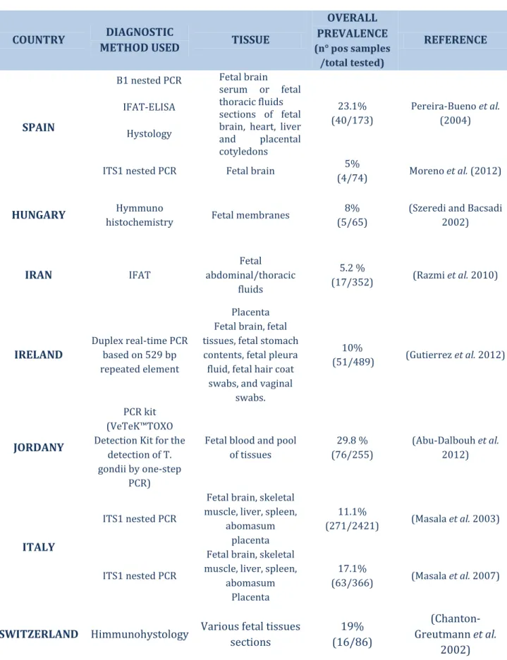
In the last decade the scientific community shown increased interest in determining the role played by T. gondii in ovine abortion and numerous studies have been conducted worldwide with the aim to investigate the presence of the parasite in placentae or aborted foetuses. The results of these studies are summarized in Table 3.
20
Table 3 Summary of the T. gondii-abortion cases reported in literature.
COUNTRY DIAGNOSTIC
METHOD USED TISSUE
OVERALL PREVALENCE (n° pos samples /total tested) REFERENCE SPAIN B1 nested PCR IFAT-ELISA Hystology Fetal brain serum or fetal thoracic fluids sections of fetal brain, heart, liver and placental cotyledons 23.1% (40/173) Pereira-Bueno et al. (2004)
ITS1 nested PCR Fetal brain 5%
(4/74) Moreno et al. (2012)
HUNGARY Hymmuno
histochemistry Fetal membranes
8% (5/65)
(Szeredi and Bacsadi 2002) IRAN IFAT Fetal abdominal/thoracic fluids 5.2 % (17/352) (Razmi et al. 2010) IRELAND Duplex real-time PCR based on 529 bp repeated element Placenta Fetal brain, fetal tissues, fetal stomach contents, fetal pleura fluid, fetal hair coat
swabs, and vaginal swabs. 10% (51/489) (Gutierrez et al. 2012) JORDANY PCR kit (VeTeK™TOXO Detection Kit for the
detection of T. gondii by one-step
PCR)
Fetal blood and pool of tissues 29.8 % (76/255) (Abu-Dalbouh et al. 2012) ITALY ITS1 nested PCR
Fetal brain, skeletal muscle, liver, spleen,
abomasum placenta
11.1%
(271/2421) (Masala et al. 2003)
ITS1 nested PCR
Fetal brain, skeletal muscle, liver, spleen,
abomasum Placenta
17.1%
(63/366) (Masala et al. 2007)
SWITZERLAND Himmunohystology Various fetal tissues
sections 19% (16/86) (Chanton-Greutmann et al. 2002)
21 These results confirm once more how T. gondi represents a common cause of abortion in sheep. In global sheep farming a more strictly application of proper prophylactic measures is urgently needed for the prevention of the infection and the economic losses due to missed lactation and lambs.
For reducing losses to the sheep industry, a life vaccine (Toxovax ™) is available in New Zealand and Europe to help prevent ovine congenital toxoplasmosis; this vaccine, initially developed in New Zealand and further studied in Scotland, consists of a modified S48 strain of T. gondii that has lost the ability to differentiate into bradyzoites (O'Connell et al. 1988) but that can induce protective cell-mediated immune responses in sheep (Buxton and Innes 1995). Toxovax™ is advised to be administered at least 3 weeks prior to mating by subcutaneous inoculation and it induces a protective immunity for at least 18 months with a meat and milk withdrawal period of six weeks following vaccination (Buxton and Innes 1995); care should be taken by those administering it as it is a zoonotic pathogen (Innes and Vermeulen 2006).
Together with the vaccination, other prophylactic measures are fundamental for reducing the environmental contamination and the infection rate of the animals, i.e. the control of the cat population (both stray and pets) in the farm, especially for the 2 months before the lamb season (Dubey et al. 1990b), the cover of the feed stock at all times to prevent contamination (Buxton 1990) and, when possible, prevent the access of cats to water sources used for drinking animals. Moreover, strict sanitation measures are necessary during an abortion outbreak, e.g. the separation of aborting ewes or the ones with vaginal discharges and the dispose of aborted fetuses and placentae (Masala et al. 2007).
The control of T. gondii infection in sheep is also important from a public health point of view since tissue cysts may persist in organs of infected sheep for several months and perhaps for life, representing a source of infection for humans (Uggla and Buxton 1990; EFSA 2013). It has been demonstrated, indeed, that T. gondii was still detectable after more than five months p.i in tissues of asymptomatic sheep experimentally infected (Dubey 1984). Regarding the distribution of the cysts, two experimental studies, which examined by PCR and histological techniques the distribution of T. gondii in various tissues of experimentally infected sheep, found that the parasite was more frequently detected in the brain, in the heart and in muscle samples (Esteban-Redondo and Innes 1998; Esteban-Redondo et al. 1999). According to European Food Safety Authority, the consumption of raw or undercooked mutton represent the main source of infected meat in Southern European countries (EFSA 2007; Kijlstra and Jongert 2008).
Little genetic typing has been performed on T. gondii isolates from sheep and published data indicated that Type II is the predominant lineage of the strains isolated. Interestingly, no Type I isolate of T. gondii has been found in sheep to date (Dubey 2009b).
Several studies have shown the presence of T. gondii DNA in ovine milk samples. Fusco et al. (2007) detected T. gondii DNA in 3.4% (4/117) of the milk samples (10 samples pooled from
22 each farm) collected from 117 farms of the Campania region in Italy; additionally, they tested milk samples for anti-Toxoplasma antibodies founding 91 positive out of the 117 samples tested. Similarly, in Brazil, T. gondii DNA was detected in the milk of 5 (3%) naturally infected sheep, out of the 70 seropositive tested animals (Camossi et al. 2011). In a recent study performed in Iran, milk samples from different animal species, including sheep, have been tested for T. gondii by cell line culture, ELISA and PCR; using the culture method, the 7.02% (13/315) of ovine milk samples were found to be contaminated, with the positivity confirmed by bioassay in cats too (Dehkordi et al. 2013). Thus far, acute toxoplasmosis has not been associated with the consumption of unpasteurized ovine milk; however these results, in particular the positivity in bioassay, suggest that the consumption of raw ovine milk, or dairy products made with raw milk, might be considered a source of infection for humans (Tenter et al. 2000).
Toxoplasma infection in goats
Similarly to sheep, T. gondii infection in goats has been reported worlwide with seropositivity values varying widely among different countries (from 4% to 77%) depending on the serological techniques used, the cut off values, the animals investigated and management conditions (Tenter et al. 2000). Though these differences, it is clear that the infection is widespread among global goats population.
Similarly to sheep, goats are supposed to acquire the infection mainly by the ingestion of oocysts from contaminated pastures or water (Mancianti et al. 2013); various studies demonstrated that risk factors associated with T. gondii infection in goats are similar to the ones described above for sheep (Tzanidakis et al. 2012; Garcia-Bocanegra et al. 2013; Kantzoura et al. 2013; Lopes et al. 2013). Furthermore, goats are thought to more susceptible to toxoplasmosis than sheep as a result of their higher activity and movement compared with sheep, which increases the probability of contact with contaminated sources (Abu-Dalbouh et al. 2012).
Congenital transmission from goats primarily infected during pregnancy to their foetuses is similar to that seen in sheep representing a likewise important cause of abortion and neonatal mortality. The stage of pregnancy when transplacental transmission of T. gondii takes place is important in determining the clinical outcome. Infection early in gestation can result in foetal death and resorption/abortion while infection in the latter part of the pregnancy may have no clinical effect, with the offspring born normal but infected and immune (Buxton 1998). In experimentally infected goats, doses as low as 10 sporulated oocysts may produce abortion (Dubey 1982). An experimental study showed that infected goats experimentally re-infected with oocysts had a repeat transplacental transmission of the parasite (Dubey 1982). On the contrary, an experiment using 11 pregnant goats, infected and re-infected with high doses of oocysts showed that none of the young born to the previously-exposed goats had congenital toxoplasmosis (Obendorf et al. 1990). More studies are required to improve the understanding of the pathogenesis of congenital toxoplasmosis in goats and if the infected animals develop protective immune response. Although abortion and neonatal mortality are
23 the main clinical signs, adult goats can develop clinical toxoplasmosis involving liver, kidneys and brain (Dubey and Beattie, 1988)
As previously reported for sheep farming (Dubey 2009b), also actual data on the impact T. gondii abortion in caprine farming are difficult to obtain and they can only be deduced from the scientific press. An 8-year period diagnostic assessment of caprine abortion cases conducted by immunohistochemical procedures in California, USA, identified T. gondii as the causative agent in the 3% (7/211) of cases (Moeller 2001). Recently, an investigation on caprine aborted foetuses, sent to a diagnostic service in Spain, revealed the presence of T.gondii DNA in 3.8% (1/6) of the examined samples whereas previously the parasite was detected in 8.3% of goat abortion (Moreno et al. 2012). Investigations on abortion during two lambing seasons in Switzerland showed that T. gondii was the responsible agent of caprine abortion in the 15% of cases (22/144) (Chanton-Greutmann et al. 2002); in Egypt, the rate of abortion due to T. gondii reported in a flock of goats was 43.7% (Ahmed et al. 2008). Although these rates of detection are noteworthy, the burden of T.gondii abortion in goats is not truly assessed as the disease is still under-detected and underreported in relation to the absence of a proper surveillance system.
Regarding the distribution of tissue cysts, early studies on distribution of Toxoplasma in tissues of goats experimentally and naturally infected demonstrated that tissue cysts frequently develop in liver and kidneys but that persist longer in skeletal muscle than in brain (Dubey 1980b; Dubey et al. 1980).
Recently, parasites have been be detected by a quantitative PCR in various tissues of experimentally infected goats, with the highest concentration of tissue cysts found in lung and brain tissue (Jurankova et al. 2013). Furthermore, the parasite has been isolated by bioassay in mice from goats' hearts destined to human consumption in USA; the isolates were subsequently grown in cell culture for genotyping studies, revealing a dominance of Type II and Type III and also the presence of atypical genotypes (Dubey et al. 2011). Genotyping data on T. gondii strains from goats worldwide is very limited. Dubey (1980a) isolated a mouse virulent strain (GT1) from muscles of a goat from Ohio, USA. Ragozo et al. (2009) isolated viable T. gondii from tissues of 12 out of 26 seropositive goats from Brazil: these 12 isolates were grouped into five atypical genotypes and clonal Types II and III were absent. Recently, Mercier et al. (2011) isolated T. gondii from 10 seropositive goats from Dienga, Gabon, Africa; all 10 isolates were avirulent for mice and Type III by T. gondii microsatellite markers.
Regardless of genotypes identified, all of these studies suggest that goats can be important hosts for T. gondii transmission to humans.
The control of toxoplasmosis is similar to that in sheep and biosecurity measures should be conducted in farms to prevent contact between cats, rodents and feed bins.
ToxovaxTM vaccine is not licensed for use in goats; however, when it has been tested,
protection was observed against abortion but the 25% of the vaccinated group had stillbirths after challenge with sporulated oocysts (Chartier and Mallereau 2001).
The excretion of T.gondii tachyzoites in caprine milk was studied for the first time in 1980 by inoculation of mice with milk collected from experimentally and naturally infected goats
24 (Dubey 1980b; Dubey et al. 1980): the parasite was successfully isolated from milk sampled from experimentally infected animals (Dubey et al. 1980) whereas no isolation was achieved from naturally infected goats (Dubey 1980b). In Iran milk's samples collected from healthy naturally infected goats have been recently tested for Toxoplasma infection by cell line culture and the positive ones confirmed by bioassay in cats: 18% (10/180) of the samples tested positive by the culture method and had positive bioassay too (Dehkordi et al. 2013). Other studies documented the presence of T.gondii DNA in the milk of naturally infected goats (Bezerra et al. 2013; Mancianti et al. 2013). These findings, in particular the positivity in bioassay, together with the reports of acute human toxoplasmosis following the consumption of milk of naturally infected goats (Sacks et al. 1982; Chiari Cde and Neves 1984; Skinner et al. 1990), highlight the importance of caprine milk as a source of infection for human. Moreover, a recent case-control study of adults infected with T. gondii in USA identified drinking unpasteurized goats' milk as a risk factor for recently-acquired toxoplasmosis (Jones et al. 2009). Nevertheless feeding of goat whey was also identified as a source of T. gondii infection in pigs in the Netherlands (Meersburg et al., 2006).
Toxoplasma infection in sheep and goats in Italy
According to the National Institute of Statistics (ISTAT), in Italy there are 7.5 million small ruminants, in particular about 6.8 million of sheep and 861,000 of goats (http://www.istat.it). Small ruminants are predominantly reared in Central-Southern Italy: Sardinia is the Italian region with the highest percentage of heads of small ruminants, owning approximately half of the total Italian sheep stock and more than 20% of the total heads of goats. Following Sardinia, Sicily, Lazio and Tuscany own respectively 10.4%, 9.4% and 8.1% of the total sheep stock whereas Calabria, Sicily and Basilicata own 15.1%, 13.2% and 10.7% of the total goats population, respectively (http://www.izs.it/IZS 2013).
In Southern Italy, the traditional handling sheep methods are represented by transhumance and by permanent handling system. Transhumance, or "big transhumance" in relation to the high number of animals (1000-3000) present in each flock, is a seasonal migration between winter and summer pastures which was very practised in the past but less common nowadays, with the exception of Sardinia. Under this system, sheep are grazed on the plains during the falling-winter season whereas they are kept on mountain pastures during the summer. In relation to the high number of heads in each flock and to the nomadic characteristic of the system, animals are unlikely to be closely supervised by the shepherd and can be uncontrolled for long-short periods (Idda et al. 2010). Under the permanent handling system, flocks of 500-1000 heads are permanently kept outside on fenced pastures, almost exclusively living on graze, receiving very little food supplement and drinking water coming from surface sources (local streams or lakes) or ground waters, like springs and wells (Vesco et al. 2007).
Throughout Italy, goats are predominantly reared under extensive conditions, using natural pastures; furthermore in relation to their capacity for adaptation to very different environments, they can be reared also under extreme conditions, as barren lands, in particular in Southern Italy (De Luca, 2004).