La Spodoptera frugiperda (J.E. Smith, 1797), comunemente nota come fall armyworm (FAW), è un lepidottero della famiglia Noctuidae originario delle Americhe che negli ultimi anni ha assunto lo status di emergenza fitosanitaria globale. La sua straordinaria capacità adattativa, la polifagia e l’elevato potenziale riproduttivo rendono questo insetto una delle sfide più complesse per l’agricoltura contemporanea.
Inquadramento tassonomico e biologico
Dal punto di vista sistematico, S. frugiperda appartiene all’ordine Lepidoptera, famiglia Noctuidae, sottofamiglia Amphipyrinae. Sono state identificate due distinte varianti di ceppo (strain): il ceppo “mais” (corn strain) e il ceppo “riso” (rice strain), morfologicamente quasi indistinguibili ma differenziabili attraverso analisi molecolari. Questa distinzione ha importanti implicazioni per la gestione fitosanitaria, poiché i due ceppi mostrano preferenze alimentari e profili di resistenza agli insetticidi parzialmente diversi.
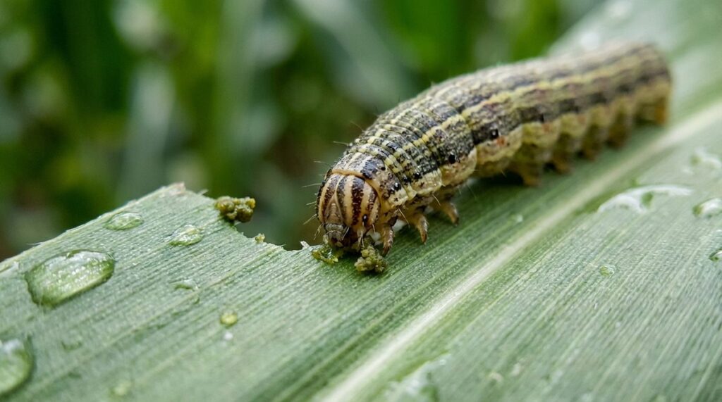
Le larve attraversano sei stadi di sviluppo e possono raggiungere i 35 mm di lunghezza. Il danno alle colture è causato principalmente dagli stadi larvali avanzati (dal terzo al sesto), che consumano grandi quantità di tessuto fogliare e possono penetrare nel cartoccio del mais, rendendo difficile il contatto con gli agenti di controllo.
Dalla cronologia dell’invasione alla situazione europea
Originaria del continente americano, dove è considerata un parassita endemico, la S. frugiperda ha compiuto un salto intercontinentale senza precedenti. La cronologia dell’invasione evidenzia la rapidità della diffusione:
- 2016: prime segnalazioni confermate nell’Africa occidentale e centrale
- 2017–2018: diffusione in oltre 40 paesi africani e prime rilevazioni in Asia meridionale
- 2019: presenza confermata in Cina, nel sud-est asiatico e nel Vicino Oriente
- 2020–2023: segnalazioni sporadiche nell’area mediterranea e monitoraggio intensificato nell’UE
L’EPPO (European and Mediterranean Plant Protection Organization) ha inserito S. frugiperda nel proprio contesto di sorveglianza fitosanitaria, fornendo note tecniche e aggiornamenti regolari sulla distribuzione dell’organismo nocivo. Le schede EPPO costituiscono il riferimento ufficiale per le autorità fitosanitarie degli Stati membri, delineando protocolli di identificazione, requisiti di notifica e raccomandazioni per la sorveglianza del territorio.
La velocità di espansione geografica della S. frugiperda non ha precedenti tra i lepidotteri invasivi: in meno di quattro anni dall’arrivo in Africa, la specie ha raggiunto quasi tutti i continenti, favorita dalla sua elevata capacità migratoria (gli adulti possono percorrere centinaia di chilometri in una singola notte) e dalla disponibilità diffusa delle colture ospiti.
Impatto economico e piante ospiti
Il mais (Zea mays) rappresenta l’ospite primario, ma S. frugiperda è in grado di attaccare oltre 350 specie vegetali, tra cui riso, sorgo, canna da zucchero, cotone e numerose orticole. Secondo le stime della FAO, le perdite di produzione di mais in Africa dovute al FAW possono raggiungere il 20–50% in assenza di interventi di controllo, con un impatto economico stimato in diversi miliardi di dollari annui.
L’impatto fitosanitario della Spodoptera frugiperda non si misura esclusivamente in termini di resa agricola: include i costi di monitoraggio, le restrizioni commerciali legate alla quarantena e gli investimenti necessari per lo sviluppo di strategie di contenimento.
Strategie di gestione integrata (IPM)
L’approccio più efficace per il contenimento di S. frugiperda è la lotta integrata (IPM — Integrated Pest Management), che combina diverse tecniche in un quadro coordinato. Come discusso nella recente review pubblicata su Frontiers in Agronomy (2025), le componenti fondamentali dell’IPM per il FAW includono:
Monitoraggio e soglie di intervento
L’utilizzo di trappole a feromoni consente di monitorare la presenza e la densità della popolazione adulta. Le soglie di intervento variano in funzione dello stadio fenologico della coltura e della densità larvale osservata. Un monitoraggio precoce e sistematico è essenziale per ottimizzare i tempi di intervento.
Controllo biologico
Diversi nemici naturali sono stati identificati come potenziali agenti di biocontrollo: parassitoidi del genere Telenomus e Trichogramma, predatori come Orius spp. e patogeni entomopatogeni (Metarhizium, Beauveria bassiana, Baculovirus). L’integrazione di questi agenti in programmi di lotta biologica aumentativa rappresenta una delle frontiere più promettenti.
Gestione della resistenza agli insetticidi
La S. frugiperda ha sviluppato resistenza documentata a diverse classi di insetticidi, compresi organofosforici, piretroidi e alcune tossine Bt. La rotazione dei principi attivi e l’adozione di strategie refuge per le colture Bt sono misure indispensabili per preservare l’efficacia degli strumenti chimici e biotecnologici disponibili.
Diagnosi e identificazione in campo
Il riconoscimento precoce della S. frugiperda è fondamentale per un intervento tempestivo. In campo, il danno si manifesta con erosioni fogliari caratteristiche, presenza di escrementi granulari nel cartoccio e, nei casi più gravi, danneggiamento delle spighe. La distinzione dalle specie autoctone di Spodoptera (come S. exigua o S. littoralis, già presenti nel bacino mediterraneo) richiede attenzione ai dettagli morfologici: la “Y” rovesciata sulla capsula cefalica della larva e i quattro punti disposti a trapezio sull’ultimo segmento addominale sono caratteri diagnostici chiave.
Per la conferma dell’identificazione, le autorità fitosanitarie raccomandano l’utilizzo di protocolli molecolari standardizzati (PCR su regioni del gene COI), mentre il monitoraggio degli adulti si avvale di trappole a feromoni con esche specifiche per il ceppo mais e il ceppo riso.
Per un quadro più ampio sulle sfide della diagnostica applicata e sul monitoraggio di fenomeni emergenti, il tema della sorveglianza fitosanitaria offre interessanti paralleli metodologici. Per approfondire, consulta anche la storia dell’alimentazione e delle colture. Per approfondire, consulta anche le tecnologie informatiche nella gestione fitosanitaria.
Fonti e riferimenti
- EPPO (European and Mediterranean Plant Protection Organization). Fall armyworm — Spodoptera frugiperda. Short note. https://www.eppo.int
- EPPO Global Database. Reporting Service: useful publications on Spodoptera frugiperda. https://gd.eppo.int/reporting/article-6266
- FAO AGRIS. Forecasting the global extent of invasion of the fall armyworm. Record 65df5a224c5aef494fe1fd69. https://agris.fao.org
- Review: Fall armyworm impacts and integrated management in Africa. Frontiers in Agronomy, 2025. DOI: 10.3389/fagro.2025.1538198