UNIVERSITY OF PISA
School of Graduate Studies
“Scienza del Farmaco e delle Sostanze Bioattive”
PhD THESIS
2006-2008“
PREPARATION AND CHARACTERIZATION OF TOPICAL
FORMULATIONS CONTAINING
6-O-ALKYL DERIVATIVES OF ASCORBIC ACID
”
Author Veronica Saino
SUPERVISOR DIRECTOR OF THE SCHOOL Prof. Patrizia Chetoni Prof. Claudia Martini
Index
Summary……….I-II
1. Introduction………1
1.1. The Skin…...………...……….……1
1.1.1. The Viable Epidermis and Epidermal Differentiation…………..2
1.1.2. The Stratum Corneum………..……….……3
1.1.3. The Dermis and the Hypodermis………..…….……4
1.1.4. Dermatological Vehicles………..……....……4
1.1.5. Permeation Routes……….………6
1.1.6. Factors Affecting Drug Permeation Through the skin….….……7
1.1.7. Strategies to increase drug penetration through the skin…….…7
1.2. The Eye……….………..9
1.2.1. Ocular anatomy and physiology……….………..9
1.2.2. Ophtalmic drug delivery systems……….………...12
1.2.3. Transcorneal drug absorption and vehicle effects…….…….….14
1.3. Alkanoyl-6-O-ascorbic acid esters……….…….…....18
1.3.1. Physicochemical properties of ASCn …...………….……….….18
1.3.2.In vitro permeation of ASCn and influence in drug absorption (enhancement properties)………...24
1.4. Ibuprofen: Technological aspects………25
1.5.Diclofenac: Technological aspects………...26
2. Materials and methods………..…..27
2.1. Chemicals………….……….27
2.2. Pharmacological aspect of the drugs………..……28
2.2.1. Dithranol……….28
2.2.3 Griseofulvin……….……… 30
2.2.4 Ibuprofen………….………..31
2.3. Methods……….…………33
2.3.1. Synthesis of ascorbyl-laurate………..…….33
2.3.2. Characterization of ascorbyl-laurate………..……. 33
2.3.3. Characterization of the ascorbyl-laurate by differential scanning calorimetry (DSC)……….………...…. 34
2.3.4. Characterization of the formulations by rheological measurements……….……34
2.4. Effect of polyethylen glycol 400 (PEG 400) on the solubility of Anthralin and Griseofulvin……….………36
2.4.1. Preparation of the formulations containing PEG 400…………..36
2.5. Evaluation of the stability of ASC16 coagels ………37
2.5.1. Preparation of the coagels for the preliminary evaluation of ASC16 derivate stability………...………...…37
2.6. Analytical method ……….…………38
2.6.1. Chromatographic apparatus ……….38
2.7. Preparation of the formulations for ex vivo skin permeation study of ibuprofen ………40
2.8. In vitro release of Ibuprofen ……….42
2.9. Ex vivo permeation of Ibuprofen through hairless rat skin …..43
2.10. Skin distribution studies of ibuprofen from coagels……… 44
2.13. Preparation of formulations containing diclofenac…...…….. 48 2.13.1. Preparation of the coagels……….. 48 2.13.2. Preparation of the aqueous suspension (DICH-D)………….…48
2.14. In vitro release of diclofenac from coagels………..………….49
2.15. Ocular in vivo studies on the rabbit eye……….………..50 2.15.1. Intraocular penetration study………...…….50 2.15.2. In vivo precorneal residence measurements…………...…….51
2.16. Analytical method for Diclofenac………...….52
2.17. Pharmacokinetic and Statistical Analysis………54
3. Results and Discussion………55
References
Summary.
Alkyl vitamin C derivatives (ASCn) are amphiphilic molecules having physicochemical
properties that depend on the alkyl chain length. The derivatives with low molecular weight (n < 11) have enough aqueous solubility to produce self-assemblies at room temperature (≈25°C), while those with longer alkyl chains possess a critical micellar temperature (CMT) higher than 30°C. Above CMT aqueous ASCn derivatives suspensions turn into
either micellar solutions or gel phases, depending on the length of the alkyl side chain. On cooling, liquid crystal structures, called coagels, are produced.
The semisolid consistency of such coagels is an interesting property that makes ASCn
coagel a promising pharmaceutical platform for drug delivery. Coagels could be able to solubilize insoluble and unstable drugs, protect them from any possible aggressive environment and enhance their permeation through skin. Furthermore the rheological properties of coagels would be adequate for topical administration.
Aim of the study was the evaluation of L-ascorbic acid lipophilic derivatives as permeation enhancers for two non-steroidal antinflammatory drugs, ibuprofen (IBU) and diclofenac (DICH), through hairless rat skin and ocular structures of rabbit eye, respectively, by means of in vitro, ex vivo and in vivo studies.
Among lipophilic derivatives the lauroyl-6-O-ascorbic acid or ascorbyl-laurate (ASC12) and
the palmitoyl-6-O-ascorbic acid or ascorbyl palmitate (ASC16) were chosen and different
vehicles for both IBU and DICH were prepared and compared with the commercially available formulations. The rheological characterization of each formulation was performed. Moreover, in order to better define the physicochemical properties of ASC12
and ASC16, the effect of the co-solvent on their drug-loading capacity was evaluated using
two insoluble model drugs, anthralin (ANTH) and griseofulvin (GRIS). Preliminary studies on the stability of ASC16 wereconducted in the presence or absence of vitamin E (Vit E),
used as antioxidant.
Results showed that both ASC12 and ASC16 increased IBU transdermal permeation through
hairless rat skin, producing a low depot in the skin of the same order of magnitude for both epidermis and dermis. Furthermore, a synergistic enhancement effect between ASCn and
II
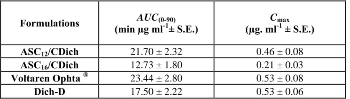
In vivo studies on rabbit eyes demonstrated that even if coagels did not increase the retention time of DICH in the tear fluid with respect to the commercial formulation, they were capable to raise drug concentration in the aqueous humour of two and three folds for ASC16 and ASC12, respectively.
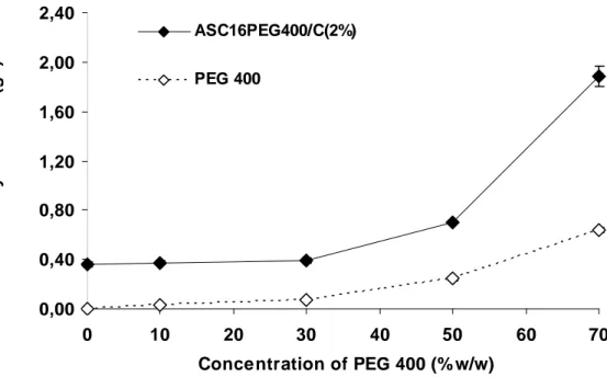
The incorporation of both IBU and DICH in the system did not produce changes on the characteristic crystalline structure of coagel, as observed from rheological measurements. The solubility profiles showed that the solubility of the model-drugs (ANTH and GRIS) was higher when the amount of the co solvent increased and a synergistic effect of PEG 400 and ASC16 coagel on the solubility of such drugs was observed.
Finally, preliminary stability studies demonstrated that the presence of Vit E as antioxidant did not improve the stability of ASC16, which showed a quicker degradation at 60°C with
respect to room temperature.
In conclusion, ASCn coagels may be considered promising pharmaceutical systems for
drugs utilizing by topical administration, even if further studies would be performed to increase their stability.
1. Introduction 1.1. The Skin
The skin has the primary function to provide a barrier against environmental influences and to protect the body against the loss of endogenous substances. Furthermore, the skin is also a major contributor to thermoregulation and performs endocrine functions, e.g. Vitamin D synthesis or peripheral conversion of pro-hormones. Macroscopically, two distinct skin layers are present, an unvascularized outer layer (epidermis) and an inner vascularized layer. The vascularized layer consists of the dermis and the hypodermis and has the function to provide physiological support to the unvascularized epidermis. This layer comprises the viable epidermis and the outermost layer of the skin, the stratum corneum, which consists of dead keratin-filled cells. Figure 1 gives an overview about the dimensions and the stratified appearance of the skin.
1.1.1. The Viable Epidermis and Epidermal Differentiation
The viable epidermis is divisible into three distinct layers, namely, from inside to outside: stratum basale, stratum spinosum and the stratum granulosum. The cells of these layers undergo continuous differentiation to produce the outermost layer of the skin, the stratum corneum, that consists of layers of dead keratin filled cells. Due to this process, histological preparations of the epidermis are of stratified appearance.
The stratum basale is a single layer of epidermal stem cells which are anchored to a basement membrane that separates the epidermal tissue from the underlying dermis. The basal cells are continually dividing, creating cells that undergo several changes while moving towards the skin surface in further course of differentiation. Above, these differentiating cells are forming the stratum spinosum. The cells are now of spiny appearance, due to abundance of desmosomes.
In addition to the cell organelles seen in the basal layer, the stratum spinosum also reveals lipid enriched lamellar bodies (also called Odland bodies) and an increase in keratin filaments, indicating the dual character of the differentiation, protein and lipid synthesis (Menon, 2002). On their way to the skin surface, the cells begin to flatten and elongate, forming the stratum granulosum. Distinct keratohyalin granules that become larger in the upper granulocytes reflect an increase in keratin synthesis, accompanied by an increasing number of lamellar bodies. In course of the terminal differentiation of the uppermost granulocytes into a corneocyte, these lamellar bodies are secreted to the extracellular domains, forming the stratum corneum extracellular bilayers. This process is accompanied by the formation of the corneocyte`s cornified envelope, the dissolution of cell organelles and the condensation of the enriched keratin filaments. The nature of the signals that initiate the irreversible process of cornification isn’t still elucidated, but it is evidenced that an extracellular calcium gradient with higher Ca2+ concentrations in the upper regions of the viable epidermis triggers the transformation from a granulocyte to a corneocyte. The deceased cells are finally shed from the skin by desquamation. The complete renewing process takes about 3-4 weeks. The viable part of the epidermis also contains melanocytes that are responsible for skin pigmentation, antigen presenting Langerhans cells and Merkelcells (mechanoreceptors) (Stenn, 1983).
1.1.2. The Stratum Corneum
The stratum corneum constitutes the outermost layer of the epidermis and represents the main barrier function, although it is the thinnest and smallest compartment of the skin. It is about 10 to 20 μm thick and consists of several layers of dead, keratin filled corneocytes that represent the final state of epidermal differentiation. These cells are embedded in a matrix of lipid lamellas that descend from the secreted content of the lamellar bodies, which gives the stratum corneum a brick and mortar organization (Menon, 2002). A rigid envelope that replaces the plasma membrane surrounds the corneocytes after terminal differentiation. This cornified envelope in turn is coated with long chain ceramides, some of them covalently attached through ester linkages to the outer surface of the envelope. A free interaction of their acid chains with unbound extracellular lipid lamellae assists establishing the lamellar organization in the extracellular lipids (Candi et al., 2005). Another specialty of the stratum corneum is the lipid composition, which differs significantly from those of the cells of the lower epidermal layers. The lipid composition of the stratum basale is comparable with that of cells of other tissues and constitutes mainly of phospholipids. In the stratum granulosum, a drastically decrease in the phospholipids occur for the benefit of sphingolipids and cholesterol. The extracellular lipid matrix of the stratum corneum finally is free of phospholipids and consists of ceramids, cholesterol and free fatty acids in roughly equimolar ratios (Menon, 2002). The ceramides are crucial for the lipid organization of the stratum corneum barrier, while the cholesterol is promoting the intermixing of different lipid species. Figure 2 points out these differences in lipid composition for the stratum basale, the stratum granulosum and the stratum corneum.
Water-holding properties of the stratum corneum are influenced by the rate of proteolysis, which generates a blend of amino acids termed as natural moisturing factors (Rawlings et al., 1994). The general degree of hydration of the stratum corneum is between 10 to 30% bound water, depending on the location within the stratum corneum, body site and environmental conditions. Under occlusive conditions (defined as complete impairment of passive transepidermal water loss), however, the stratum corneum may bind a 2.75 fold amount of water compared to its dry weight (Nitsche et al., 2006).
1.1.3. The Dermis and the Hypodermis
The dermis is connected by the basement membrane to the stratum basale. This junction is of folded appearance, supporting the exchange of nutrients and other physiological substances with the unvascularized epidermis. Main components of the dermis are collagen and elastin fibres that form a vast network of filamentous and amorphous connective tissue that prevents strength and flexibility to the skin (Schaefer and Redelmaier, 1996). Furthermore, the dermis accommodates cellular residents such as fibroblasts, endothelial cells, mast cells and, under conditions of inflammation, macrophages, lymphocytes and leucocytes. The tissue of the dermis has an extensive vascular network that is involved in nutrition, thermal regulation and immune responses. A variety of appendages, such as hair follicles or sweat and sebaceous glands are also derived from this tissue. Underneath the dermis is the hypodermis situated that contains, in contrast to the dermis, loose connective tissue and adiposities as the main cellular exponent which represent an energy source for the body.
1.1.4 Dermatological Vehicles
In clinical situations, a drug is rarely applied to the skin in form of a pure chemical but, instead, is normally incorporated in a carrier system, the vehicle (Surber and Smith, 2005). Such preparations vary in their physicochemical nature from powders through semisolids to liquids. However, a uniform and comprehensive classification for dermatological vehicles is currently not yet available. A formulation may be classified
by its pharmaceutical nomenclature used in pharmacopoeias (e.g. cream, ointment, gel), by the principle of the structural matrix (e.g. emulsion, liposomal dispersion) or by macroscopic appearance (e.g. milk, foam, shake). Table 1 gives a simplified possible classification of dermatological vehicles.
Table 1 Simple classification of dermatological vehicles
It is obvious that this classification system is a raw simplification of the diversity of external formulations. It does not account for many of the newer external formulations (e.g. liposomes, microcapsules, microemulsions, ect.) (Surber and Smith, 2005).
emulsion (o/w, w/o) often designated as milk, lotion, shake, ect. suspension
often designated as paint, shake, ect. nonpolar solution,
often designated as oil polar solution
often designated as paint, lotion, ect
emulsion (o/w/o)
often designated as milk, lotion, shake, ect.
suspension
often designated as paint, shake, ect. Liquid
Semisolid water-free polar or nonpolar ointment water containing polar or nonpolar gel
emulsion (o/w, w/o) often designated as washable (o/w), nonwashable (w/o)or amphiphilic (o/w, w/o) cream
suspension
often designated as paste.
emulsion with powder (o/w,w/o)
often designated as cream
pastes
Solid powder transdermal patch
System Monophasic Diphasic Tri-(multi-)phasic
1.1.5. Permeation Routes
The permeation of drugs through the skin includes the diffusion through the intact epidermis and trough skin appendages, for example sweat glands and hair follicles. However, the appendages occupy only 0.1% of the human skin surface so that the contribution of these shunt pathways to transdermal permeation is usually considered to be small (Moser et al., 2001).
Theoretically, there are two pathways through this layer (Figure 3): a transcellular (across the corneocytes and the lipid matrix) and an intercellular way (via the intercellular lipid domains between the corneocytes). In both cases, however, the permeant has to pass the intercellular lipid matrix.
Intercellular lipids are arranged in multiple layers, containing both, polar and nonpolar components. This suggests that hydrophilic and lipophilic penetrants diffuse through different domains in the lipid matrix. The transcellular pathway is generally the unfavoured route for a drug to permeate through the stratum corneum because of the high diffusional resistance of the cornified cells (Hadgraft, 1983). Only hydrophilic molecules such as water or short-chain alcohols seem to prefer the transcellular pathway (Barry, 1983).
Figure 3 . Schematic representation of stratum corneum and its intercellular and transcellular pathways of drug permeation (Moghimi et al., 1999).
1.1.6 Factors Affecting Drug Permeation Through the Skin
Several biological and physicochemical parameters may influence drug permeation across the skin (Barry, 1983). The physicochemical factors that control the passive diffusion of a substance from a vehicle into and across the skin are determined by the molecular properties of the substance, the vehicle and the skin. Skin hydration is a crucial and possibly the most frequently investigated factor affecting drug permeation. Alterations the vehicle following application, commonly due to evaporation of volatile components, represent a further crucial parameter that may affect drug permeation. Furthermore, skin metabolism may have additional impact on transdermal delivery of drugs. As skin undergoes many structural and functional changes with increasing age it would be useful to know whether such alterations affect the transdermal diffusion of drugs. Studies have shown that the effect of age is rather due to the smaller surface-to-volume ratio in case of newborn infants compared with adults than to an effect of the lower barrier function of younger skin (Walters, 1986). In practice, there is often a combination of these factors that contribute to transdermal drug permeation.
• Skin Hydration and Occlusion
• Evaporation of Volatile Vehicle Compounds Following Application • Drug-Skin Interactions
• Drug-Vehicle Interactions • Vehicle-Skin Interactions
1.1.7. Strategies to increase drug penetration through the skin.
Since many drugs do not permeate the skin well enough to be suitable for a transdermal formulation several technological approaches have been studied in the recent decades to enhance percutaneous drug penetration
Methods for improving cutaneous delivery may consist either on the use of chemical penetration enhancers, novel vehicle systems (i.e. microemulsions, liposomal-based delivery systems), or physical enhancement strategies (iontophoresis, sonophoresis, and electroporation). non-toxic, non-allergenic, compatible with drugs and excipients and
and fully and they should not have pharmacological activity. Many enhancers exhibit these attributes, e.g. azone and its analogues (Michniak et al., 1993), fatty acids (Aungst et al., 1990) alcohols (Takahash et al., 1991), pyrrolidones (Babu et al., 2008), sulfoxides (Choi et al., 1991), 6-O-ascorbic acid alkanoates (Palma et al., 2006), urea and its analogues and terpenes (Williams and Barry, 1991).
Referring to Fick’s first law three permeation enhancement strategies may be postulated:
1) Increasing the diffusion coefficient.
A reduction of the diffusional resistance of the skin can be obtained by disordering the stratum corneum lipids. Fatty acids are generally believed to increase diffusivity across the stratum corneum. Oleic acid, for example, has been shown to induce phase separation in the stratum corneum lipid domains, thereby reducing barrier function. Azone, dimethyl sulfoxide (DMSO) and different terpenes may provoke lipid disorder in the stratum corneum increasing the drug diffusivity (Moser et al., 2001).
2) Increasing drug solubility in the skin.
Agents typically thought to act in this way are propylene glycol, ethanol, Transcutol® and N-methyl pyrrolidone.
Ethanol seems to enhance the solubility of some drugs both by increasing the apparent partition coefficient and by extraction of stratum corneum lipids. (Moser et al., 2001) Transcutol® and some terpenes have also been investigated due to their potential to increase permeation by improving partitioning into the skin.
1.2. The Eye.
1.2.1. Ocular anatomy and physiology.
Although it is small in size, the eye (Figure 4) provides us with the most important of the five senses: vision.
Light enters the eye through the cornea. The cornea is spherically shaped and it forms the first and most important refracting surface due to the large change in index of refraction encountered from air to the corneal tissue. In a spectral sense it heavily attenuates optical radiation below 320 nm, such that at about 300 nm the eye is virtually opaque to optical radiation. Next, the light passes the aqueous humor, which is optically very transparent, except in the far infrared. The lens is another optically important component. It forms the second largest refracting element, and it further spectrally filters the light, mainly as an additional block of the short wavelengths that are dangerous to the retina. The lens is optically limited in size by the circularly shaped pupil, the diameter of which depends on the intensity of the light falling on the retina. Light passing the lens enters the vitreous, like the aqueous humor an almost optically transparent element. The refraction of light by both the cornea and the lens forms an image of the world outside at the retina. The lens can adjust its shape to have a sharp view nearby, or at a distance; a property called accommodation. This property decreases with age and is lost at middle age.
The most important part of the eye is the retina, as it contains the light receiving elements, the cones and the rods. The cones are most densely packed in a central area used for vision with the highest acuity, called the fovea. A somewhat larger central area
is called the macula. The rods are absent in the central fovea, but very abundant in the peripheral retina. Linked to the spatially peaked cone distribution is a layer of a
yellow substance, the macular pigment, which mainly consists of lutein and zeaxanthin. The main function of macular pigment probably is its antioxidant property in the fight against free radicals, generated by the combination of high oxygen tension and light. Optical effects are the spectral filtering of dangerous blue light passing the cornea and eye lens filters, and possibly, reducing chromatic aberration in this area of very detailed vision.
back side from stray light from deeper layers. The RPE is attached to Bruch’s membrane, a mechanically strong structure. The blood-brain barrier is formed by close contacts (so called tight junctions) between RPE cells. Posterior to Bruch’s membrane is first a thin fenestrated layer of blood vessels, the choriocapillaris. Apart from supplying nutrients, it also serves to stabilize the temperature in case of high levels of illumination absorbed by the RPE. The sclera is a very strong structure at the outside of the eye; it has a whitish appearance. Between the sclera and the choriocapillaris, we find the choroidal space filled with blood vessels, melanin particles, and tissue.
A brief description of the most important structures of the human eye is reported below:
• Aqueous. A water-like fluid which fills the front part of the eye between the lens and cornea. This fluid is produced by the ciliary body and drains back into the blood circulation through channels in the chamber angle. It is turned over every 100 minutes.
• Chamber Angle. Located at the junction of the cornea, iris, and sclera, the anterior chamber angle extends 360 degrees at the perimeter of the iris. Channels here allow aqueous fluid to drain back into the blood circulation from the eye. May be blocked in glaucoma.
• Choroid. A very vascular layer between the sclera and retina which serves to nourish the outer portions of the retina. Has one of the highest blood flows in the body.
• Ciliary Body. A structure located behind the iris (rarely visible) which produces aqueous fluid that fills the front part of the eye and thus maintains the eye pressure. It also allows focusing of the lens.
• Conjunctiva. A thin lining over the sclera, or white part of the eye. This also lines the inside of the eyelids. Cell in the conjunctiva produce mucous, which helps to lubricate the eye.
• Cornea. The clear window through which we see. Actually, this is a very vital part of the eye's focusing, and the curvature of the cornea itself accomplishes about 80% of the focusing of the eye.
• Episclera. A fibrous layer between the conjunctiva and sclera. Sometimes lumps (pingueculum) will form in this layer on the surface of the eye near the inside or outside corners.
• Extraocular Muscles. Six muscles control eye movement. Five of these originate from the back of the orbit and wrap around the eye to attach within millimeters of the cornea. Four of these move the eye roughly up, down, left and right. Two muscles (one originating from the lower rim of the orbit) control the twisting motion of the eye (when the head is tilted).
• Iris. This is the part of the eye which gives it color. It contains muscles which open or close the pupil in response to the brightness of surrounding light. A blue
• Lens. This is located just behind the iris, and helps to focus light. A "capsule" surrounds the lens "nucleus". The nucleus can become cloudy, and this is termed cataract.
• Macula. The part of the retina which is most sensitive, and is responsible for the central (or reading) vision. It is located near the optic nerve directly at the back of the eye (on the inside). This area is also responsible for color vision.
• Optic Nerve. This contains visual information from the eye, and has about 1.2 million nerve fibers. The optic disc is visible on the inside of the eye, where the nerve is viewed "end on". The sheath around the optic nerve is continuous with that of the brain, and the nerve connects directly into the brain.
• Orbit. The boney socket containing the eye, fat, extraocular muscles, nerves, and blood vessels. The floor and inside walls of the orbit are paper thin, and are easily fractured by trauma.
• Pupil. Essentially, a hole in the iris. This is the black opening in the center of the eye. Its size is controlled by the iris muscles.
• Retina. This thin layer lines the inside of the eye and receives light rays, processes them, and sends signals to the brain via the optic nerve. The retina is like the "film of a camera". It is separated from the very vascular choroid by the "retinal pigment epithelium". Sometimes breakdowns in this pigmented layer allow macular degeneration.
• Sclera. The white, tough wall of the eye. Few diseases affect this layer. It is covered by the episclera and conjunctiva, and eye muscles are connected to this. • Uveal tract. A group of similar eye structures including the choroid, ciliary body
and iris. May be prone to inflammatory conditions (uveitis or iritis).
• Vitreous. A jelly-like, clear fluid which fills most of the eye (from the lens back). This tends to liquefy with age, and its separation from the retina can lead to retinal tears and detachment.
1.2.2 Ophthalmic drug delivery systems.
Ophthalmic drug delivery is one of the most interesting and challenging endeavors facing the pharmaceutical scientist. The anatomy, physiology, and biochemistry of the eye render this organ highly impervious to foreign substances. Development of newer,
more sensitive diagnostic techniques and novel therapeutic agents continue to provide ocular delivery systems with high therapeutic efficacy. Conventional ophthalmic solution, suspension, and ointment dosage forms no longer constitute optimal therapy for this indications.
The specific aim of designing a therapeutic system is to achieve an optimal concentration of a drug at the active site for the appropriate duration. Ocular disposition and elimination of a therapeutic agent is dependent upon its physicochemical properties as well as the relevant ocular anatomy and physiology. A successful design of a drug delivery system, therefore, requires an integrated knowledge of the drug molecule and the constraints offered by the ocular route of administration.
Nowadays, there are, on the market or undergoing clinical trials, ocular drug delivery systems to sustain drug release. Most of them are for the treatment of long-term diseases that affect the posterior segment of the eye. The important indications for ocular drug delivery systems include ocular degeneration, viral infections (like CMV infections), glaucoma, ocular inflammations, dry eye syndrome, and retinal degenerations. There are many important indications for ocular drug delivery systems depending on the site of application and the delivered drug.
The challenge is to provide a system with improved ocular drug bioavailability and prolonged duration of activity, but still with a minimum risk of ocular complications (Table 2). The mode of administration plays an important role in defining the safety of the device and further, in achieving patient compliance and acceptance. In general the highest risk of ocular complications is with invasive ocular drug delivery. The risk is smaller with periocular drug administration. When ocular complications are rare for the systemic drug administration, there is a prevalence of systemic adverse effects.
Table 2 Classification of the routes for ocular drug administration Invasive drug administration to intraocular cavities
y Intravitreal surgery (at the oars plana) y Repeated (*) intravitreal injections y Intracameral surgery (capsular bag) y Subretinal injections
y Repeated (*) intracameral injections
Invasive periocular and sclera modes of drug administration y Intrascleral surgery
y Episcleral surgery
y Repeated (*) periocular injections y Repeated (*) subconjuctival injections
y Transscleral diffusion from controlled release systems
Non-invasive methods
y Topical administration on the eye
Systemic administration
y Intravenous infusion and injection y Per oral
(*) The repetition is needed to accomplish long-term ocular treatment. (Del Amo and Urtti , 2008).
1.2.3 Transcorneal drug absorption and vehicle effects.
Owing to easy accessibility of the eyeball, topically applied drugs are widely employed in ophthalmology for both diagnostic and therapeutic purposes.
In spite of a variety of defense mechanisms which protect the eye from noxious substances in the environment (as the continuous secretion of tears which coats an impermeable surface epithelium), topical administration of drugs on the eyes surfaces allows persistence of therapeutic levels of medications for clinically effective durations of time and minimized unwanted collateral systemic effects, though these substances can rapidly spread to all parts of the body.
On their way from the outside to the inside of the eye medications may encounter several obstacles
The first impact is on the lachrymal film: the characteristics of tear distribution and flow greatly affect one of the most important factors involved in intraocular drug penetration. Contact-time may be prolonged by means of various techniques, such as increased
viscosity of drops or other engineered devices (e.g. viscous formulations, mucoadhesive formulations, in situ active gel-forming systems, inserts, shields systems or particulates) (Vincent H.L. Lee, 1990). However it is mainly influenced by the tear flow, which is known to be variable for different subjects and in different situations.
As a general rule, the rapid washout with drops instilled into the conjunctival sac leads to immediate loss of a great part of the drug.
In fact, the total volume of fluid the eye can hold being 10 µl, only about 20% of a drop delivered by commercial eye droppers (the volume of which is about 50 µl) can be retained by the eye. Additional loss of medications usually occurs since a faster turnover rate may result from reflex tearing caused by the irritation of the drop.
The composition of tears includes various substances such as proteins, electrolytes, vitamins, enzymes, glucose and others, which may play a rule in the transfer of drugs to the cornea.
In normal conditions when the eye is open the lachrymal fluid is divided into three separate compartments: a) the pre-corneal film which covers the exposed cornea and conjunctiva; b) a mostly stagnant layer moistening the conjunctiva under the lids; c) the marginal tear strips along which the tear flow occurs.
The pre-corneal film in turn is composed of three layers: the superficial oily layer is primarily derived from the meibomian glands and restricts the evaporation of the underlying watery layer which is mostly produced by the lachrymal glands. The innermost mucoid layer is derived from the conjunctival globet cells and is effective in stabilizing the tear film.
During a blink the mucin layer is spread over the cornea, thus maintaining its normal wetting and producing a hydrophilic surface for the tear film.
Drugs dissolved in the lachrymal film penetrate the globe almost exclusively through the cornea, most of the material which crosses the conjunctiva being rapidly lost to the blood stream, and only small portions diffusing through the limbal zone and the sclera. In fact the conjunctival epithelium acts as a barrier against penetration of substances under the conjunctiva, but it appears to be weaker than the corneal epithelium. When in the sub-conjunctival space, drugs are rapidly lost to the blood before having the possibility to penetrate the deeper tissues. This could give reason for unpleasant systemic side effects sometimes occurring after instillation of drugs into the conjunctival sac, and for reduces therapeutic efficacy of topical medications in diseases
The cornea may act as a pathway, a barrier or a reservoir of drugs. Of the five layers in which the cornea is classically divided, only the sandwich consisting of the stromal connective tissue covered by the epithelial and endothelial cellular layers is relevant to trans-corneal drug penetration. The barrier is mainly represented by the epithelium, the reservoir by the stroma (particularly for hydrophilic substances), while the role of the endothelium is negligible for both aspects.
The epithelium and the endothelium, rich in lipids and cells, are mostly permeably to substances possessing a fat-soluble phase, while the stroma, characterized by relative acellularity and a high water content, is mostly permeable to substances possessing a water-soluble phase. Therefore, to be able to pass through the intact cornea, substances must be soluble in both fats and water, since purely water-soluble substances cannot penetrate the epithelium and purely fat-soluble substances cannot penetrate the stroma. Epithelium occupies about 10% of total corneal thickness and is composed of five to six layers of cells, increasing to eight to ten at the corneal periphery.
Three groups of cells are usually identified in the epithelium: a single row of basal cells, an intermediate zone of two or three layers of polygonal cells (wing cells), and a couple of superficial layers of large, flattened cells, which are characteristically joined by tight junctions, and present surface microplicae and microvilli which could play a role in the retention of pre-corneal film. The barrier effect of corneal epithelium is mainly due to the presence of tight junctions, which probably represent the most difficult hurdle to penetration of ionic solutes.
In fact the dissociated ions such as Na+ and Cl- pass the cellular layers slowly through the paracellular pathway, like other ionized substances (as for example fluorescein). The arrangement of superficial epithelial cells leads to an increase of the path distance, owing to flattening and overlapping of these cells, the large diameter of which allows relatively few intercellular spaces which open on the outer surface of the cornea.
Undissociate salts on the other hand are fat-soluble and can pass more rapidly through cell membranes (Saettone et al., 1987).
The fat solubility of the substance depends, among other factors, on its non-polar nature, i.e. on possessing an extremely symmetric atomic structure. These substances therefore penetrate more rapidly into the eye when the epithelium is intact than when it is disrupted or absent (the contrary, of course, occurring with water-soluble substances).
The permeability of corneal epithelium to different substances varies considerably according to their dissociation, which is mainly dependent upon the hydrogen ion concentration (pH) of the solution in which they are dissolved.
Moreover epithelial permeability has been shown to decreased in anoxic conditions, as can occur in patients using extended wear contact lenses. (Saettone et al., 1987).
1.3 Alkanoyl-6-O-ascorbic acid esters
Ascorbic acid and its derivatives play an important role in many biological processes as reducing agents or radical scavengers. Triplet excited state carbonyl compounds react with ground state oxygen to give the highly reactive singlet excited molecular oxygen, that reacts with proteins, nucleic acids, and cellular lipids, and induce severe damages in biological systems . When water is exposed to ionizing radiation, different radicals are formed, such as .OH and .OOH. The current increment of radical species in the atmosphere (particularly nitrogen oxides), the seasonal partial loss of the ozone layer, and the increase of the ultraviolet irradiation over the Earth, justify the importance of using antioxidant species in protecting terrestrial life from free radicals’ attack, or in minimizing their dangerous activity.
Cellular protection against singlet oxygen damage may be granted by a number of quenchers, such as ascorbic acid, carotenoids and phenols, notably tocopherols. Ascorbate behaves as a weak singlet oxygen quencher, it is, in fact, a better one-electron reductant than tocopherol ( E°’= +0.23 V). Its lipophilic derivative 6-O-ascorbyl-palmitate is a good antioxidant in model systems and is also effective in cellular systems or against Epstein-Barr virus activation. The presence of one or more hydrophobic chain in vitamin C-based surfactants leads to the formations of supramolecular self-assembled aggregates in water, such as micelles, where an inner lipophilic core is surrounded by a hydrophilic shell made up of the polar headgroups (Palma et al., 2002a).
1.3.1 Physicochemical propierties of ASCn
Alkanoyl-6-O-ascorbic acid esters were synthesized in order to transfer the peculiar properties of vitamin C in lipophilic media. Since a hydrophobic portion (aliphatic chain) and a polar group (ascorbic acid) are present in their structure, these esters behave as anphiphilic molecules but, due to their low water solubility, their use as surfactant agents is limited, especially when the side tail is a long hydrocarbon chain (more than ten carbon atoms) and at low temperatures. When the surfactant concentration reaches the critical micellar concentration (CMC) value, solubility increases, due to the formation of supramolecular aggregates and the solubilization of
monomers in these structures (Palma et al., 2007). Further addition of monomers to a micellar dispersions increases the number of the supramolecular aggregates, and may result in structural (shape and size) changes, while the monodispersed amphiphiles concentration remains unchanged. The temperature dependence of surfactans solubility indicates the existence of the so-called Krafft temperature or Krafft point (CMT) (Myers, 1994; Clint, 1992).
R = (CH2)x – CH3
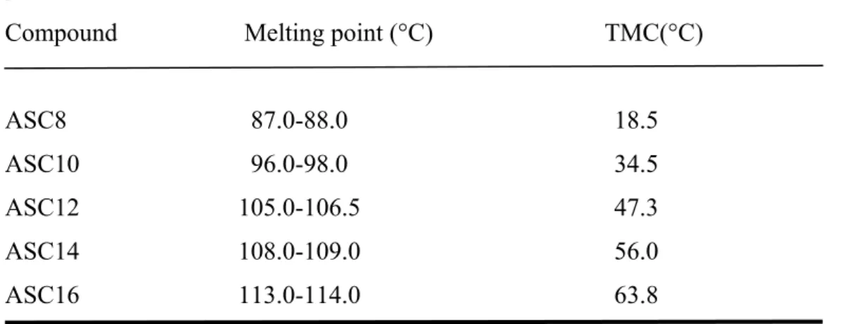
Once the CMT has been reached (Table 3), cooling down the sample results in the formations of a liquid crystal, and a semisolid viscous system known as “coagel” is obtained, due to the little hydration of the aggregates (micelles, lamella and vesicles).
Figure 5. a-Schema of ASCn derivative; b- Structure of ASCn
a b x ASCn 6 ASC 8 8 ASC 10 9 ASC 11 10 ASC 12 12 ASC 14 14 ASC 16
Table 3: Melting point and TMC of ASCn aqueous dispersions
Compound Melting point (°C) TMC(°C)
ASC8 87.0-88.0 18.5 ASC10 96.0-98.0 34.5 ASC12 105.0-106.5 47.3 ASC14 108.0-109.0 56.0 ASC16 113.0-114.0 63.8 (from Palma et al, 2002a)
Structural differences of coagels can be tracked also at a microscopic level. Figure 6 shows the ESEM micrographs of coagels from ASC8, ASC10, ASC12 and ASC16. For ASC8 and ASC10, the coagel structures can be depicted as a planar continuous lamellar phase, where bilayers are ordered in sheets. However, important size differences in the plates are observed (see Figure 6a and 6c) (Palma et al., 2003a).
In Fig 7, where are shown shear rate vs. shear stress flow curves, the rheological behavior of the coagels is reported. All coagels show a non-Newtonian flow. The differences are directly related to their structure.
Coagels from ASC10 and ASC11 (Figure 7b and c) showed pseudo-plastic flow and in the case of ASC11 thixotropy was detected as well.
Conversely, coagels from ASC8, ASC12, ASC14 and ASC16 exhibit a much more complex rheology (Figure 7a,d–f). The rheograms form a hysteresis loop in which the ‘down’ curve is displaced to the left of the ‘up’ curve. A characteristic feature of many such rheograms is the presence of a ‘spur’ point on the ascending curve. The rheology of these coagels displays a high yield or “spur” value. This can be taken as a measure of the strength necessary to break the ordered structure of the system before a significant flow occur. The shear stress at spur point is called the ‘static yield value’ (σS). For such systems an irreversible structure breakdown is observed when the shear stress exceeds σS. When the spur value is reached, the secondary structure of coagels is broken and ASC12, ASC14 and ASC16 coagels acquire pseudo plastic flow (Palma el at, 2007). Figure. 7 Rheograms for ASCn coagels: (a) ASC8; (b) ASC10; (c) ASC11; (d) ASC12; (e) ASC14; and (f) ASC16.
Coagels from ASC12, ASC14 and ASC16 have a rheological behavior similarly to ASC8 samples. However, ESEM images ( Figure 6e–h) show a very different arrangement of the lamellae that form a ‘house of cards’ structure. Swelling and strengthening of the semisolid network is owed to the presence of strongly bound water regions between the amphiphilic bilayers. On the contrary, for ASC10 and ASC11 coagels, this kind of arrangement is apparently not possible. When coagels of ASC12, ASC14 and ASC16 are subjected to shear stress (τ>σS), the structure breaks irreversibly and the viscosity decreases significantly. The sample acquires a less ordered pattern, similar to that of ASC10. Figure 7f indicates that the ASC16 coagel acquires a pseudo plastic behavior after shear stress as in the case of ASC10 and ASC11 coagels. Such rheology is a characteristic feature of some coarse systems commonly used in pharmacy.
The spur value can be changed with the addition of compounds as polyethylen glycol 400 (PEG 400) (Saino et al., 2004). Increasing the amount of PEG 400 doesn’t produce changes in the rheological behavior of ASC16, as shown in Figure 8 where flow curves (shear rate vs shear stress) are reported. Furthermore it is evident from rheograms profile that the spur values is present in every curve and σS become higher increasing the amount of PEG 400, owing to the co-solvent improvement of coagel structure.
Figure 8 Rheograms for ASC16 coagels with PEG400 10, 30, 50 and 70 % w/w. 0.00 100.00 200.00 300.00 400.00 500.00 600.00 0.00 10.00 20.00 30.00 40.00 50.00 TAU D ( 1 /S )
PEG 400 10%/ ASC16 PEG 400 30%/ASC16 PEG 400 50%/ ASC16 PEG 400 70%/ ASC16
1.3.2 In vitro permeation of ASCn and influence in drug absorption (enhancement
properties)
The permeation of ASCn (ASC12 and ASC16) and its effect on “in vitro” and “in vivo” drug (Anthralin, ANTH) diffusion through rat skin were evaluated (Palma et al., 2006). Penetration of ASCn through rat skin epidermis was very fast and quantitatively significant. ASC12 appears to be the compound that possesses the highest capacity to enhance the penetration of the drug maybe because of self-penetration through the epidermis. The ability of these compounds to permeate the rat skin is related to their chemical composition, since the flux of ASCn decreases as alkyl chain length increases. Furthermore, a burst effect was observed with ASC12.
1.4. Ibuprofen: Technological aspects.
Ibuprofen (IBU) is a potent non-steroidal anti-inflammatory (NSAID) drug often used for the treatment of acute and chronic arthritic conditions (Busson, 1986). IBU can cause gastric mucosal damage which may results in ulceration. For this reason in past it was interesting to develop topical dosage forms of IBU. Indeed, many new topical formulations of IBU have been studied and evaluated in order to increased the bioavailability of the drug and minimized the side effects. Topical drug delivery of IBU in various formulations has been described (Muktadir et al.,1986; Chang et al., 1997; Cilurzo et al., 2005).
In the experimental work of Yun-Seok Rhee et al., (2008), the in vitro IBU permeation from several topical formulations through hairless mouse skin was studied. The three factors chosen for the factorial design were the concentration of the drug, of polyoxyethylene(5)cetyl/oleyl (POE(5)cetyl/oleyl) ether and of ethanol; the level of each factor was low, medium and high. The permeation rate of IBU significantly increased in proportion to the drug concentration, but decreased in proportion to POE(5)cetyl/oleyl ether concentration. Ethanol concentration was inversely proportional to the lag time.
In another experimental work (Herkenne et al., 2006) the effect in vivo of propylene glycol (PG) on IBU absorption through human skin, was studied. IBU Formulations with several amounts of PG (from 0 to 100 % v/v), a common co-solvent in topical formulations, were assessed by tape-stripping. Dermatopharmacokinetic parameters, characterizing drug amount in and diffusivity through the stratum corneum (SC), were derived. The solubility of the drug increased when the PG concentration increased, the values of viscosity increased in formulations with higher co-solvent concentration, but PG did not affect the diffusivity of the IBU across the SC.
1.5. Diclofenac: Technological aspects.
Diclofenac (DICH), an NSAID, has been found to be a viable alternative to steroids in treating ocular inflammation. It has poor aqueous solubility. Aqueous solutions of DICH sodium (DICHNa) commonly applied topically in the eye were 0.1% w/v. A major impediment to the bioavailability of topically applied ophthalmic drugs is incomplete absorption due to nasolacrimal drainage ( Ahuja et. al, 2007).
To overcome this, several dose ophthalmic forms have been studied to maximize the amount of the DICH in the site of action. Attama (2007) studied the delivery of DICHNa to the eye using bio-engineered human cornea from solid lipid nanoparticles (SLNs) prepared with a combination of homolipid from goat (goat fat) and phospholipid. Results obtained in this work showed that permeation of DICHNa through the cornea construct was improved by formulation as SLN modified with phospholipids. In another work, Valls et al., (2008), tested different formulations of DICHNa and demonstrated that corneal permeation was remarkably enhanced when diclofenac was formulated as a ß-cyclodextrin complex.
2. Materials and methods 2.1. Chemicals
Palmitoyl-6-O-ascorbic acid or ascorbyl palmitate (ASC16), L-ascorbic acid (AA), Anthralin (Anth), Griseofulvin (Gris), S-(+)-Ibuprofen (IBU), Diclofenac acid (DICH), lauric acid (LA), polyethylen glycol 400 (PEG 400), sodium lauryl sulphate (SLS) and disodium hydrogen phosphate (Na2HPO4) were purchased from Fluka (Milan, Italy), their purity was 99% according to the manufacturer. Lauroyl-6-O-ascorbic acid or ascorbyl-laurate (ASC12) was synthesized in our laboratories according to the procedure already reported in literature (Capuzzi, 1996).
Sulfuric acid 95%, diethyl ether, petroleum ether, and sodium sulphate (analytical grade) were purchased from Fluka (Milan, Italy) and used without purification.
D(+) glucose monohydrate (Glu), methanol (MeOH), acetonitrile (AcN), isopropyl alcohol both of HPLC-grade, were purchased from Carlo Erba (Milan, Italy). Milli-Q water was used in all experiments.
A commercial gel containing 10% w/w of ibuprofen lysine (Arfen®, Lisapharma, Erba, CO, Italy) was used as references in the permeation study through hairless rat skin, and the commercial Voltaren Ophta® (Ciba Vision) was used as reference in the ocular pharmacokinetics tests.
2.2. Pharmacological aspect of the drugs
2.2.1 Dithranol (ANTH)
Dithranol or Anthralin, 1,8-dihydroxy-9,10-dihydroanthracen-9-one (C14H10O3), is a hydroxyanthrone, anthracene derivative, medicine applied to the skin of people with psoriasis. It is commercially available as 0.1 to 2% w/w/ creams, ointment or pastes. Dithranol has a slower onset of action in controlling psoriasis, typically several weeks, compared to glucocorticoid steroids, but is without the potential for rebound reaction on withdrawal. Dithranol accumulates in mitochondria where it interferes with the supply of energy to the cell, probably by the oxidation of dithranol releasing free radicals. This impedes DNA replication and so slows the excessive cell division that occurs in psoriatic plaques. In addition Dithranol may act by reducing the elevated levels of cGMP that occurs in psoriasis.
The application of Dithranol formulation temporarily stains the skin a yellowy-brown. It may cause a local burning sensation and irritation, this may be minimised by careful attention to the details of treatment and only gradually stepping up through the strengths of dithranol formulations..
2.2.2. Diclofenac (DICH)
Diclofenac, 2-[2-[(2,6-dichlorophenyl)amino]phenyl]acetic acid (C14H11Cl2NO2) is a non-steroidal anti-inflammatory drug (NSAID) with inflammatory and analgesic properties, reducing pain in conditions such as arthritis or acute injury.
There is some evidence that diclofenac inhibits the lipoxygenase pathways, thus reducing formation of the leukotrienes (also pro-inflammatory autacoids). There is also speculation that diclofenac may inhibit phospholipase A2 as part of its mechanism of action and these additional actions may explain the high potency of diclofenac.
The action of one single dose is much longer (6 to 8 hours) than the very short half-life that the drug indicates. This could partly be due to a particular high concentration achieved in synovial fluids.
Diclofenac is used for musculoskeletal complaints, especially arthritis (rheumatoid arthritis, osteoarthritis, spondylarthritis, ankylosing spondylitis), gout attacks, and pain management in case of kidney stones and gallstones. An additional indication is the treatment of acute migraines.
As long-term use of diclofenac and similar NSAIDs predisposes for peptic ulcer, many patients at risk for this complication are prescribed a combination of diclofenac and misoprostol, a synthetic prostaglandin analogue, to protect the gastric mucosa.
An external, gel-based formulation containing 3% of diclofenac is available for the treatment of facial actinic keratosis which is caused by over-exposure to sunlight. Some countries have also approved the external use of diclofenac 1% gel to treat musculoskeletal conditions.
The use of diclofenac is not indicated in the case of hypersensitivity against the drug, in patient with history of allergic reactions (bronchospasm, shock, rhinitis, urticaria) following the use of Aspirin or another NSAID or in patient with active stomach and/or duodenal ulceration or gastrointestinal bleeding, inflammative intestinal disorders such as Crohn's disease or ulcerative colitis and severe insufficiency of the heart (Goodman & Gilman, 2006).
Figure 10 - Structure of DICH
2.2.3 Griseofulvin (GRIS)
Griseofulvin, (2S,6'R)-7-chloro-2',4,6-trimethoxy-6'-methyl-3H-spiro[1-benzofuran-2,1'-cyclohexan]-2'-ene-3,4'-dione, is an antifungal drug. It is used both in animals and in humans, to treat ringworm infections of the skin and nails. The drug binds to tubulin, interfering with microtubule function, thus inhibiting mitosis.
It binds to keratin in keratin precursor cells and makes them resistant to fungal infections. It is only when hair or skin is replaced by the keratin-griseofulvin complex that the drug reaches its site of action. Griseofulvin will then enter the dermatophyte through energy dependent transport processes and bind to fungal microtubules. This alters the processing for mitosis and also underlying information for deposition of fungal cell walls.
Known side effects of griseofulvin include: hives, skin rashes, confusion, dizziness, diarrhea, fatigue, headache, nausea and vomiting (Goodman & Gilman, 2006).
Figure 11 - Structure of GRIS
2.2.4 Ibuprofen (IBU)
Ibuprofen is a non-steroidal anti-inflammatory drug (NSAID) originally marketed as Brufen and used as an analgesic and anti-inflammatory for relief of symptoms of arthritis, primary dysmenorrhea, fever. Ibuprofen is a core medicine in the World Health Organization's "Essential Drugs List", which is a list of minimum medical needs for a basic health care system.
Low doses of ibuprofen (200 mg, and sometimes 400 mg) are available over the counter (OTC) in most countries. Ibuprofen has a dose-dependent duration of action of approximately 4–8 hours, which is longer than suggested by its short half-life. Ibuprofen is an NSAID which is believed to work through inhibition of cyclooxygenase (COX), thus inhibiting prostaglandin synthesis. There are at least 2 variants of cyclooxygenase (COX-1 and COX-2). Ibuprofen inhibits both COX-1 and COX-2. It appears that its analgesic, antipyretic, and anti-inflammatory activity are achieved principally through COX-2 inhibition; whereas COX-1 inhibition is responsible for its unwanted effects on platelet aggregation and the GI mucosa. The role of the individual COX isoforms in the analgesic, anti-inflammatory, and gastric damage effects of NSAIDs is uncertain and different compounds cause different degrees of analgesia and gastric damage.
Ibuprofen appears to have the lowest incidence of gastrointestinal adverse drug reactions of all the non-selective NSAIDs. However, this only holds true at lower doses of ibuprofen.
Common adverse effects include: nausea, dyspepsia, gastrointestinal ulceration/bleeding, raised liver enzymes, diarrhea, epistaxis, headache, dizziness, unexplained rash, salt and fluid retention, and hypertension. (Goodman & Gilman, 2006).
2.3 Methods
2.3.1 Synthesis of ascorbyl-laurate
Ascorbyl laurate (ASC12) was synthesized by condensation reaction between lauric acid (LA) and C6OH primary group of L-ascorbic acid in sulfuric acid at 45°C (Capuzzi, 1996). A 100 ml sample of 95% sulfuric acid was placed in an Erlenmeyer flask, and a gentle stream of nitrogen was passed for 30 min under magnetic stirring at room temperature. Then, ascorbic acid (4.41 g, 25 mmol, AA) was completely dissolved and after LA (5.01 g, 25 mmol) was added. The flask was then equipped with a rubber stopper and kept in a nitrogen atmosphere. A water bath at 45°C was used to dissolve LA completely. After 16 h, the mixture was poured into a beaker containing 600 ml of ice and stirred until it reached room temperature. The solution was treated with diethyl ether several time, after the organic phase was treated with anhydrous sodium sulfate for 30 min and then filtered. The white-yellow solid was obtained after evaporation under reduced pressure of the solvent which was washed in petroleum ether and finally recrystallized three times from diethyl ether/petroleum ether (50:50) mixtures.
2.3.2 Characterization of ascorbyl-laurate
Purity was conveniently assessed through TLC and chemical-physical analysis. TLC (silica gel, diethyl ether): only one spot with Rf= 0.41 was observed. The melting point
was 104.5-105.5°C , and the UV-VIS spectroscopy showed an assorbance at λ max,= 236 nm (CH3CN) for an ASC12 organic solution.
The ASC12 was characterized after preparation of a 2% w/w aqueous dispersion measuring the critical micellar temperature (CMT) by DSC analysis.
DSC analysis was carried out using a Pyris DSC6 differential scanning calorimeter (Perkin-Elmer), connected to an MC480 cooler circulator (FTS, Stone Ridge, NY, USA), using airtight closed aluminum pans. The critical micellar temperature (CMT) was taken as the temperature of the endothermic peak obtained for a 10% w/w aqueous dispersion of ASC12 (ASC12/C).
The preparation of ASC12/C was reported in section 2.3.3. and its composition was indicated in Table 4.
2.3.3 Characterization of the ascorbyl-laurate by differential scanning calorimetry (DSC)
The critical micellar temperature (CMT) for ASC12 was obtained through DSC runs with a Pyris DSC6 differential scanning calorimeter (Perkin-Elmer), connected to an MC480 cooler circulator (FTS, Stone Ridge, NY, USA), using airtight closed aluminum pans. The CMT was taken as the temperature of the endothermic peak obtained for 10% w/w aqueous dispersion of ASC12.
Coagel was prepared by heating 10% w/w of ASC12 aqueous suspensions above its phase transition temperature (CMT = 47.3 °C, ASC12, Palma, 2003b) until the gel phase was formed. Then, it was permitted to reach room temperature to obtain the ASC12 coagel (ASC12/C).
2.3.4 Characterization of the formulations by rheological measurements
The rheological measurements were carried out on the coagels prepared without , and on the “disrupted coagels”, characterized by a structure where the typical form as “house of cards” was broken (Palma et al., 2003a). The composition of the analyzed formulations are reported in Table 5.
Rheological assays were performed using a Haake RS1 Rheometer, equipped with the cone plate combination ( C60/4°) on a samples placed into the sensor plate and kept for 5 min at 20°C.
The measurements were carried out between 0 and 200 s-1 in 300 s. Data were acquired and analyzed by the Rheo Win Pro software (Haake).
Preparation of samples analyzed.
• Coagels were prepared by heating aqueous suspensions containing 2% w/w of ASC12 and ASC16 and glucose mono hydrate (5% w/w) above their phase transition temperature (CMT = 47.3 °C, ASC12; 63.8 °C, ASC16, Palma , 2003b) and adding
DICH (0.1% w/w) until the gel phase was formed. The systems were kept to of over TMC for 2 hours in order to obtain the complete solubilization of the drug, and then, they were permitted to reach room temperature to obtain the respective ASC12 and ASC16 coagels (ASC12/CGDich and ASC16 /CGDich). Glucose mono hydrate was added to obtain isotonic formulations.
Coagels without DICH but containing Glucose (ASC12/CGlu and ASC16/CGlu) and coagels without DICH and Glucose (ASC12/C(2%) and ASC16 /C(2%)) were prepared using the same method and used as references in the ocular pharmacokinetic studies.
• Coagels were prepared by heating aqueous suspensions containing 5% w/w of ASC12 and ASC16 above their phase transition temperature (CMT = 47.3 °C, ASC12; 63.8 °C, ASC16, Palma, 2003b) and adding Ibu (0.85% w/w) until the gel phase was formed. The systems were kept to of over TMC for 2 hours in order to obtain the complete solubilization of the drug. Then, they were permitted to reach room temperature to obtain the respective ASC12 and ASC16 coagels (ASC12/CIbu and ASC16 /CIbu). Coagels without Ibu (ASC12/C(5%) and ASC16 /C(5%)) were prepared using the same method and used as references in the cutaneous permeation studies.
• “Disrupted coagel” coagel was prepared by heating a 5% w/w ASC16 aqueous suspension above their phase transition temperature (CMT = 63.8 °C, ASC16, Palma., 2003b) and adding Ibu (0.85% w/w) until the gel phase was formed. The system was kept to of over TMC for 2 hours in order to obtain the complete solubilization of the drug, and then, it was permitted to reach room temperature with stirrer (600 rpm) to obtain the ASC16 disrupted coagel (ASC16/Ibu).
2.4 Effect of polyethylen glycol 400 (PEG 400) on the solubility of Anthralin and Griseofulvin.
The evaluation of the effect of different amount of cosolvent (PEG 400) on the solubility of ANTH and GRIS in presence or in absence of ASC16 were performed by UV–VIS measurements in the range of 200–400 nm at 25°C.
2.4.1. Preparation of the formulations containing PEG 400.
An excess amount of drug was added to 5 ml water containing or not 2% w/w of ASC16 (ASC16PEG400 /C(2%) and PEG 400 respectively), with increasing amounts of PEG 400 (10 30, 50 and 70 % w/w). The preparations were shaken for 3 h in a water bath above their phase transition temperature (CMT = 63.8 °C, ASC16, Palma, 2003b).
After equilibrium was reached, suspensions were centrifuged at 13000 rpm for 10 min to remove un-dissolved drug. An aliquot from each solution was adequately diluted with ethanol ( 1: 20 ratio) and analyzed at 365 and 324 nm for ANTH and GRIS respectively (UV–VIS Shimadzu UV-160 spectrophotometer). The experiments were carried out in triplicate (Palma et al., 2003c).
2.5 Evaluation of the stability of ASC16 coagels
To evaluate the chemical stability of ASCn derivatives after preparation of Coagels a preliminary study was performed on a formulation containing 2% w/w ASC16 derivative alone and in presence of 8 % w/w of Vitamin E, as antioxidant (Spiclin P. et al., 2001).
2.5.1. Preparation of the coagels for the preliminary evaluation of ASC16 derivate
stability
Coagels were prepared by heating 2% w/w ASC16 aqueous dispersions, with and without Vit.E (8% w/w) in a water bath above their phase transition temperature (CMT = 63.8 °C, ASC16 ) for about 120 minutes. Then the samples were kept to room temperature to obtain ASC16/C(2%) and ASC16/C(2%)VitE.
The composition of the preparations were reported in Table 6.
Aliquots of both formulations were maintained in closed vials at room temperature (RT) and 60° C and at predetermined times the samples were submitted, after appropriate dilution with methanol to HPLC analysis for quantitative analysis of ASC16 derivatives. The HPLC method was reported in section 2.5.
2.6 Analytical method
2.6.1 Chromatographic apparatus
The amount of ASCn derivatives, IBU and DICH in the formulations and in the different experimental samples were assayed by HPLC.
The chromatographic system consisted of a LC 6A Pump, SPD-10A array detector and computer integrating system (Shimadzu Corp., Kyoto, Japan). The column (150 x 4 mm) was C18 Synergi 4μm Fusion–RP 80A (Milford, MA, USA) to analyzed ASCn derivatives and IBU. The column used to analyze DICH samples was a C18 Bondclone 10μm (30 cm x 3.9 mm, Phenomenex). The mobile phase was filtered, in all cases, through a 0.45 μm filter (Millipore™ Durapore) and degassed by vacuum prior to use. The different conditions, mobile phase, flow rate, retention time and wavelength are reporter in the Table 7.
Preparation of standard solutions
A stock standard solutions of ASCn (SS/ASCn), IBU, (SS/Ibu) and DICH (SS/Dich) were prepared by solubilization in methanol of an appropriate amount of each product exactly weighed. Then, several dilutions in of isotonic buffer phosphate solution pH 7.4 were prepared and injected (20 μl) into the HPLC using a manual system.
Table 7. HPLC condition
Compound Mobile phase (ratio) flow-rate (ml/min) Retention time (min) Wavelength (nm) ASC12 ASC16 MeOH:CH3CN: phosphate buffer 0.05M adjusted to pH 2.5 with phosphoric acid (65:14:21) 1.0 13.5 (ASC12) 22.5 (ASC16) 265 IBU MeOH:CH3CN: phosphate buffer 0.05M adjusted to pH 2.5 with phosphoric acid (65:14:21) 1.0 4.2 225 DICH MeOH:CH3CN: phosphate buffer 0.05M adjusted to pH 7.4 with phosphoric acid (46:13:41) 1.2 8.5 273
2.7 Preparation of the formulations for ex vivo skin permeation study of ibuprofen
Preparation coagels (ASC12/CIbuand ASC16 /CIbu).
Coagels were prepared by heating at 52°C and 68°C a 5% w/w of ASC12 and ASC16 aqueous suspensions above their phase transition temperature (CMT = ASC12: 47.3 °C, (ASC16,: 63.8 °C; Palma , 2003b) and adding IBU (0.85% w/w) until the gel phase was formed. The systems were kept over TMC for 2 hours in order to obtain the complete solubilization of IBU, and then, they were permitted to reach room temperature to obtain the respective ASC12 and ASC16 coagels (ASC12/CIbu and ASC16 /CIbu). The composition of the preparations were reported in Table 8.
Preparation of coagel based on ASCn withPEG 400 (ASC12 PEG400/CIbu and ASC16
PEG400/CIbu).
Coagels were prepared the method previously reported but adding in the aqueous suspensions of ASCn the PEG 400 30% w/w to obtained the final formulations ASC12 PEG400/CIbu and ASC16 PEG400/CIbu. The composition of the preparations were reported in Table 8.
Preparation of “Disrupted coagel“, based on ASC16 (ASC16 /Ibu)
Coagels were prepared by heating (5% w/w of ASC16 aqueous suspensions above its phase transition temperature (CMT = 63.8 °C, ASC16, Palma, 2003b) and adding Ibu (0.85% w/w) until the gel phase was formed. The systems were kept to of over TMC for 2 hours in order to obtain the complete solubilization of the drug, and then, they were permitted to reach room temperature with stirrer (600 rpm) to obtain the ASC16 with its structure disrupted (ASC16/Ibu).
Preparation of the reference suspension based on PEG 400 (PEG-D)
A suspension based on 0.85 % w/w of Ibu (PEG-D) was prepared in a mixture constituted with PEG 400 30% in water. The suspension was maintained under magnetic stirrer for hour at room temperature.
Preparation of the reference solution (Ibu-S)
A solution containing 0.85 w/w Ibu (Ibu-S) prepared by dissolving Ibu in a mixture isopropanol/pH 7.4, 66.7 mM phosphate buffer (20:80 ratio) under magnetic stirrer and it was used as reference. The composition was reported in Table 8.
2.8 .In vitro release of Ibuprofen from coagels.
The in vitro release of ibuprofen from coagels ASC12/CIbu, ASC16 /CIbu, ASC16PEG400/CIbu, ASC12PEG400/CIbu and from the reference formulations (Ibu-S, PEG-D, , and Arfen®) was assessed using dialysis bags (cut off=3000- 3500 Da) filled with a fixed amount of each formulation (about 10.0 g). The bags were put into 10.0 ml of isotonic buffer phosphate solution 66.7 M, pH 7.4 (receptor phase). The receptor phase was stirred and thermostated at 37 °C. At fixed time 1.0 ml of the receptor phase were withdrawn and replaced with fresh fluid. The drug released was assayed by HPLC.
2.9. Ex vivo permeation of Ibuprofen through hairless rat skin
Permeation tests of Ibu were performed using excised skin of 6-8 week-old hairless rat (OFA-hr/hr, Charles River Italia SpA). The animals were killed by cervical dislocation immediately before the experiments, the skin was carefully excised and the adhering fat and subcutaneous tissue were removed.
The perfusion apparatus consisted of Gummer-type diffusion cells with an available diffusion area of 1.23 cm2 and stratum corneum facing the donor compartment (Monti et al., 2002). The donor phase consisted of about 1.0 g of coagels (ASC12/CIbu , ASC16/CIbu, ASC16 PEG/CIbu and ASC12PEG/CIbu), or Ibu-S, ASC16 /Ibu and of 0.14 ml of Arfen®. The dose of drug used for the experiment was corresponding to 8.5 mg of IBU
The receiving compartment contained 5.0 ml of receiving isotonic 66.7 mM pH =7.4 phosphate buffer solution maintained at 37°C and stirred at 600 rpm. At predetermined intervals of time, 5.0 ml samples of receiving phase were withdrawn for HPLC analysis and replaced with the same volume of fresh fluid. All experiments lasted 20 hours and were performed four times (Tampucci, 2005).
Figure 12.- Schema of the cell Gummer type. (1,2: receiving phase; 3,4: thermostated water).
1
2 4