A Three-protein Charge Zipper Stabilizes a Complex
Modulating Bacterial Gene Silencing
*
Received for publication, December 4, 2014, and in revised form, June 16, 2015Published, JBC Papers in Press, June 17, 2015, DOI 10.1074/jbc.M114.630400 Tiago N. Cordeiro‡§1, Jesu´s García¶, Pau Bernado´§2, Oscar Millet!3, and Miquel Pons‡4
From the‡Biomolecular NMR Laboratory, Department of Organic Chemistry, University of Barcelona, 08028 Barcelona, Spain, §Centre de Biochimie Structurale, INSERM U1054, CNRS UMR 5048, Universite´ Montpellier 1 and 2, 34092 Montpellier, France, ¶Institute for Research in Biomedicine (IRB-Barcelona), 08028 Barcelona, Spain, and the!Structural Biology Unit, Center for Cooperative Research in Biosciences (CIC-bioGUNE), 48160 Elexalde, Derio, Spain
Background: The complex between Hha and H-NS selective represses genes in Enterobacteria acquired by horizontal
transfer.
Results: A structural model for the regulatory complex is described.
Conclusion: A charge zipper formed by interdigitation of residues from three proteins stabilizes the complex.
Significance: Charge zippers provide selectivity to electrostatic protein complexes. Understanding selective gene silencing may
help fighting antibiotic resistance.
The Hha/YmoA nucleoid-associated proteins help selectively silence horizontally acquired genetic material, including patho-genicity and antibiotic resistance genes and their maintenance in the absence of selective pressure. Members of the Hha family contribute to gene silencing by binding to the N-terminal dimerization domain of H-NS and modifying its selectivity. Hha-like proteins and the H-NS N-terminal domain are unusu-ally rich in charged residues, and their interaction is mostly elec-trostatic-driven but, nonetheless, highly selective. The NMR-based structural model of the complex between Hha/YmoA and the H-NS N-terminal dimerization domain reveals that the ori-gin of the selectivity is the formation of a three-protein charge zipper with interdigitated complementary charged residues from Hha and the two units of the H-NS dimer. The free form of YmoA shows collective microsecond-millisecond dynamics that can by measured by NMR relaxation dispersion experiments and shows a linear dependence with the salt concentration. The number of residues sensing the collective dynamics and the pop-ulation of the minor form increased in the presence of H-NS. Additionally, a single residue mutation in YmoA (D43N) abol-ished H-NS binding and the dynamics of the apo-form, suggest-ing the dynamics and bindsuggest-ing are functionally related.
Antibiotic resistance and the appearance of new virulent bac-terial strains constitute a major threat to human health (1). The problem is aggravated by the transfer of resistance and viru-lence genes between bacteria (horizontal gene transfer) (2–4). In this context, a detailed knowledge of the mechanisms allow-ing bacteria to tolerate the acquisition of foreign DNA is lack-ing, and it may open the way to new sustainable strategies to fight infectious diseases. Proteins Hha and YmoA were first identified as environmental regulators of the expression of vir-ulence factors in Escherichia coli (5) and Yersinia sp. (6), respec-tively. Hha/YmoA bind to the nucleoid-associated protein H-NS (7), enhance its capacity to silence newly acquired genes, and facilitate the smooth integration of foreign genes in the existing genome (8, 9). The key role of members of the Hha/ YmoA family in enterobacteria is supported by their conserva-tion in obligate endosymbionts (10) and their presence in con-jugative plasmids (11). Hha is also directly involved in the formation and dispersal of biofilms, a microbial life-style that is responsible of many chronic infections and is associated with increased antibiotic tolerance and with resistance dissemina-tion (12–14). Therefore, Hha/YmoA constitutes promising tar-gets in the development of new antimicrobial drugs.
Hha and YmoA are homologous and functionally replaceable proteins (15, 16) with high sequence identity/homology (82.1/ 94.0%) and very similar structures formed by four helices sepa-rated by loops (17, 18). Interestingly, !30% of their primary structure corresponds to charged residues. The high degree of conservation (Fig. 1) and extensive mutational studies (19–21) suggest the relevant role of charged amino acids in Hha func-tion. The N-terminal region of H-NS interacting with Hha is also unusually rich in charged residues (39%), reinforcing the notion that electrostatic interactions are essential for this interaction (19). The role of electrostatics is also supported by the ionic strength sensitivity of genes regulated through Hha!H-NS (22).
The putative role of electrostatic interactions in the Hha/ YmoA complex with H-NS raises the problem of selectivity, as electrostatic interactions are often associated with nonspecific contacts in contrast to van der Waals interactions that display
*This work was supported by the Spanish Ministry of Economy and Competi-tivity (BIO2010-15683, BIO2013– 45793R, and CTQ2012–32183), the Gen-eralitat de Catalunya (2009SGR1352), the European Commission (BioNMR, contract 261863), and by the Department of Industry, Tourism, and Trade of the Government of the Autonomous Community of the Basque Country. The authors declare that they have no conflicts of interest with the con-tents of this article.
The atomic coordinates and structure factors (code2mw2) have been deposited in the Protein Data Bank (http://wwpdb.org/).
1Recipient of a fellowship from the Fundac¸a˜o Cieˆncia e Tecnologia (Portugal). 2Supported by the CHEX SPIN-HD project of the Agence National de la
Recherche and ATIP-Avenir.
3To whom correspondence may be addressed: Structural Biology Unit, Cen-ter for Cooperative Research in Biosciences (CIC-bioGUNE), Parque Cientí-fico Tecnolo´gico de Bizkaia Bldg. 801A, 48160 Elexalde Derio, Spain. Tel.: 34-946572504; E-mail: [email protected].
4To whom correspondence may be addressed: Biomolecular NMR Labora-tory, University of Barcelona, Cluster Building. Barcelona Science Park, Baldiri Reixac, 10-12 08028 Barcelona. Spain. Tel.: 34-934034683; E-mail: [email protected].
at Biblioteca de la Universitat de Barcelona on August 31, 2015
http://www.jbc.org/
high selectivity by means of an exquisite shape complementar-ity. The interaction of Hha/YmoA with H-NS is highly specific (19, 20).
In this article we have investigated the interplay between electrostatic forces and the constitutional and functional prop-erties of Hha/YmoA. A detailed analysis of the structures of the Hha!H-NS complex in crystals (21) and in solution (first reported here) unraveled a charge zipper with conserved resi-dues contributed by three proteins (Hha/YmoA and the two units of an antiparallel H-NS dimer). This feature explains the high selectivity of the Hha/YmoA interaction with H-NS. In addition, electrostatic repulsive interactions within YmoA are also responsible for the presence of collective slow motions (!s-ms) in the free form that, we argue, may also contribute to shape the biological function of Hha/YmoA.
Experimental Procedures
Protein Preparation—Unlabeled and isotopically enriched H-NS46C21S(19),H-NS64
(23),Hhavariants(23),andYmoAvari-ants (18) were expressed and purified as previously described. Single cysteine mutants were prepared by site-directed mutagenesis after substitution of the native cysteine by ser- ine(H-NS)orisoleucine(Hha).Site-directedmutagenesiswascar-ried using the QuikChange site-directed mutagenesis kit. All con-structswereverifiedbyDNAsequencing.Fluorescenceanisotropy titrations were carried out to evaluate the affinity of YmoA toward H-NS64(24) at different ionic strengths.
NMR Samples for PRE5Experiments—Experimental
inter-molecular PRE data were obtained on samples containing "100 !M15N-labeled H-NS46C21S and 15–30 !MHhaC18I
deriva-tized with MTSL at positions 37 or 66.
Additional experiments were carried out by observing15
N-labeled HhaC18I in the presence of H-NS46 dimer with an
EDTA-Mn2#tag placed in the C21 residue of each monomer. A
third set of PRE experiments was carried out using15N-labeled
H-NS46C21S with an additional C-terminal cysteine tagged with
4-(2-iodoacetamide)-2,2,6,6-tetramethyl-1-piperidinyloxy radi-cal (TEMPO). This sample was used to confirm the antiparallel topology of the H-NS46dimer. In all the PRE experiments the
buffer was 20 mMHEPES (pH 7.0), 150 mMNaCl, and 0.01%
(w/v) NaN3. Adding a 2–3 molar excess of ascorbic acid
gener-ated diamagnetic reference samples of nitroxide radicals. EDTA-Ca2#-tagged dimers were used as diamagnetic
refer-ence for the manganese-containing samples.
Spin Labeling—Derivatization reagents were 1-oxyl-2,2, 5,5-tetramethyl-3-pyrroline-3-methyl)-methanethiosulfon-ate (MTSL) or N-[S-(2-pyridythiol)cysteaminyl]-EDTA.
Proteins containing a single cysteine residue were incubated with 10 mMDTT at room temperature to ensure the reduction
of any intermolecular disulfide bonds. The excess of DTT was removed by passing 2 times the reaction mixture through a
PD-10 column (GE Healthcare) (99.9% desalting capacity). After elution, protein solutions in 10 mMTris (pH 7.4) ("100
!M) were mixed with a 10-fold molar excess of the
derivatiza-tion reagent an incubated for 3 h in the dark at room tem-perature. To remove any unreacted tag, the resulting protein solution was passed 2$ through a PD-10 column, and the
buffer was exchanged to 20 mM HEPES (pH 7.0), 150 mM
NaCl, and 0.01% (w/v) NaN3. Complete incorporation of
MTSL was confirmed by mass spectrometry, and the oligo-meric state of the conjugated protein was checked by analyt-ical gel filtration.
In the case of the cysteaminyl-EDTA adduct, the reaction was carried out in the presence of 1 mMMnCl2or CaCl2to
generate the paramagnetic and diamagnetic samples. PRE mea-surements for the backbone amides were carried out at low molar fractions of complex (0.15–0.3) to reduce binding-in-duced broadening.
NMR Samples for CPMG Experiments—All 15N-CPMG
relaxation dispersion experiments were recorded on samples containing 1.1 mMYmoA protein (or variant) in 20 mMsodium
phosphate, 1 mM (tris(2-carboxyethyl)phosphine), 0.2 mM
EDTA, 0.01% (w/v) NaN3, and 8% D2O at pH 7.5 and 285 K and
the stated concentration of NaCl and H-NS64.
NMR Assignments—HhaC18I had been assigned previously (24). Assignments for the1H,15N amide resonances of H-NS
46
C21S were obtained using a combination of three-dimensional
1H,15N-edited NOESY-HSQC and 1H,15N-edited
TOCSY-HSQC experiments acquired, with mixing times of 120 and 60 ms, respectively. Assignments have been deposited in Biologi-cal Magnetic Resonance Bank (25296).
YmoA backbone amide assignments were obtained based on published data (Biological Magnetic Resonance Bank entry 15486) (24) at pH 6.0 and 298 K.1H,15N HSQC spectra of YmoA
were stepwise recorded at pH 7.5, 7.0, 6.5, and 6.0 and then at 285, 292, and 298 K. Small1H,15N chemical shift differences
were observed for most residues and varied linearly with the temperature, such that the assignments at 298 K and pH 6.0 could be propagated to the other temperatures and pH values.
1H,15N HSQC NMR spectra of YmoA variants are similar to
those of YmoA wild type and consistent with a well folded, monomeric protein with a similar overall structure.
PRE-guided Modeling—1H,15N HSQC spectra of
diamag-netic and paramagdiamag-netic samples were acquired at 298 K with a recycling delay between scans of 2.5 s to ensure that magneti-zation recovery levels are identical for both states and using the same concentration and number of scans for both samples. NMR spectra were acquired on 600 MHz or 800 MHz Bruker spectrometers equipped with a TCI cryo-probe.
The paramagnetic contribution to the relaxation rate %2was
determined using the ratio of peak intensities in the paramag-netic, and diamagnetic state (25),
Ipara Idia "R2 diaexp(&% 2t) R2dia# %2 (Eq. 1)
where t is the evolution time during the INEPT transfer (set to "10.63 ms; based on a1H,15N scalar coupling of 94 H
Z); R2diais
transverse relaxation rate in the absence of paramagnets, which
5The abbreviations used are: PRE, paramagnetic relaxation enhancement; MTSL, 1-oxyl-2,2,5,5-tetramethyl-3-pyrroline-3-methyl)-methanethiosul-fonate; TEMPO, 2,2,6,6-tetramethyl-1-piperidinyloxy; CPMG, Carr-Purcell-Meiboom-Gil; HSQC, heteronuclear single quantum correlation; AIR, ambiguous intermolecular distance restraint; T, tesla; SAXS, small-angle x-ray scattering; NTD, N-terminal dimerization domain; r.m.s.d., root mean square deviation.
at Biblioteca de la Universitat de Barcelona on August 31, 2015
http://www.jbc.org/
can be estimated for each residue from the line-width of the peak at half-maximum height.
Calculated PRE rates were converted into intermolecular distances using the equation,
d "6
"
K%2,expDD /pb
!
#
4 # 31 # $H2%c2
$
(Eq. 2)
where K is 1.23 10&32cm6s&2, $
His the Larmor frequency of
the proton, %2is the experimental determined PRE rate, pbis
the fraction of complex, %cis the correlation time estimated
from Stokes-Einstein equation. The values of %2were corrected
by 1/pbto obtain the expected values for fully bound proteins.
The correlation time for the electron-nuclear interaction was assumed to be equal to the global correlation time of the complex.
PRE effects from spin labels located at positions 37 and 66 were integrated as distance restraints together with mutagene-sis data and chemical shift mapping (Fig. 3) in the HADDOCK docking approach (26). Interfacial residues were selected based in the fulfillment of at least three of the following criteria: (i) significant chemical shift perturbation upon the addition of small amounts of the interacting partner, (ii) at least 30% sur-face accessible area, (iii) as part of a cluster of residues that forms a plausible binding patch, (iv) mutation of the residue to give rise to a properly folded protein but with null or decreased binding ability. The selected residues were defined as “active” and their solvent accessible surface neighbors or residues that were in agreement with criteria (i) and (ii) as “passive.” Haddock encodes these interfacial residues in terms of ambiguous intermolecular distance restraints (AIRs) to drive the dock-ing (26). AIRs correspond to ambiguous distance between all atoms of the source residue (active) to all the atoms of all target residues (active and passive). The AIR definitions are provided in Table 1.
The known structures of free Hha (PDB 1JW2) and H-NS46
dimer (PDB code 1NI8) were used as starting structures in the protein docking protocol. MTSL-modified cysteines were com-putational designed onto the surface of Hha starting structure. The mobility of the MTSL tags was taken into account by per-forming ensemble averaging of the PRE-driven restraints with four different MTSL orientations. All docking runs were per-formed using the HADDOCK standard docking protocol of three consecutive steps: (it0) rigid body energy minimization, (it1) semi-flexible refinement in the torsion angle space, and a final water refinement in explicit solvent (water). Hinges and flexible regions, paramagnetic tags, and/or residues identified as being part of the binding interface of a given model were considered explicitly as flexible in the it1- and water MD-sim-ulated annealing refinement steps. The hinge prediction server HingeProt (27) was used to define the hinge regions of the flex-ible monomers. The number of structures was increased to
2000, 400, and 400 for it0, it1, and water, respectively. Random removal of AIRs was turned on. Other parameters were left to their default values. Scoring and clustering were performed according to standard HADDOCK procedures (28).
PRE effects on15N-HhaC18I caused by H-NS
46with
EDTA-Mn2#placed in each Cys-21 residue were not used in the
mod-eling and, therefore, provided an independent validation of the generated structures. Theoretical intermolecular PRE rates were back-calculated from the structural models using the Sol-omon-Bloembergen approximation by representing the para-magnetic label by an ensemble of states and calculating the order parameters accounting for the motion of the dipolar pro-ton-electron interaction vector (29). The %cof each pose was
estimated using HydroNMR (30). PRE rates were scaled by the population of the bound state and compared with the experi-mental values.
Side chain-directed HCACO Experiments—The standard HCACO experiment was modified to detect correlations between side-chain carbonyls and the adjacent & or ' methy-lene groups. In particular C&/C' excitation was achieved with a 320-!s Q5 pulse centered at 35 ppm. The experiments were run at 14.1 T and 298 K in a sample containing 0.7 mM13C-YmoA,
20 mMsodium phosphate buffer (pH 7.5) with 100 mMNaCl, 1
mM(tris(2-carboxyethyl)phosphine), 0.2 mMEDTA, and 0.01%
(w/v) of NaN3in the absence or in the presence of 0.3 mM
H-NS64to obtain about 40% of bound YmoA. We used a
two-dimensional version of the experiment with 256 increments and 32 scans. Assignments were based on published data (Bio-logical Magnetic Resonance Bank entry 15486).
CPMG Relaxation Dispersion Experiments—15N-CPMG
sin-gle-quantum relaxation dispersion experiments for YmoA were performed at 285K (calibrated with a methanol sample). Data sets were recorded always with two identical aliquots of the same freshly prepared protein sample on Bruker Avance III spectrometers operating at static magnetic fields of 14.1 and 18.8 T, the latter equipped with a cryo-probe.
All 15N-CPMG relaxation dispersions were collected as a
series of two-dimensional planes with 16 interleaved frequency (cpvalues, ranging from 25 to 1000 Hz, including 2 repeats for error analysis and using a constant-time version (31) of the relaxation-compensated TROSY CPMG pulse scheme (32). Each two-dimensional spectrum was recorded with 180 and 200 complex points in t1 dimension at 14.1 T and 18.8 T,
respectively, and a delay between scans of 1.2 s. The total con-stant-time-CPMG relaxation Trelaxdelay was set to 80 ms. A
compensation element was applied at the beginning of each scan to suppress artifacts due to (cp-dependent changes in
sam-ple temperature.
Effective relaxation rates, R2,eff, were calculated according to
the relation (33), R2,eff'(cp( " & 1 Trelaxln
#
I'(cp( I0$
(Eq. 3)where I0is the intensity of a peak in the reference spectrum
recorded without CPMG relaxation delay, and I((cp) is the
cor-responding peak intensity in the spectrum measured at a fre-quency of (cp.
TABLE 1
Interface restraints
Protein Active residues Passive residues
Hha 8, 25, 48 22, 29, 44, 45, 49, 52, 68
H-NS46a 9, 10, 12, 28 6, 11, 13, 15, 16, 31, 32, 35
aResidues of both chains were taken into account.
at Biblioteca de la Universitat de Barcelona on August 31, 2015
http://www.jbc.org/
Dispersion profiles for each15N were individually and
glob-ally analyzed to obtain values of kex)k##k&by fitting the
Carver-Richards equation (34) using in-house Matlab scripts either to one field alone or simultaneously using data from two fields.
Electrostatic Potentials and Clustering—We used Poisson-Boltzmann electrostatic calculations and homology modeling to quantitatively identify regions of conserved electrostatic character (35, 36) in the Hha/YmoA family of proteins. Homol-ogy modeling was used to generate structures for the Hha ho-mologues with unsolved structures based on PDB code 1JW2 using SWISS-MODEL (37). All protein sequences were taken for UniProt (38).
All electrostatic potential calculations were performed using DelPhi (39) with 129 $ 129 $ 129 grid points, considering solvent dielectric constant of 80 with an ion concentration of 200 mM, whereas the protein dielectric constant was 4. A probe
with a radius of 1.4 Å was used to define the dielectric boundary. Each electrostatic potential calculation was centered on each structure to ensure proper alignment of electrostatic potentials before similarity calculations.
Similarity matrices were generated based on the electrostatic similarity index as defined elsewhere (35, 36). Cumulative dis-tributions of electrostatic conservation index (ECI) were calcu-lated using (40),
ECI "1 N
%
i ) 1 N
* &+A&&&+B,i&
max'&+A&, &+B,i&( (Eq. 4)
Here +Arepresents the electrostatic potential of E. coli Hha to
which all other potentials +B,iwere compared. The electrostatic
conservation index is calculated at each grid point and normal-ized by N, the number of electrostatic potentials comparisons, which are in total 12. The electrostatic conservation only describes the similarity of the electrostatic potential of a set of proteins to one particular protein at a given grid point. The surface projections of electrostatic conservation were gener-ated using UCSF Chimera (41).
Small-angle X-ray Scattering (SAXS) Experiments—SAXS data were measured in the beam-line BM29 (ESRF-Grenoble). Data were collected at multiple concentrations from 0.9 to 10 mg/ml at 285 K in 20 mMphosphate buffer (pH 7.5), 100 mM
NaCl, 1 mM (tris(2-carboxyethyl)phosphine), 0.2 mM EDTA
and 0.01% NaN3. A momentum transfer range of 0.032–4.93
Å&1 was measured. Repetitive measurements indicated that
samples did not present radiation damage.
Because the relative populations of species in solution change as a function of the concentration, the final SAXS pro-files were directly used without merging them. Initial points of the curves were discarded for the analysis to minimize interpar-ticle interaction effects. Data processing was performed using PRIMUS (42). The theoretical SAXS curves of full-length YmoA, H-N64, and YmoA!H-NS64complexes were computed
using the program CRYSOL (43). All theoretical curves were computed with 101 points and a maximum scattering vector of 0.5. Flexibility was accounted for by averaging ensembles of "1000 structures. Theoretical curves for YmoA!H-NS64
mix-tures were computed by linear combination of the theoretical curves of the individual species weighted by their predicted population in the mixture. The binding constant was obtained from fluorescence anisotropy titrations. Experimental and pre-dicted curves were directly compared with no adjustable parameter fitting.
Results
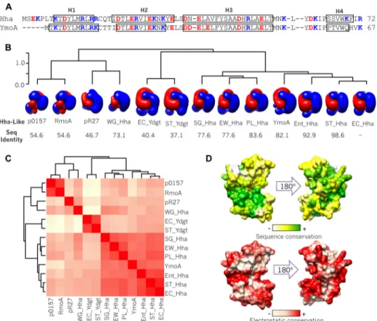
Electrostatics in the Hha/YmoA Family of Proteins—To investigate the role of electrostatic forces in the function of Hha-like proteins, we first examined the conservation of the charge distribution across this family of proteins. For this pur-pose we chose 13 Hha homologues differing in sequence (37.1– 98.6% identity with E. coli Hha) and net charge (&0.8 to #6) and computed their electrostatic potentials using structural templates generated by homology modeling from the Hha structure (PDB code 1JW2). The isopotential surfaces shown in Fig. 1B clearly show a similar asymmetrical electrostatic poten-tial distribution. Quantitative pairwise electrostatic similarity calculations (35) show that chromosomic Hha paralogues clus-ter together, whereas Hha-like proteins from plasmids and the obligate endosymbiont Wigglesworthia glossinidia form a sep-arate cluster (Fig. 1C). Fig. 1D shows the local degree of conser-vation of the electrostatic potential between the 13 homo-logues, mapped on the Hha structure compared with the conservation in the amino acid sequence. Thus, the abundance of charged residues and their distribution seems to be a con-served feature of members of the Hha/YmoA family, suggesting a functional role.
Solution Structure of the Hha!H-NS46Complex; a
Three-pro-tein Charge Zipper—To unravel the role of electrostatics in the molecular recognition associated to Hha, we determined the solution structure of Hha bound to H-NS. H-NS contains an oligomerization and a DNA binding domain separated by a linker. Two dimerization regions connected by a long ,-helix form the oligomerization domain (44). Hha interacts exclu-sively with the N-terminal dimerization domain (NTD) of H-NS. The NTD is completely included in a construct formed by residues 2–47 (H-NS46) (19, 45). The solution structure of
the complex formed between Hha and H-NS46could not be
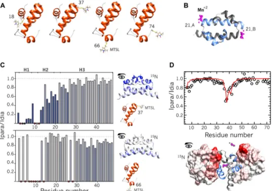
determined by classical NOE-based methods because of exten-sive broadening of key NMR signals upon complex formation. Therefore, we turned to PRE NMR experiments as a source of structural restraints (46). This approach allows the observation of residues distant from the paramagnetic tag, even at low molar fractions of complex where intrinsic broadening associ-ated to complex formation is minimized. Paramagnetic tags were introduced by reaction MTSL in four single cysteine vari-ants of Hha: wild type Hha (C18) and three single cysteine mutants at positions 37, 66, and a non-native C-terminal posi-tion 74. In these mutants cysteine 18 was mutated to isoleucine. The C18I mutation had been previously shown not to affect H-NS binding (24). Paramagnetic effects were measured on
15N-labeled H-NS
46C21S. The native cysteine in H-NS46
(Cys-21) was mutated to serine to avoid MTSL exchange. The labeled positions (Fig. 2A) are located outside of the H-NS binding interface inferred by perturbations mapping (Fig. 3). The addi-tion of Hha spin-labeled at posiaddi-tions 37 and 66 to15N-labeled
at Biblioteca de la Universitat de Barcelona on August 31, 2015
http://www.jbc.org/
H-NS46 C21S resulted in selective intermolecular PRE
dis-tance-dependent broadening of H-NS46 1H,15N resonances
(Fig. 2B), whereas no effect was observed when MTSL was attached at positions 18 or 74 of Hha. PRE data, binding-in-duced broadening, and mutagenesis information were con-verted into structural restraints and used in a flexible Haddock docking protocol starting with the known structures of unbound Hha (PDB code 1JW2) (17) and H-NS46antiparallel
dimer (PDB code 1NI8) (47). The solution structures were val-idated with an additional set of independent PREs. The struc-tural models correctly predicted the intermolecular PRE obtained by incorporating a paramagnetic tag, EDTA-Mn2#, in
each Cys-21 residue of H-NS46dimer (Fig. 2C) and measuring
the paramagnetic effects on15N-labeled Hha (Fig. 2D).
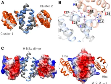
Structural statistics are given in Table 2. An ensemble of structures derived from PRE-driven modeling has been depos-ited in the PDB (PDB code 2mw2, RCSB number RSCB104115, Biological Magnetic Resonance Bank accession code 25296). The most relevant feature shared by all the models in the ensemble is the presence of an interdigitated array of residues with alternating charges, forming an electrostatic zipper (Fig. 4). The involved residues belong to three molecules, Hha and the two molecules (A and B) that constitute the H-NS46dimer:
Glu-25 (Hha)-Arg-12 (H-NS46-A)-Asp-48 (Hha)-Lys-32
(H-NS46-B). Fig. 3B shows a close-up view of the electrostatic
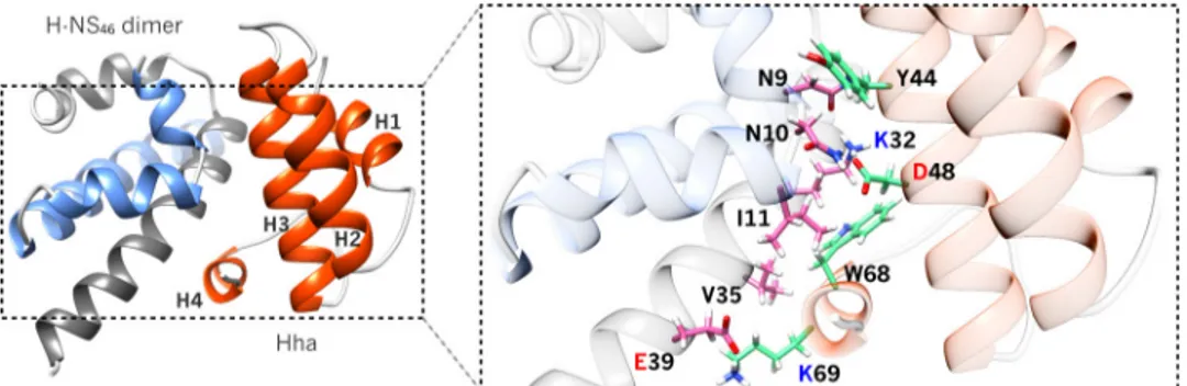
zip-per in the Hha!H-NS model structure. Although the electro-static interactions in the charge zipper probably dominate, additional contacts that also contribute to the complex stability are shown in Fig. 5.
The charge zipper model explains the known effect of muta-tions in Hha (D48E and E25Q) and H-NS (R12K) (19, 20). It also predicted that Lys-32 of H-NS should be essential for the inter-action. Indeed, this prediction was experimentally confirmed: mutation to glutamine of Lys-32 in H-NS leads to the complete loss of Hha binding (Fig. 6). The K32Q mutant and the wild type form have nearly identical circular dichroism spectra, confirm-ing that the mutation does not affect the foldconfirm-ing of H-NS (data not shown). Similar effects were observed in YmoA and YdgT (20), suggesting that H-NS complexes formed by other mem-bers of the Hha/YmoA family involve a similar charge zipper.
We confirmed the participation of aspartic and glutamic acid side chains of YmoA in its complex with H-NS64, a truncated
H-NS construct comprising residues 1–64, using a HCACO experiment modified to detect the correlation between side-chain carbonyl groups and their adjacent methylene groups (Fig. 6C). The experiments were done with13C-labeled YmoA
because, in comparison with Hha, it has one more acidic group predicted to participate in the charge zipper (Asp-43, Glu-36, Glu-20 in YmoA; Asp-48, Ala-41, Glu-25 in Hha).
FIGURE 1. Electrostatics conservation in Hha/YmoA family. A, sequence and secondary structure of Hha/YmoA. Open boxes denote ,-helical regions. B, clustering of 13 Hha-like proteins according to the similarity of their electrostatic potential: EC-Hha (E. coli Hha), ST-Hha (Salmonella enterica serovar Typhimu-rium Hha), Ent-Hha (Enterobacter cloacae Hha), YmoA (Yersinia spp. YmoA), PL-Hha (Photorhabdus luminescens Hha), EW-Hha (Erwinia carotovora Hha), WG-Hha (W. glossinidia Hha), SG-Hha (Sodalis glossinidius Hha), Rmoa (plasmid R100 RmoA), p0157 (plasmid p0157 Hha), pR27 (plasmid R27 Hha), EC-YdgT (E. coli YdgT), and ST-YdgT (S. enterica serovar Typhimurium YdgT). Negative (red) and positive (blue) isopotential contours of each protein are represented. The percentage of sequence identity with respect to EC-Hha is shown below. C, heat-map showing the pairwise electrostatic similarities. The scale is relative from low (light yellow) to high (red) ES. D, the local conservation of sequence (top) and electrostatic potential (bottom) of the ensemble of 13 proteins with respect to E. coli Hha is mapped on the surface of Hha.
at Biblioteca de la Universitat de Barcelona on August 31, 2015
http://www.jbc.org/
As predicted by the charge zipper model, the addition of H-NS64results in large changes in Asp-43 and Glu-36. Glu-20 is
in a crowded region. Residue Glu-29, located in a loop and not predicted to interact directly or indirectly with H-NS, is not affected by the addition of H-NS64and acts as a negative
con-trol. Residue Asp-56 is also located in a loop, but it forms an intramolecular salt bridge that contributes to positioning of helix 4 that is in contact with H-NS in the complex. Therefore, the intramolecular salt bridge is indirectly perturbed by the addition of H-NS64and Asp-56 provides a positive control. FIGURE 2. Paramagnetic relaxation enhancement experiments. A, MTSL-tagging of singled-cysteine Hha variants. Nitroxide spin labels located at different points in Hha structure are rendered in sticks. B, EDTA-Mn2#tagging positions on H-NS
46dimer. Mn2#(paramagnetic centers) atoms are displayed as magenta spheres, representing the flexibility of the tag. C, intermolecular PRE restraints. The histograms show the experimental intensity ratios of each amide resonance of15N-H-NS
46C21S in the presence of substoichiometric amounts of Hha at natural isotopic abundance, with MTSL conjugated at D37C or S66C. Ipara is the intensity in the paramagnetic sample, and Idia is the intensity in the corresponding diamagnetic control. Signals that disappear in paramagnetic conditions are indicated by black dots. Red dots identify residues that could not be accurately measured due to broadening caused by complexation. White circles indicate missing signals. Stronger intermolecular PREs are displayed on H-NS46structure and highlighted on the PRE profiles in blue scale. PRE ratios were converted into intermolecular distance restraints. D, intermolecular paramagnetic effects (open circles) induced on the backbone amide resonance of15N-HhaC18I by EDTA-Mn2#attached to H-NS
46The solid red line represents the average PRE profile predicted from the best structures. The inset displays the PREs effects mapped on the surface of representative Hha structures of both clusters.
FIGURE 3. Hha!H-NS interaction mapping. A, Hha residues most affected by broadening in the presence of 0.5 eq of H-NS64are highlighted in yellow on ribbon and surface representations of Hha structure. C18 (in red) mutants do not affect H-NS binding. Asp-48 and Glu-25 mutants show null or reduced affinity toward H-NS. These residues are located in the same side of Hha structure, whereas C18 is located on the opposite side of H-NS binding site. B, H-NS46residues most affected by the addition of Hha are highlighted in yellow. The Hha binding region is located around the first two helices of H-NS and R12 is essential for Hha binding. N9 mutants also strongly reduce Hha binding.
at Biblioteca de la Universitat de Barcelona on August 31, 2015
http://www.jbc.org/
Our solution structural model of the Hha!H-NS complex can be compared with the recently published crystal structure of an equivalent complex (21). Despite the fact that the crystal struc-ture has missing electron density for atoms in 50 side chains, including most of the interface region, the x-ray and NMR models have very similar backbone structures. The closest structures have a root-mean-square difference (r.m.s.d.) of "1.6 Å, confirming the overall geometry of the complex. Mod-eling the missing side chains in the x-ray structure also leads to a charge zipper (results not shown).
Fig. 7 shows a comparison of the NMR and x-ray models. The x-ray structure was obtained with an excess of Hha to saturate the two binding sites in the H-NS dimer. Based on the measured binding constant, the population of the species with two Hha molecules bound to the H-NS dimer should be very low under our experimental NMR conditions.
The structure of the YmoA!H-NS64complex was predicted
by homology based on the Hha!H-NS structure (Fig. 6D). SAXS of YmoA in the presence of H-NS64was in good agreement with
predictions based on the structures of YmoA, H-NS64, and
models of the YmoA!H-NS64 complexes (one or two YmoA
molecules per H-NS dimers) weighted according to the popu-lations predicted by the experimental binding constant, assumed to be identical for the two sites in the H-NS dimer (Fig. 8). These results confirm the structure of the Hha!H-NS com-plex, the independent binding to the two sites, and the similar-ity between the complexes formed by Hha and YmoA.
The two bound Hha molecules in the crystal structure are not equivalent by symmetry. Although all the PRE-based models present the charge zipper, they also fall into two structural clus-ters. One of the x-ray determined sites is similar to one of the clusters, but it has a r.m.s.d. higher than 2.7 Å to the members of the second cluster. The second x-ray model shows intermediate r.m.s.d. values to the PRE-based models. The major difference between the various models and the structure of free Hha involves changes in the position of helix 4 that is displaced to allow H-NS binding and conformational changes in residues spatially close to Asp-48 (Tyr-44 and Trp-68) to allow key res-idues to interact with H-NS. The observed structural variabil-ity, the missing electronic density within the crystallographic Hha!H-NS interface, and previous NMR data (23) suggest the existence of dynamics in the Hha protein family.
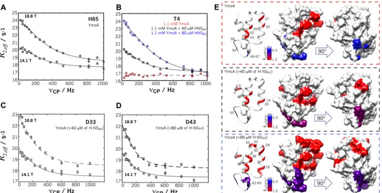
YmoA Shows Concerted Pervasive Microsecond-Millisecond Dynamics—Dynamics of free YmoA was studied using CPMG relaxation dispersion measurements for the backbone amide group and at two magnetic fields (14.1 and 18.8 T). Hha samples tend to aggregate at the concentrations required for these experiments, whereas the YmoA construct used by McFeeters et al. (18) is highly soluble and stable over long periods at 1.1 mMin 20 mMsodium phosphate, 200 mMNaCl, pH 7.5 and
285 K.
Fig. 9 shows representative dispersion curves. CPMG relax-ation dispersion curves were analyzed as an equilibrium between two sites characterized by resonance frequencies $A
and $Bwith populations pAand pB, respectively, exchanging at
a rate kex)k##k&(34). A preliminary analysis of the
relax-ation dispersion curves of individual residues showed dynamics in 12 residues with similar apparent kexvalues distributed along
the entire sequence: Leu-7 in helix1, Glu-24 and Lys-27 in helix 2, Asp-33, Tyr-39, Ala-42, and Asp-43 in helix 3, Tyr-55 and Ile-58 in a loop, and Val-62, Gln-64, and His-65 in the C-termi-nal helical region. The spatial distribution of the dynamic resi-dues and the similar kexvalues found for the exchanging
resi-dues suggest concerted dynamics affecting large regions or the entire protein. Therefore, the dispersion curves were analyzed collectively assuming that groups of residues move in a con-certed way (i.e. with the same kexand pB). A protocol for residue
incorporation, based on F-test analysis, was created. Two groups were identified, a first group including residues 55, 58, 64, and 65 that can be fitted collectively with a kex)267 * 85
s&1and p
B)6 * 2%. These residues are located in helix 4 and
the loop that connects it to helix 3, a region showing major structural differences among the different existing structures of the Hha/YmoA family.
TABLE 2 Structural statistics Cluster 1 Interface restraints PRE restraints 31 AIRSa 7 Structure statistics Violations AIR deviations 1.36 * 0.48 PRE deviations 1.51 * 0.54
Deviations from idealized geometry
Bond lengths (Å) 0.0030 * 0.0001 Bond angles (°) 0.65 * 0.05 Average pairwise r.m.s.d.b(Å) Backbone 0.85 * 0.13 All atoms 1.41 * 0.10 Ramachandran analysisb
Most favored regions 95.99 * 1.57%
Additional allowed regions 2.23 * 1.46%
Generously allowed regions 0.84 * 0.55%
Disallowed regions 0.98 * 0.36%
aSee Table 1.
bRamachandran analysis was performed using PROCHECK.
FIGURE 4. Hha!H-NS charge zipper. A, superposition of the 10 lowest energy PRE-derived solution complex structures. Hha (orange) and H-NS46dimer (light blue and gray) are shown in ribbon representations. All models satisfy the experimental data clustered into two equivalent solutions. B, close-up of the charge zipper interface. The solid and dashed-line boxes denote residues, whose mutation completely or nearly abolishes the binding, respectively. C, surface representations of the electrostatics potential of Hha and H-NS46 show that the complex is stabilized by charge complementary.
at Biblioteca de la Universitat de Barcelona on August 31, 2015
http://www.jbc.org/
A second group of dynamic residues (24, 27, 39, 42, and 43) is located in helices 2 and 3, constituting the structural core region of the Hha/YmoA family, well conserved in Hha, YmoA, and YdgT. These residues could be collectively fitted using kex)1200 * 130 s&1and pB)2 * 1%. Under the assumption
of the two-state model, the derived kinetic constants for the process are: k#)1177 * 65 s
&1and k
&)23 * 8 s
&1.
Residues that could not be incorporated in any of the two groups were further analyzed using a three-state model based on the simultaneous occurrence of the two previously defined processes. A good fit was obtained, but an F-test analysis
showed that the improvement with respect to an independent two-state model was not significant.
The existence of concerted dynamics raises the question of the functional coupling between YmoA dynamics and H-NS binding. To address this question, we measured CPMG relax-ation dispersion of YmoA in the presence of increasing amounts (40 and 80 !Mresulting in 3 and 6% bound YmoA,
respectively) of H-NS64. The low concentrations of H-NS64had
little effect in the observed spectra but significantly affected the dynamics observed in the CPMG experiment. In the presence of 40 (80) !MH-NS64the number of residues showing millisec-FIGURE 5. Additional hydrophobic contacts in Hha!H-NS46complex. Ribbon representation of the atomic model obtained by combining PRE, chemical shift
perturbations, and mutagenesis data showing Hha in orange and H-NS46dimer in gray/blue. Residues at the interface are shown in pink (H-NS46) or green (Hha) sticks. In addition to electrostatics complementary (Fig. 3), the complex is also stabilized by hydrophobic contacts, flanking the salt bridge formed by Asp-48 (Hha) and Lys-32 (H-NS46).
FIGURE 6. Corroborating experimental evidence for the charge zipper. A,1H,15N HSQC spectra of15N-labeled Hha in the presence of 0.5 eq of wild-type H-NS64(left panel) or 1 eq of H-NS64K32Q mutant (right panel). Residues showing !75% reduction in their intensities upon the addition of H-NS64are indicated. The spectra of YmoA free and in the presence of NS64K32Q are identical, indicating that the K32Q mutation completely prevents the interaction. B, modified HCACO spectra of13C-YmoA showing the correlation between side-chain carbonyls of Asp and Glu residues and the corresponding & and ' protons in the absence (blue) and in the presence of H-NS64. C, close-up view of the structure of the Hha!H-NS complex. The residue Lys-32 is located in the center of the charge zipper at the complex interface, forming a salt bridge with Asp-48 of Hha. D, close-up view of the structure of the YmoA!H-NS complex generated by homology modeling from the Hha!H-NS complex. YmoA side chains that are perturbed in the presence of H-NS64(panel B) are indicated.
at Biblioteca de la Universitat de Barcelona on August 31, 2015
http://www.jbc.org/
ond dynamics increased from 12 to 21 (38) residues of YmoA. Although H-NS64binding in the presence of intrinsic dynamics
in the apo form would in principle call for at least a three-state model, an F-test analysis showed that the improvement of the fit was not statistically significant with respect to the two-state model. The small concentration of complex present thus appears as a perturbation of the apoprotein dynamics, and data measured in the presence of H-NS were analyzed with a two-state model with concerted dynamics, as in the free YmoA but incorporating additional residues to the group in the core region that participate in the concerted dynamics. The similar-ity of the structures of the core regions of free and bound forms in the case of Hha support the idea that H-NS binding affects mostly the dynamics of YmoA.
Residues in helix 4 showed a complex response to the addi-tion of H-NS. The structural variability in the posiaddi-tion of helix 4 in free YmoA is probably aggravated by the formation of the complex with H-NS. The effect of H-NS64addition on the side
chain of a key residue in the loop that connects helices 3 and 4 supports the idea of more complex structural changes in this region, although a three-state model did not improve the fit with respect to the two-state one, and no further analysis was attempted.
The population of the minor species sensed by the core res-idues increased from 2% in the free form to 5–6% in the pres-ence of 6% H-NS-bound species (Table 3). Remarkably, these
populations agree, within experimental error, with the complex concentrations derived from the association constant. Finally, although the apparent kex value remained largely unaltered
within experimental error (1200 * 130 s&1(free), 1162 * 104
s&1(40 !MH-NS
64), 1240 * 116 s&1(80 !MH-NS64)), the
resulting kinetic constants showed a constant increase in k&,
whereas k# remained constant within experimental error
(Table 3). The simplest model explaining the experimental observations is that binding of H-NS is coupled with the intrin-sic dynamics of YmoA, which may also have an electrostatic origin.
Ionic Strength Effects and Mutagenesis Show That YmoA Dynamics and H-NS Binding Are Related—Because of the elec-trostatic character of the YmoA!H-NS interface, we character-ized the stability of the complex as a function of ionic strength (200 mM, 100 mM, and 50 mMNaCl). The dissociation constants
(KD) for the YmoA!H-NS46complex increases linearly with the
salt concentration (Fig. 9), suggesting that electrostatic effects are important for complex formation but through their effect on the off-rate, as an effect of the ionic strength in the on-rate should result in strong deviations from linearity (48).
The increase in stability of the complex at low ionic strength is consistent with the charge zipper motif identified in the NMR structure of the Hha!H-NS complex. The high charge density in free YmoA, when not compensated by the interaction with H-NS, is expected to create intramolecular electrostatic inter-actions, which may be the origin for the dynamics observed in YmoA. To test this hypothesis we compared CPMG relaxation dispersion experiments of free YmoA at 200 mM, 100 mM, and
50 mMNaCl (Fig. 10 and Table 3). When the salt concentration FIGURE 7. Comparison of x-ray and solution models of Hha!(H-NS46)2
complexes. A, superimposition of the two Hha molecules in the x-ray
struc-ture of Ali et al. (21) using the H-NS46molecules with which they interact as a reference. Hha molecules are shown in red and light blue. B, comparison of the location of Hha in representative structures of both clusters shown in blue and pink ribbon representations. C, r.m.s.d. of the 400 best Hha models derived from solution experiments to the two Hha models derived from x-ray diffrac-tion. The color code is the same. Using the x-ray model depicted in light blue as a reference, the solution models fall into two clusters with low (1.73 * 0.12 Å) and high (2.99 * 0.23 Å) r.m.s.d. In contrast, using the “red” x-ray model, the solution models fall in a single cluster with intermediate r.m.s.d. (1.9 –2.7 Å).
FIGURE 8. SAXS data confirm the homology models of YmoA!H-NS64
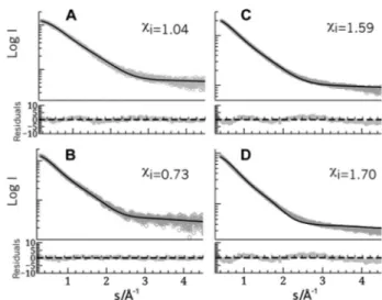
com-plexes. Comparison of experimental (circles) and calculated (continuous lines)
SAXS curves. A, pure YmoA. The theoretical curve was based on the NMR structure (18) allowing for flexibility in the connection between helices 3 and 4. B, pure H-NS64. The theoretical curve of the H-NS64dimer was extracted from the x-ray structure of an H-NS oligomer and adding the flexible His tag. C and D, YmoA and HNS64mixtures. The YmoA!H-NS64complexes were mod-eled using the Hha!H-NS structure presented in this study and allowing the same level of flexibility that the free partners. The molar fractions of the spe-cies, calculated on the basis of the binding constant and the actual concen-trations, were 0.73 (YmoA), 0.03 (H-NS64dimer), 0.12 H-NS64-YmoA (2:1), and 0.12 (H-NS64-YmoA (2:2) (C) and 0.46 (YmoA), 0.10 (H-NS64dimer), (0.26 (H-NS64-YmoA (2:1)), and 0.16 (H-NS64-YmoA (2:2)) (D). The quality of the agreement can be evaluated from the residuals shown below each curve and their individual figure of merit -i).
at Biblioteca de la Universitat de Barcelona on August 31, 2015
http://www.jbc.org/
was reduced from 200 mMto 50 mMNaCl, the number of core
residues showing collective motion increased from 5 to 16 res-idues. 11 of these residues belong to helices 2 and 3, and the short loop that connects them and 4 residues belong to helix 1. Thus, by reducing the ionic strength the collective motion was extended to embrace helix 1. The increased number of residues involved in the collective dynamics is expected to result in a reduction in the timescale of the motional process. Indeed, the exchange rate was progressively reduced nearly 2-fold and 8-fold when NaCl concentration was reduced to 100 and 50 mM, respectively.
Thus, electrostatic interactions are responsible for both the intrinsic dynamics of YmoA and the association to H-NS, sta-bilized through a charge zipper. This connection is strength-ened by the observation that mutation of residue Asp-43 to asparagine completely abolished dynamics in the microsecond-millisecond time scale even in the presence of 80 !MH-NS64.
Residue Asp-43 plays a crucial role in the charge zipper, and mutation of the equivalent residue in Hha (Asp-48) completely abolished binding to H-NS (data not shown).
Discussion
Hha-like proteins modify the capacity of H-NS to silence specific DNA regions (8). H-NS binds and silences DNA
FIGURE 9. CPMG dispersion profiles and mapping of affected residues. A, H65 in helix 4 of free YmoA at two magnetic fields. B, residue T4 dynamics becomes observable only in the presence of H-NS64. C and D, residues 33 and 43 of the core region in the presence of 40 !Mand 80 !Mof H-NS64, respectively. The solid lines represent the simultaneously curve fitting to the data from the two magnetic fields. B, residues showing exchange are colored in the YmoA structure according to the exchange rates. The three boxes show the results in the absence (red dashed box) or presence of 40 !M(black dashed-box), 80 !M(blue dashed-box) of H-NS64.
TABLE 3
CPMG kinetic parameters
YmoA kex pB k! k" +NaCl,
s&1 % s&1 s&1 mM
Apo 1200 * 130 2 * 1 1177 * 65 23 * 8 200 Apo 500 * 71 5 * 3 493 * 20 3 * 2 100 Apo 152 * 37 5 * 2 142 * 16 7 * 5 50 #40 !MH-NS 1162 * 104 5 * 3 1104 * 72 58 * 15 200 #80 !MH-NS 1240 * 116 6 * 2 1162 * 69 78 * 10 200 D43N 0 0 200
FIGURE 10. Ionic strength dependence of YmoA dynamics H-NS binding. A, YmoA residues showing ms-!s exchange at different ionic strength. B, fluorescence anisotropy titrations of YmoA with H-NS64 at various ionic strength values. The inset shows the linear dependence of KDwith ionic
strength.
at Biblioteca de la Universitat de Barcelona on August 31, 2015
http://www.jbc.org/
through the simultaneous interaction of the DNA binding domains from individual H-NS molecules assembled into linear oligomers. DNA selectivity, which in turn determines which genes are ultimately silenced, is achieved by indirect readout; DNA binding domains of H-NS sense local DNA distortions, and the formation of H-NS oligomers restricts the possible rel-ative locations of adjacent DNA binding domains (49, 50). Hha binds to H-NS dimers formed by the interaction of N-terminal dimerization domain of two H-NS molecules. H-NS oligomer-ization involves a second dimeroligomer-ization site (residues 57–83) (44). A functional result of this interaction is that the Hha!H-NS complexes preferentially repress horizontally acquired genes.
The mechanism by which Hha binding induces changes in selectivity with respect to the genes that are repressed is not completely clear. Hha-like proteins and the NTD of H-NS con-tain an unusually large proportion of charged residues and a marked electrostatic asymmetry. The positive region of H-NS NTD has been suggested to interact with DNA according to the model of the DNA-H-NS complexes presented by Arold et al. (44) on the basis of the x-ray structure of the H-NS oligomers. According to this model, Hha-like molecules bind and mask the positively charged region of H-NS NTD. However, because of the electrostatic asymmetry of Hha itself, a new positively charged patch (from Hha) is displayed (19, 20), although at a different position with respect to the H-NS oligomer axis. The presence of Hha-like proteins is believed to change the “match-ing condition” between DNA bind“match-ing and H-NS oligomeriza-tion and, therefore, the DNA silencing specificity (20, 21, 50). Mutagenesis in the positively charged region of Hha clearly demonstrates that this region is critical for transcriptional repression but not for H-NS binding (21, 24).
Binding of Hha to a strongly charged region of H-NS imposes the need for an electrostatic complex. However, electrostatic complexes are usually associated with a poor selectivity. In con-trast, the tight network of multiple complementary interactions from three different proteins in the Hha complex with a H-NS dimer, conforming the observed charge zipper, ensures an exquisite selectivity (19, 20). A charge zipper combining intra-and intermolecular interactions stabilizing the translocation pore of TatA protein has been recently described (51).
Although electrostatic interactions play a clear role in inter-molecular interactions (with H-NS or DNA), intrainter-molecular electrostatic interactions may induce forces distorting the low energy packing of the helical core. The pervasive dynamics observed in the free form of YmoA may originate from the balance between electrostatically driven distortions and the hydrophobic interactions restoring forces. The fact that dynamics has been preserved by evolution suggests it may result in functional advantages.
Our results do not provide direct information on the possible conservation of the Hha/YmoA dynamics in the complexes, although the missing residues and structural variations between the two sites in the x-ray structure suggest that this may be the case. A direct functional correlation between dynamics and complex formation is supported by the changes observed in the number of residues involved in the microsec-ond-millisecond dynamics and the population of the minor conformation. This correlation is also supported by the effect of
the D43N mutation of YmoA that completely abolished H-NS binding as well as !s-!s dynamics of the free form.
We can only speculate about how Hha/YmoA dynamics may influence function. We suggest at least three possible effects. (a) Due to its positively charged surface, a direct interaction between Hha and DNA was suggested (21). The existence of pervasive motions in the free molecule would increase the entropic penalty upon binding and may decrease spurious binding of Hha to DNA. (b) Hha binding to the NTD domain and the intrinsic dynamics of Hha-like proteins may change the flexibility of H-NS oligomers (52), thus modifying the selectiv-ity. The fact that horizontally acquired DNA regions silenced by H-NS tend to be longer than those in the core genome (53) could make the flexibility of H-NS oligomers an important control element (50). (c) YmoA/Hha dynamics may facilitate binding to and/or dissociation of charge zipper complexes. Considering that electrostatic interactions are long range, the formation of the charge zipper may be compromised by local minima and dynamics may facilitate the optimization of the complex. Similarly, complete dissociation of an electrostatic complex may require large displacements of the charged ele-ments before the interaction is effectively lost, and dynamics, if retained in the complex, may facilitate its dissociation. The sim-ilarity in the exchange rates observed in the presence and in the absence of H-NS would be compatible with a dynamic coupling between the intrinsic motions of Hha-like molecules and the formation or dissociation of the complex with H-NS.
Author Contributions—T. N. C. performed experiments leading to the determination of the structure of the complex and did the elec-trostatic analysis of the Hha family. J. G. generated and measured mutants to test the models and prepared complexes. P. B. contrib-uted to SAXS measurement and analysis and to the general discus-sion. O. M. measured and analyzed CPMG relaxation dispersion experiments and contributed to the design and interpretation of experiments. M. P. conceived and coordinated the study and wrote the paper with input from all the other authors.
Acknowledgments—We thank O. Marimon, I. Latorre, and X. Roa for help in sample preparation, Prof. A. Juarez and the members of the consolidated group 2014SGR1260 (Generalitat de Catalunya) for useful discussions, and Dr. D. S. Waugh (NCI-Frederick) for the plas-mid encoding for YmoA.
References
1. WHO (2014) Antimicrobial Resistance. Global Report on Surveillance, World Health Organization Press
2. Ochman, H., Lawrence, J. G., and Groisman, E. A. (2000) Lateral gene transfer and the nature of bacterial innovation. Nature 405, 299–304 3. Davies, J. (1994) Inactivation of antibiotics and the dissemination of
resis-tance genes. Science 264, 375–382
4. Davies, J., and Davies, D. (2010) Origins and evolution of antibiotic resis-tance. Microbiol. Mol. Biol. Rev. 74, 417–433
5. Nieto, J. M., Carmona, M., Bolland, S., Jubete, Y., de la Cruz, F., and Jua´rez, A. (1991) The hha gene modulates haemolysin expression in Escherichia coli. Mol. Microbiol. 5, 1285-1293
6. Cornelis, G. R., Sluiters, C., Delor, I., Geib, D., Kaniga, K., Lambert de Rouvroit, C., Sory, M. P., Vanooteghem, J. C., and Michiels T. (1991) ymoA, a Yersinia enterocolitica chromosomal gene modulating the ex-pression of virulence functions. Mol. Microbiol. 5, 1023–1034
at Biblioteca de la Universitat de Barcelona on August 31, 2015
http://www.jbc.org/
7. Nieto, J. M., Madrid, C., Miquelay, E., Parra, J. L., Rodríguez, S., and Jua´rez, A. (2002) Evidence for direct protein-protein interaction between mem-bers of the enterobacterial Hha/YmoA and H-NS families of proteins. J. Bacteriol. 184, 629–635
8. Ban˜os, R. C., Vivero, A., Aznar, S., García, J., Pons, M., Madrid, C., and Jua´rez A. (2009) Differential regulation of horizontally acquired and core genome genes by the bacterial modulator H-NS. PLoS Genet. 5, e1000513 9. Aznar, S., Paytubi, S., and Jua´rez, A. (2013) The Hha protein facilitates incorporation of horizontally acquired DNA in enteric bacteria. Microbi-ology 159, 545–554
10. Madrid, C., García, J., Pons, M., and Jua´rez, A. (2007) Molecular evolution of the H-NS protein: interaction with Hha-like proteins is restricted to Enterobacteriaceae. J. Bacteriol. 189, 265–268
11. Takeda, T., Yun, C.-S., Shintani, M., Yamane, H., and Nojiri, H. (2011) Distribution of genes encoding nucleoid-associated protein homologs in plasmids. Int. J. Evol. Biol. 2011, 685015
12. García-Contreras, R., Zhang, X. S., Kim, Y., and Wood, T. K. (2008) Pro-tein translation and cell death: the role of rare tRNAs in biofilm formation and in activating dormant phage killer genes. PLoS ONE 3, e2394 13. Hong, S. H., Lee, J., and Wood, T. K. (2010) Engineering global regulator
Hha of Escherichia coli to control biofilm dispersal. Microb. Biotechnol. 3, 717–728
14. Sharma, V. K., and Bearson, B. L. (2013) Hha Controls Escherichia coli O157:H7 biofilm formation by differential regulation of global tran-scriptional regulators FlhDC and CsgD. Appl. Environ. Microbiol. 79, 2384 –2396
15. Mikulskis, A. V., and Cornelis, G. R. (1994) A new class of proteins regu-lating gene expression in enterobacteria. Mol. Microbiol. 11, 77–86 16. Balsalobre, C., Jua´rez, A., Madrid, C., Mourin˜o, M., Prenafeta, A., and
Mun˜oa, F. J. (1996) Complementation of the hha mutation in Escherichia coli by the ymoA gene from Yersinia enterocolitica: dependence on the gene dosage. Microbiology 142, 1841–1846
17. Yee, A., Chang, X., Pineda-Lucena, A., Wu, B., Semesi, A., Le, B., Ramelot, T., Lee, G. M., Bhattacharyya, S., Gutierrez, P., Denisov, A., Lee, C. H., Cort, J. R., Kozlov, G., Liao, J., Finak, G., Chen, L., Wishart, D., Lee, W., McIntosh, L. P., Gehring, K., Kennedy, M. A., Edwards, A. M., and Arrow-smith, C. H. (2002) An NMR approach to structural proteomics. Proc. Natl. Acad. Sci. U.S.A. 99, 1825–1830
18. McFeeters, R. L., Altieri, A. S., Cherry, S., Tropea, J. E., Waugh, D. S., and Byrd R. A. (2007) The high-precision solution structure of Yersinia mod-ulating protein YmoA provides insight into interaction with H-NS. Bio-chemistry 46, 13975–13982
19. García, J., Madrid, C., Jua´rez, A., and Pons, M. (2006) New roles for key residues in helices H1 and H2 of the Escherichia coli H-NS N-terminal domain: H-NS dimer stabilization and Hha binding. J. Mol. Biol. 359, 679–689
20. de Alba, C. F., Solo´rzano, C., Paytubi, S., Madrid, C., Juarez, A., García, J., and Pons, M. (2011) Essential residues in the H-NS binding site of Hha, a co-regulator of horizontally acquired genes in Enterobacteria. FEBS Lett.
585, 1765–1770
21. Ali, S. S., Whitney, J. C., Stevenson, J., Robinson, H., Howell, P. L., and Navarre, W. W. (2013) Structural Insights into the Regulation of Foreign Genes in Salmonella by the Hha/H-NS Complex. J. Biol. Chem. 288, 13356–13369
22. Paytubi, S., García, J., and Jua´rez, A. (2011) Bacterial Hha-like proteins facilitate incorporation of horizontally transferred DNA. Open Life Sci-ences 6, 879–886
23. García, J., Cordeiro, T. N., Nieto, J. M., Pons, I., Jua´rez, A., and Pons, M. (2005) Interaction between the bacterial nucleoid associated proteins Hha and H-NS involves a conformational change of Hha. Biochem. J. 388, 755–762
24. Cordeiro, T. N., Garcia, J., Pons, J. I., Aznar, S., Jua´rez, A., and Pons, M. (2008) A single residue mutation in Hha preserving structure and binding to H-NS results in loss of H-NS mediated gene repression properties. FEBS Lett. 582, 3139–3144
25. Battiste, J. L., and Wagner, G. (2000) Utilization of site-directed spin la-beling and high-resolution heteronuclear nuclear magnetic resonance for global fold determination of large proteins with limited nuclear
Over-hauser effect data. Biochemistry 39, 5355–5365
26. Dominguez, C., Boelens, R., and Bonvin, A. M. (2003) HADDOCK: a pro-tein-protein docking aproach based on biochemical or biophysical infor-mation. J. Am. Chem. Soc. 125, 1731–1737
27. Emekli, U., Schneidman-Duhovny, D., Wolfson, H. J., Nussinov, R., and Haliloglu, T. (2008) HingeProt: automated prediction of hinges in protein structures. Proteins 70, 1219–1227
28. de Vries, S. J., van Dijk, A. D., Krzeminski, M., van Dijk, M., Thureau, A., Hsu, V., Wassenaar, T., and Bonvin, A. M. (2007) HADDOCK versus HADDOCK: new features and performance of HADDOCK2.0 on the CAPRI targets. Proteins 69, 726–733
29. Iwahara, J., Schwieters, C. D., and Clore, G. M. (2004) Ensemble approach for NMR structure refinement against1H paramagnetic relaxation en-hancement data arising from a flexible paramagnetic group attached to a macromolecule. J. Am. Chem. Soc. 126, 5879–5896
30. García de la Torre, J., Huertas, M. L., and Carrasco, B. (2000) HYDRONMR: prediction of NMR relaxation of globular proteins from atomic-level structures and hydrodynamic calculations. J. Magn. Reson.
147, 138–146
31. Tollinger, M., Skrynnikov, N. R., Mulder, F. A., Forman-Kay, J. D., and Kay, L. E. (2001) Slow dynamics in folded and unfolded states of an SH3 do-main. J. Am. Chem. Soc. 123, 11341–11352
32. Loria, J. P., Rance, M., and Palmer, A. G. (1999) A TROSY CPMG se-quence for characterizing chemical exchange in large proteins. J. Biomol. NMR 15, 151–155
33. Mulder, F. A., Skrynnikov, N. R., Hon, B., Dahlquist, F. W., and Kay, L. E. (2001) Measurement of slow (micros-!s) time scale dynamics in protein side chains by15N relaxation dispersion NMR spectroscopy: application to Asn and Gln residues in a cavity mutant of T4 lysozyme. J. Am. Chem. Soc. 123, 967–975
34. Carver, J. P., and Richards, R. E. (1972) A general two-site solution for the chemical exchange produced dependence of T2upon the Carr-Purcell pulse separation. J. Magn. Reson. 6, 89–105
35. Gabdoulline, R. R., Stein, M., and Wade, R. C. (2007) qPIPSA: relaing enzymatic kinetic parameters and interaction fields. BMC Bioinformatics
8, 373
36. Richter, S., Wenzel, A., Stein, M., Gabdoulline, R. R., and Wade, R. C. (2008) webPIPSA: a web server for the comparison of protein interaction properties. Nucleic Acids Res. 36, W276–W280
37. Biasini, M., Bienert, S., Waterhouse, A., Arnold, K., Studer, G., Schmidt, T., Kiefer, F., Cassarino, T. G., Bertoni, M., Bordoli, L., and Schwede, T. (2014) SWISS-MODEL: modeling protein tertiary and quaternary struc-ture using evolutionary information. Nucleic Acids Res. 42, W252–W258 38. UniProt Consortium (2014) Activities at the Universal Protein Resource.
Nucleic Acids Res. 42, D191–D198
39. Rocchia, W., Sridharan, S., Nicholls, A., Alexov, E., Chiabrera, A., and Honig, B. (2002) Rapid grid-based construction of the molecular surface and the use of induced surface charge to calculate reaction field energies: applications to the molecular systems and geometric objects. J. Comput. Chem. 23, 128–137
40. Kieslich, C. A., and Morikis, D. (2012) The two sides of complement C3d: evolution of electrostatics in a link between innate and adaptive immunity. PLoS Comput. Biol. 8, e1002840
41. Pettersen, E. F., Goddard, T. D., Huang, C. C., Couch, G. S., Greenblatt, D. M., Meng, E. C., and Ferrin, T. E. (2004) USCF Chimera: a visualization system for exploratory research and analysis. J. Comput. Chem. 25, 1605–1612
42. Konarev, P. V., Volkov, V. V., Sokolova, A. V., Koch, M. H. J., and Svergun, D. I. (2003) PRIMUS: a Windows PC-based system for small-angle scat-tering data analysis. J. Appl. Cryst. 36, 1277–1282
43. Svergun, D. I., Barberato, C., and Koch, M. H. J. (1995) CRYSOL: a pro-gram to evaluate x-ray solution scattering of biological macromolecules from atomic coordinates. J. Appl. Cryst. 28, 768–773
44. Arold, S. T., Leonard, P. G., Parkinson, G. N., and Ladbury, J. E. (2010) H-NS forms a superhelical protein scaffold for DNA condensation. Proc. Natl. Acad. Sci. U.S.A. 107, 15728–15732
45. García, J., Madrid, C., Cendra, M., Jua´rez, A., and Pons, M. (2009) N9L and L9N mutations toggle Hha binding and hemolysin regulation by
at Biblioteca de la Universitat de Barcelona on August 31, 2015
http://www.jbc.org/
richia coli and Vibrio cholerae H-NS. FEBS Lett. 583, 2911–2916 46. Clore, G. M., and Iwahara, J. (2009) Theory, practice, and applications of
paramagnetic relaxation enhancement for the characterization of tran-sient low-population states of biological macromolecules and their com-plexes. Chem. Rev. 109, 4108–4139
47. Bloch, V., Yang, Y., Margeat, E., Chavanieu, A., Auge´, M. T., Robert, B., Arold, S., Rimsky, S., and Kochoyan, M. (2003) The H-NS dimerization domain defines a new fold contributing to DNA recognition. Nat. Struct. Biol. 10, 212–218
48. Zhou, H. X. (2001) Disparate ionic-strength dependencies of on and off rates in protein-protein association. Biopolymers 59, 427–433
49. Cordeiro, T. N., Schmidt, H., Madrid, C., Jua´rez, A., Bernado´, P., Griesinger, C., García, J., and Pons, M. (2011) Indirect DNA readout by an H-NS related protein: structure of the DNA complex of the C-terminal domain of Ler. PLoS Pathog. 7, e1002380
50. Ferna´ndez-de-Alba, C., Berrow, N. S., Garcia-Castellanos, R., García, J.,
and Pons, M. (2013) On the origin of the selectivity of plasmidic H-NS towards horizontally acquired DNA: linking H-NS oligomerization and cooperative DNA binding. J. Mol. Biol. 425, 2347–2358
51. Walther, T. H., Gottselig, C., Grage, S. L., Wolf, M., Vargiu, A. V., Klein, M. J., Vollmer, S., Prock, S., Hartmann, M., Afonin, S., Stockwald, E., Heinzmann, H., Nolandt, O. V., Wenzel, W., Ruggerone, P., and Ulrich, A. S. (2013) Folding and self-assembly of the TatA translocation pore based on a charge zipper mechanism. Cell 152, 316–326
52. Renault, M., García, J., Cordeiro, T. N., Baldus, M., and Pons, M. (2013) Protein oligomers studied by solid-state NMR: the case of the full-length nucleoid-associated protein histone-like nucleoid structuring protein. FEBS J. 280, 2916–2928
53. Kahramanoglou, C., Seshasayee, A. S., Prieto, A. I., Ibberson, D., Schmidt, S., Zimmermann, J., Benes, V., Fraser, G. M., and Luscombe, N. M. (2011) Direct and indirect effects of H-NS and Fis on global gene expression control in Escherichia coli. Nucleic Acids Res. 39, 2073–2091
at Biblioteca de la Universitat de Barcelona on August 31, 2015
http://www.jbc.org/
Bernadó, Oscar Millet and Miquel Pons
Tiago N. Cordeiro, Jesús García, Pau
Silencing
Complex Modulating Bacterial Gene
doi: 10.1074/jbc.M114.630400 originally published online June 17, 20152015, 290:21200-21212. J. Biol. Chem.
10.1074/jbc.M114.630400
Access the most updated version of this article at doi:
JBC Affinity Sites.
Find articles, minireviews, Reflections and Classics on similar topics on the Alerts:
When a correction for this article is posted
• When this article is cited •
to choose from all of JBC's e-mail alerts
Click here
http://www.jbc.org/content/290/35/21200.full.html#ref-list-1
This article cites 52 references, 15 of which can be accessed free at
at Biblioteca de la Universitat de Barcelona on August 31, 2015
http://www.jbc.org/