UNIVERSITÀ POLITECNICA DELLE MARCHE
Scuola di Dottorato in Medicina e Chirurgia
Curriculum: “Medicina sperimentale”
XIV Cycle (New Series)
Doctoral Thesis
R
AFK
INASEI
NHIBITORP
ROTEIN(RKIP)
EXPRESSION AND FUNCTION IN UTERINE LEIOMYOMACoordinator : Prof. Saverio Cinti, MD
Tutor: Prof. Mario Castellucci, MD, PhD
Direct Supervisor: Pasquapina Ciarmela, PhD
Ph.D. candidate: Dr. Milijana Janjusevic
To my beloved grandpa Djuro, grandma Saja, parents and Jelena
Contents
Summary (English)...5. Summary (Italian)...6. List of publications...7. 1. General introduction...8. 1.1. Leiomyoma ...8.1.2. Definition and location ...8.
1.3. Etiology ...10.
1.4. Epidemiology ...12.
1.5. Signs and symptoms...12.
1.6. Treatments...13.
2. The Raf kinase inhibitor protein (RKIP)...15.
2.1. Protein structure of RKIP...15.
2.2. RKIP : An inhibitory modulator protein...16.
2.3. Prevalence of RKIP...17.
3. Materials and Methodes...19.
3.1. Sample collection...19.
3.2. Immunohistochemistry...19.
3.3. Western blotting...20.
3.4. Myometrial and leiomyoma cell culture...20.
3.5. RNA extraction and real-time PCR...21.
3.6. Immunocytochemistry...21.
3.7. Cell proliferation assay...22.
3.9. Data analysis...23.
4. Results...24.
4.1. Localisation of RKIP in leiomyoma and myometrim...24.
4.2. Expression of RKIP in leiomyoma and myometrim...25.
4.3. RKIP inhibition by locostatin induces ECM expresion...27
4.4. RKIP inhibition by locostatin reduses cell proliferation...30.
4.5. RKIP inhibition by locostatin reduces cell migration...32.
4.6. RKIP inhibition by locostatin reduses GSK3β...34.
5. Discussion...35.
6. References...38.
Summary (English)
Uterine leiomyomas (fibroids, myomas) are benign (non-cancerous) tumors that origin from the smooth muscle layer of the uterus (myometrium), and are the most common indication for hysterectomy in the world. Uterine leiomyomas affect about 77% of women of reproductive-age, and approximately 25% of them bear clinically apparent tumors with symptoms like heavy or abnormal uterine bleeding, pelvic pain or pressure, infertility, and recurrent pregnancy loss. It is commonly known that these tumors are characterized by increased cell proliferation and excessive deposition of extracellular matrix (ECM). Growth of leiomyoma is thought to be dependent on ovarian hormones activity through intermediate elements such as cytokines and growth factors.
Raf Kinase Protein Inhibitor (RKIP) has emerging roles as regulator of multiple signaling networks and is associated with an increasing number of diseases through its involvement with signal transduction pathways.
The aim of the present thesis was to investigate the presence and the role of RKIP in leiomyoma.
We demonstrated that RKIP is expressed in human myometrial and leiomyoma tissue. In order to define the RKIP role, we performed in vitro experiments with the chemical compound locostatin, known to bind and block RKIP. We showed that locostatin treatment results in the activation of the MAPK signal pathway (ERK phosphorylation), providing a powerful validation of our targeting protocol. Further, we showed that RKIP inhibition by locostatin reduces ECM components, including collagen1A1, fibronectin, and versican. Moreover, the inhibition of RKIP by locostatin impairs cell proliferation and migration in both leiomyoma and myometrial cells. Finally, we demonstrated that locostatin treatment reduced GSK3β expression. Therefore, even if the activation of MAPK pathway should increase proliferation and migration, the destabilization and inactivation of GSK3β leads to the reduction of proliferation and migration of myometrial and leiomyoma cells.
Summary (Italian)
I leiomiomi uterini (detti anche fibromi, miomi) sono tumori benigni che originano dallo strato muscolare dell’utero (miometrio) e rappresentano la principale indicazione dell’isterectomia nel mondo. I leiomiomi uterini colpiscono circa il 77% delle donne in eta fertile e circa il 25% di esse presenta tumori con sintomatologia clinica evidente, tra cui la presenza di forte o anomalo sanguinamento uterino, dolore o pressione pelvica, infertilità e aborti ricorrenti.
È comunemente noto che questi tumori sono caratterizzati da una elevata proliferazione cellulare ed una eccessiva deposizione di matrice extracellulare (ECM). Si ritiene che la crescita dei leiomiomi dipenda dall’azione degli ormoni ovarici mediante elementi intermedi come citochine e fattori di crescita.
La Proteina Inibitore della Raf Chinasi (RKIP) ha un ruolo emergente come regolatore in diversi pathway molecolari ed è associato a un numero crescente di malattie, essendo coinvolto indiverse vie di trasduzione del segnale.
Lo scopo della presente tesi è stato quello di indagare la presenza e il ruolo dell’RKIP nel leiomioma.
Abbiamo dimostrato che l’RKIP è espresso nel miometrio e nel leiomioma. Per individuare il ruolo dell’RKIP, abbiamo eseguito esperimenti in vitro con un composto chimico quale la locostatina, capace di legarsi all’RKIP bloccandolo. Abbiamo dimostrato che il trattamento con la locostatina porta all’attivazione della via di segnale MAPK (fosforilazione di ERK), fornendo una opportuna validazione dell’efficacia nel bloccare l’RKIP. Inoltre, abbiamo dimostrato che l'inibizione dell’RKIP con la locostatina riduce le componenti della ECM, tra cui il collagene 1A1, la fibronectina, e il versican. In aggiunta, l'inibizione dell’RKIP con la locostatina riduce la proliferazione cellulare e la migrazione sia nelle cellule miometriali che di leiomioma. Infine, abbiamo dimostrato che il trattamento con la locostatina riduce l’espressione del GSK3β. Pertanto, anche se l'attivazione delle MAPK dovrebbe far aumentare la proliferazione e la migrazione, la destabilizzazione e l’inattivazione del GSK3β porta alla riduzione della proliferazione e della migrazione delle cellule miometriali e di leiomioma.
List of publications
1. Ciarmela P, Carrarelli P, Islam MS, Janjusevic M, Zupi E, Castellucci M, Petraglia
F. 2014. Ulipristal acetate modulates activin A expression and functions in leiomyoma cells. Reproductive Sciences (ISSN: 1933-7191). 21(9):1120-1125 IF: 2.230
2. Islam MS, Akhtar MM, Ciavattini A, Giannubilo SR, Protic O, Janjusevic M,
Procopio AD, Segars JH, Castellucci M and Ciarmela P. 2014. Use of dietary phytochemicals to target inflammation, fibrosis, proliferation and angiogenesis in uterine tissues: promising options for prevention and treatment of uterine fibroids?
Molecular Nutrition & Food Research (ISSN: 1613-4125). 58(8):1667-1684. IF:
4.603
3. Islam MS, Catherino WH, Protic O, Janjusevic M, Gray PC, Giannubilo SR,
Ciavattini A, Lamanna P, Tranquilli AL, Petraglia F, Castellucci M and Ciarmela P.
2014. Role of activin-A and myostatin and their signaling pathway in human
myometrial and leiomyoma cell function. Journal of Clinical Endocrinology &
Metabolism (ISSN: 0021-972X). 99(5):E775–E785. IF: 6.209
4. Islam MS, Greco S, Janjusevic M, Ciavattini A, Giannubilo SR, D'Adderio A,
Biagini A, Fiorini R, Castellucci M and Ciarmela P. 2015. Growth factors and pathogenesis.Best Practice & Research Clinical Obstetrics & Gynaecology.
1.General introduction
1.1. Leiomyoma
Uterine leiomyoma, or fibroid, is the most common benign, monoclonal, gynecological tumor in women originating from the smooth muscle layer of the uterus (myometrium) and represents the single most common indication for hysterectomy [1, 2]. They are composed of large amounts of extracellular matrix (ECM) containing collagen, fibronectin and proteoglycan. Uterine leiomyomas affect about 77% of women of reproductive-age, and 25% of them women bear clinically apparent tumors and they cause significant morbidity [3, 4], including heavy or abnormal uterine bleeding, pelvic pain or pressure, infertility, and reproductive dysfunction in rare cases. Hysterectomy is associated with morbidity and mortality and also associated with a substantial economic impact on healthcare systems that amounts to approximately $2.2 billion/year in the US alone [5]. Therefore, both the economic cost and the effect on quality of life are substantial.
1.2. Definition and location
Leiomyoma is usually spherical masses of tissue that can vary from a few millimeters to many centimeters in diameter (Figure 1).
Figure 1. Leiomyoma.
The blood supply enters at the periphery of the tumor and the core is relatively avascular, which is the probable reason for the various forms of degeneration and necrosis that are seen in the center of some leiomyomas. There are three primary types of uterine fibroids, classified primarily according to location in the uterus [6-8] (Figure 2).
*Subserosal: These fibroids develop on the outer part of the uterus, just under the covering of the outside of the uterus and continue to grow outward.
*Intramural: This is the most common type of fibroids. These fibroids develop completely within the muscular wall of the uterus, which make the uterus feel larger than normal (which may cause "bulk symptoms").
*Submucosal: These fibroids develop just under the lining of the uterine cavity. These are
the fibroids that have the most effect on heavy menstrual bleeding and the ones that can cause problems with infertility and miscarriage.
Figure 2. Location of uterine leiomyomas.
Uterine leiomyoma can be also classified in three different groups based on histological characteristics such as usual leiomyoma (Figure 3b), cellular leiomyoma (Figure 3c) and bizarre leiomyoma (Figure 3d). Other histological features have also been described such as haemorrhagic cellular (apoplectic) leiomyoma, lipoleiomyomas, vascular leiomyomas, leiomyristioma with haematopoietic elements, myxoid leiomyomas, epithelioid leiomyomas, clear cell and granular cell leiomyomas, intravenous leiomyomatosis, benign metastasizing leiomyoma, perinodular hydropic leiomyoma, multinodular hydropic leiomyoma and cotyledonoid dissecting leiomyoma [9, 10].
Figure 3. Normal myometrium and different histological types of uterine leiomyoma [11].
1.3. Etiology
The cause of uterine leiomyomas remain unknown, but the current understanding is that genetic and epigenetic factors, sex steroids, growth factors, cytokines, chemokines, and extracellular matrix components are known factors involved in the development and growth of leiomyoma [1, 11] (Figure 4).
A leiomyoma arises from a single neoplastic cell within the smooth muscle of the myometrium. Multiple myomas in the same uterus develop de novo rather than through a metastatic mechanism. The factors responsible for the initial neoplastic transformation of the myometrium to leiomyoma have yet to be elucidated. Neoplastic transformation probably involves somatic mutations of normal myometrial cells and the complex interaction of sex steroids and local growth factors.
Recently, stem cell research has opened new possibilities of understanding the growth of uterine fibroids and new potential treatment pathway [12-15]. It is reported that side population in human uterine myometrium displays phenotypic and functional characteristics of myometrial stem cells [12] and leiomyoma-derived side populations, which have stem cell characteristics, are necessary for in vivo growth of leiomyoma xenograft tumors [13].
Genetic study showed that about 50% of leiomyomas contain chromosomal (karyotypic, cytogenetic) abnormalities [16-18]. Somatic mutations such as translocations, duplications, and deletions have been identified in almost one half of the leiomyoma studied by cytogenic analysis. The genetic alterations of several chromosomes (2, 3, 6, 7, 8, 10, 11, 12, 13, 14, a nd
a) Normal myometrium b) Usual leiomyoma
22) with several candidate genes (MED12, HMGA2, HMGA1, FH, BHD, TSC2, PCOLCE, ORC5L and LHFPL3) are associated with leiomyoma development [1, 19-27]. Outstandingly, MED12 gene has been found to be mutated at high frequency (70%) in uterine leiomyomas [23, 24, 28-31]. Recent studies documented the involvement of epigenetic mechanisms (DNA methylation, histone modification, and microRNAs) in uterine leiomyoma [32-34].
Figure 4. Etiology of uterine fibroids. Leiomyomas are heterogenous in their natural history and etiology. Hereditary defects in the FH, BHD, and TSC2 genes and somatic alterations affecting HMG2A genes contribute to the development of fibroids, as do risk factors such as obesity, parity, and race. Tumor growth occurs by an increase in tumor cell number and
ECM production and is promoted by both endocrine and autocrine growth factors [1].
Estrogens [35, 36] and progesterone [35-37] are considered as the most important regulators of leiomyoma growth. The actions of estrogens and progesterone on their target tissues are partly mediated by growth factors, cytokines and chemokines [36, 38]. Estrogen and progesterone act as physiologic regulators of gene expression by activating nuclear receptors that are themselves transcription factors. In this way estrogen and progesterone play a key role in regulating genes that direct cell growth. Some studies have shown that both steroids are important in leiomyoma growth, but it is progesterone that influences the proliferation of leiomyoma more than estrogen. Several growth factors (EGF, HB-EGF, PDGF, IGF, TGF-α, TGF-β, VEGF, aFGF, bFGF, activin-A and myostatin) [36, 39-43], cytokines (1, 6, IL-11, IL-13, IL-15, TNF-α, GM-CSF, and erythropoietin) [38, 44-49] as well as chemokines and their receptors (MIP-1α, MIP-1β, RANTES, Eotaxin, Eotaxin-2, IL-8, CCR1, CCR3, CCR5, CXCR1, CXCR2, and MCP-1) [38, 49, 50] have been demonstrated to play key roles in myometrial and leiomyoma biology. Extracellular matrix is an important factor of
leiomyoma growth and it may serve as a reservoir for growth factors, cytokines, chemokines, angiogenic and inflammatory response mediators [1, 38, 51]. The ECM of fibroids contains the predominance of collagen subtypes, fibronectin and proteoglycans [51-54].
1.4. Epidemiology
Ethnicity has been suggested as an important risk factor for uterine leiomyomas. The overall incidence of uterine leiomyomas is estimated to be 3-4 times higher in African American women compared to Caucasian women [55-59]. The molecular mechanism underlying this ethnic disparity is not fully understood. Polymorphism of genes that are involved in estrogen synthesis and/or metabolism (COMT,CYP17), variations in the expression levels or function of estrogen and progesterone receptors or retinoic acid nuclear receptors (retinoid acid receptor-α, or retinoid X receptor-α), or the aberrant expression of micro-RNAs are some of the molecular mechanisms that may be involved [60, 61]. In addition, several other factors are considered to be associated with uterine leiomyoma such as early menarche, age (late reproductive years), nulliparity, obesity, ethnicity, increasing parity, menopause, alcohol, smoking, oral contraceptives, hormone replacement therapy, dietary factors. [39, 62]
1.5. Signs and symptoms
Leiomyoma can be asymptomatic or symptomatic. Although uterine leiomyomas are common, most are asymptomatic and require no treatment. When patients are symptomatic, the nature of their complaints is often attributable to the number, size, and/or location of their fibroids [63]. Symptomatic women may experience several significant health complications [1, 60, 64, 65]. Common symptoms associated with leiomyomas are menorrhagia, pelvic pressure or pain, and reproductive symptoms. The cause of leiomyoma-associated menorrhagia is unknown. Rarely, leiomyoma may present as a result of pressure on neighboring organs, such as urinary frequency or urinary obstruction secondary to urethral obstruction. Rectal symptoms also are rare, but blockage or irritation of the rectum and rectal sigmoid can occur. Acute pain associated with a low-grade fever and uterine tenderness may be observed with degeneration of leiomyomas or with torsion or a pedunculated subserous myoma. The major clinical issues involved with leiomyomas include the differential diagnosis, management of the asymptomatic pelvic mass, and appraisal of the role of the fibroid in various clinical problems such as abnormal bleeding, pain, infertility, and pregnancy.
1.6. Treatments
Most leiomyomas are asymptomatic and require no treatment. The management of symptomatic uterine myomas has traditionally involved surgery. Hysterectomy is frequently used for women with symptomatic fibroids but it is unsuitable for women, wishing to remain fertile [66]. Myomectomy is alternative of hysterectomy which is performed via laparotomy, hysteroscopy, or laparoscopy to remove uterine fibroids preserving their uterus [67]. Unfortunately, myomectomy is associated with significant health complications including haemorrhage, adhesion formation, leiomyoma recurrence, blood transfusion, bowel injury and rarely hysterectomy [68]. Fibroid treatment via myolysis/cryomyolysis [69-71], uterine artery embolization [72], laparoscopic uterine artery occlusion [73], doppler-guided uterine artery occlusion [74], are being studied and those are not well adopted.
The medical management of leiomyomas should aim to improve a patient's quality of life by reducing signs and symptoms, while also minimizing side effects. Medical treatment may be preferred over a surgical approach, especially in patients who would like to avoid risks inherent to surgery or who desire uterine preservation [63].
Considering the central role of ovarian steroids in leiomyoma growth, gonadotrophin-releasing hormone agonist has been approved by FDA for reducing fibroid volume and related symptoms, however, the benefits of gonadotropin releasing hormone agonists (GNRHa) are tempered by several ways. The introduction of GnRH agonists offered temporary relief to women with myomas who do not desire surgery and are approaching menopause. GnRHa is effective for shrinking the fibroid size up to 50% of their initial volume and temporary control of bleeding [75, 76]. Unfortunately, this treatment is restricted to a 3- to 6-month interval and when treatment is stopped, fibroids come back to their pretreatment size [77, 78]. Moreover, this treatment is associated with side effects include menopausal symptoms and bone loss [78-80].
Furthermore, a recent study demonstrated a pivotal role of progesterone in stimulating the growth of smooth muscle cells and the deposition of the extracellular matrix (ECM), giving rise to new therapeutic implications for the use of antiprogestins. The role of excessive growth of a disorganized ECM in fibroid symptomatology has led to the ECM being considered as a novel target for medical treatment; thus, therapies aimed at reducing the deposition of collagen have been developed . Nonhormonal medications, such as pro-coagulating agents and nonsteroidal anti-inflammatory drugs, are aimed at controlling symptoms of abnormal uterine bleeding and pain, but they have marginal effects on fibroid growth or size [63].
Ulipristal acetate, belongs to this group of compounds collectively known as selective progesterone receptor modulators. It reversibly blocks the progesterone receptor in its target tissues (uterus, cervix, ovaries, and hypothalamus) and acts as a potent, orally active antiprogestin [81]. In 2012 UPA (5mg) was approved by European Medicines Agency (EMA) for the treatment of moderate to severe symptoms-limited to 3 months and pre-surgery [82].
Currently several therapeutic options (mifepristone, asoprisnil, proellex, aromatase inhibitors, epigallocatechin gallate and pirfenidone) are under clinical trial. Besides, several synthetic (especially tranilast) and natural compounds (especially curcumin and Vitamin D), growth factor inhibitors are currently under laboratory investigation which would be useful to manage leiomyoma. Furthermore, gene therapy and epigenetic therapy would be alternative medical treatment option in near future [83] (Figure 5).
Figure 5. Hypothetical representation of possible therapies to inhibit uterine fibroid formation and growth [83].
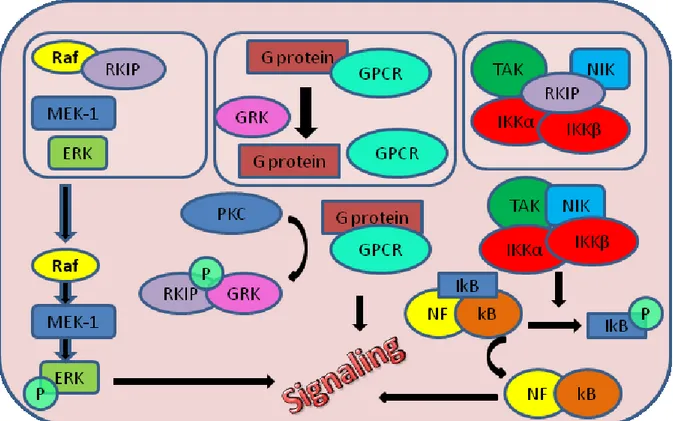
The Raf kinase inhibitor protein (RKIP) has emerging roles as regulator of multiple signaling networks, including the modulation of a) the activity of Raf-1 kinase, b) G protein-coupled receptor kinase 2 (GRK2), and c) proteins involved in nuclear factor κB activation, including inhibitor of κB kinase α (IKKα). The number of signaling cascades known to be modulated by RKIP is constantly increasing [84, 85].
2.1.Protein structure of RKIP
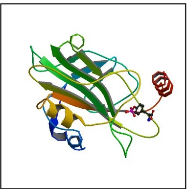
RKIP is a member of the evolutionary conserved phosphatidylethanolamine-binding protein (PEBP) family. The PEBP family of proteins is highly conserved and does not share significant homology with any other protein family [86]. Most members of this protein family, including RKIP, posses a conserved ligand-binding pocket formed by highly conserved amino acid residues with high affinity for small anionic groups. This pocket is required for its inhibitory activity of c-Raf kinase to bind and activate MAP/ERK kinase [85]. Indeed, RKIP was indentified as first physiologic inhibitor of the RAF-MEK-ERK pathway [87]. The structure of the RKIP/PEBP proteins is characterized by a central β-sheet surrounded by more variable β-stands and α-helices (Figure 6). Now, this small 21kDa protein, is suggested as a major modulator of kinases involved in signal transduction due to the ability of interfering with various signaling cascades and cellular functions .
Figure 6: Human Raf Kinase Inhibitor Protein (RKIP) in complex with o-phosphotyrosine.
2.2. RKIP : An inhibitory modulator protein
RKIP is identified as a direct interaction partner of some components of Raf-MEK-ERK pathway: Raf1, MEK1 end ERK2, which belong to the mitogen activated protein (MAP) kinase cascade [84, 85]. More precisely, RKIP binds to and inhibits Raf1, thereby inhibiting Raf dependent activation of MEK and ERK [87]. Locostatin, a non antibacterial oxazolidinone derivative, is a small molecule that covalently binds RKIP. Making this protein–protein interaction, locostatin inhibits and abrogates the ability of RKIP to bind and inhibit Raf-1 kinase [84, 87]. The Raf-MEK-ERK is a highly conserved signaling pathway that regulates cell cycle progression, growth, differentiations, migrations and apoptosis. Mutation or aberrant expression of some components of this pathway is associated with many types of agressive and metastatic cancers including breast cancer, prostate cancer, colorectal cancer and over 50% of acute myelogenous leukemia and acute lymphocytic leukemia [88, 89]. These are the reasons why the Raf/MEK/ERK pathway itself and all proteins involved in its regulation are so important as targets for therapeutic intervention (Figure 7).
Figure 7. RKIP signaling pathway.
In general, protein phosphorylations promote conformational changes and may thereby change protein stability, affect self-assosiation, generate new binding sites, influence
protein-protein interactions, induce activation or inhibition of enzyme activity, or in this cese, PKC changes the affinity of RKIP which predominantly binds and inhibits Raf1, from Raf1 to GRK-2. RKIP phosphorylation by PKC relieves a key inhibitor of the Raf/MAP kinase signaling cascade and may represent a general mechanism for the regulation of MAP kinase pathways [90, 91] (Figure 7).
RKIP may also interfere with glycogen synthase kinase 3β (GSK3β) and influence cell division and apoptosis. GSK3β is a well-established downstream component of the phosphatidylinositol 3-kinase (PI3K) signalling pathway, also known as a key enzyme in signalling pathway and cyclin D1 activation. It has been shown that RKIP is required for the posttranscriptional maintenance of GSK3β in its active form and that RKIP loss induce GSK3β destabilisation and inactivation [85, 88, 92].
2.3. Prevalence of RKIP
In addition, biological activities of RKIP can not be explained by its effects on a single pathway because this small protein plays cardinal roles in cells by regulating cell signaling, growth, and survival through its expression and activity. RKIP expression has been detected in many different tissues, such as brain, heart, liver, stomach, spleen, lung bronchioles, mesenteric lymph node, oviduct, ovary, lactating mammary glands, uterus, prostate epithelium, thyroid and muscle of human, cow, rat, and chicken and it is mainly localized in the cytoplasm and in inner periplasmic membranes [93, 94]. It has been shown by immunohistochemical staining high expression of RKIP in normal prostate and primary prostate tumors, whereas low or undetectable levels were observed in metastasis. Also significant down-regulation of RKIP has been found in melanoma cell lines in comparison with normal melanocytes. Additionally, restoration of RKIP expression in a metastatic prostate cancer cell line does not affect primary tumor growth, but it does inhibit prostate cancer metastasis. Also restoration of RKIP reduced spontaneous lung metastasis.. These parameters identify RKIP as a metastasis suppressor gene [94-96]. Moreover, RKIP can promote intercellular adhesion and inhibits the migration of cancer cells through regulation of the extracellular matrix [97]. Furthermore, RKIP is considered to play a pivotal role in cancer, regulating apoptosis induced by drugs or immune-mediated stimuli.
All the above considerations suggested us that RKIP might also play an important role in leiomyoma. In the present study we aimed to evaluate the localization and the expression level of RKIP in myometrium and leiomyoma. Next goal of this research was to evaluate the role of the RKIP in the cells of the myometrium and leiomyoma treating cells with locostatin, a molecule that covalently binds and inhibits RKIP and to show impact of RKIP in migration, proliferation and on the levels of expression of the several ECM components.
3. Materials and Methodes
3.1. Sample collection
This study included samples of fibroid and adjacent normal myometrium e xcised from women undergoing myomectomy or hysterectomy. Considering the high variability that could occur with different age, race, hormonal milieu, tumor size and location of tumors, we included in the study the most homogenous possible sample. All patients were Caucasian, age range: 41-49 years. Fibroid tissue was defined based on the well established histopathologic criteria. The location of the fibroid was predominantly intramural and their size range was 3-10 cm of diameter. Tissue samples were only taken from women who had not received exogenous hormones for the previous 3 months. All patients gave their informed consent and the permission of the Human Investigation Committee was granted.
For this study we provided three different sample collections: frozen tissue of leiomyoma and miometrium for RNA and protein extraction, formalin fixed tissue of leiomyoma and miometrium for immunohistochemistry, and fresh tissue of leiomyoma and miometrium for cell cultures.
3.2 Immunohistochemistry
Paraffin sections were deparaffinized and rehydrated via xylene and a graded series of ethyl alcohol. Most formalin-fixed tissue required an antigen retrieval step before immunohistochemical procedures. This is due to the formation of methylene bridges during fixation, which cross-link proteins and therefore mask antigenic sites. To break the methylene bridges and expose the antigenic sites in order to allow the antibodies to bind , the section were immersed in a container containing 1 mM EDTA buffer pH8, and allowed in a water bath set to 100 ° C for 20min.
To inhibit endogenous peroxidase activity, sections were incubated for 60 min with 3% hydrogen peroxide in methanol. Sections were then washed in PBS. To block non-specific background, the sections were incubated, 20 min at room temperature (RT), with normal goat serum (NGS), for RKIP, diluted 1:75. Sections were then incubated overnight at 4-C with RKIP rabbit polyclonal antibody (Upstate biotechnology, Lake Placid, NY) diluted 1:500 and 1:200.
On second day of immunohistochemistry, after washing in PBS, the sections were incubated with biotinylated anti-rabbit IgG made in goat diluted 1:200 (Vector Laboratories, Burlingame, CA) for 30min. The peroxidase ABC method (Vector Laboratories) was
performed for 1 h at RT using 3,3' diaminobenzidine hydrochloride (DAB) (Sigma, St Louis, MO) as chromogen. Sections were counterstained in Mayer’s hematoxylin, dehydrated and mounted with Eukitt solution (Kindler GmbH and Co., Freiburg, Germany).
3.3. Western blotting
Proteins extraction was performed according to the manufacturer’s instructions. Soluble protein was quantified using a Bradford protein assay (Bio-Rad, Richmond, CA), and equal amounts of proteins were loaded onto 4-12% NuPAGE gels (Invitrogen, Life Technologies, Carlsbad, CA) and resolved by SDS-PAGE under reducing conditions. Proteins were transferred to 0.2-µm nitrocellulose membranes in an X-cell II apparatus (Invitrogen) according to the manufacturer’s instructions. Ponceau S solution (Euroclone, Milan, Italy) was used for the detection of proteins on nitrocellulose membranes. After blocking the membrane with 5% (wt/vol) non-fat milk powder in Tris-buffered saline with Tween 20 (TBST) [50 mm Tris-HCl (pH 7.4), 150 mm NaCl, 0.05% Tween 20], membranes were incubated overnight with a primary antibody: 1:2000 dilutions for polyclonal rabbit against RKIP (Upstate biotechnology), 1:10000 dilution for monoclonal mouse against fibronectin (Sigma), 1:1000 dilution for monoclonal mouse against pERK (Sigma) and 1:1000 dilution for polyclonal rabbit against GSK3β. Next day, membranes were washed four times in 5% (wt/vol) non-fat milk powder in TBST and incubated with 1:10,000 dilutions of horseradish peroxidase-conjugated antirabbit IgG (Pierce, Rockford, IL) against RKIP and GSK3β or 1: 10000 horseradish peroxidase linked anti-mouse (Amershan, Milan, Italy) against fibronectin and pERK for 2 h. Membranes were washed four times in 5% (wt/vol) non-fat milk powder in TBST, and immunoreactive proteins were visualized using Super Signal West Pico chemiluminescent substrate (Pierce). Protein levels were assessed by densitometric analysis using Chemidoc and Quantity One software (Bio-Rad Laboratories, Milan, Italy).
3.4. Myometrial and leiomyoma cell culture
Immediately after surgery, the myometrial and leiomyoma samples were placed into Hanks’ balanced salt solution (HBSS) (Euroclone) and transported to the laboratory for necessary procedures in order to perform primary cultures. Samples were washed several times with Dulbecco’s PBS (Invitrogen) to remove excess of blood. After cutting myometrial and leiomyoma tissue into small pieces, samples were minced in 0.1% type 2 (Invitrogen) or type
8 collagenase (Serva, Heidelberg, Germany) in serum free Dulbecco Modified Eagle medium (DMEM) (Sigma) and incubated at 37 °C for 5-6 h in water bath with manual shaking. Digested cell suspensions were then centrifuged at 1200 rpm for 10 min and washed several times. Finally, cell pellet was dispersed in DMEM containing 10% fetal bovine serum (FBS) (Sigma), 1% antibiotic (penicillin-streptomycin; Euroclone), 1% fungizone (Amphotericin B; Lonza, Verviers, Belgium) and 1% glutamine (Gibco, Life Technologies, Carlsbad, CA) in plastic dishes at 37 °C in 95% air-5% CO2. The growth medium was
changed after 24 h or 48 h to remove unattached cells and subsequently twice a week. The purity of cells was assessed by staining with a specific smooth muscle cells marker (anti-α-smooth muscle actin). The lower passage number (P0-P4) of cells was used for experiments to avoid changes in phenotype and gene expression.
3.5. RNA extraction and real-time PCR
Total RNA was extracted using TRIZOL reagent (Invitrogen) according to the manufacturer’s instructions. Samples were digested with a ribonuclease-free deoxyribonuclease (PromegaCorp., Madison,WI), and the RNA was cleaned up and concentrated using an RNeasy microkit (QIAGEN, Milan, Italy). We performed the reverse transcriptase (RT) using the high-capacity cDNA RT kit (Applied Biosystems, Foster City, CA) with 1 µg RNA, and we performed the TaqMan real-time PCR for all the genes analyzed. We used the TaqMan gene expression assays (Applied Biosystems): (Hs00164004_m1), fibronectin (Hs00365052_m1), versican (Hs00171642_m1), performing the following thermal cycle protocol (initial denaturation at 95 °C for 20 sec, followed by 40 cycles of 95 °C for 1 sec and 60 °C for 20 sec) using 100 ng cDNA in a final reaction volume of 10 µl. The blank for each reaction, consisting of amplifications performed in the absences of RT enzyme, was performed.
3.6. Immunocytochemistry
Myometrial and leiomyoma cells were seeded in chamber tissue culture slides and allowed to divide. Cells were washed 3 times with PBS, treated with 0.2% triton X-100 in PBS for 5 min and washed 3 times with PBS. To inhibit endogenous peroxidase activity, cells were incubated for 10 min with 3% hydrogen peroxide in deionised water. Cells were washed 3 times with PBS, and to block non-specific background, cells were incubated for 20 min at
room temperature (RT) with Normal Goat Serum diluted 1:75 in 1% bovine serum albumin (BSA) in PBS. Cells were then incubated with RKIP polyclonal rabbit antibody at 1:500 and 1:200 dilutions for 1 h at RT. After washing with PBS, cells were incubated with biotinylated anti-rabbit IgG made in goat diluted 1:200. The peroxidase ABC method was performed for 1 h at RT using 3′, 3′ diaminobenzidine tetrahydrochloride (DAB) as chromogen. The peroxidase ABC method was performed for 1 h at RT using 3,3' diaminobenzidine hydrochloride (DAB) as chromogen. Sections were counterstained in Mayer’s hematoxylin, dehydrated and mounted with Eukitt solution.
3.7. Cell proliferation assay
Myometrial and leiomyoma cells were seeded in 6-well plates at initial densities of 4000 cells per well in total volume of 1.5ml DMEM supplemented with 10%FBS. Cells were treated after with locostatin (10μM) (Upstate biotechnology), and/or with the growth factor EGF (10ng/ml) (Sigma), or both, while no treated cells were used as control. After 7 days of treatment, images were taken by optical microscope, and cells were counted directly after trypsinization.
3.8. Wound-healing assay
The cultured human leiomyoma and myometrium cells were seeded in six-well plates. When almost 100% cell confluence was reached, the cells were rendered quiescent by incubation for 24 h in DMEM. Confluent monolayer cells were scratched in a straight line with a 200µl yellow tip and the cells debris and suspended cells were removed by washing the cells with serum-free DMEM. The cells were then incubated in serum-free DMEM in absence or presence of DMSO (0.05%), or locostatin (10 µM) and/or EGF (10ng/ml) for 24h. After 24 h of incubation at 37C in 95% air-5% CO2 additional images were then captured by aligning
the dishes with the reference point made at 0 h. The distance between one side of the scratch and the other was measured using Image Pro-Plus software (Media Cybernetics, Inc., Warrendale, PA, USA). Cell migration was indicated as wound exposure and expressed as percentage of unhealed distance of treated cells versus that of control cells. Closure percentage was calculated as
3.9. Data analysis
Statistical analyses were performed using GraphPad Prism version 4.01 for Windows (GraphPad, San Diego, CA). The data were analyzed using Kruskal-Wallis test, followed by a post hoc Dunn's multiple comparison test when comparing two related groups of results and Wilcokson test when comparing more than two related groups of results . Differences were considered significant when p < 0,05. All of the experiments were done in triplicate and were repeated two or three times, except for the cell proliferation assay in which n = 4.
4. Results
4.1. Localisation of RKIP in leiomyoma and myometrim
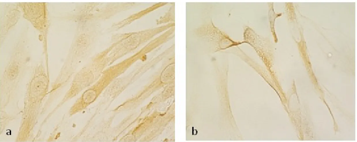
Immunohistochemistry staining was performed to demonstrate the expression of RKIP and its localization in human myometrium and leiomyoma (Figure8).
RKIP immunostaining showed that the localization of the RKIP in leiomyoma was predominantly cytoplasmatic (Figure 8a) and also a weak presence of RKIP in vascular endothelium was observabile (Figure 8b). In coresponding myometrial tissues the RKIP expression was lower respect leiomyoma and in some cases was absent (Figure 8c,d).
Figure 8. Immunohistochemical staining for RKIP in leiomyoma and myometrium. RKIP expression in leiomyoma was high and predominantly citoplasmatic (a), also was observable the small presence of RKIP in vascular endotelium (b). In myometrium the RKIP expression appears lower (c,d).
4.2.Expression of RKIP in leiomyoma and myometrim
In order to evaluate the expression level of RKIP we performed Western blot and Real time PCR analysis. Western blot results showed that the expression of RKIP was variable and it was greater in leiomyoma than in the equivalent myometrium, although the difference was not statistically significant (Figure 9). Measuring mRNA expression of RKIP in the same samples we obtained similar results (Figure 10). We concluded that mRNA expression of RKIP is a little bit higher in leiomyoma respect equivalent myometrium although the difference is not statistically significant.
Figure 8. Western blotting showing the expression of RKIP protein in myometrium and leiomyoma tissues. Represetative gel (A) and quantification of Western blotting results represented as line graphs (B) linking paired specimens and as box-and-whisker plots (C), n=6.
Figure 9. Levels of mRNA expression of RKIP in paired leiomyoma and myometrium. Results represented as line graphs (A) linking paired specimens and as box-and-whisker plots (B), n=6.
Our next objective was to evaluate the role of RKIP both in cells of myometrium and of leiomyoma.
4.3. RKIP inhibition by locostatin induces ECM expresion
In order to evaluate the role of RKIP in leiomyoma and myometrial cells, we examined whether the cultured cells retained the expression of RKIP. Immunostaing of RKIP was mainly in the cytoplasm (Figure 11).
Figure 11. .Immunocytochemical staining of RKIP in leiomyoma (a) and myometrial (b) cells.
Therefore, we treated the cells with locostatin, a synthetic substance already known as a small molecule that covalently binds and inhibits RKIP. Since RKIP binds and inhibits the Raf1, thereby inhibits Raf dependent activation of MEK and ERK. Blocking RKIP with locostatin, the protein expression of pERK should increase. To confirm RKIP inhibition by locostatin, we treated the leiomyoma and myometrium cells with 10μM of locostatin for 20 and 60 min and we observed the induction of ERK phosphorylation. The Western blot results showed increase of pERK after 20 min of treatment (Figure 12).
Figure 12. Western blotting showing the expression of pERK protein in the myometrium and leiomyoma cells. Represetative gel (a) and results of Western blot for the relative amounts of pERK in response to 10μM locostatin treatment for 20 and 60min (b). Locostatin treatment for 20min significantly increased concentration of pERK respect the intreated control, p<0.05, n=4.
Next we tested whether RKIP has influence on various components of extracellular matrix (ECM). Leiomyoma is a tumor with fibrotic characteristic and accumulates excessive amount of ECM mainly composed of collagens, fibronectin, and versican. After treatment of
leiomyoma and miometrium cells with 10μM of locostatin for 24h the expression levels of ECM components were evaluated by Western blot and Real time PCR.
Western blot results showed that locostatin slightly reduced, although non-statistically significant, the levels of fibronectin both in leiomyoma and myometrium cells (Figure 13). We obtained similar results measuring mRNA expression of fibronectin.
Figure 13. Western blotting showing the expression of fibronectin in myometrium and leiomyoma cells. Represetative gel (A) and results of Western blot for fibronectin relative amounts in response to 10μM locostatin treatment for 24h (B,C), n=4.
On the another hand, measuring mRNA expression of collagen and versican, we found that locostatin significantly reduced expression levels of collagen and versican in both leiomyoma and myometrium cells (Figure 14).
Figure14. Results of real–time PCR for relative amounts of collagen, fibronectin and versican mRNA in myometrial and leiomyoma cells in responce to 10μm locostatin for 24h. Locostatin significaly reduce the level of collagen and verican mRNA respect the untreated control (A,C), p<0.05, n=4.
4.4. RKIP inhibition by locostatin re duses cell prolife ration
To demostrate the effect of RKIP on cell proliferation, the leiomyoma and myometrial cells were treated with locostatin (10 μM), or epidermal growth factor EGF (10 ng/ml), or both, or left untreated. EGF was used as positive control.
After seven days of treatment, the images were taken during microscopic observation (Figure 15). Locostatin clearly reduced cell proliferation in leiomyoma and myometrium cells, both in the presence and in the absence of EGF treatment.
Figure 15. Cell observation. Representative photographs showing the effect of 10µM locostatin and 10ng/ml EGF on myometrial (a) and leiomyoma (b) cells proliferation after 7 days of treatmant. Locostatin clearly reduced cell proliferation in leiomyoma and myometrium cell, both in the presence or absence of EGF treatment.
In order to quantify the effect of locostatin in cell proliferation the cells were directly counted after trypsinization (Figure 16), confirming the above observations.
Figure 16. Quantification of proliferation in miometrium (a) and leiomyoma (b) cells after treatment with 10μM locostatin and/or 10ng/ml EGF for 7days. Locostatin significally reduce the cell proliferatin, in the presence and absence of EGF treatment, both in myometrial and leiomyoma cells respect untreated control, p<0.05, n=4.
4.5. RKIP inhibition by locostatin reduces cell migration
We used a wound assay to test the effects of RKIP on leiomyoma and myometrial cell motility. Cells were grown to confluence, scrape wounded and then treated for 24h with either locostatin (10μM), RKIP antagonist, EGF (10ng/ml), which was used as positive control for cell migration, or both, while no treated cells were used as a control.[84] Locostatin inhibited wound closure both in the presence or absence of EGF treatment (Figure17).
Figure 17. Wound closure experiments. Representative photographs (A) and graphs showing the effect of EGF treatment anhanced wound closure, locostatin inhibited wound closure when applied alone and abolished the effect of EGF in the samples with both compounds in myometrium (B) and leuiomyoma (C), p<0.05, n=4.
4.6. RKIP inhibition by locostatin reduses GSK3β
Finally, we evaluated the expression level of GSK3β in leiomyoma and myometrium cells treated with locostatin since it is already known that RKIP is required for the posttranscriptional maintenance of GSK3β in its active form and that RKIP loss induces GSK3β destabilisation and inactivation. Western blotting showed that GSK3β decreased significantly after locostatin treatment (Figura18).
Figure 18. Western blotting showing the expression of GSK3β in myometrium and leiomyoma cells. Represetative gel (A) and results of Western blot for the relative amounts GSK3β in response to 10μM locostatin treatment for 24h (B,C). Locostatin treatment reduced significantly the concentration of GSK3β respect the untreated control, p< 0.05, n=4.
5. Discussion
In the present paper, we demonstrate that RKIP is expressed in human myometrial and leiomyoma tissue and we showe the impact of RKIP inhibition by locostatin in migration, proliferation and on the levels of expression of several ECM components.
Immunohistochemical staining show the present of RKIP predominantly cytoplasmatic and it is also observable in vascular endothelium. Interestingly, in all examined leiomyoma tissue we observed the expression of RKIP while in respective myometrial tissue the expression of RKIP was variable and in some cases was absent. These data are interesting because several studies have shown RKIP expression in many normal and healthy tissues while RKIP is frequently downregulated in cancers [94-96]. The finding that RKIP expression tends to be higher expressed in leiomyoma compared to healthy myometrial tissue fits with the concept that leiomyoma, a non cancer tissue, is a tumor with fibrotic characteristics.
After establishing that leiomyoma and myometrium express RKIP mRNA and protein, we performed in vitro experiments to study the effects of RKIP inhibition by the chemical compound locostatin. Locostatin is known as an inhibitor of cell migration and cell-substratum adhesion that covalently binds RKIP and disrupts the interaction of RKIP with Raf-1 kinase [98-100]. Our demonstration that locostatin treatment results in activation of the MAPK signal pathway (ERK phosphorylation) provides a powerful validation of our targeting protocol.
We found that inhibition of RKIP by locostatin is able to reduces ECM components, including collagen1A1, fibronectin, and versican in leiomyoma cells. Uterine fibroids accumulate excessive amount of extracellular matrix (ECM) components mainly composed mainly of collagens, fibronectin, and versican. These ECM components play important roles in regulating adhesion, proliferation, migration, growth, differentiation, survival as well as wound healing and fibrotic processes [101, 102].
We also showed that locostatin reduces leiomyoma and myometrium cell proliferation. This data were not expected because locostatin binding RKIP should lead to proliferation since phosphorylation of Raf-1 and ERK is one of the signal pathways that leads to it. As long as the ERK/MAPK pathway is suppressed, proliferation should be restrained.
Further, we showed that RKIP inhibition by locostatin impairs cell migration both in leiomyoma and myometrium cells. This finding contrasts with the largely accepted role for RKIP in which it suppresses cancer metastasis by inhibiting cell migration. [94, 96] However, opposite effects of RKIP on migration have also been described in different cell types, for instance reduction in the invasiveness of human prostate cancer cells in vitro [103], rat hepatic stellate cells [104] and promotion of cell migration in Madin-Darby canine kidney (MDCK) epithelial cells [105].
Recently, Al-Mulla et al. (2011) reported that RKIP may also influence cell division and apoptosis by interference with glycogen synthase kinase 3β signaling. GSK-3 is active in a number of central intracellular signaling pathways, including cellular proliferation, migration, glucose regulation, and apoptosis. We took advantage of these recent findings to explain the role of RKIP in proliferation and migration in leiomyoma and myometrium. Since RKIP seems to be more expressed in leiomyoma and its inhibition by locostatin, reduces leiomyoma cell proliferation, migration and ECM apposition, one explanation of downstream signalling could be trough GSK3β promoting migration by controlling the basic machinery of the cytoskeleton.[106, 107] Our results has shown that locostatin significantly reduces the level of GSK3β confirming that RKIP is required for the posttranscriptional maintenance of GSK3β in its active form and that RKIP loss induced GSK3β destabilisation and inactivation (Figure 19).[85, 88, 92]
Figure 19. RKIP is able to bind Raf1 inhibing the MAPK pathway. RKIP is also able to stabilize GSK3β. When RKIP is blocked by locostatin, the MAPK pathway is activated. At the same time, reduction of active RKIP brings to destabilization of GSK3β. Although the activation of MARK pathway should increased proliferation and migration, reduced concentration of GSK3β lead to reduction of proliferation and migration.
6. References
1. Walker, C.L. and E.A. Stewart, Uterine fibroids: the elephant in the room. Science, 2005.
308(5728): p. 1589-1592.
2. Stewart, E.A., Uterine fibroids. Lancet, 2001. 357(9252): p. 293-298.
3. Cramer, S.F. and A. Patel, The frequency of uterine leiomyomas. American Journal of Clinical Pathology, 1990. 94(4): p. 435-438.
4. Buttram Jr, V.C. and R.C. Reiter, Uterine leiomyomata: etiology, symptomatology, and management. Fertility and Sterility, 1981. 36(4): p. 433-445.
5. Flynn, M., et al., Health care resource use for uterine fibroid tumors in the United States. American Journal of Obstetrics and Gynecology, 2006. 195(4): p. 955-964.
6. Krysiewicz, S., Infertility in women: diagnostic evaluation with hysterosalpingography and other imaging techniques. American Journal of Roentgenology, 1992. 159(2): p. 253-261. 7. Murase, E., et al., Uterine Leiomyomas: Histopathologic Features, MR Imaging Findings,
Differential Diagnosis, and Treatment1. Radiographics, 1999. 19(5): p. 1179-1197.
8. Ahmadi, F., et al., Uterine Leiomyoma: Hysterosalpingographic Appearances. International Journal of Fertility and Sterility, 2008. 1(4): p. 137-144.
9. Al-Nafussi, A., Uterine smooth-muscle tumours: practical approach to diagnosis. Current Diagnostic Pathology, 2004. 10(2): p. 140-156.
10. Toledo, G. and E. Oliva, Smooth muscle tumors of the uterus: a practical approach. Archives of Pathology & Laboratory Medicine, 2008. 132(4): p. 595-605.
11. Islam, M.S., et al., Uterine leiomyoma: available medical treatments and new possible therapeutic options. Journal of Clinical Endocrinology & Metabolism, 2013. 98(3): p. 921– 934.
12. Ono, M., et al., Side population in human uterine myometrium displays phenotypic and functional characteristics of myometrial stem cells. Proceedings of the National Academy of Sciences of the United States of America, 2007. 104(47): p. 18700-18705.
13. Ono, M., et al., Role of Stem Cells in Human Uterine Leiomyoma Growth. PloS one, 2012.
7(5): p. e36935.
14. Chang, H.L., et al., Uterine leiomyomas exhibit fewer stem/progenitor cell characteristics when compared with corresponding normal myometrium. Reproductive Sciences, 2010.
17(2): p. 158-167.
15. Mas, A., et al., Identification and characterization of the human leiomyoma side population as putative tumor-initiating cells. Fertility and Sterility, 2012.
16. Kiechle-Schwarz, M., et al., Nonrandom cytogenetic changes in leiomyomas of the female genitourinary tract. A report of 35 cases. Cancer Genetics and Cytogenetics, 1991. 53(1): p. 125-36.
17. Rein, M.S., et al., Cytogenetic abnormalities in uterine leiomyomata. Obstetrics and Gynecology, 1991. 77(6): p. 923-6.
18. Meloni, A.M., et al., Uterine leiomyomas: cytogenetic and histologic profile. Obstetrics and Gynecology, 1992. 80(2): p. 209-17.
19. Cha, P.C., et al., A genome-wide association study identifies three loci associated with susceptibility to uterine fibroids. Nature genetics, 2011. 43(5): p. 447-50.
20. Ligon, A.H., et al., PCOLCE deletion and expression analyses in uterine leiomyomata. Cancer Genetics and Cytogenetics, 2002. 137(2): p. 133-7.
21. Ptacek, T., et al., Physical mapping of distinct 7q22 deletions in uterine leiomyoma and analysis of a recently annotated 7q22 candidate gene. Cancer Genetics and Cytogenetics, 2007. 174(2): p. 116-20.
22. Velagaleti, G.V., et al., Fusion of HMGA2 to COG5 in uterine leiomyoma. Cancer genetics and cytogenetics, 2011. 202(1): p. 11-6.
23. Mäkinen, N., et al., MED12, the mediator complex subunit 12 gene, is mutated at high frequency in uterine leiomyomas. Science, 2011. 334(6053): p. 252-255.
24. Mäkinen, N., et al., MED12 exon 2 mutations are common in uterine leiomyomas from South African patients. Oncotarget, 2011. 2(12): p. 966–969.
25. Nezhad, M.H., et al., 6p21 rearrangements in uterine leiomyomas targeting HMGA1. Cancer Genetics and Cytogenetics, 2010. 203(2): p. 247-52.
26. Sandberg, A.A., Updates on the cytogenetics and molecular genetics of bone and soft tissue tumors: leiomyoma. Cancer Genetics and Cytogenetics, 2005. 158(1): p. 1-26.
27. El-Gharib, M.N. and E.S. Elsobky, Cytogenetic aberrations and the development of uterine leiomyomata. Journal of Obstetrics and Gynaecology Research, 2010. 36(1): p. 101-107. 28. Je, E.M., et al., Mutational analysis of MED12 exon 2 in uterine leiomyoma and other
common tumors. International Journal of Cancer, 2012. 131(6): p. E1044–E1047.
29. Markowski, D.N., et al., MED12 mutations in uterine fibroids-their relationship to cytogenetic subgroups. International Journal of Cancer, 2012. 131(7): p. 1528–1536.
30. McGuire, M.M., et al., Whole Exome Sequencing in a Random Sample of North American Women with Leiomyomas Identifies MED12 Mutations in Majority of Uterine Leiomyomas. PloS one, 2012. 7(3): p. e33251.
31. Pérot, G., et al., MED12 Alterations in Both Human Benign and Malignant Uterine Soft Tissue Tumors. PloS one, 2012. 7(6): p. e40015.
32. Marsh, E.E., et al., Differential expression of microRNA species in human uterine leiomyoma versus normal myometrium. Fertility and Sterility, 2008. 89(6): p. 1771-76.
33. Navarro, A., et al., Genome-Wide DNA Methylation Indicates Silencing of Tumor Suppressor Genes in Uterine Leiomyoma. PloS one, 2012. 7(3): p. e33284.
34. Greathouse, K.L., et al., Environmental Estrogens Differentially Engage the Histone Methyltransferase EZH2 to Increase Risk of Uterine Tumorigenesis. Molecular Cancer Research, 2012. 10(4): p. 546-557.
35. Maruo, T., et al., Sex steroidal regulation of uterine leiomyoma growth and apoptosis. Human Reproduction Update, 2004. 10(3): p. 207-20.
36. Ciarmela, P., et al., Growth factors and myometrium: biological effects in uterine fibroid and possible clinical implications. Human Reproduction Update, 2011. 17(6): p. 772-790.
37. Kim, J.J. and E.C. Sefton, The role of progesterone signaling in the pathogenesis of uterine leiomyoma. Molecular and Cellular Endocrinology, 2011. 358(2): p. 223-31.
38. Chegini, N., Proinflammatory and profibrotic mediators: principal effectors of leiomyoma development as a fibrotic disorder. Seminars in Reproductive Medicine, 2010. 28(3): p. 180-203.
39. Flake, G.P., J. Andersen, and D. Dixon, Etiology and pathogenesis of uterine leiomyomas: a review. Environmental Health Perspectives, 2003. 111(8): p. 1037-54.
40. Ciarmela, P., et al., Activin-A and Myostatin Response and Steroid Regulation in Human Myometrium: Disruption of Their Signalling in Uterine Fibroid. Journal of Clinical Endocrinology & Metabolism, 2011a. 96(03): p. 755-65.
41. Ciarmela, P., E. Wiater, and W. Vale, Activin-A in myometrium: characterization of the actions on myometrial cells. Endocrinology, 2008. 149(5): p. 2506-16.
42. Ciarmela, P., et al., Presence, actions, and regulation of myostatin in rat uterus and myometrial cells. Endocrinology, 2009. 150(2): p. 906-14.
43. Sozen, I. and A. Arici, Interactions of cytokines, growth factors, and the extracellular matrix in the cellular biology of uterine leiomyomata. Fertility and Sterility, 2002. 78(1): p. 1-12. 44. Hatthachote, P. and J.I. Gillespie, Complex interactions between sex steroids and cytokines in
the human pregnant myometrium: evidence for an autocrine signaling system at term. Endocrinology, 1999. 140(6): p. 2533-40.
45. Litovkin, K.V., et al., Interleukin-6 -174G/C polymorphism in breast cancer and uterine leiomyoma patients: a population-based case control study. Experimental Oncology, 2007.
29(4): p. 295-98.
46. Luo, X., et al., Gene expression profiling of leiomyoma and myometrial smooth muscle cells in response to transforming growth factor-beta. Endocrinology, 2005. 146(3): p. 1097-1118.
47. Ding, L., X. Luo, and N. Chegini, The expression of IL-13 and IL-15 in leiomyoma and myometrium and their influence on TGF-β and proteases expression in leiomyoma and myometrial smooth muscle cells and SKLM, leiomyosarcoma cell line. Journal of the Society for Gynecologic Investigation, 2004. 11: p. 319A.
48. Kurachi, O., et al., Tumor necrosis factor-α expression in human uterine leiomyoma and its down-regulation by progesterone. Journal of Clinical Endocrinology & Metabolism, 2001.
86(5): p. 2275-2280.
49. Syssoev, K.A., et al., Expression of mRNA for chemokines and chemokine receptors in tissues of the myometrium and uterine leiomyoma. Bulletin of Experimental Biology and Medicine, 2008. 145(1): p. 84-89.
50. Sozen, I., D.L. Olive, and A. Arici, Expression and hormonal regulation of monocyte chemotactic protein-1 in myometrium and leiomyomata. Fertility and Sterility, 1998. 69(6): p. 1095-1102.
51. Malik, M., et al., Why leiomyomas are called fibroids: the central role of extracellular matrix in symptomatic women. Seminars in Reproductive Medicine, 2010. 28(3): p. 169-179.
52. Wolanska, M., et al., Extracellular matrix components in uterine leiomyoma and their alteration during the tumour growth. Molecular and cellular biochemistry, 1998. 189(1): p. 145-52.
53. Arici, A. and I. Sozen, Transforming growth factor-beta3 is expressed at high levels in leiomyoma where it stimulates fibronectin expression and cell proliferation. Fertility and Sterility, 2000. 73(5): p. 1006-1011.
54. Norian, J.M., et al., Transforming growth factor beta3 regulates the versican variants in the extracellular matrix-rich uterine leiomyomas. Reproductive Sciences, 2009. 16(12): p. 1153-1164.
55. Witherspoon, J.T. and V.W. Butler, The etiology of uterine fibroids with special reference to the frequency of their occurrence in the Negro: an hypothesis. Surgery, Gynecology & Obstetrics, 1934. 58: p. 57-61.
56. Torpin, R., E. Pund, and W.J. Peeples, The etiologic and pathologic factors in a series of 1741 fibromyomas of the uterus. American Journal of Obstetrics and Gynecology, 1942. 44: p. 569-74.
57. Wilcox, L.S., et al., Hysterectomy in the United States, 1988-1990. Obstetrics and Gynecology-New York, 1994. 83: p. 549-549.
58. Amant, F., et al., Ethnic variations in uterine leiomyoma biology are not caused by differences in myometrial estrogen receptor alpha levels. Journal of the Society for Gynecologic Investigation, 2003. 10(2): p. 105-109.
59. Marshall, L.M., et al., Variation in the incidence of uterine leiomyoma among premenopausal women by age and race. Obstetrics and Gynecology, 1997. 90(6): p. 967-73.
60. Sabry, M. and A. Al-Hendy, Innovative Oral Treatments of Uterine Leiomyoma. Obstetrics and Gynecology International, 2012. 2012(2012).
61. Othman, E.-E.R. and A. Al-Hendy, Molecular genetics and racial disparities of uterine leiomyomas. Best Practice & Research Clinical Obstetrics & Gynaecology, 2008. 22(4): p. 589-601.
62. Okolo, S., Incidence, aetiology and epidemiology of uterine fibroids. Best Practice & Research Clinical Obstetrics & Gynaecology, 2008. 22(4): p. 571-588.
63. Kashani, B., et al., Role of Medical Management for Uterine Leiomyomas. Best Practice & Research Clinical Obstetrics & Gynaecology.
64. Marsh, E.E. and S.E. Bulun, Steroid hormones and leiomyomas. Obstet Gynecol Clin North Am, 2006. 33(1): p. 59-67.
65. Evans, P. and S. Brunsell, Uterine fibroid tumors: diagnosis and treatment. American Family Physician, 2007. 75(10): p. 1503-1508.
66. Farquhar, C.M. and C.A. Steiner, Hysterectomy rates in the United States 1990-1997. Obstetrics and Gynecology, 2002. 99(2): p. 229-234.
67. Frishman, G.N. and M.W. Jurema, Myomas and myomectomy. Journal of Minimally Invasive Gynecology, 2005. 12(5): p. 443-456; quiz 457-458.
68. Olufowobi, O., et al., Are the anticipated benefits of myomectomy achieved in women of reproductive age? A 5-year review of the results at a UK tertiary hospital. Journal of Obstetrics and Gynaecology, 2004. 24(4): p. 434-40.
69. Donnez, J., et al., Laparoscopic myolysis. Human reproduction update, 2000. 6(6): p. 609-613.
70. Phillips, D.R., et al., Laparoscopic leiomyoma coagulation. The Journal of the American Association of Gynecologic Laparoscopists, 1996. 3(4, Supplement): p. S39.
71. Ciavattini, A., et al., Laparoscopic cryomyolysis: an alternative to myomectomy in women with symptomatic fibroids. Surgical Endoscopy, 2004. 18(12): p. 1785-1788.
72. Spies, J.B., et al., Long-term outcome of uterine artery embolization of leiomyomata. Obstetrics & Gynecology, 2005. 106(5, Part 1): p. 933-39.
73. Liu, L., et al., Laparoscopic transient uterine artery occlusion and myomectomy for symptomatic uterine myoma. Fertility and Sterility, 2011. 95(1): p. 254-258.
74. Vilos, G.A., et al., Transvaginal Doppler-guided uterine artery occlusion for the treatment of symptomatic fibroids: summary results from two pilot studies. Journal of Obstetrics and Gynaecology Canada, 2010. 32(2): p. 149-154.
75. Friedman, A.J., et al., A randomized, placebo-controlled, double-blind study evaluating leuprolide acetate depot treatment before myomectomy. Fertility and Sterility, 1989. 52(5): p. 728-33.
76. Andreyko, J.L., et al., Use of an agonistic analog of gonadotropin-releasing hormone (nafarelin) to treat leiomyomas: assessment by magnetic resonance imaging. American Journal of Obstetrics and Gynecology, 1988. 158(4): p. 903-10.
77. Schlaff, W.D., et al., A placebo-controlled trial of a depot gonadotropin-releasing hormone analogue (leuprolide) in the treatment of uterine leiomyomata. Obstetrics & Gynecology, 1989. 74(6): p. 856-62.
78. Friedman, A.J., et al., Treatment of leiomyomata uteri with leuprolide acetate depot: a double-blind, placebo-controlled, multicenter study. The Leuprolide Study Group. Obstetrics and Gynecology, 1991. 77(5): p. 720-725.
79. Leather, A.T., et al., The prevention of bone loss in young women treated with GnRH analogues with" add-back" estrogen therapy. Obstetrics & Gynecology, 1993. 81(1): p. 104-107.
80. Stovall, T.G., et al., GnRH agonist and iron versus placebo and iron in the anemic patient before surgery for leiomyomas: A randomized controlled trial. The Leuprolide Study Group. Obstetrics & Gynecology, 1995. 86(1): p. 65-71.
81. Talaulikar, V.S. and I.T. Manyonda, Ulipristal Acetate: a Novel Option for the Medical Management of Symptomatic Uterine Fibroids. Advances in Therapy, 2012: p. 1-9.
82. Ciarmela, P., et al., Ulipristal acetate modulates the expression and functions of activin A in leiomyoma cells. Reproductive Sciences, 2014. 21(9): p. 1120-1125.
83. Islam, M.S., et al., Complex networks of multiple factors in the pathogenesis of uterine leiomyoma. Fertility and Sterility, 2013b. Submitted.
84. Ciarmela, P., et al., Possible role of RKIP in cytotrophoblast migration: immunohistochemical and in vitro studies. Journal of Cellular Physiology, 2012. 227(5): p. 1821-1828.
85. Deiss, K., et al., Raf kinase inhibitor protein (RKIP) dimer formation controls its target switch from Raf1 to G protein-coupled receptor kinase (GRK) 2. Journal of Biological Chemistry, 2012
287(28): p. 23407-23417.
86. Banfield, M.J., et al., Function from structure? The crystal structure of human phosphatidylethanolamine-binding protein suggests a role in membrane signal transduction. Structure, 1998. 6(10): p. 1245-1254.
87. Yeung, K., et al., Suppression of Raf-1 kinase activity and MAP kinase signalling by RKIP. nature, 1999. 401(6749): p. 173-177.
88. Al-Mulla, F., et al., Raf kinase inhibitor protein RKIP enhances signaling by glycogen synthase kinase-3β. Cancer research, 2011


![Figure 3. Normal myometrium and different histological types of uterine leiomyoma [11]](https://thumb-eu.123doks.com/thumbv2/123dokorg/2969796.27200/10.893.276.688.105.453/figure-normal-myometrium-different-histological-types-uterine-leiomyoma.webp)

![Figure 5. Hypothetical representation of possible therapies to inhibit uterine fibroid formation and growth [83]](https://thumb-eu.123doks.com/thumbv2/123dokorg/2969796.27200/14.893.201.761.537.898/figure-hypothetical-representation-possible-therapies-inhibit-uterine-formation.webp)