https://doi.org/10.1007/s13304-018-0536-2
ORIGINAL ARTICLE
Surgical treatment of hepatic metastases from gastric cancer
Silvia Ministrini1 · Leonardo Solaini2 · Chiara Cipollari3 · Silvia Sofia4 · Elisabetta Marino5 · Alessia D’Ignazio6 · Maria Bencivenga3 · Guido A. M. Tiberio1
Received: 14 January 2018 / Accepted: 13 May 2018 / Published online: 29 May 2018 © Italian Society of Surgery (SIC) 2018
Abstract
The purpose of the study was to investigate the clinical factors influencing the prognosis of patients submitted to hepa-tectomy for metastases from gastric cancer and their clinical role. We conducted a retrospective multicentre review. We evaluated how survival from surgery was influenced by patient-related, tumour-related and treatment-related prognostic factors. We analysed data on 144 patients submitted to hepatectomy for metastases from gastric cancer, in the synchronous and metachronous setting. In 117 cases, an R0 resection was achieved, while in 27 an R + hepatic resection was performed. Chemotherapy was administered to 55 patients. Surgical mortality was 2.1% and morbidity 21.5%. One-, 3-, and 5-year OS rates after surgery were 49.9, 19.4 and 11.6%, respectively, with a median OS of 12.0 months. T4 gastric cancer, H3 hepatic involvement, non-curative resection, recurrence after surgery, and abstention from chemotherapy were associated with a worse prognosis. Factor T and H displayed a clear (p < 0.001) cumulative effect. Our data show that R0 resection must be pursued whenever possible. The treatment of T4 gastric cancer with hepatic bilateral and diffuse metastasis (H3) should be considered carefully or it should be probably avoided. Finally, a multimodal treatment associating surgery and chemotherapy offers the best survival results.
Keywords Gastric cancer · Hepatic metastasis · Curative surgery
Introduction
In recent years, surgical treatment of hepatic metastases from gastric cancer has been widely discussed in an effort to ameliorate the oncologic results offered by palliative chemo-therapy or supportive care [1–18].
We contributed to the debate through previous publica-tions, showing that simple clinical elements may help in the selection of good candidates for surgery, both in the
metachronous [19] and in the synchronous [20] setting, and that the possibility to achieve a curative resection is the main prognostic element in this group of patients. Moreover, we found that administration of chemotherapy positively influ-enced the overall survival of metastatic patients treated with surgery.
In this invited paper, included in an almost monographic issue addressing gastric cancer through different hot top-ics, we want to present the most recent data concerning the surgical treatment of hepatic metastases from gastric cancer obtained from the analysis of the experience of the major Institutions participating in the Italian Gastric Cancer Research Group.
Methods
We retrospectively reviewed the data of 144 gastric cancer patients submitted to surgical treatment for both synchronous and metachronous hepatic metastases from 1990 to Janu-ary 2017. The data were extrapolated from a prospectively collected multicentric database, shared by six institutions, The article is part of topical collection on Gastric Cancer Surgery.
* Silvia Ministrini
1 General Surgery, University of Brescia, piazzale Spedali Civili, 1, 25123 Brescia, Italy
2 General Surgery, Forlì Hospital, Forlì, Italy 3 General Surgery, University of Verona, Verona, Italy 4 General Surgery, University of Torino, Turin, Italy 5 General Surgery, University of Perugia, Perugia, Italy 6 General Surgery, University of Siena, Siena, Italy
members of the Italian Research Group on Gastric Cancer. Data were managed according to institutional rules with patient consent.
Preoperative workup systematically included computed tomography, while hepatic MRI and staging laparoscopy were not routinely employed, particularly for patients treated before 2010. Patients with preoperative evidence of direct infiltration of the hepatic parenchyma from the gastric pri-mary were not considered as well as those with extrahepatic metastases identified before surgery.
Pathologic data concerning the gastric primary were col-lected as suggested by the General Rules of the IGCA and classified following the 7th AICC-TNM system. The extent of hepatic involvement was classified according to the JGCA H grading of liver involvement [21].
Follow-up was structured as already described [22] and stopped on 1 June 2017. We evaluated how survival from diagnosis was influenced by patients-, gastric cancer-, metas-tasis-, and treatment-related prognostic factors, as detailed in Table 1.
Statistical analysis
Descriptive statistics are presented as median and inter-quartile range (IQR 25–75%) or confidence interval (CI). Comparisons between groups were obtained through Chi square analysis for discrete variables and through t Student’s test analysis for continuous variables. Overall survival (OS) was measured from the date of resection to the date of death or the latest follow-up. Survival curves were generated by the Kaplan–Meier method, and statistical significance was determined using the log-rank test. Only variables that were statistically significant (p < 0.05) at univariate analysis were considered for multivariate analysis with Cox proportional hazards model.
Results
In this study, we considered 144 patients, 94 males and 50 females. Median age was 68 years (IQR 59.5–75 years). One-hundred and twelve patients (77.8%) presented syn-chronous hepatic metastases at the diagnosis of gastric cancer, while 32 (22.2%) developed metachronous hepatic metastases after curative gastrectomy.
Gastric cancer was treated in all patients with radical surgery, associated in 75.7% of cases to D2 lymphadenec-tomy; D1 lymphadenectomy was performed in 11 patients (7.6%), as well as D3 lymphadenectomy. Hepatic resec-tion was achieved by single or multiple metastasectomy in 108 cases (75.0%), segmentectomy/bisegmentectomy in 24 cases (16.7%), and major hepatic resection in 12 (8.3%). In the synchronous setting, 13 patients (11.6%) also had
other extrahepatic metastases, detected at laparotomy: in 6 cases limited peritoneal metastases and in 7 cases dis-tant intraabdominal lymphonodal metastases. In five cases, these extrahepatic metastases were resected.
Table 1 Clinical characteristics of the population
Characteristics n %
Sex
Male 94 65.3
Female 50 34.7
Gastric cancer location
Upper 48 33.3 Body 45 31.3 Distal 25 17.3 Linitis 2 1.4 Stump 3 2.1 x 21 14.6 pT T1–T2 23 16.0 T3 27 18.8 T4 66 45.8 x 28 19.4 pN N0–N1 48 33.3 N2 14 9.7 N3 54 37.5 x 28 19.4 Histology Intestinal 77 53.5 Diffuse 24 16.7 Other 16 11.1 x 27 18.8 Grading G1 0 0.0 G2 13 9.0 G3 22 15.3 x 10⁹ 75.7
Timing of hepatic metastases
Synchronous 112 77.8
Metachronous 32 22.2
Extent of hepatic involvement
H1 100 69.5 H2 29 20.1 H3 15 10.4 Extrahepatic metastases Yes 13 9.0 No 131 91.0
Location of extrahepatic metastases
Peritoneal 7 4.9
A radical resection (R0) was obtained in 117 cases (81.2%), while a microscopically (R1) or macroscopically (R2) non-curative resection was obtained in 14 (9.7%) and 13 cases (9.1%), respectively. The most common reasons for non-curative resection were unexpected intraoperative upstaging of the factor H, the critical position of hepatic lesion/s, and the presence of other extrahepatic metastasis.
Postoperative complication rate was 21.5% and three patients (2.1%) died during the postoperative period (Table 2).
In the synchronous setting, preoperative chemotherapy was administered in 16 cases (14.3%); 28 patients (25.0%) received postoperative chemotherapy, while 4 patients (3.6%) received both pre- and postoperative chemotherapy. Among patients treated with postoperative chemotherapy (alone or associated with preoperative treatment), seven (21.2%) had not received a curative resection. With regard to patients with metachronous hepatic metastases, chemo-therapy was administered only in seven cases (22.6%).
Twenty-six patients (18%) were alive at the time we stopped follow-up; 22 of them had no tumour relapse, while 4 had haematogenous recurrence. Five patients died due to other causes than tumour recurrence and six patients were lost at follow-up. One-hundred and eleven patients (77.1%) died because of gastric cancer recurrence. This was haema-togenous in 42 cases, peritoneal in 5, and in the regional nodes in 4; the details of recurrence were unknown in 60 cases.
One-, 3-, and 5-year OS rates after surgery were 49.9, 19.4, and 11.6%, respectively (Fig. 1), with a median OS of 12.0 months (CI 95% 8.7–15.3).
At univariate analysis, the factors that proved to have an impact upon survival were: factor T of gastric primary, the extent of hepatic involvement (factor H), the timing of hepatic metastases (synchronous vs metachronous), the cura-tive effect of surgery, the recurrence of the disease, and the administration of chemotherapy. At multivariate analysis, all these factors except the timing of hepatic metastases were confirmed (Table 3). In detail, T4 gastric cancer, H3 hepatic involvement, non-curative resection, recurrence after sur-gery, and the abstention from chemotherapy were associated with a worse prognosis. Factors T and H are the sole clini-cal factors that may be considered in a preoperative phase in the selection of patients to be submitted to surgery; they displayed a clear (p < 0.001) cumulative effect.
Discussion
Our analysis shows that the extension of gastric primary (factor T) and the extension of hepatic disease (factor H) are the only clinical factors that influence survival in the subgroup of gastric cancer patients with hepatic
metastasis. These factors were already found to have a prognostic role in this subgroup of patients [3, 9, 11, 20]; we confirm that there are very few clinical factors that can drive the selection of the best candidates for surgery in the metastatic setting of gastric cancer. The other prognos-tic factors that emerge from our analysis are the curative
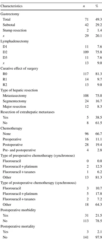
Table 2 Details of treatment
Characteristics n % Gastrectomy Total 71 49.3 Subtotal 42 29.2 Stump resection 2 1.4 x 29 20.1 Lymphadenectomy D1 11 7.6 D2 109 75.8 D3 11 7.6 x 13 9.0
Curative effect of surgery
R0 117 81.3
R1 14 9.7
R2 13 9.0
Type of hepatic resection
Metastasectomy 108 75.0
Segmentectomy 24 16.7
Major resection 12 8.3
Resection of extrahepatic metastases
Yes 5 38.5 No 8 61.5 Chemotherapy None 96 66.7 Preoperative 16 11.1 Postoperative 28 19.4
Pre- and postoperative 4 2.8
Type of preoperative chemotherapy (synchronous)
Fluorouracil 0 0.0
Fluorouracil + platinum 2 12.5
Fluorouracil + taxanes 1 6.2
Other 13 81.3
Type of postoperative chemotherapy (synchronous)
Fluorouracil 3 10.7 Fluorouracil + platinum 5 17.8 Fluorouracil + taxanes 2 7.2 Other 18 64.3 Postoperative morbidity Yes 31 21.5 No 113 78.5 Postoperative mortality Yes 3 2.1 No 141 97.9
effect of surgery, recurrence of disease, and administration of chemotherapy.
In a recent meta-analysis from Markar et al. [23] that ana-lysed 39 studies on 991 patients treated with hepatic resec-tion for gastric cancer metastases, the number of hepatic metastases and the type of liver involvement, the factor T and N of gastric primary, the vascular invasion, and the cura-tive effect of surgery were reported as prognostic factors. The prognostic role of the factor T of the gastric primary, negative when rated T4, is widely recognized [3–8]. Indeed, the serosal involvement is a watershed between a neoplasm still theoretically limited into the gastric wall and one with diffusive potential to the peritoneal cavity. As regard the prognostic role of the hepatic tumour burden (factor H), a general consensus exists. Besides all biological considera-tions, it is worth noting that in H3 patients the percentage of non-curative resection was higher than in the H1–H2 group (40 vs 17.3%, respectively). Factors T and H displayed a cumulative negative impact upon survival: it dropped actu-ally from 30 months in the case of T1–T3 and H1–H2 dis-ease to 7.3 months for T4 and H3 patients (p < 0.001).
Our data confirm that the complete surgical removal of tumor bulk (both gastric tumor and hepatic metastasis) is a major prognostic factor and it suggests that no efforts must be spared to achieve it, also referring to non-surgical abla-tive techniques [4, 24, 25]. Median survival is 17 months
Fig. 1 Overall survival
Table 3 Prognostic factors: univariate and multivariate analysis (only variables significant at univariate analysis are shown) Prognostic variables n Median survival
(months) 95% CI Univariate p value Multivariate p value Hazard ratio 95% CI
pT 0.001 0.025 3.8 1.4–10.8
T1–T2 21 5.0 16.9–53.1
T3 27 20.0 6.4–33.5
T4 65 10.2 6.5–13.8
Timing of hepatic metastases 0.004 n.s.
Synchronous 109 11.2 7.6–14.7 Metachronous 32 31.0 22.6–39.3 Hepatic involvement < 0.001 0.021 2.8 1.3–6.1 H1 98 17.0 9.6–24.3 H2 28 14.4 2.8–25.9 H3 15 7.3 3.9–10.7 Curative effect < 0.001 0.010 5.8 1.8–15.8 R0 15 17.0 12.1–21.9 R1 13 6.6 5.6–7.5 R2 13 8.0 4.9–11.1 Chemotherapy 0.030 < 0.001 3.8 2.0–7.4 Yes 55 24.0 16.1–31.9 No 86 13.0 9.9–16.1 Recurrence < 0.001 0.002 10.3 2.3–46.5 No 19 116.0 99.3–132.7 Yes 61 11.2 6.9–15.4
after curative surgery, but drops to 7 months in the case of R + resection, which is similar to the median survival of patients submitted to palliative surgery or to supportive treatments [19, 20]. Our data on the role of curative surgery confirms those reported by some European authors [12, 15,
26], but are in contrast with those reported by Cheon et al. [24], who observed no significative differences in survival performances after R0 or R + resections. This can be prob-ably explained by the positive impact of chemotherapy that was administered to a higher percentage of patients (88 vs 33.3%) in the Korean series. Moreover, we should consider the role of the different biologies of gastric cancer in Eastern and Western countries. Due to the retrospective nature of our study, it was difficult to analyse factors related to tumour biology, but we are well aware that further study is needed to examine this point [23, 27]. It is important to highlight that the surgical treatment of these patients should be referred to tertiary centres as the ones involved in our study, where post-operative morbidity and mortality rate can be minimized.
The beneficial role of chemotherapy upon survival can be explained by the fact that hepatic metastases are expression of haematogenous diffusion; therefore, they can take advan-tage of systemic treatment before or after surgical debulking. It is well known that a multidisciplinary approach is needed for the treatment of metastatic patients [15, 24, 28], but the timing of chemotherapy is still a matter of discussion, both for the treatment of non-metastatic and metastatic patients.
Chemotherapy displayed in this study a clear survival benefit (24.0 vs 13 months) and all the long-term survival patients had received chemotherapy. In Fig. 2 we show the huge impact of chemotherapy upon survival, dividing the study population into two groups: those who received chemotherapy and those who did not. Chemotherapy ame-liorates survival also for patients with negative prognostic
factors as T4 and H3 tumors, pushing the survival curve at the level of patients without risk factors who did not receive chemotherapy. At contrary, there are no survival chances for patients presenting risk factors if they do not receive systemic treatment.
Due to lack of data, we could not analyse the role of response to chemotherapy but we would like to prompt the collection of these data for further studies.
As could be expected, recurrence after surgery impacts unfavourably upon survival. Interestingly, 55.7% of patients who developed recurrence had not received chemotherapy and the absence of chemotherapy proved to be a risk factor for the development of disease recurrence (HR 2.9 CI 95% 2.7–4.9; p < 0.001).
Our data do not allow an insight into the respective role of neoadjuvant and adjuvant chemotherapy in this subset of patients. This point will be focused on soon.
Conclusion
We can state that the surgical treatment of hepatic metasta-sis from gastric cancer should be taken into consideration after careful evaluation of each single case, as only a radical approach with curative intent is worthy. Moreover a multi-disciplinary treatment is necessary, as chemotherapy plays a fundamental role in the metastatic setting of the disease.
Compliance with ethical standards
Conflict of interest The authors declare that they have no competing
interests.
Research involving human participants and/or animals The study was approved by Institutional Review Board.
Informed consent All patients signed at the admission a consent to anonymous data treatment for retrospective studies.
References
1. Adam R, Chiche L, Aloia T et al (2006) Hepatic resection for noncolorectal nonendocrine liver metastases: analysis of 1452 patients and development of a prognostic model. Ann Surg 244:524–535
2. Lehner F, Ramackers W, Bektas H et al (2009) Liver resection for noncolorectal, non-neuroendocrine liver metastases—is hepatic resection justified as part of the oncosurgical treatment? Zentralbl Chir 134:430–436
3. Ochiai T, Sasako M, Mizuno S et al (1994) Hepatic resection for metastatic tumours from gastric cancer: analysis of prognostic factors. Br J Surg 81:1175–1178
4. Ueda K, Iwahashi M, Nakamori M et al (2009) Analysis of the prognostic factors and evaluation of surgical treatment for syn-chronous liver metastases from gastric cancer. Langenbecks Arch Surg 394:647–653
Fig. 2 Influence of chemotherapy on survival according to the
5. Okano K, Maeba T, Ishimura K et al (2002) Hepatic resection for metastatic tumors from gastric cancer. Ann Surg 235:86–91 6. Hwang S-E, Yang D-H, Kim C-Y (2009) Prognostic factors for
survival in patients with hepatic recurrence after curative resec-tion of gastric cancer. World J Surg 33:1468–1472
7. Makino H, Kunisaki C, Izumisawa Y et al (2010) Indication for hepatic resection in the treatment of liver metastasis from gastric cancer. Anticancer Res 30:2367–2376
8. Ambiru S, Miyazaki M, Ito H et al (2001) Benefits and limits of hepatic resection for gastric metastases. Am J Surg 181:279–283 9. Miyazaki M, Itoh H, Nakagawa K et al (1997) Hepatic resection
of liver metastases from gastric carcinoma. Am J Gastroenterol 92:490–493
10. Shirabe K, Shimada M, Matsumata T et al (2003) Analysis of the prognostic factors for liver metastasis of gastric cancer after hepatic resection: a multi-institutional study of the indications for resection. Hepatogastroenterology 50:1560–1563
11. Sakamoto Y, Sano T, Shimada K et al (2007) Favorable indica-tions for hepatectomy in patients with liver metastasis from gastric cancer. J Surg Oncol 95:534–539
12. Thelen A, Jonas S, Benckert C et al (2008) Liver resection for metastatic gastric cancer. Eur J Surg Oncol 34:1328–1334 13. Tsujimoto H, Ichikura T, Ono S et al (2010) Outcomes for patients
following hepatic resection of metastatic tumors from gastric can-cer. Hepatol Int 4:406–413
14. Garancini M, Uggeri F, Degrate L et al (2012) Surgical treatment of liver metastases of gastric cancer: is local treatment in a sys-temic disease worthwhile? HPB 14:209–215
15. Schildberg CW, Croner R, Merkel S et al (2012) Outcome of operative therapy of hepatic metastatic stomach carcinoma: a ret-rospective analysis. World J Surg 36:872–878
16. Miki Y, Fujitani K, Hirao M et al (2012) Significance of surgical treatment of liver metastases from gastric cancer. Anticancer Res 32:665–670
17. Takemura N, Saiura A, Koga R et al (2012) Long-term outcomes after surgical resection for gastric cancer liver metastasis: an analysis of 64 macroscopically complete resections. Langenbecks Arch Surg 397:951–957
18. Liu J, Li JH, Zhai RJ et al (2012) Predictive factors improving sur-vival after gastric and hepatic surgical treatment in gastric cancer
patients with synchronous liver metastases. Chin Med J (Engl) 125:165–171
19. Tiberio GAM, Coniglio A, Marchet A et al (2009) Metachronous hepatic metastases from gastric carcinoma: a multicentric survey. Eur J Surg Oncol 35:486–491
20. Tiberio GAM, Baiocchi GL, Morgagni P et al (2015) Gastric can-cer and synchronous hepatic metastases: is it possible to recognize candidates to R0 resection? Ann Surg Oncol 22:589–596 21. Japanese Gastric Cancer Association (2011) Japanese
classifica-tion of gastric carcinoma: 3rd English ediclassifica-tion. Gastric Cancer 14:101–112. https ://doi.org/10.1007/s1012 0-011-0041-5 22. Marrelli D, De Stefano A, de Manzoni G et al (2005) Prediction
of recurrence after radical surgery for gastric cancer: a scoring system obtained from a prospective multicenter study. Ann Surg 241:247–255
23. Markar SR, Mikhail S, Malietzis G et al (2016) Influence of surgi-cal resection of hepatic metastases from gastric adenocarcinoma on long-term survival: systematic review and pooled analysis. Ann Surg 263(6):1092–1101
24. Cheon SH, Rha SY, Jeung H-C et al (2008) Survival benefit of combined curative resection of the stomach (D2 resection) and liver in gastric cancer patients with liver metastases. Ann Oncol 19:1146–1153
25. Oki E, Tokunaga S, Emi Y et al (2016) Kyushu Study Group of Clinical Cancer (2015) Surgical treatment of liver metasta-sis of gastric cancer: a retrospective multicenter cohort study (KSCC1302). Gastric Cancer 19(3):968–976
26. Chiche L, Ducreux M, Lebreton G et al (2005) Metastases hepa-tiques des cancers de l’estomac. In: Adam R, Chiche L (eds) Chi-rurgie des metastases hepatiques de cancers non colorectaux non endocrine. Monographies de l’association Franc¸aise de Chirur-gie. Arnette, Paris, pp 45–49
27. Choi AH, Kim J, Chao J (2015) Perioperative chemotherapy for resectable gastric cancer: MAGIC and beyond. World J Gastro-enterol 21(24):7343–7348
28. Koga R, Junji Y, Shigekazu O et al (2007) Liver resection for metastatic gastric cancer: experience with 42 patients including eight long-term survivors. JJCO 37:836–842