Università degli studi di Pisa
FACOLTA’ DI MEDICINA E CHIRURGIADottorato di ricerca in Fisiopatologia e Clinica dell’Apparato Cardiovascolare e Respiratorio
Direttore Prof. A. Mussi
Tesi di dottorato
Fisiopatologia e trattamento chirurgico
dell’insufficienza mitralica ischemica
cronica
(Pathophysiology and surgical treatment of chronic
ischemic mitral regurgitation)
Relatore: Dottorando:
Chiar.mo Prof. Alfredo Mussi Dott. Riccardo Codecasa
a.a. 2005
2 ABSTRACT
Background. If, when and how to treat patients with chronic ischemic mitral regurgitation remains at present controversial. An encouraging surgical approach is represented by performing under-sized mitral annuloplasty in most part of patients with mitral regurgitation due to annular dilation or papillary muscle displacement. However, the modifications at mid-term follow-up of the diastolic function in these patients, hasn’t been definitely established yet. So, in this study we focused on the functional results observed in patients who underwent undersized mitral annuloplasty, paying particular attention to diastolic function.
Methods. We retrospectively investigated all available data from 112 patients affected by chronic ischemic mitral regurgitation who underwent surgical myocardial revascularization associated to implantation of a prosthetic (mitral) annular ring. Mean follow-up time was 20 ± 10 months
Results. Mitral repair was possible in every patient, 30-day mortality was 5.4%. The strongest associations with hospital mortality at univariate analysis were a poor ejection fraction (p = 0.002) and an high pulmonary pressure (p = 0.001). At late follow-up no residual regurgitation was present in 71% of patients, trivial regurgitation in 24% and mild in 5%. Tenting area reduced from 3.4 ± 1.1 cm2 to 1.9 ± 0.5cm2 at discharge (p<0.001), and to 1.9 ± 0.5cm2 and 1.9 ± 0.3cm2 at early and late follow-up. Systolic and diastolic sphericity index significantly reduced at discharge (p = 0.002 and p < 0.001, respectively) and at early follow-up (p < 0.001). Considering at least a 15% decrease of the end-systolic volume of the left ventricle as an index of left ventricular reverse remodelling (LVRR), 61% of the patients (n. = 64) showed LVRR at discharge, 78% (n. = 61) at early follow up (χ2 = 10.1, p = 0.006) and 87.5% (n. = 79) at late follow-up (χ2 = 0.57, p = ns). Part of the patients recovered a normal diastolic pattern at discharge (46%), at early follow-up (50%) and at late follow-up (54%).
Conclusions. Adding an undersized annular ring implantation to complete myocardial revascularization yields to good functional results in terms of freedom from mitral regurgitation recidivism, reverse left ventricular
remodelling, and improvement of both systolic and diastolic left ventricular function.
4 AIM OF THE STUDY
The treatment of a patient affected by chronic ischemic mitral regurgitation (CIMR) is still controversial for diagnostic techniques, indications to therapy and choice of surgical technique.
A very encouraging surgical approach is represented by the implantation of an undersized mitral prosthetic ring. This would be able to solve most part of CIMR secondary to annular dilation and/or displacement of the papillary muscle/s. However, the real efficacy of such an option is at present a much debated question because the effects in terms of ventricular response to mitral repair and clinical consequences are not well known. An important paragraph in this kind of patients, at present really very few investigated, is represented by the modifications of diastolic function both immediately and at longer follow-up.
In the present paper we searched for functional results obtained in an unselected group of patients with at least moderate CIMR who underwent a combined operation of complete myocardial revascularization and under-sized annuloplasty. In particular we studied: mitral regurgitation recidivism, mitral valve deformation, global remodelling, local remodelling and reverse remodelling of the left ventricle, and the mid-term follow-up function of the left ventricle.
INTRODUCTION
Ischemic mitral regurgitation: definition
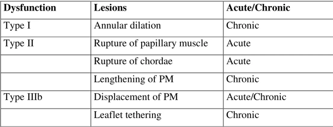
A patient with previous myocardial infarction can develop mitral regurgitation that can be defined “ischemic” as a consequence of ventricular remodelling 1. This condition is clearly separate from “organic” mitral regurgitation (secondary to rheumatic disease, valve degeneration, post-endocarditic regurgitation …). The classification of the mitral regurgitation proposed by Carpentier 2 is very useful to better understand CIMR and is based on aetiology, valvular lesions and consequent valvular dysfunction. This classification bases on opening and closing movements of the mitral leaflets (Fig. 1). In type I dysfunction there’s normal motion of the leaflets and the cause of the regurgitation is the annular dilatation. In type II dysfunction the excessive motion (prolapse) causes the regurgitation mostly secondary to lengthening and/or rupture of a chorda tendinea or a papillary muscle. In type III dysfunction the restricted motion leads to the lost of leaflet coaptation. In the type IIIa dysfunction motion is restricted both in systole and in diastole, and major causes are leaflet retraction, chordal shortening and fusion, and commissural fusion. In the type IIIb dysfunction the motion is restricted only in systole and the mechanism of this consists in dilation and dysfunction of the left ventricle with displacement of the papillary muscles. Ischemic mitral regurgitation can be consequence of a type I, II or IIIb lesion (Tab. I). In type I dysfunction the annular dilation can be secondary to a basal infarction or to a progressive enlargement of the left ventricular chamber. In type II dysfunction the regurgitation is secondary to acute ischemic rupture of a papillary muscle 3. In type IIIb dysfunction (most frequent form of ischemic mitral regurgitation) we have more components: left ventricle modifications (wall motion abnormal condition, dilation, increased sphericity), sub-valvular modifications (papillary muscles displacement and leaflet tethering), and annular modifications (dilatation and distortion) 1.
Type IIIb mitral regurgitation is defined by: 1) symptomatic multivasal coronaropathy, with or without previous myocardial infarction; 2) mitral regurgitation ≥ 2+ at echocardiogram or angio-ventriculogram; 3) normal
6 mitral leaflets; 4) absence of stenosis. It can be defined as “functional” relatively to the integrity of the mitral valve.
In the present paper we refer to “chronic” ischemic mitral regurgitation, excluding the acute II type due to papillary rupture because the implications of the acute onset of this condition would lead out of the target of this study.
Clinical impact of ischemic mitral regurgitation
The interest on CIMR is related to negative impact it have on mid-term and late survival of patients with coronary disease. In patients with myocardial infarction, mild mitral regurgitation represents and independent risk factor for 1-year mortality 4-6. Grigioni demonstrated, in well matched patients, the important prognostic value of the presence or the worsening of CIMR: Effective Regurgitant Orifice (ERO) > 20 mm2 are directly and independently associated to higher mortality (Fig. 2). Therefore, the assessment of the mitral regurgitation is basic not only during the acute and soon post-infarctual phase, but also in the chronic phase in order to stratify the risk of death. Adler et al. 7, observed a double risk of late death when mitral regurgitation was not fixed in a wide population of more than 2000 patients.
It seems obvious that the suppression of the regurgitation could represent a goal of the treatment of the patient with cardiac failure. This goal can be reached pursued both medically (reducing pre- ad afterload) and surgically.
Mitral valve anatomy
The mitral valve apparatus consists of 6 elements: posterior wall of the left atrium, annulus, leaflets, chordae, papillary muscles, and left ventricular free wall. Integrity and interdependency of all of these elements causes normal coaptation. The appearance of mitral regurgitation into a dilated and hypo-kinetic left ventricle requires, in absence of valvular lesions, the abnormal function of at least another component of the mitral apparatus.
Doppler echocardiography has been previously used to demonstrate that the incomplete coaptation of the mitral leaflets causes the mitral regurgitation. Being the amount of the area of the mitral leaflet about double the orifice
area, functional abnormalities have been hypothesised to interpret the regurgitation.
Leaflets
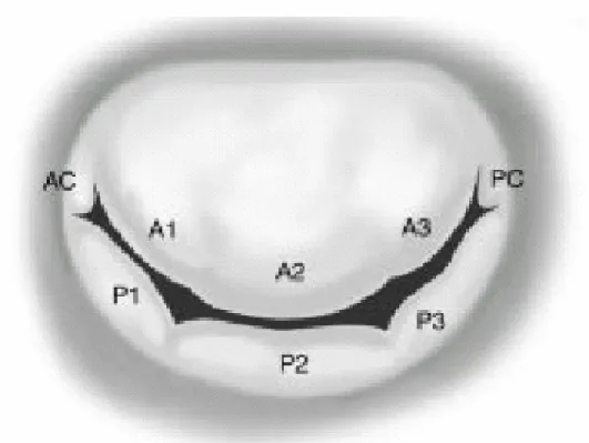
Anatomically, mitral valve is monocuspid, the leaflet is continuous along the annular circumference. Nevertheless, surgically (and commonly) commissures separate and anterior leaflet (or aortic leaflet) from a posterior or mural leaflet. The anterior leaflet takes about 1/3 of the annulus, the posterior leaflet 2/3. The posterior leaflet is subdivisible (by secondary commissures) into 3 scallops: lateral (P1), central ( P2), and medial (P3). Surgical strategies can vary accordingly to the different scallop prolapsing. The anterior leaflet is also subdivisible in 3 parts: A1, A2, A3 corresponding to the posterior scallop (Fig. 3). The 2 leaflets face each other along an “apposition zone” (from the free edge of the leaflet a few millimetres toward the body of the leaflet). The leaflet coaptation along the apposition zone reduces the pressure across the valve during the systole because it acts simultaneously onto the facing portions of the leaflet, cancelling each other out.
Papillary muscles
A normal kinetics and sequence of contraction of the papillary muscles (PMs) is basic for a optimal valve competence. The contraction of PMs follows the developing of endoventricular pressure, their maximal shortening is reached during the opening period and continues throughout the entire period of isovolumetric relaxing, and the maximal lengthening is reached during the closing period. Contrary to past beliefs, isolate dysfunction of a PM is not sufficient to give mitral regurgitation in patients with ischemic left ventricular dysfunction.
Indeed in 1968 Burch et al. 8 advocated the so called “papillary muscle syndrome” where a reduced or absent systolic shortening of the ischemic PM could give mitral regurgitation by leaflet prolapse. Subsequent studies have demonstrated how leaflet prolapse is relatively rare in patients with ischemic mitral regurgitation. Mittal et al. 9observed that the ischemic damage of both PMs and contiguous ventricular wall could give mitral regurgitation, but not the ischemic damage of one PM.
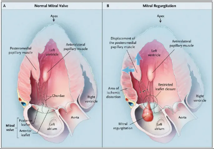
8 Kaul et al. 10 experimentally showed the relation between functional mitral regurgitation and alteration of one of the papillary muscle, both in presence of regional and global left ventricular dysfunction. A single PM dysfunction (with its adjacent portion of free wall) in presence of global good contractility couldn’t give mitral regurgitation. Global reduction of left ventricular function seems necessary for the development of mitral regurgitation, and the severity of it is proportional to the degree of cardiac failure. This author11reported a strict correlation between the distance of the point of coaptation of the mitral leaflets from the mitral annulus plane and the degree of mitral regurgitation. Indeed, the symmetric or asymmetric misalignment of the papillary muscles (PMs), and the consequent increase of the traction force exerted by the chordae (under the tension originated from the ventricular wall) on the valve may cause incomplete closure of the mitral leaflets. Such misalignment would be consequence of the left ventricular remodelling, both regional and global, causing displacement of the base of the PMs postero-laterally and apically, that is outward and inferiorly the mitral orifice (Fig. 4 and 5).
Mitral annulus
The mitral annulus is the scaffolding of the valve. It’s a fibrous ellipsoidal structure with a shorter septum-lateral diameter and a longer inter-commissural diameter. The antero-medial portion of the annulus gives insertion to the anterior leaflet and it’s non well anatomically distinguished. It’s part of the fibrous continuity that takes part between the non coronary and the left coronary cusp of the aortic root and it’s delimited anteriorly and posteriorly by 2 fibrous trigones. The postero-lateral portion of the annulus is instead a fibrous ring that separates atrial myocardium from ventricular myocardium. This anatomical connection (annulus-myocardium) causes a sphyncteric action in systole capable to approximate the leaflets so reducing the annular area of about 25%, supporting valve closure.
Three-dimensional echocardiography has shown an important relationship between the aortic root and the mitral annulus. Kaplan et al. 12 have demonstrated that because the aortic root leans on the hollow portion of the annulus, during systole the flow through the aorta dilates it and press the
annulus causing a decrease of the septum-lateral diameter and supporting leaflet coaptation. This mechanism is however incomplete in patients with dilated cardiomyopathy where the septum-lateral diameter increases and the aortic dilation is reduced by the low cardiac output. The mitral annulus changes its shape and diameter during systole. The annular diameter strictly correlates to the size of the left ventricular chamber and in particular to the end-diastolic volume. Authors have considered the dilatation of the mitral annulus a main cause of genesis and maintenance of the functional mitral regurgitation in patients with left heart failure 13,14. Boltwood 13 compared annular area and circumference of healthy people to measures obtained from patients with dilated cardiomyopathy (with or without mitral regurgitation). The group of patients with mitral regurgitation presented greater annular dimensions than both healthy subjects and patients without mitral regurgitation. Studying a variety of parameters they found that annular dilatation represented the main determinant of the functional mitral regurgitation. However the sole annular dilation shouldn’t be capable to effectively reduce the closing surface of the leaflets. Otsuji et al. 15 recently compared the echocardiographic dimensions of the mitral annulus in patients with lone atrial fibrillation (with no ventricular dilation nor dysfunction) to those obtained in patients with idiopathic or post-ischemic cardiomyopathy. Under the same annular diameter patients with lone atrial fibrillation showed a milder degree of mitral regurgitation in confront of the dilated patients who presented more moderate-to-severe degrees of mitral regurgitation. They concluded that functional mitral regurgitation needs both annular dilatation consistent with dilated and dysfunctional left ventricle. This would cause an altered balance of forces acting to the mitral leaflets. The pathophysiologic assumption of the treatment of the functional mitral regurgitation in patients with dilated cardiomyopathy is represented by the reduction of the annular diameter. Nevertheless such an intervention, in the absence of a reduction of the left ventricular size could be not enough to resolve regurgitation and, as a consequence, to improve late-term prognosis 16,17.
10 Ventricular geometry
It’s been demonstrated that under the same left ventricular dilation and dysfunction (volumes and ejection fraction), mitral regurgitation is present in those ventricles that reshape loosing the physiologic ellipsoidal shape toward a more spherical morphology 18. The sphericity index (SI = left ventricular transverse diameter/ longitudinal diameter, in systole and in diastole) has been calculated to better quantify the deformation of the left ventricle. When SI is 1 the shape is spherical. Otsuji 15 created an in vivo pharmacological model of left ventricular dysfunction where the integrity of the pericardium didn’t let the ventricle dilate. Evaluation of mitral regurgitation was achieved by means of 3D-echocardiography measuring spatial relations between the tips of the PMs and the mitral leaflets. They demonstrated that in presence of sever left ventricular dysfunction, in absence of dilatation (pericardium integral) mitral regurgitation was not significant whilst in presence of dilatation (pericardium opened) mitral regurgitation associated was moderate. The 3D reconstruction shoed how the regurgitant volume and the area of the orifice didn’t correlate with ejection fraction (i.e. contractility) but with the increased distance between the PM and the anterior portion of the annulus.
Mitral regurgitation can be explained in terms of altered balance between the forces acting on the leaflets during the systole. It has been in fact observed that the incomplete systolic closure of the mitral leaflets results from: geometrical deformation of the left ventricle combined with the displacement of PMs with consequent leaflet tethering and decrease of the closing force secondary to the contractile failure (Fig. 6). There would be a shift of the coaptation point toward the left ventricular apex causing a limitation of the closing motion during systole (leaflet tenting) 10. In theory, being the mass of the leaflet quite little they shouldn’t need much force for closure although in presence of left ventricular dysfunction. Nevertheless Otsuji 15 proposed that in presence of left ventricular dilation/dysfunction a modified geometry of the mitral apparatus would request a great increase of the closing forces. Yiu et al. 19 measured in patients with different type of regional remodelling different effective regurgitant orifice (ERO). They reported a strong relationship between a postero-apical displacement of PMs and greater EROs, a weaker relationship with a lateral shift. In conclusion the
major determinant of the ERO seems to be the systolic tenting area under the deformed mitral leaflets and the plane of the mitral annulus.
Regurgitant orifice area and trans-mitral pressure
The idea of regurgitant orifice was first introduced by Gorlin and Dexter in 1952 20 as a quantitative method of measure of mitral regurgitation, whose computation was achieved by hydraulics assumptions considering the orifice as fast during the entire cardiac cycle. It was later demonstrated that this parameter changes dynamically as a consequence o loading and size modifications of the left ventricle and the mitral annulus 21. Moreover it’s been recently demonstrated that the temporal pattern o the trans-mitral flow varies with the aetiology of the valve disease. In patients with rheumatic disease the orifice area is constant. In patients with degenerative disease and prolapse the orifice increases during mid- and end-systole 22. In patients with functional mitral regurgitation we can notice an increase of the orifice area in proto- and in end-systole, with a reduction in meso-systole. Therapeutic tools could be able to support or prolong the intermediate period of reduction of the regurgitant orifice.
This temporal pattern can be explained as follows:
• The regurgitant orifice area changes accordingly to the annular area that reduces in mid-systole and re-increases in end-systole as a consequence of atrial filling.
• The regurgitant orifice area changes as a consequence of cyclic variations of the trans-mitral pressure, acting as a pro-coapting force with its peak in mid-systole.
Studies have so been designed to investigate the role of the temporal variations of annulus and trans-mitral pressure into the genesis of the dynamic change of the regurgitant orifice area. He et al. 14 invented an in vitro model of a mitral apparatus that mimicking a ventricular failure showed a flow pattern of the functional mitral regurgitation characterised by a main proto– and end-systole, with a reduction in mid-systole consistent with the peak of trans-mitral pressure. The contractile dysfunction and the increase of the left atrial pressure causes a decrease of the trans-mitral pressure. The increased tension of the mitral leaflets due to the apical displacement of the PMs delays
12 the valve closure, but in mid-systole, the greatest endo-ventricular pressure produces a force strong enough to reduce the dimensions of the regurgitant orifice that re-dilates at the end of systole (Fig. 7).
Hung et al.16, estimating with the PISA method (see later) the regurgitant volume, have studied an interesting group of patients with low ejection fraction and functional mitral regurgitation some of them already operated on by positioning of a Carpentier-Edwards PhysioRing in order to evaluate the temporal pattern of the regurgitant area in presence of a fast annulus. Notwithstanding the relevance of both factors for the dynamic variation of the regurgitant area, the trans-mitral pressure is the major determinant of the functional mitral regurgitation in comparison with annular area.
The regurgitant orifice area presents cyclic variation specular to those of trans-mitral pressure also in patients with fixed annulus. A plot describing regurgitant orifice for systole duration shows, in patients with left ventricular dysfunction and apical-posterolateral displacement of the PMs with annular dilatation a bi-modal pattern of the regurgitant orifice area, with decrease in mid-systole. Therefore, in presence of systolic dysfunction without dilation and deformation of the left ventricle, the sole reduction of the closing force cannot cause an appreciable mitral regurgitation. The combination of an altered wall kinetics with an increased sphericity causes incomplete coaptation and the generation of an effective regurgitant orifice (ERO), whose wideness strictly correlates with the severity of the mitral regurgitation. The strict inter-dependence between the ERO and the trans-mitral pressure gives reason to argue the presence, in dilated left ventricles, of forces that oppose to valve leaflet closure (tethering forces), Fig. 8 - A.
The systolic tethering of the mitral leaflets delays the coaptation till the left intra-ventricular pressure increases enough to close the valve. The delayed closure reveals a left ventricular dysfunction that hampers the developing of the intra-cavity pressure. We must consider the functional mitral regurgitation a dynamic lesion. The dimensions of the orifice and the regurgitant volume can considerably vary as a consequence of therapeutic tools capable to reduce the left ventricular dimensions, to produce a reverse remodelling towards an ellipsoidal shape, or to modify the atrio-ventricular pressure gradient. It’s possible to under-estimate the entity of the regurgitation during the
pre-operative assessment, especially during the trans-oesophageal echocardiography made intra-operatively due to hemodynamic changes of pre- and after-load during general anaesthesia 23.
14 Echocardiographic evaluation of the functional mitral regurgitation
Mono-bidimensional echocardiography
The echocardiographic examination, mono-, bidimensional, and Doppler, is the most used technique to evaluate the functional mitral regurgitation. This relatively cheap diagnostic tool makes it clear most part of pathophysiologic mechanisms at the base of this disease. Many methods have been described in the world literature each of them addressing the estimation of the severity of the regurgitation. None of them, alone, is probably reliable. Indeed using a pool of methods based upon different theoretical assumptions makes it possible to apply a step-by-step strategy of evaluation and comparison: the greater the coherence between them, the greater the reliability of the evaluation. The functional mitral regurgitation has a single flow pattern, with an proto-systolic increase and a reduction in end-systole. 2-D echocardiography allows the calculation of a lot of parameters and a morphologic evaluation:
1. Lost of coaptation of the mitral leaflets.
2. Tenting area of the mitral valve (a triangular area whose base is the annular plane and the height is the distance between the point of coaptation of the leaflet in proto- and end-systole). This measure tells us the entity of the displacement of the coaptation point toward the ventricular apex (Fig. 8 – B).
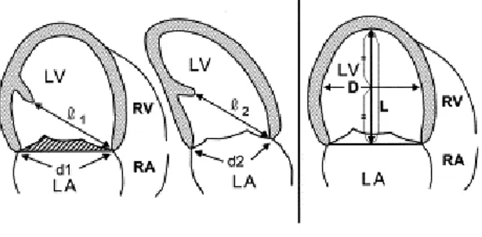
3. Mitral annular diameter, from whom it’s possible to deduce the annular area using an ellipsoidal assumption (d1xd2xπ/4) where d1 and d2 are the distances between the point of insertion of the mitral leaflets to the annulus measured in the apical 4 chambers and 2 chambers views.
4. Dys-alignment of the papillary muscles, measuring the distance between the papillary muscles from the short-axis para-sternal view and the distance between the apex of the PM and the point of insertion to the annulus of the contra-lateral mitral leaflet, both in apical 4 chambers and 2 chambers view (Fig. 9).
5. Sphericity Index: calculated as the ratio between the transverse and the longitudinal diameters both from the long-axis para-sternal view and in short axis, in end-systole.
In order to estimate the severity of the mitral regurgitation, the non-invasive determination with echo-colour-Doppler has widely shown a good correlation with the data obtained by ventriculography. A variety of methods, both qualitative and quantitative have been developed to assess the degree (severity) of the regurgitation. The echocardiographic gold standard method (accurate, routine-usable, clinical-correlated) however seems to be far.
Quantitative methods
Quantitative measures include: regurgitant volume, regurgitant fraction, effective regurgitant orifice (ERO), obtained with bidimensional and Doppler echocardiography and with the measurement of the proximal isovelocity surface area (PISA), and the vena contracta (VC). The regurgitant volume is calculated by the assumption that the stroke-volume (SV) through an orifice is given by the product of the area of the orifice (πr2) by the corresponding velocity-time integral (VTI) at pulsed-Doppler; in absence of regurgitation, the SVs calculated in different region (left ventricle outflow tract, mitral, pulmonary valves) are equal. In presence of valvular regurgitation, the flow through this valve is greater than the flow of another region: the difference between the two SVs represents the regurgitant volume (RV) 24. In mitral regurgitation, the regurgitant volume is calculated as the difference between trans-mitral flow and trans-aortic flow (measured at the aortic outflow tract); from this it can be calculated the regurgitant fraction ( RF) 24:
RV=SVregurg valv-SVcomp valv
RF= (SVregurg valv-SVcomp valv)/ SVregurg valv
ERO area is obtained dividing the RV by the velocity-time integral of the continuous Doppler flow profile of the regurgitant mitral jet 24:
ERO=RV/VTI regurg jet
The PISA analysis, bases upon the observation that closed to the regurgitant orifice the flow produces concentric surfaces of isovelocity, having decreasing extension and progressively increasing velocities. By the principle
16 of conservation of the mass, the flow through each layer must pass through the regurgitant orifice. Thanks to the Doppler colour mapping it’s possible to identify the different layers by a selected velocity, or differently to create a profile of the isovelocity range as a digital map. Because of the hemispheric shape of the surfaces of isovelocity, the flow through each of them is equal to to its area (2πr² where r is the ray of the PISA) multiplied by the velocity that identifies that layer, defined as velocity of aliasing 24:
Regurgitant flow=2πr²x Va
The ERO is then measured as the ratio between the regurgitant flow and the peak velocity of the regurgitation obtained by continuous Doppler 24:
ERO= regurgitant flow/V peak of regurgitant jet
The Vena Contracta (VC) is defined as the most narrow portion of the regurgitant jet, localised downstream the orifice. VS is characterised by a laminar flow with high velocity and is a few more narrow of the anatomical regurgitant orifice 24. The area of the transverse section of the VC represents a measure of ERO. But in dynamic lesions the VC can change with hemodynamic conditions or during the cardiac cycle.
Semi-quantitative methods
Qualitative, or semi-quantitative analysis is based on a lot of variables such as colourimetrical characteristics of the regurgitant jet, the wideness of jet and the area (derived by colour-Doppler application), jet intensity (by continuous-Doppler), and the flow pattern into the pulmonary veins (obtained with the pulsed Doppler). The flow mapping of the regurgitant jet with colour Doppler analysis by different projections, makes it possible to three-dimensionally reconstruct the wideness of the turbulence of the flow. This approach however deserves a specification: the turbulence pointed out into the left atrium by colour-Doppler is not the regurgitant volume, rather being the chromatic representation of the direction of flow (and consequently of the red
cells), of its velocity, with a different colour brilliance, and of type of flow, laminar or turbulent (the latter represented by a “mosaic effect”. Therefore no feature of the jet is directly correlated with the severity of the mitral regurgitation: in fact if just few red cells regurgitate through the orifice at very high velocity, they will determine a large area of turbulence, though a negligible effective regurgitant volume. Despite this theoretic limitation it’s been demonstrated that in most cases the greater the regurgitant volume the greater the area of turbulence into the left atrium 22. On this basis the regurgitation can be estimated by a discrete scale depicting severity from 0 to 4 +, where 1+ means mild, 2-3+ moderate and 4+ severe degree of regurgitation.
Shape and direction of the regurgitant jet depend on the geometry of the regurgitant orifice and therefore on the aetiology of mitral regurgitation. It’s been widely observed that patients with left ventricular and mitral annular dilation have a central and symmetric regurgitant jet. Another evaluation of the jet is based on the measurement of its length and area, expressed as a fraction of the area of the left atrium; however the measurement of the area by colour-Doppler, even so very sensible (about 90%) and specific (almost 100%) and well-correlated to the degree of regurgitation at the angiogram, can be influenced by a lot of physical, geometrical, instrumental and operator-depending factors all of them limiting its accuracy. The length of the jet depends on the pressure gradient between left atrium and ventricle, that is related to the compliance of he chambers: the grater the gradient the longer the jet. If left atrium pressure is high, jet length and area can result reduced also in presence of a great regurgitant orifice; moreover, in presence of atriomegalia the fraction area of left atrium taken by the jet could appear relatively small. It’s been demonstrated indeed that a regurgitant jet area into the left atrium >8 cm2, classically considered index of severe mitral regurgitation, can coincide with relatively small regurgitant volumes and/or fractions, actually over-estimating the severity of regurgitation really in patients affected by functional mitral regurgitation 25.
The analysis of the flow-pattern into the pulmonary veins with pulsed-Doppler, represents an useful index of severity of the regurgitation. When regurgitant flow enters the left atrium part of the blood already into the
18 chamber necessarily shifts, and it’s been observed that in presence of severe mitral regurgitation an inversion of the systolic flow into the pulmonary veins is present. However false negatives are possible in presence of important left atriomegalia and atrial compliance, that make it possible the entire volume of regurgitant blood be accepted by left atrium without flow-reverse. And false positives are also possible in presence of an eccentric jet whose direction is oriented toward a pulmonary vein, with a consequent flow-reverse although in absence of a severe regurgitation.
Taking advantage of pulsed-Doppler examination it is possible to take a map of the mitral regurgitation: by positioning of the sample volume into the atrium of mitral valve it is possible to obtain information about presence an length of regurgitation. By continuous-Doppler furthermore we can obtain a lot of information: first, being the intensity of the received signal proportional to the number of red cells, it’s possible to compare intensity produced by the regurgitant flow with that of antegrade mitral flow. By this it’s possible to estimate severity of mitral regurgitation24. Besides, increasing antegrade flow velocity through mitral valve, the more severe the regurgitation, the greater the antegrade velocity obtained by continous-Doppler24.
Accordingly with ASE/ESC guidelines of 200324, parameters used to quantify mitral regurgitation are reported in Tab. II.
Role of trans-oesophageal echocardiography
Trans-oesophageal echocardiography (TEE) plays an important role in determination of aetiology and severity of mitral regurgitation 26. Such approach gives greater accuracy than trans-thoracic (p<0,001) both in patients with good and poor acoustic window, to more precisely research anatomical details (in particular leaflet details) 27. During the last years, as a consequence of growing interest into mitral sparing procedures, intra-operative TEE is playing an important role in all patients with coronary artery disease and some a degree of mitral regurgitation, undergoing coronary artery bypass grafting operations. This because by TEE it’s possible to display the presence of an
entire set of anatomical features/alterations like chordal rupture, leaflet flail and so on, supporting the surgeon to decide surgical strategy.
It’s been however observed that intra-operatively patients with functional mitral regurgitation can be underestimated 28. Hemodynamic effects of anaesthetics (by reducing afterload) can reduce severity of mitral regurgitation 23. Underestimation and decision to not correct mitral regurgitation can lead to worst prognosis: a retrospective analysis assembling more than 2000 patients operated on with only CABG demonstrated how not corrected mitral regurgitation correlated to doubled risk of late death 7. Moreover, an adding risk of about 10% is calculated when delayed mitral surgery is performed.
Surgical techniques for the treatment of chronic ischemic mitral regurgitation
Surgery for mitral regurgitation is based on valve replacement and repair. In this chapter it will not further report about replacement. During the last years some wall-stress reducing procedures have been celebrated. These techniques act on some determinants of the geometrical alterations of the left ventricle, in particular on the radius of the minor diameter of the left ventricle. Bolling et al. 29 proposed and alternative approach in patients with cardiomyopathy complicated by functional mitral regurgitation: undersized annuloplasty. A smaller ring, remodelling the base of the heart, would reduce left ventricular wall-stress and re-establish an elliptical shape to the chamber
29
. In a paper from Tibayan et al. 30 undersizing annuloplasty not only reduced global sphericity but also the end-systolic bend radius of left ventricle (an important determinant of wall-stress). This important effect involved not only the base (as before assumable) but also the equatorial and apical portions of the left ventricle. Correction of leaflet tethering is achieved in most patient by annuloplasty alone, nevertheless in some other surgical steps are necessary. These are represented by so called “edge-to-edge” suture between the leaflets, both in the central part of the leaflet (symmetric displacement) and closed to the (posterior) commissure (asymmetric displacement); shortening of secondary chordae to favour mobilisation of the anterior leaflet and its
20 coaptation with the posterior one; leaflet extension; wall resection, plication and remodelling; PM repositioning all represent very complex manoeuvres that can be added in order to achieve good valve competence.
METHODS
Population
Data of all patients who underwent combined myocardial revascularization (CABG) and mitral annuloplasty in our cardiac surgery unit (SOD Cardiochirurgia – Dipartimento del cuore e dei vasi – Direttore Dott. P. Stefàno) since September 2001 to September 2004 have been retrospectively analysed and integrated with prospective follow-up for most of them.
Selection of patients was based on the following items: • Presence of moderate to severe mitral regurgitation • Previous myocardial infarction (more than 2 weeks)
• More than 75% stenosis of one or more coronary arteries with corresponding asynergic area
• Absence of mitral regurgitation due to organic mitral disease. Exclusion criteria were:
• Post-ischemic rupture of a papillary muscle or chorda tendinea • Accompanying congenital cardiac disease
• Presence of other valve disease
• Presence of atrial fibrillation or cardiac frequency more than 100 bpm (because possibly related to another cause of mitral regurgitation)
• Previous ventriculoplasty
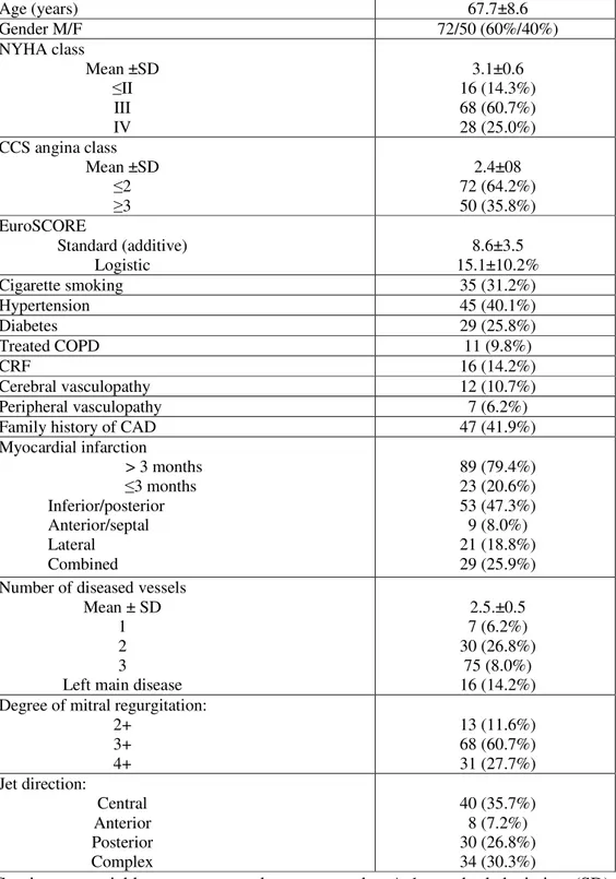
A total of 112 patients has been enrolled whose preoperative characteristics are presented in Tab. III. Sixty-eight were male and 44 female, mean age was 68 ± 10 years with a range between 37 and 88 years. Mean functional class (expressed accordingly to the classification of heart failure proposed by the New York Heart Association) was 3.4 ± 0.4. Expressing mitral regurgitation on a semi-qualitative scale between 1 and 4, mean regurgitation was 3.3 ± 0.5. Each patient was affected by multivessel coronary disease.
It was proposed to discharged patients to adhere to a follow-up program (clinical and echocardiographic) spliced into three main moments: discharge,
22 early follow-up and late follow-up. Data of discharge have been collected by means of the unit database, containing clinical and echocardiographical data. For patients who presented echocardiographic examination already performed into an external medical facility in a period of time corresponding to our early and/or late follow up, completeness and accuracy of data was tested. If quality was acceptable, the external echocardiography was included, on the contrary further internal examination was performed in all patients with sub-optimal echocardiography. This way the major number of examinations have been collected. Clinical data have been mostly collected by case history and direct examination of the patient. For most patients with good external echocardiographic data, clinical information was gathered by phone contact and report from own cardiology.
They were eventually characterised three periods: discharge, early follow-up (between 1 and 6 months, mean 4 ± 1.9 months) and late follow-up , between 8 and 40 months (mean 20 ± 10.4 months).
Echocardiographic measurements
Every patient underwent trans-thoracic echocardiography (TTE) and trans-oesophageal peri-operative echocardiography (TEE) and TTE at discharge and other follow-up. All the peri-operative examination and most of follow-up echocardiograms were performed using Acuson Sequoia with a 3.5 MHz transducer (Acuson Corporation, Mountain View, CA). Standard projections defined by ASE/ESC 24 have been used: long-axis and short axis para-sternal, apical 2-4-5 chambers for TTE. Every measurement have been evaluated onto at least 3 cardiac cycles.
M-mode and B-mode echocardiographic parameters:
The measurements of end-systolic and end-diastolic transverse diameter of the left ventricle and diameters of left atrium have been obtained by long- axis para-sternal M-mode projection. End-diastolic and end-systolic volumes of the left ventricle, and left ventricular ejection fraction have been measured in apical 4-chamber projection using modified Simpson’ method and area-length method 31. A more than 15% decrease of end-systolic left ventricular volume compared with preoperative values was considered a index of reverse remodelling of the left ventricle 32. The sphericity index (SI) was calculated as the ration between longitudinal and transverse diameter both in systole and in diastole. The closest is SI to 1, the greatest the spherical remodelling of the left ventricle 18.End-systolic wall stress was calculated on the base of arterial pressure 33. Mitral tenting area (TA) is an index of systolic deformation of the leaflets and can be defined as the triangular area whose base corresponds to the plane of the annulus and the height is the distance between the point of leaflet coaptation and the annular plane in proto- and end-systole, measured by para-sternal long-axis view. The systolic shift of the point of coaptation of mitral leaflets was expressed by difference between systolic and diastolic distance between the point of coaptation and the left ventricular apex 19,13. Systolic and diastolic mitral area has been deduced by measurement of diastolic mitral annulus (at greatest opening of the leaflets) and systolic one
24 (at closure) by 4-chambers and 2-chambers apical view 34. Regional wall kinetics has been described by means of “wall motion index” (WMI) with 17 segments 35. To quantify kinetics a score from 1 to 4 has been used being 1 = normo-kinetic, 2 = hypo-kinetic, 3 = a-kinetic, 4 = dys-kinetic area. The ratio between the sum of the scores of the segments and the number of the total number of analysed segments is the WMI. It can vary form 1, no regional a-synergy, to 3 35. PMs displacement has been directly measured by the distance between PMs (para-sternal short-axis view), or by means of the distance between the head of the PM and the insertion of the contra-lateral leaflet to the mitral annulus, both by apical 4-chambers ad by apical 2 –chambers view. By apical 5-chambers view, apical displacement of the posterior PM has been measured as the distance between the head of the PM and the inter-valvular mitro-aortic fibrosa 19.
Evaluation of the mitral regurgitation
Parameters of quantitative evaluation
Doppler method of volumes:
stroke-volume (SV) through and orifice is given by the product of the area of the orifice (πr2) by the correspondent velocity-time integral (VTI) at pulsed Doppler; in absence of regurgitation SVs from different spots (left ventricular outflow tract, mitral, pulmonary) are equal. In presence of a valvular regurgitation the flow across that valve is greater than across another spot: the difference between the 2 SV represents the Regurgitant Volume (RV) 24.
For mitral regurgitation RV is calculated as the difference between the mitral output and the aortic output; it’s then possible calculate the Regurgitant Fraction (RF) 24:
RV=SVregurg valv-SVcomp valv
RF= (SVregurg valv-SVcomp valv)/ SVregurg valv
The area of effective regurgitant orifice (ERO) was obtained dividing the RV by the velocity-time integral of the flow profile of the mitral jet at continuous-Doppler 24:
ERO=RV/VTI reg jet
PISA method ( proximal isovelocity surface area):
based upon the analysis of the proximal flow of convergence. The regurgitant flow is calculated by means of the formula 2π x r 2 x Va where r is the radius of the PISA (the distance expressed in mm between the point of aliasing of the flow of convergence and the mitral valve plane) and Va is the velocity of aliasing of the colour scale for the sending away flow 24. In this case ERO can be calculated as the ratio between the regurgitant flow and the peak velocity of the regurgitant flow:
26 ERO= Regurgitant flow/peak velocity (of the regurgitant flow) 24
Accordingly with ASE/ESC 24 guidelines, mitral regurgitation has been classified in mild, moderate and severe for values of RV respectively < 30 ml, 30-59 ml, and ≥ 60 ml/beat, for RF < 30%, 30-49% and ≥50% ad for a value of ERO area < 20 mm2, 20-39 mm2 and ≥ 40 mm2.
For patients with trivial mitral regurgitation at colour-Doppler examination ERO has been considered as null.
Evaluation of the diastolic function
The diastole has been evaluated by measurement of the trans-mitral flow. By pulsed-Doppler it’s possible to obtain the peak velocity of the E and A waves, E/A ratio, and deceleration time of the E wave 36.
Isovolumetric relaxation time (IVRT) is obtained by continuous-Doppler with sample volume positioned anteriorly and medially to mitral valve toward the left ventricular outflow tract by apical 5-chabers view. It’s possible to discriminate 3 patterns that describe an alteration of the diastolic function: • Altered relaxation: defined by E/A ratio < 1, DT > 220 msec, and IVRT >
100 msec;
• Pseudo-normal pattern: defined by E/A ratio between 1 and 2, DT between 150 and 220 msec, and IVRT between 60-100 msec;
• Restrictive pattern: defined by E/A ratio > 2, DT < 150 msec, and IVRT < 60 msec 37.
28 Surgical procedure
The trans-oesophageal probe was positioned in every patient after oro-tracheal intubation, before surgical draping. The echo examination was then performed during different hemodynamic conditions to better state entity, anatomy, variations and surgical peculiarities in order to achieve a good repair.
For patients with left ventricular ejection fraction < 25-30%, complex coronary artery disease and absence of severe peripheral vascular disease intra-aortic balloon pump was used peri-operatively as a prophylactic measure. In those patients a 40cc counter-pulsation balloon was positioned per-cutaneously via a femoral artery just after the surgical draping and before the surgical incision. 1:1 counter-pulsation was then started (triggered upon arterial pressure) and maintained during graft harvesting, it was stopped during cardiopulmonary bypass and restarted (triggered upon pressure or EKG/temporary PM), for at least 24-48 hours (in absence of leg complications). Eventually after IABP weaning 1:2 and then 1:3, the device was removed per-cutaneously by manual compression and strict monitoring.
The surgical procedures were performed via median complete sternotomy in all patients. Internal thoracic arteries were harvested using the skeletonizing technique, the radial artery was harvested with open technique and the saphena was harvested with open technique, skin tunnelling or endo-surgery. Distal tract of the ascending aorta and both venae cavae separately were cannulated for bypass management. A left ventricular vent catheter through the right superior pulmonary vein and an aortic vent catheter (also used for cardioplegia delivery) were positioned. For the first 78 patients cardioplegic arrest was achieved with cold blood cardioplegia initially infused into the aortic root followed by retrograde infusion (through the coronary sinus) and then, every 20 minutes of cross-clamping time, maintained with 2 minutes lasting only retrograde infusions. For the remaining patients, due to a general shift of the heart protection technique toward crystalloid cardioplegia in our department, a single dose of 20 ml/kg (of patient’s body weight)
HKT® cold antegrade cardioplegia was administered for 180 minutes of protection during cross-clamping.
In order to avoid dislocation of the heart once positioned the prosthetic ring, marginal branches and inferior wall arteries were revascularized first. The indications for operation in this group of patient were the same than for all the other multivessel coronary patients. All branches affected by ≥ 70% stenosis (50% for left main) and with a good run-off demonstrated by angiogram (usually equivalent to a vessel diameter ≥ 1-1,5mm) were revascularized. Saphena was in preference used as a sequential graft in order to obtain the greatest run-off by performing anastomoses to 2-3 coronary branches. All vascular anastomoses were performed using surgical telescopes, with a polypropylene running suture. The revascularization of the left anterior descending artery (and diagonal branches) was performed after the closure of atriotomy using the left internal thoracic artery in all patients but one because previous thoracic irradiation for lymphoma with patent mammary but not-satisfactory arterial wall. The right thoracic artery in addition to the left was used only in patients younger than 70years, in good clinical conditions and with good prognosis. The percentage of bilateral mammaries in this group of patients was lesser in general CAD patients because the relative higher risk. The use of radial artery for revascularization was motivated by: juvenile age, contraindications to the use of bilateral thoracic arteries, previous saphenectomy. Allen test, followed by Doppler examination for doubtful results let us discriminate contraindications to the use of the radial artery. Proximal anastomoses on the ascending aorta (when needed) were performed during cross-clamping with a polypropylene running suture. A mean of 2.6 ± 1.2 anastomoses per patient was performed with a mean of 1.2 ±0.07 arterial grafts.
The surgical approach to the mitral valve was obtained through a direct left atriotomy at inter-atrial groove just underneath the confluence of the venae cavae, or in alternative through a right atriotomy and trans-atrial incision. After the positioning of an atrial spreader, morphologic and functional analysis of the valve was done, assisted by intra-operative trans-oesophageal echocardiography because of the complete integrity of the valve in this group of patients. The pathophysiologic assessment of the mitral valve
30 was done accordingly with the criteria proposed by Carpentier 2. The size of the ring used for the annuloplasty was chosen on the basis of echocardiographic measurements and direct measurements with a mitral obturator of the inter-trigonal distance and the length of the anterior leaflet. Two different types of mitral ring were used (Carpentier classic, Carpentier Physio [Edwards Lifesciences, Irvine, CA]). Mean diameter of the rings was 27.6 ± 1.6 mm. Lone annuloplasty was sufficient for 69% of patients (77), and it was indispensable to add secondary chordal resection in 11 (10%) and para-commmissural edge-to-edge suture in 17 (15%). Nine patients underwent circular endoventriculoplasty because of the presence of a left ventricular aneurysm conditioning significant left ventricular dys-synergy.
Initial evaluation of the conservation of mitral valve symmetry and of the correct level of coaptation of the leaflets was achieved by ventricular filling with saline. Mean bypass time and cross-clamping time were 123 ± 31 minutes and 94 ± 25 minutes respectively. Final intra-operative evaluation was performed with trans-oesophageal echocardiography in order to evaluate the presence and the degree of possible residual mitral incompetence, diastolic trans-mitral gradient, valve area and coaptation depth of the leaflets.
A residual mitral jet ≤ 1 +, a coaptation depth of 0.5 – 0.9 cm, and a mitral area > 2 cm2 were considered signs of good surgical repair.
Statistical analysis
Statistical analysis was performed using a computer software [SPSS per Windows (SPSS, Inc, Chicago, IL)] to search for associations between preoperative characteristics of the patients and early clinical events, and to analyse the results of the repair using the serial echocardiographic examinations. Measures are expressed as mean values ± DS and are compared with Student’s t-test for paired and unpaired data. Repeated measures have been compared using the ANOVA method of analysis and the post-hoc Sheffè method for multiple groups. It was considered statistically significant p<0.05 and highly significant p < 0.001. The confront of proportions was performed using the χ² test and the Mantel Haenszel test.
32 RESULTS
Surgical mortality
One patient (0.9%) died for intra-operative low cardiac output not responding to maximal inotropic therapy and intra-aortic counter-pulsation. 30-day mortality was 5.4% with 6 deaths of whom one for intra-operative low cardiac output (see above), 3 for postoperative low cardiac output syndrome, one for complications correlated to acute renal insufficiency (although treated with CVVH), and one for respiratory distress.
Of the 105 patients discharged, 82 have been addressed to a cardiological rehabilitation centre where a protocol of at least 2 weeks (depending on the individual health conditions) have been completed, 2 patients have been transferred to another ICU, the rest of the patients have been moved to a cardiology ward to ameliorate the medical therapy.
One patient moved to an extra-unit ICU died after 7 weeks because of neurological complications while the other patient was later transferred to a rehabilitation unit with good functional recovery.
Univariate analysis for 30-day mortality showed an association with advanced age (73 ± 10 vs 67 ± 10 years, p = 0.05), minor ejection fraction (27 ± 7% vs 36 ± 11 %, p = 0.002), redo operation (2 redo vs 4 redo, p = 0.04), end-systolic left ventricular volume (89 ± 7 vs 83 ± 6 mL, p = 0.02), systolic pulmonary artery pressure (48 ± 11 vs 36 ± 9 mmHg, p = 0.001), preoperative NYHA class (3.6 ± 0.4 vs 3.1 + 0.5, p = 0.03). Gender and ERO area were not significantly associated. None of the parameters above resulted independent risk factor for 30-day mortality at multivariate analysis.
Specific morbidity secondary to trans-oesophageal echocardiography and to peri-operative prophylactic aortic counter pulsation
All interventions were performed under TEE surveillance with probe insertion after the induction of anaesthesia and removal after chest closure. No complication was observed relative to extensive use of TEE.
In one patient IABP was inserted (and removed) by open surgical technique because of contextual femoral false-aneurysm secondary to angiogram, in presence of contra-lateral stenotic iliac, uneventfully. Another patient with prophylactic IAPB showed enzymatic increase (mainly muscular) without critical state of the omolateral (to IABP) lower limb. In this patient it was preferred to remove the device with open technique, uneventfully. A patient with clinically indicated (not prophylactic) IABP developed severe ischemia of the lower limb that necessitated surgical removal, fomoro-popliteus bypass graft, fasciotomy and continuous veno-venous hemofiltration to reduce the negative effects of the great enzymatic release specific of myonecrosis.
Follow-up events
Two late deaths were reported: one 12 months after the operation due to therapy refractory heart failure, and another 18 months after the operation due to a major neurologic event. Remaining patients are in fairly good clinical state with 94 patients in I-II NYHA class and 9 patients in III class. For patients in III class careful evaluation is going on for possible implantation of bi-ventricular pacemaker whenever possible. Because the present few late events no statistical analysis upon them has been performed.
Follow-up echocardiography
Via telephone contact with the patient or his/her practitioner, the higher number of early echocardiographs (1-6 months) has been collected, and another follow-up echocardiography at our division has been proposed. Nineteen patients didn’t come because excessive distance from the hospital, of these: 4 patients have mailed a private echocardiography and 2 have
34 communicated it by phone. A total of 112 preoperative echocardiograms has been collected, 112 intra-operative examinations, 106 at discharge, 78 at early follow-up, and 90 at late follow-up.
Residual mitral regurgitation and mitral deformation
Trans-thoracic echocardiography at discharge (Tab. IV) pointed out mild residual mitral regurgitation in 4 patients (3.6%), trivial mitral regurgitation in 29 (25.9%), no regurgitation in the remaining 79 patients (70.5%). Mean coaptation depth was 0.8 ± 0.2 cm ( p < 0.001 vs pre-operative). Mean diastolic trans-mitral pressure gradient was 2.5 ± 0.5 mmHg and the mitral valve area in systole was 2.6 ± 0.7 cm², in diastole 3.3 ± 0.5 cm².
At early follow-up in 72 patients (68%) no residual regurgitation was present, in 28 (26%) regurgitation was trivial, and in 6 (6%) it was mild. Mean diastolic trans-mitral pressure gradient was 2.4 ± 0.3 mmHg (p = ns vs discharge), mean coaptation depth was 0.8 ± 0.2 cm (p = ns vs discharge), Mitral valve area in systole was 2.6 ± 0.4 cm² and in diastole was 3.4 ± 0.6 cm² (p = ns vs discharge).
Similar values were observed at later follow-up: no residual regurgitation in 64 cases (71%), trivial regurgitation in 22 (24%) and mild in 4 (5%). Mean diastolic trans-mitral pressure gradient (2.3 ± 0.3mmHg), coaptation depth (0.8 ± 0.2cm) and mitral valve area in systole (2.6 ± 0.7cm2) and in diastole (3.4 ± 0.9cm2) were nearly the same of early follow-up (p = ns). Tenting area reduced from 3.4 ± 1.1 cm2 to 1.9 ± 0.5cm2 at discharge (p<0.001), and to 1.9 ± 0.5cm2 and 1.9 ± 0.3cm2 at early and late follow-up.
Global left ventricular remodelling
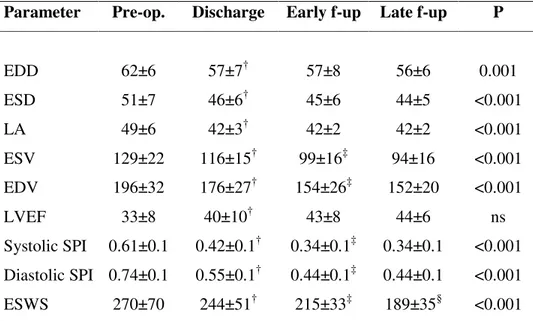
As shown in Tab. V, mean end-systolic and end-diastolic dimensions of the left ventricle and the size of the left atrium reduced at discharge (p<0.001, p = 0.005, p = 0.002, respectively), with a further decrease at early and late follow-up. End-diastolic and end-systolic mean left ventricular volume
significantly reduced at discharge (p = 0.001 and p = 0.01, respectively) and at early follow-up (p = 0.004 and p = 0.02), it didn’t at later follow-up (p = ns). Mean left ventricular ejection fraction showed an increase at discharge (p < 0.001) and remained stable in later examinations. Systolic and diastolic sphericity index significantly reduced at discharge (p = 0.002 and p < 0.001, respectively) and at early follow-up (p < 0.001). No further amelioration was noticed at later follow-up. End-systolic wall stress significantly reduced at discharge (p < 0.001), at early follow-up (p < 0.001), and at late follow-up (p < 0.001).
Reverse left ventricular remodelling
Considering at least a 15% decrease of the end-systolic volume of the left ventricle as an index of left ventricular reverse remodelling (LVRR), 61% of the patients (n. = 64) showed LVRR at discharge, 78% (n. = 61) at early follow up (χ2 = 10.1, p = 0.006) and 87.5% (n. = 79) at late follow-up (χ2 = 0.57, p = ns). In 11 patients (12.5 %) no reverse remodelling was observed. In patients with LVRR end-systolic volume reduced from 125.0 ± 15 ml to 87.4 ± 8.1ml at late follow-up (p<0.001). In patients without LVRR, mean end-systolic volume was 131 ± 10 ml pre-operatively, 97 ± 9 ml (p<0.001) at late follow-up (p<0.001 vs. patients with LVRR). In patients without LVRR mean end-systolic left ventricular diameter was 52.4 ± 3.2 mm vs 49.6 ± 2.9 mm (p = 0.01) and end-diastolic left ventricular diameter was 63.0 ± 2.1 mm vs 59.6 ± 3.0 mm, (p = 0.005), and left ventricle was more “spherical”([systolic SI 0.5 ± 0.07 vs 0.7 ± 0.04, p<0.001], [ SPI diastolic 0.7 ± 0.02 vs 0.8 ± 0.03, p = 0.006) in confront of patient with early and late LVRR.
Regional left ventricular remodelling
Regional remodelling (Tab. VI) consisted of significant posterior displacement of both PMs (p<0.001), significant apical displacement (distance between PM and contra-lateral leaflet fibrosa, p<0.001), separation
36 between PMs (p=0.001) and WMI referred to PMs (p=0.01) significantly reduced at discharge, not further varying at later follow-up. Lateral displacement of both PMs didn’t showed significant changes however.
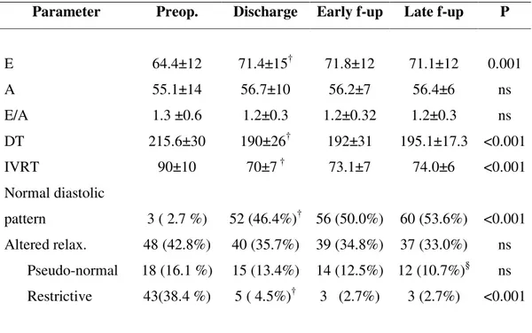
Left ventricular diastolic function
Part of the patients recovered a normal diastolic pattern at discharge (46%), at early follow-up (50%) and at late follow-up (54%), Tab. VII. A time-related decrease of the number of patients with a restrictive pattern was noticed (p<0.001) and, at late follow-up, only 3 patients (3%) presented such a pattern of diastolic filling (p<0.001 vs pre-operatively). Trans-thoracic echocardiography at discharge showed a significant increase of the E-wave (p=0.04) and a concomitant decrease both of deceleration time (p=0.006) and of iso-volumetric relaxation time (p=0.01). No further variations were observed in later follow-up. A-wave and E/A ratio didn’t vary significantly.
DISCUSSION
Ischemic mitral regurgitation is a functional disease without structural alterations of the leaflets and of the chordal apparatus, it correlates with altered geometry and function of the left ventricle and, as a result, of the mitral valve. A lot of studies such as those performed by the Mayo Clinic echo-lab importantly aided in identification of the mechanisms causing ischemic mitral regurgitation 19,6. However, in spite of a better comprehension of these mechanisms of reduced leaflet coaptation, the outcome following mitral repair are not satisfactory because a recidivism rate of mitral regurgitation near to 30% at one year 38. On the contrary, very interesting outcomes are described when myocardial surgical revascularization is associated to undersized mitral annuloplasty, and this represents at present the standard treatment of patients with (chronic) ischemic mitral regurgitation. Undersized annuloplasty is a physiological correction that, notwithstanding the restoration of a good coaptation of the mitral leaflets (by reducing the annular diameter), it cannot address the ventricular deformation determining the tethering of the leaflets.
Nonetheless, papers including heterogeneous groups of patients, let us completely comprehend the real efficacy of the combined CABG to undersized annuloplasty. Bolling et al. in 1995 29 demonstrated feasibility of the combined operation and then evaluated the mid-term and long-term survival of left ventricular wall stress via a remodelling of the base of the heart and via a more elliptical reshaping.
Bax et al. 39 described a group of patients treated with the combined intervention in whom mitral regurgitation had been virtually always corrected and 2-year survival was 84%.
Overall surgical risk
This particular group of patients represents a population at high risk of intra-operative, early post-operative and late postoperative complications. Despite a well defined strategy oriented to minimise the complications
38 secondary to postoperative low cardiac output syndrome, mortality remains significantly high but probably susceptible of further improvements. An encouraging future challenge might be represented by the clinical aid provided by preoperative cardiac re-synchronisation with bi-ventricular pacing into a wider population. In our centre we treated several patients with ischemic mitral regurgitation associated to ventricular dys-synchronism by implantation of a bi-ventricular pacemaker, optimisation of medical therapy and peri-operative intra-aortic counter-pulsation, so achieving encouraging (but not statistically significant because of number) results.
At present the strategy chosen in our centre for the therapy of patients affected by more then mild chronic ischemic mitral regurgitation does not provide for the possibility of randomise patients to lone revascularization or to revascularization associated to annular procedure. We believe in fact that, on the basis of the present literature, this could be represent a risk of under-treatment unjustifiable for an basically high surgical risk population. For this we cannot provide complete original data to confront a more conservative treatment with the complete one. A meta-analysis involving wider populations and with greater statistical power could be useful to better clarify this comparison.
Recidivism of mitral regurgitation
Hung and co-workers 16 described the relapse of mitral regurgitation in 30 patients who underwent mitral annuloplasty for ischemic mitral regurgitation (4-year follow-up) and it was correlated to continual left ventricular remodelling and to presence of leaflet tethering. Moreover, Tahta et al. 38 reported 24% of mild mitral regurgitation and 5% of moderate regurgitation after 3 years of follow-up of a wide population of patients. In these patients however annuloplasty wasn’t always undersized. In a more recent paper McGee et al. 40 reported about 585 patients subjected to mitral annuloplasty plus CABG in whom 6-months recidivism of at least moderate mitral regurgitation was 28%. Yet, contrarily to our group of patients, in 79% of cases annuloplasty had been performed using a flexible band (68%) or a bovine pericardial strip (11%). Bax et al 39 on the other hand reported
excellent results at 1,5-year follow-up in a group of 51 patients who underwent restrictive annuloplasty with undersized (2 sizes) ring: 34 (75.5%) patients without relapse of mitral regurgitation, 22.2% (n=10) trivial regurgitation and 2.2% (n=1) moderate regurgitation. In a more recent report, Geidel 41 and co-workers reported data about restrictive annuloplasty and CABG performed in 38 patients with ischemic mitral regurgitation: interestingly in this group of patients restrictive annuloplasty was performed in a “dynamic” fashion, that is related to left ventricular function (ring 2, 3 or 4 in presence o ejection fraction respectively >30, 20-30%, < 20%), using both flexible (n=8) or semi-rigid rings (n=30). At late follow-up (13±7 months) 5 patients (17%) had mild mitral regurgitation, 9 moderate. At discharge it was observed a decrease by 10% of end-diastolic diameter of the left ventricle in 47% of patients, and of end-systolic diameter in 86% of them. Our survey reports data concordant to these latter papers: relapse of mitral regurgitation at discharge and at early follow-up was respectively 0.3±0.3 e 0.3±0.4. At late follow-up 71.4 % of patients didn’t showed mitral regurgitation at all, 24.1% had a trivial mitral regurgitation and 4.5% a mild one. None of the patients required a re-operation for recidivism of mitral regurgitation.
Left ventricular remodelling
A significant restraint to geometrical left ventricular remodelling represents an important aim of the treatment of ischemic mitral regurgitation. As suggested by a lot of studies upon remodelling, on the basis of Laplace’s law the left ventricular volume is considered as an index of the remodelling 42; a 15% decrease of the end-systolic volume (in confront of the basal) has been considered as an index of reversed remodelling. Braun et al. 43 investigated the possible preoperative variables predictive of reverse remodelling into a group of 87 patients who underwent CABG associated to restrictive annuloplasty. At a 18-month follow-up 60.5% (n=45) of the patients developed a significant reverse remodelling (giving significance to a reduction ≥ 10% of the end-diastolic diameter). The cut-off values predictive of reversion of the