UNIVERSITÀ DEGLI STUDI DI SALERNO
Dipartimento di Farmacia
Dottorato di ricerca
in Scienze Farmaceutiche
Ciclo XII NS — Anno di discussione 2014 Coordinatore: Chiar.mo Prof. Gianluca SbardellaDESIGN AND SYNTHESIS OF PEPTIDES THAT
MODULATE APOPTOTIC PROCESS
settore scientifico disciplinare di afferenza
: CHIM/08
Dottorando Tutore
Dott. Ermelinda Vernieri Chiar.mo Prof. Pietro Campiglia
I
INDEX
Chapter 1: Programmed cell death pathways in cancer governed by the balanced action of PTKs and PTPs...1-26
Chapter 2: Synthesis, characterization and biological evaluation of PTPRJ (protein tyrosine phosphatase receptor like-j) agonists... 27-79
Chapter 3: SAR studies and conformational analysis of a series of novel peptides G protein–coupled receptor kinase 2 (GRK2) inhibitors...80-127
Chapter 4: Characterization of a selective CaMKII peptide inhibitor...128-159
Appendix A: Automated Peptide Synthesizers...160-166
CHAPTER 1
Programmed cell death pathways in cancer governed by the
balanced action of PTKs and PTPs
- 1 -
Abstract Oncogenic activation of tyrosine kinases is a common feature in
cancer, and its regulation represents an excellent antitumoral target. Tyrosine phosphorylation is also controlled by protein-tyrosine phosphatases (PTPs). Recent evidence has shown that PTPs can function as tumour suppressors. An improved understanding of how these enzymes function and how they are regulated might aid the development of new anticancer agents.
It has been shown that cross-regulation of kinases/phosphatases and caspases allows for fine-tuning of the apoptotic threshold, as well as the opportunity to amplify apoptotic signals. The signaling pathways involved in the control of cell proliferation, adhesion and migration are governed by the balanced action of protein tyrosine kinases (PTKs) and protein-tyrosine phosphatases (PTPs).
Keywords caspases, pathway, kinases, phosphatases.
Abbreviations IAPs, Inhibitor of Apoptosis Proteins; TRADD, TNF
receptor-associated death domain; FADD, Fas-associated death domain; DISC, death-inducing signaling complex; AIF, apoptosis inducing factor; Smac, second mitochondria-derived activator of caspase; TRAF2, TNF receptor associated factor 2; Erk, extracellular signal-regulated kinase; Cdk1, cyclin-dependent kinase; PTKs, protein tyrosine kinases; PTPs, protein-tyrosine phosphatases; PTPRJ, Protein Tyrosine Phosphatase receptor like-j; CaMKII, Ca2+/Calmodulin-dependent protein kinase II; GRK2, G-protein-coupled receptor kinase 2; FasL, Fas ligand; LOH, loss of heterozygosity; S2ED, heparin sulfate proteoglycan Syndecan-2; TSP1, Thrombospondin- 1; GPCRs, G protein coupled receptors; 2AR,
2-adrenergic receptor.
1. Introduction
Cancer can be viewed as the result of a succession of genetic changes during which a normal cell is transformed into a malignant one while evasion of cell death is one of the essential changes in a cell that cause this malignant transformation.1,2 As early as the 1970’s, Kerr et al. had linked apoptosis to the elimination of potentially malignant cells, hyperplasia and tumour
- 2 -
progression.3 Hence, reduced apoptosis or its resistance plays a vital role in carcinogenesis.
Apoptosis is a form of programmed cell death that eliminates individual cells within an organism while preserving the overall structure of surrounding tissue.4
However, it was not until the mid-1990s that apoptosis was linked to the activation of the cysteine-dependent aspartate driven proteases (caspases),5,6 which cleave key intracellular substrates to promote cell death. Given the critical role that caspases play in dismantling the cell during apoptosis, their activation and subsequent activity are highly regulated. Failure of a cell to properly modulate caspase activity can cause aberrant or untimely apoptotic cell death, potentially leading to carcinogenesis, autoimmunity, neurodegeneration, and immunodeficiency.7,8
There are many ways through which a malignant cell can acquire reduction in apoptosis or apoptosis resistance. Generally, the mechanisms of the evasion of apoptosis can be broadly dividend into: 1) disrupted balance of pro-apoptotic and anti-pro-apoptotic proteins (like Bcl-2 family, p53 and Inhibitor of Apoptosis Proteins (IAPs); 2) reduced caspase function and 3) impaired death receptor signaling.
1.1 Caspases, a family of cysteine proteases
The caspases are a family of cysteine proteases that are constitutively present in most mammalian cells, and they reside in the cytosol as single chain proenzymes.
The primary structure of a caspase is an amino-terminal prodomain and a carboxy-terminal protease domain, which contains the key catalytic cysteine residue. Caspases are categorized as initiator or effector caspases, based on their position in apoptotic signaling cascades. The initiator caspases (caspase-2, -8, -9, and -10) act apically in cell death pathways and all share long, structurally similar prodomains.9,10 This group of enzymes is activated through “induced proximity” when adaptor proteins interact with the prodomains and promote caspase dimerization. 11 , 12 In contrast, the effector caspases
- 3 -
(caspase-3, -6, and -7) have shorter prodomains and exist in the cell as preformed, but inactive, homodimers. Following cleavage mediated by an initiator caspase, effector caspases act directly on specific cellular substrates to dismantle the cell. Although many individual caspase substrates have been implicated in specific aspects of cellular destruction (e.g., lamin cleavage is required for the efficient packaging of nuclei into small membrane-bound vesicles), recent proteomic approaches have greatly expanded the known repertoire of proteolytic products generated during apoptosis.13 Further work will be needed to confirm these findings and to determine how (or if) all of these substrates participate in the apoptotic process,14 especially as new details emerge on the relationship between posttranslational modifications, like phosphorylation, and caspase cleavage.15
1.2 Apoptotic pathways
There are three pathways by which caspases can be activated. The two commonly described initiation pathways are the intrinsic (or mitochondrial) and extrinsic (or death receptor) pathways of apoptosis (Figure 1). Both pathways eventually lead to a common pathway or the execution phase of apoptosis. A third less well-known initiation pathway is the intrinsic endoplasmic reticulum pathway.16
1.2.1 The extrinsic death receptor pathway
The extrinsic death receptor pathway, as its name implies, begins when death ligands bind to a death receptor. Although several death receptors have been described, the best known death receptors is the type 1 TNF receptor (TNFR1) and a related protein called Fas (CD95) and their ligands are called TNF and Fas ligand (FasL) respectively.17 These death receptors have an intracellular death domain that recruits adapter proteins such as TNF receptor-associated death domain (TRADD) and Fas-receptor-associated death domain (FADD), as well as cysteine proteases like caspase 8.18 Binding of the death ligand to the death receptor results in the formation of a binding site for an adaptor protein and the whole ligand-receptor-adaptor protein complex is known as the death-inducing signaling complex (DISC). DISC then initiates the assembly and activation of pro-caspase 8. The activated form of the enzyme, caspase 8
- 4 -
is an initiator caspase, which initiates apoptosis by cleaving other downstream or executioner caspases.
1.2.2 The intrinsic mitochondrial pathway
As its name implies, the intrinsic pathway is initiated within the cell. Internal stimuli such as irreparable genetic damage, hypoxia, extremely high concentrations of cytosolic Ca2+ and severe oxidative stress are some triggers of the initiation of the intrinsic mitochondrial pathway. Regardless of the stimuli, this pathway is the result of increased mitochondrial permeability and the release of pro-apoptotic molecules such as cytochrome-c into the cytoplasm.19 The intrinsic pathway is closely regulated by a group of proteins belonging to the Bcl-2 family, named after the BCL2 gene originally observed at the chromosomal breakpoint of the translocation of chromosome 18 to 14 in follicular non-Hodgkin lymphoma. There are two main groups of the Bcl-2 proteins, namely the pro-apoptotic proteins (e.g. Bax, Bak, Bad, Bcl-Xs, Bid, Bik, Bim and Hrk) and the anti-apoptotic proteins (e.g. Bcl-2, Bcl-XL, Bcl-W, Bfl-1 and Mcl-1).20 While the anti-apoptotic proteins regulate apoptosis by blocking the mitochondrial release of cytochrome-c, the pro-apoptotic proteins act by promoting such release. It is not the absolute quantity but rather the balance between the pro- and anti-apoptotic proteins that determines whether apoptosis would be initiated. Other apoptotic factors that are released from the mitochondrial intermembrane space into the cytoplasm include apoptosis inducing factor (AIF), second mitochondria-derived activator of caspase (Smac), direct IAP Binding protein with low pI (DIABLO) and Omi/high temperature requirement protein A (HtrA2). Cytoplasmic release of cytochrome c activates caspase 3 via the formation of a complex known as apoptosome which is made up of cytochrome c, Apaf-1 and caspase 9.21 On the other hand, Smac/DIABLO or Omi/HtrA2 promotes caspase activation by binding to inhibitor of apoptosis proteins (IAPs) which subsequently leads to disruption in the interaction of IAPs with caspase-3 or -9.22
- 5 -
1.2.3 The common pathway
The execution phase of apoptosis involves the activation of a series of caspases. The upstream caspase for the intrinsic pathway is caspase 9 while that of the extrinsic pathway is caspase 8. The intrinsic and extrinsic pathways converge to caspase 3. Caspase 3 then cleaves the inhibitor of the caspase-activated deoxyribonuclease, which is responsible for nuclear apoptosis. In addition, downstream caspases induce cleavage of protein kinases, cytoskeletal proteins, DNA repair proteins and inhibitory subunits of endonucleases family. They also have an effect on the cytoskeleton, cell cycle and signaling pathways, which together contribute to the typical morphological changes in apoptosis.23
- 6 -
1.2.4 The intrinsic endoplasmic reticulum pathway
This intrinsic endoplasmic reticulum (ER) pathway is a third pathway and is less well known. It is believed to be caspase 12-dependent and mitochondria-independent.24 When the ER is injured by cellular stresses like hypoxia, free radicals or glucose starvation, there is unfolding of proteins and reduced protein synthesis in the cell, and an adaptor protein known as TNF receptor associated factor 2 (TRAF2) dissociates from procaspase-12, resulting in the activation of the latter.
1.3 The bidirectional communication between caspases and kinases/phosphatases
Although the cleavage of many caspase substrates is required for the structural packaging of cellular contents during apoptosis, a subset of caspase substrates are signaling molecules whose cleavage alters their signaling properties to affect the internal environment of the dying cell. In turn, signaling molecules can modulate caspase function to positively or negatively alter the trajectory of the cell death program. Given the millions of reversible phosphorylation events necessary to maintain cellular homeostasis and to allow cells to adapt nimbly to changing internal and external environments, the bidirectional communication between caspases and the kinases/phosphatases that control the cellular phosphoproteome is of particular interest.25
With respect to phosphorylation, both the caspase activation process and intrinsic enzymatic activity are under the control of modifying kinases and phosphatases. This permits cellular flexibility in setting a threshold for the induction of apoptosis in response to alterations in the cellular environment (for example, after growth factor stimulation or changes in cellular metabolism) through changes in the activity of pro- or antiapoptotic kinases and phosphatases. Phosphorylation may also control caspase activity indirectly by controlling other apoptotic modulators (including caspase binding partners). If we consider that the control of caspases can be exerted via their direct phosphorylation, focused on caspase 9 we find that of caspase 9 may result in the failure of apoptotic induction. In particular the phosphorylation of caspase 9 by both Erk (extracellular signal-regulated kinase) and Cdk1
- 7 -
(cyclin-dependent kinase 1) suppresses caspase 9 activation.26 Given that the Erk pathway is upregulated in a variety of cancers, this inhibitory phosphorylation of caspase 9 may contribute to apoptotic (and therefore chemotherapeutic) resistance. It’s note that phosphorylation and inhibition of caspase 9 by ERK promotes cell survival during development and tissue homeostasis suggesting that phosphorylation of Thr 125 on caspase 9 may be an important mechanism through which growth factor and survival signals that activate the ERK-MAPK pathway can inhibit apoptosis.
Therefore the apoptosis pathway could be regulated by a phosphorylation and a dephosphorylation event of apoptotic effectors.
Cross-regulation of kinases/phosphatases and caspases allows for fine tuning of the apoptotic threshold, as well as the opportunity to amplify apoptotic signals.
In the apoptotic process, the regulatory role of protein kinases is clearly established instead of the regulatory role of protein phosphatases is not clear. For this reason we are interested in the regulation of signaling responses of phosphatases.
However, it has recently become apparent that protein phosphatases can no longer be viewed as passive housekeeping enzymes, but they partner with kinases in the regulation of signaling responses. The distinct but complementary function of these enzymes is emphasized by recent studies, in which kinases have been implicated in controlling the amplitude of signaling responses, whereas phosphatases are thought to have an important role in controlling the rate and duration of the response.27,28
In base to these considerations, my doctoral thesis aimed to identify chemical entities able to regulate the apoptotic process through the modulation of phosphatase and kinase activities. Therefore, I studied 1) PTPRJ, a protein tyrosine phosphatase receptor like j; 2) GRK2, G protein-coupled receptor kinase 2; and 3) CaMKII, Ca2+/Calmodulin-dependent protein kinase II.
1.4 Protein Phosphatases
The first PTP was purified in 1988, approximately 10 years after the discovery of tyrosine kinases. It is now known that PTPs constitute a large, structurally diverse family of tightly regulated, highly specific enzymes with important regulatory roles.29,30
- 8 -
It is also clear that PTPs have both inhibitory and stimulatory effects on cancer associated signaling processes, and that deregulation of PTP function is associated with tumorigenesis in different types of human cancer.
Among all protein tyrosine phosphatases, PTPRJ is of particular interest for its role in human and experimental tumorigenesis.31 In fact, after its discovery,32a consistent body of literature supports its tumor suppressor activity in several models. PTPRJ (also named DEP-1, HPTPη, or CD148) is down-regulated in mammary cancer cells, and its restoration blocks their proliferation; a similar behavior was described in both human and experimental models of thyroid tumorigenesis, where PTPRJ overexpression was able to interfere with cancer cells malignant phenotype both in vitro and in vivo.33 Moreover, down-regulation of PTPRJ expression operated by miR-328 increases cell proliferation in HeLa and SKBr3 cell lines.34
PTPRJ has been successfully used as a therapeutic gene in cancer gene therapy preclinical models of thyroid and pancreatic cancer.35 The role of PTPRJ in human tumorigenesis was also highlighted by Ruivenkamp et al., 36 who demonstrated that PTPRJ was affected by loss of heterozygosity (LOH) in colon, lung, and mammary tumors; a significant percentage of LOH was also described in human thyroid tumors.37 Moreover, a polymorphism in the mouse Ptprj locus (Scc1) is associated with colon cancer development, although Ptprj genetic ablation did not induce spontaneous tumors in mice.38,39
The biochemical pathways negatively regulated by PTPRJ have been partly elucidated. Various reports indicate an inhibitory effect of PTPRJ on several players of the mitogenic signals in both normal and cancer cells. In fact, CD148 interacts with and dephosphorylates numerous receptor tyrosine kinases (RTKs) including PDGFR, HGFR, 40,41 RET, and EGFR42,43 whose aberration in cancer cells is responsible for self sufficiency cell growth.44 PTPRJ is also a negative modulator of the signaling mediated by cytosolic transducers, including phospholipase Cγ1 (PLCγ1), LAT, PKB/Akt,45and PI3K.46,47 Noteworthy, the role of PTPRJ on the inhibition of RTKs was also extended to VEGFR-2,48,49 whose activity is required for the formation of new vessels in tumor progression. Lately, PTPRJ ligands have been discovered, they include the heparin sulfate proteoglycan Syndecan-2 (S2ED)50 and Thrombospondin-1 (TSP1).51 CD148 acts as key intermediary in S2ED mediated cell adhesion, by modulating downstream β1 integrin mediated adhesion and cytoskeletal organization, whereas TSP1, a glycoprotein that
- 9 -
mediates cell-cell and cell-matrix interactions, increases PTPRJ activity, influencing the dephosphorylation of its substrates. These findings make PTPRJ an interesting candidate for the generation of novel therapeutic strategies.
1.4.1 PTPRJ regulates cell growth and cell cycle
The tumor-suppressive role of PTPRJ was clearly displayed in experiments where highly malignant thyroid cells, after transduction by a retrovirus carrying CD148, appeared to acquire a normal phenotype and showed increased adhesion, restoration of differentiation, reduced proliferation and decreased tumorigenicity. The same report established that PTPRJ induced a G1 growth arrest by suppressing the degradation of the cyclin-dependent kinase inhibitor p27kip1 protein.
Therefore, synthetizing PTPRJ agonists could be one of a possible approach in cancer therapy.
The receptor-like PTP phosphatase DEP-1 opposes various oncogenic receptor tyrosine kinases. Recently, DEP-1 agonists were isolated from a random peptide phage-display library.52
Intriguingly, these agonists circularize in vitro via an intramolecular disulfide bridge and form stable dimers that induce the dimerization-mediated activation of DEP-1 (Figure 2). Consistent with the activation of DEP-1 as a tumor suppressor, these peptides reduced proliferation and triggered apoptosis of cancer cells.
Figure 2. Small-molecule activators of protein phosphatases.
Starting from these observations during my PhD I focused the attention on two peptides, recently identified by means of a phage display library, able to bind and activate PTPRJ ([Cys-His-His-Asn-Leu-Thr-His-Ala-Cys]-OH and [Cys-Leu-His-His-Tyr-His-Gly-Ser-Cys]-OH) designing and synthetizing new PTPRJ agonists with the aim to identify a more stable and active peptide
- 10 -
sequence.53 These informations could help the design of peptidomimetics with PTPRJ agonistic properties.
1.5 Protein kinases as drug targets
Nearly every cellular process is controlled by phosphorylation of key regulatory proteins on specific serine (Ser), threonine (Thr), or tyrosine (Tyr) residues. The covalent attachment of a bulky and negatively charged phosphate group by a kinase usually induces a conformational change that affects protein function. Mammalian genomes harbor more than 500 genes that encode protein kinases. Almost all of these belong to the same protein kinase superfamily and have a highly similar catalytic core and mechanism, implying that they originate from a common ancestor. In recent years protein kinases have become one of the most popular drug targets.54
There are more than 518 human protein kinases recognized through their conserved sequence motifs. These constitute the third most populous protein family and represent 1.7% of the human genome. Of the total, 478 protein kinases are typical kinases, and 40 are atypical. The typical kinases are divided into those that phosphorylate serine or threonine residues (388 kinases) and those that phosphorylate tyrosine residues (90 kinases). These proteins have biochemical kinase activity but lack sequence similarity to the conventional eukaryotic kinases.
A distinguishing feature of the protein kinase family is the different structures that they adopt between the active and inactive states. This family characteristic was first appreciated following the determination of the first protein kinase structures of protein kinase A (PKA) [Protein Data Bank (PDB) code 1ATP] in the active conformation and cyclin-dependent protein kinase 2 (Cdk2) (PDB code 1HCK) in an inactive conformation.
Deregulation of protein kinase activity through mutation to constitutively active forms, loss of negative regulators, and chromosomal rearrangements that lead to the formation of active fusion proteins are associated with a number of disorders. Protein kinases have become major targets for therapy, and protein kinase structures have had a significant impact on the development of selective and specific targeted therapy. Phosphorylation of protein substrates can have profound effects. Phosphorylation can result in enzyme activation, enzyme inhibition, the creation of recognition sites for recruitment
- 11 -
of other proteins, and transitions in protein state from order to disorder or disorder to order.
1.5.1 Protein kinases in cancer
During my PhD I studied two different protein kinases: Ca2+ /Calmodulin-dependent protein kinase II and GRK2, G-protein coupled receptor kinase 2.
Aberrant protein kinase activity is linked to a range of diseases, most notably cancer.55 Recent large scale sequencing projects revealed that kinases are indeed the most frequently mutated proteins in tumors.56,57
Why is that? Cancer is the malignant growth of cells and tissues, a development that requires independence from organism control in a spectrum of processes; as for example the proliferation, survival and migration of cells. All these functions are orchestrated by protein kinase signaling networks, making kinases a likely starting point for dysregulation. This can be achieved by alteration of expression levels, but also by introducing activity modulating mutations in the kinase domain. Hence it appears logical to fight cancer with kinase activity modulating drugs.
The general structure of CaMKs includes an N-terminal kinase domain, an autoregulatory domain, an overlapping CaM-binding domain and, in phosphorylase kinase and CaMKII, a C-terminal association domain that is essential for multimerization and targeting.58 The best characterized CaM Kinase is CaMKII.
1.5.1.1 CaMKII,Ca 2+/Calmodulin-dependent protein kinase II
CaMKII is a serine/threonine protein kinase which plays an essential role in central nervous system function.59 , 60There are many types of CaMKII molecules and each tissue expresses at least one type of CaMKII.61 , 62 Therefore, CaMKII plays a variety of functions in different cell types.63
One of the functions CaMKII plays is the control of apoptosis.64,65
However, the mechanism by which CaMKII controls apoptosis remains to be determined. p27 was first identified as a cell cycle inhibitor and it was later revealed that p27 also functions as a positive cell cycle controller. Phosphorylation of serine 10 of p27 induces the translocation of p27 from
- 12 -
nucleus to cytoplasm.66 , 67 Cytoplasmic p27 may control apoptosis.68 In particular CaMKII can directly bind to MEK1, activate the kinase activity of MEK/ERK, and enhance the phosphorylation of p27 protein, therefore promoting the S-G2/M transition of the cell cycle progression and then the tumor cell growth (Figure 3).
Figure 3. Models depicting the roles of MEK/ERK signaling in CaMKII induced
promotion of cell cycle progression.
CaMKII also regulates apoptosis by inactivating Bad. One phosphorylation site on Bad, Ser170, is a potential CaMKII target, raising the possibility that CaMKII phosphorylates Bad directly.
The mechanism by which CaMKII inactivates Bad involves multiple signaling pathways, and differs among cell types. CaMKII also suppresses nuclear translocation of histone deacetylase, thereby promoting neuronal survival.69 Indeed, CaMKII has been shown to activate the pro-survival transcriptional regulator NF-κB in T lymphocytes and in neurons.70 Because dominant-negative CREB constructs do not reduce the pro-survival effect of CaMKII, it is unlikely that CREB is the nuclear target of CaMKII.
Starting from these observations synthetizing CaMKII antagonists could be one of a possible approach in cancer therapy.71
- 13 -
1.5.1.2 GRK2,G-protein coupled receptor kinase 2
Different functions of G-protein coupled receptor kinase 2. Cellular proliferation is regulated by specific membrane receptors, including receptor-tyrosine kinases and G protein coupled receptors (GPCRs), among others. GPCRs initiate a variety of intracellular signaling cascades that can modulate cell division involving both G protein-dependent and independent mechanisms.72 Regarding the latter, G protein-coupled receptor kinases (GRKs) and β-arrestins, first identified as key molecules involved in the agonist-induced desensitization of multiple GPCRs, are emerging as alternative signal transducers with a direct or potential impact on cell growth and proliferation.
Whereas β-arrestins can bring different signaling molecules into the receptor complex, 73 a growing number of non-GPCR substrates and interacting proteins are being identified for GRKs, particularly for the ubiquitous GRK2 isoform.
These include PDGF and EGF receptors, and a variety of proteins involved in pathways controlling cell migration and proliferation, such as p110-PI3K, p38Mapk, GIT, MEK, or AKT.74
Consistently, GRK2 expression has been reported to have distinct impacts on cell proliferation, depending on both the cell type and the mitogenic stimuli analyzed.
Such canonical role of GRK2 are to inhibit TGF mediated cell growth arrest and apoptosis in human hepatocarcinoma cells; to attenuate thyroid stimulating hormone- and PDGF-dependent proliferation of thyroid cancer cell lines and smooth muscle cells, respectively, whereas it increases mitogenic signaling pathways in response to EGF in osteoblasts or upon activation of the Smoothened receptor in fibroblasts. Increased GRK2 levels also potentiate migration of epithelial cells toward fibronectin and sphingosine-1-phosphate.75
Besides these canonical role, GRK2 can also initiate alternative signaling pathways and participate in cellular processes related to cell cycle progression, survival, or cell migration by phosphorylating and interacting with non-GPCR partners.76 , 77 Interestingly, the GRK2 “interactome” in different cell types includes several actors in vascular homeostasis and remodeling.78,79
Besides regulating chemokine GPCRs, GRK2 phosphorylates PDGF receptors80 and also modulates TGF-β signaling in epithelial cells81 or cardiac
- 14 -
fibroblasts. 82 GRK2 inhibits PDGF-dependent chemotactic signaling in VSMCs83 and modulates both vasoconstrictory and vasodilatory responses of VSMCs,84 whereas increased GRK2 attenuates NO production by sinusoidal ECs in the context of liver injury.85 However, the role of GRK2 in vessel formation and stability in other pathophysiological settings has not been addressed. Its dosage is important in determining how ECs (Endothelial Cells) integrate different relevant physiological stimuli and to balance TGF-β signaling to downstream pathways. Furthermore, ablation of GRK2 compromises postnatal angiogenesis and vascular remodeling in the retina and vasculogenesis during embryonic development as a result of defective vessel maturation, whereas GRK2 down regulation in ECs reduces pericyte coverage of tumoral vessels and potentiates tumor progression.
Rivas et al.,78 identified that GRK2 is involved in counterbalancing the action of different angiogenic growth factors responsible for the activation of ECs (Endothelial cells).
GRK2 down regulation in ECs alters the endothelial barrier function and causes defective tube formation on Matrigel, a process that depends on profuse cellular polarization and morphogenetic rearrangements. Therefore, manipulation of tumor-associated angiogenesis represents a promising strategy to limit cancer progression and GRK2 down modulation could represent a novel marker for pathological vasculature. In particular GRK2 down regulation would be a relevant event in the angiogenic switch triggered by tumor cells by favoring a permissive microenvironment for tumor progression.
Notably, GRK2 levels are also altered in different malignant mammary cell lines86 and enhanced GRK2 levels increase epithelial cell motility,87 , 88 stressing that cell type–specific modulation of this kinase may participate in cancer development. As tumor progression is the result of the interplay between malignant cells themselves and their surrounding microenvironment, recent results suggest that opposite changes in GRK2 expression may occur in transformed epithelial cells and in the tumor endothelium to synergistically promote tumor cell invasiveness and intravasation.
Degradation of G-protein-coupled receptor kinase 2. Such functional complexity predicts that alterations in GRK2 levels and/or activity may have important effects on cell signaling. Interestingly, several pathological conditions such as congestive heart failure, hypertension89 and rheumatoid arthritis (RA), among others, display altered GRK2 expression and
- 15 -
function.90 ,91 Salcedo et al., 86 described that GRK2 is rapidly degraded by the proteasome pathway, and that 2-adrenergic receptor (2AR) activation
enhances GRK2 ubiquitination and turnover. It has been shown that agonist-dependent binding of -arrestin to GPCRs supports GRK2 degradation by allowing the recruitment of c-Src and the phosphorylation of GRK2 on critical tyrosine residues.92 MAPK-mediated GRK2 phosphorylation also triggers GRK2 degradation in a process that is again dependent on-arrestin function.93
Proteasome degradation requires the orchestrated activities of the ubiquitin-activating enzyme (E1), ubiquitin conjugating enzymes (E2) and ubiquitin ligases (E3).94 The specificity of target protein selection is determined by ubiquitin ligases, which interact with their substrates either directly or by means of adaptor molecules. Interestingly, -arrestins are able to interact with Mdm2, a RING domain-containing E3-ubiquitin ligase involved in the control of tumor suppressor p53 activity.95-Arrestin-mediated recruitment of Mdm2 to several GPCR complexes leads to different -arrestin ubiquitination patterns, 96 which controls the characteristics of MAPK activation and receptor internalization.97,98 In this report, Alicia Salcedo et al.,86 identify Mdm2 as an E3-ubiquitin ligase for GRK2 that is critically involved in kinase ubiquitination and degradation. Moreover, they put forward a new Mdm2- mediated pathway for the modulation of GRK2 cellular levels by IGF-1. The Mdm2 oncoprotein is an E3-ubiquitin ligase best known by its role in controlling p53 degradation and transcriptional activity.99,100Growing evidence indicates that other proteins interact with Mdm2101,102 and can be regulated by this ligase.103,104 It has been identified GRK2 as a new Mdm2 target.
Several lines of evidence support the notion that Mdm2 serves as an E3 ligase for GRK2 ubiquitination and is critical for modulating its degradation and cellular levels.
Modulation of Mdm2 by the PI3K/Akt pathway upon IGF1-R stimulation alters GRK2 degradation and augments kinase cellular levels, putting forward a new mechanism for controlling GRK2 expression. Mdm2 has been shown to control ubiquitination and stability of different molecules in response to membrane receptor occupancy. 105, 106
2AR stimulation leads to the recruitment of Mdm2 to the receptor complex in a -arrestin-dependent manner. Both -arrestin1 and -arrestin2 are able to interact with Mdm2 and therefore to bring this ligase to the vicinity
- 16 -
of activated receptors. It has been reported that -arrestin1 also plays a role in GRK2 degradation by facilitating the -agonist-triggered GRK2/Mdm2 association. -arrestin might deliver Mdm2 to the receptor complex in an active form that is suitable to interact with and promote GRK2 ubiquitination, as has been reported for other targets. Mdm2 shuttles back and forth between the nucleus and the cytoplasm as a result of its interaction with different proteins. In this regard, it has been reported that -arrestin2 promotes Mdm2 cytoplasmic relocation, whereas -arrestin1 might cooperate with -arrestin2 in the maintenance of an accessible cytosolic pool of Mdm2 for degradation of targets such as GRK2 in response to specific stimuli.
GRK2 and cancer. Interestingly, in addition to its reported alteration in cardiovascular and inflammatory pathologies emerging data indicate changes in GRK2 expression in certain tumors.107 The fact that general but not cardiac-specific GRK2 knockout mice are embryonic lethal108 further supports the notion that this protein could play a central, general role in key cellular processes as proliferation or migration. However, the potential involvement of this kinase in the cell cycle has not been addressed. Given that the control of the turnover of key kinases throughout the cell cycle represents a major regulatory mechanism in cell proliferation,109 recently Penela et al. reported that phosphorylation of GRK2 by cyclin-dependent kinase 2 (CDK2) and subsequent interaction with the Pin1 prolyl-isomerase promotes its transient down-regulation in the G2 phase, and that this event is critical for adequate cell cycle progression and control.92
In particular GRK2 protein levels progressively decay during G2 as a result of a degradation process triggered by the CDK2-dependent phosphorylation of GRK2 at residue S670. Inhibition of endogenous GRK2 phosphorylation by CDK2 or expression of a mutant unable to be phosphorylated at this residue (GRK2-S670A) completely prevents GRK2 down-regulation and delays cell cycle progression. Importantly, such “default” down-regulation is blocked upon activation of the G2 checkpoint, and the resultant accumulation of GRK2 protein levels inversely correlates with the extent of activation of the p53-dependent apoptotic responses.
In the last year, identification of GRK2 modulators/inhibitors is a very active fields of research.110,111
- 17 -
Different small molecules inhibitors of GRK2 activity are currently available,112 , 113 even if they are characterized by low sensitivity and specificity.114,115 Strategies to selectively inhibit the GRK2 activity have been attempted116 using shorter peptides117 , 118 or RNA aptamers.119 In particular, Anis et al. demonstrated that myristyl or lauryl glycine derivatives of short peptides derived from HJ loop of GRK2 are potent inhibitor of the kinase and possess hypoglycemic effect in animal models of Type 2 diabetes.
Starting from these peptides during my PhD I focused the attention on synthesis of new compounds with the aim to identify more potent and selective GRK2 antagonists.
1Hanahan, D.; Weinberg, RA. The hallmarks of cancer. Cell 2000, 100, 57-70. 2
Wong, R. SJ. Apoptosis in cancer: from pathogenesis to treatment. Journal
of Experimental & Clinical Cancer Research 2011, 30, 87.
3
Kerr, JFR.; Wyllie, AH.; Currie AR. Apoptosis: a basic biological phenomenon with wide-ranging implications in tissue kinetics. Br J Cancer
1972, 26, 239-257.
4
Parrish, AB.; Freel, CD.; Kornbluth, S. Cellular Mechanisms Controlling Caspase Activation and Function. Cold Spring Harb Perspect Biol 2013, 5.
5
Cerretti, DP.; Kozlosky, CJ.; Mosley, B.; Nelson, N.; Van Ness, K.; Greenstreet, TA.; March, CJ.; Kronheim, SR.; Druck, T.; Cannizzaro, LA. Molecular cloning of the interleukin-1 converting enzyme. Science 1992, 256, 97-100.
6
Thornberry, NA.; Lazebnik, Y. Caspases: Enemies within. Science 1998, 281, 1312-1316.
7
Yuan, J.; Yankner, BA. Apoptosis in the nervous system. Nature 2000, 407, 802-809.
8
Li, J.; Yuan, J. Caspases in apoptosis and beyond. Oncogene 2008, 27, 6194-6206.
9
Boatright, KM.; Renatus, M.; Scott, FL.; Sperandio, S.; Shin, H.; Pedersen, IM.; Ricci, JE.; Edris, WA.; Sutherlin, DP.; Green, DR.; Salvesen, GS. A unified model for apical caspase activation. Mol Cell 2003, 11, 529-541.
10
Baliga, BC.; Read, SH.; Kumar, S. The biochemical mechanism of caspase-2 activation. Cell Death Differ caspase-2004, 11, 1caspase-234-1caspase-241.
11
Pop, C.; Timmer, J.; Sperandio, S.; Salvesen, GS. The apoptosome activates caspase-9 by dimerization. Mol Cell 2006, 22, 269-275.
12
Riedl, SJ.; Salvesen, GS. The apoptosome: Signaling platform of cell death.
- 18 -
13
Mahrus, S.; Trinidad, JC.; Barkan, DT.; Sali, A.; Burlingame, AL.; Wells, JA. Global sequencing of proteolytic cleavage sites in apoptosis by specific labeling of protein N termini. Cell 2008, 134, 866-876.
14
Poreba, M.; Strozyk, A.; Salvesen, GS.; Drag, M. Caspase substrates and inhibitors. Cold Spring Harb Perspect Biol 2013, 5, a008680.
15
Dix, MM.; Simon, GM.; Wang, C.; Okerberg, E.; Patricelli, MP.; Cravatt, BF. Functional interplay between caspase cleavage and phosphorylation sculpts the apoptotic proteome. Cell 2012, 150, 426-440.
16
O’ Brien, MA.; Kirby, R. Apoptosis: a review of pro-apoptotic and antiapoptotic pathways and dysregulation in disease. J Vet Emerg Crit Care
2008, 18, 572-585.
17
Hengartner, MO. Apoptosis: corralling the corpses. Cell 2000, 104, 325-328.
18
Schneider P.; Tschopp, J. Apoptosis induced by death receptors. Pharm
Acta Helv 2000, 74, 281-286.
19
Danial, NN.; Korsmeyer, SJ. Cell death: critical control points. Cell 2004, 116, 205-219.
20
Reed, JC. Bcl-2 family proteins: regulators of apoptosis and chemoresistance in haematologic malignancies. Semin Haematol 1997, 34, 9-19.
21
Kroemer, G.; Galluzzi, L.; Brenner, C. Mitochondrial membrane permeabilisation in cell death. Physiol Rev 2007, 87, 99-163.
22
LaCasse, EC.; Mahoney, DJ.; Cheung, HH.; Plenchette, S.; Baird, S.; Korneluk, RG. IAP-targeted therapies for cancer. Oncogene 2008, 27, 6252-6275.
23
Ghobrial, IM.; Witzig, TE.; Adjei, AA. Targeting apoptosis pathways in cancer therapy. CA Cancer J Clin 2005, 55, 178-194.
24
Szegezdi, E.; Fitzgerald, U.; Samali, A. Caspase-12 and ER stress mediated apoptosis: the story so far. Ann NY Acad Sci 2003, 1010, 186-194.
25
Kurokawa, M.; Kornbluth, S. Caspases and Kinases in a Death Grip. Cell
2009, 138.
26
Allan, LA.; Clarke, PR. Phosphorylation of caspase-9 by CDK1/cyclin B1 protects mitotic cells against apoptosis. Mol Cell 2007, 26, 301-310.
27
Heinrich, R.; Neel, BG.; Rapoport, TA. Mathematical models of protein kinase signal transduction. Mol Cell 2002, 9, 957-970.
28
Hornberg, JJ.; Bruggeman, FJ.; Binder, B.; Geest, CR.; de Vaate, AJ.; Lankelma, J.; Heinrich, R.; Westerhoff, HV. Principles behind the multifarious control of signal transduction. ERK phosphorylation and kinase/phosphatase control. FEBS J 2005, 272, 244-258.
29
Alonso, A.; Sasin, J.; Bottini, N.; Friedberg, I.; Osterman, A.; Godzik, A.; Hunter, T.; Dixon, J.; Mustelin, T. Protein tyrosine phosphatases in the human genome. Cell 2004, 117, 699-711.
- 19 -
30
Anderson, JM.; Van Itallie, CM.; Fanning, AS.; Setting up a selective barrier at the apical junction complex. Curr Opin Cell Biol 2004, 16, 140-145.
31
Ostman, A.; Hellberg, C.; Bohmer, FD. Protein tyrosine phosphatases and cancer. Nat Rev Cancer 2006, 6, 307-320.
32
Ostman, A.; Yang, Q.; Tonks, NK. Expression of DEP-1, a receptor-like protein-tyrosine-phosphatase, is enhanced with increasing cell density. Proc
Natl Acad Sci U.S.A. 1994, 91, 9680-9684.
33
Trapasso, F.; Iuliano, R.; Boccia, A.; Stella, A.; Visconti, R.; Bruni, P.; Baldassarre, G.; Santoro, M.; Viglietto, G.; Fusco, A. Rat protein tyrosine phosphatase eta suppresses the neoplastic phenotype of retrovirally transformed thyroid cells through the stabilization of p27(Kip1). Mol Cell Biol
2000, 20, 9236-9246.
34
Paduano, F.; Dattilo, V.; Narciso, D.; Bilotta, A.; Gaudio, E.; Menniti, M.; Agosti, V.; Palmieri, C.; Perrotti, N.; Fusco, A.; Trapasso, F.; Iuliano, R. Protein tyrosine phosphatase PTPRJ is negatively regulated by microRNA-328. FEBS J 2013, 280, 410-412.
35
Trapasso, F.; Yendamuri, S.; Dumon, KR.; Iuliano, R.; Cesari, R.; Feig, B.; Seto, R.; Infante, L.; Ishii, H.; Vecchione, A.; During, MJ.; Croce, CM.; Fusco, A. Restoration of receptor-type protein tyrosine phosphatase eta function inhibits human pancreatic carcinoma cell growth in vitro and in vivo.
Carcinogenesis 2004, 25, 2107-2114.
36
Ruivenkamp, C.; Hermsen, M.; Postma, C.; Klous, A.; Baak, J.; Meijer, G.; Demant, P. LOH of PTPRJ occurs early in colorectal cancer and is associated with chromosomal loss of 18q12−21. Oncogene 2003, 22, 3472-3474.
37
Iuliano, R.; Le Pera, I.; Cristofaro, C.; Baudi, F.; Arturi, F.; Pallante, P.; Martelli, M.L.; Trapasso, F.; Chiariotti, L.; Fusco, A. The tyrosine phosphatase PTPRJ/DEP-1 genotype affects thyroid carcinogenesis. Oncogene
2004, 23, 8432-8438.
38
Trapasso, F.; Drusco, A.; Costinean, S.; Alder, H.; Aqeilan, RI.; Iuliano, R.; Gaudio, E.; Raso, C.; Zanesi, N.; Croce, C. M.; Fusco, A. Genetic ablation of Ptprj, a mouse cancer susceptibility gene, results in normal growth and development and does not predispose to spontaneous tumorigenesis. DNA Cell
Biol 2006, 25, 376-382.
39
Zhu, JW.; Brdicka, T.; Katsumoto, TR.; Lin, J.; Weiss, A. Structurally distinct phosphatases CD45 and CD148 both regulate B cell and macrophage immunoreceptor signaling. Immunity 2008, 28, 183-196.
40
Kovalenko, M.; Denner, K.; Sandstrom, J.; Persson, C.; Gross, S.; Jandt, E.; Vilella, R.; Bohmer, F.; Ostman, A. Site-selective dephosphorylation of the platelet-derived growth factor beta-receptor by the receptor-like protein-tyrosine phosphatase DEP-1. J Biol Chem 2000, 275, 16219-16226.
- 20 -
41
Palka, HL.; Park, M.; Tonks, NK. Hepatocyte growth factor receptor tyrosine kinase met is a substrate of the receptor protein-tyrosine phosphatase DEP-1. J Biol Chem 2003, 278, 5728-5735.
42
Iervolino, A.; Iuliano, R.; Trapasso, F.; Viglietto, G.; Melillo, RM.; Carlomagno, F.; Santoro, M.; Fusco, A. The receptor type protein tyrosine phosphatase J antagonizes the biochemical and biological effects of RET-derived oncoproteins. Cancer Res 2006, 66, 6280-6287.
43
Berset, TA.; Hoier, EF.; Hajnal, A. The C. elegans homolog of the mammalian tumor suppressor Dep-1/Scc1 inhibits EGFR signaling to regulate binary cell fate decisions. Genes Dev 2005, 19, 1328-1340.
44
Hanahan, D.; Weinberg, RA. The hallmarks of cancer. Cell 2000, 100, 57-70.
45
Baker, JE.; Majeti, R.; Tangye, SG.; Weiss, A. Protein tyrosine phosphatase CD148-mediated inhibition of T-cell receptor signal transduction is associated with reduced LAT and phospholipase C gamma1 phosphorylation. Mol Cell
Biol 2001, 21, 2393-2403.
46
Omerovic, J.; Clague, MJ.; Prior, IA. Phosphatome profiling reveals PTPN2, PTPRJ and PTEN as potent negative regulators of PKB/Akt activation in Ras-mutated cancer cells. Biochem J 2010, 426, 65-72.
47
Tsuboi, N.; Utsunomiya, T.; Roberts, RL.; Ito, H.; Takahashi, K.; Noda, M.; Takahashi, T. The tyrosine phosphatase CD148 interacts with the p85 regulatory subunit of phosphoinositide 3-kinase. Biochem J 2008, 413, 193-200.
48
Lampugnani, GM.; Zanetti, A.; Corada, M.; Takahashi, T.; Balconi, G.; Breviario, F.; Orsenigo, F.; Cattelino, A.; Kemler, R.; Daniel, TO.; Dejana, E. Contact inhibition of VEGF induced proliferation requires vascular endothelial cadherin, betacatenin, and the phosphatase DEP-1/CD148. J Cell Biol 2003, 161, 793-804.
49
Chabot, C.; Spring, K.; Gratton, JP.; Elchebly, M.; Royal, I. New role for the protein tyrosine phosphatase DEP-1 in Akt activation and endothelial cell survival. Mol Cell Biol 2009, 29, 241-253.
50
Whiteford, JR.; Xian, X.; Chaussade, C.; Vanhaesebroeck, B.; Nourshargh, S.; Couchman, JR. Syndecan-2 is a novel ligand for the protein tyrosine phosphatase receptor CD148. Mol Biol Cell 2011, 22, 3609-3624.
51
Takahashi, K.; Mernaugh, RL.; Friedman, DB.; Weller, R.; Tsuboi, N.; Yamashita, H.; Quaranta, V.; Takahashi, T. Trombospondin-1 acts as a ligand for CD148 tyrosine phosphatase. Proc Natl Acad Sci U.S.A. 2012, 109, 1985-90.
52
Paduano, F.; Ortuso, F.; Campiglia, P.; Raso, C.; Iaccino, E.; Gaspari, M.; Gaudio, E.; Mangone, G.; Carotenuto, A.; Bilotta, A.; Narciso, D.; Palmieri, C.; Agosti, V.; Artese, A.; Gomez-Monterrey, I.; Sala, M.; Cuda, G.; Iuliano,
- 21 -
R.; Perrotti, N.; Scala, G.; Viglietto, G.; Alcaro, S.; Croce, CM.; Novellino, E.; Fusco, A.; Trapasso, F. Isolation and functional characterization of peptide agonists of PTPRJ, a tyrosine phosphatase receptor endowed with tumor suppressor Activity. ACS Chem Biol 2012, 7, 1666-1676.
53Ortuso, F.; Paduano, F.; Carotenuto A.; Gomez, IM.; Bilotta, A.; Gaudio, E.;
Sala, M.; Artese, A.; Vernieri, E.; Dattilo, V.; Iuliano, R.; Brancaccio, D.; Bertamino, A.; Musella, S.; Alcaro, S.; Grieco, P.; Perrotti, N.; Croce, CM.; Novellino, E.; Fusco, A.; Campiglia, P.; Trapasso, F. Discovery of PTPRJ Agonist Peptides That Effectively Inhibit in Vitro Cancer Cell Proliferation and Tube Formation. ACS Chem Biol 2013, 8, 1497-1506.
54Endicott, JA.; Noble, ME.; Johnson, LN. The structural basis for control of
eukaryotic protein kinases. Annu Rev Biochem 2012, 81, 587−613.
55Blume-Jensen, P.; Hunter, T. Oncogenic kinase signalling. Nature 2001,
411, 355-365.
56
Lin, J.; Gan, CM.; Zhang, X.; Jones, S.; Sjöblom, T.; Wood, LD.; Parsons, DW.; Papadopoulos, N.; Kinzler, KW.; Vogelstein, B.; Parmigiani, G.; Velculescu, VE. A multidimensional analysis of genes mutated in breast and colorectal cancers. Genome Research 2007, 17, 1304-1318.
57
Wood, LD.; Parsons, DW.; Jones, S.; Lin, J.; Sjöblom, T.; Leary, RJ.; Shen, D.; Boca, SM.; Barber, T.; Ptak, J.; Silliman, N.; Szabo, S.; Dezso, Z.; Ustyanksky, V.; Nikolskaya, T.; Nikolsky, Y.; Karchin, R.; Wilson, PA.; Kaminker, JS.; Zhang, Z.; Croshaw, R.; Willis, J.; Dawson, D.; Shipitsin, M.; Willson, JK.; Sukumar, S.; Polyak, K.; Park, BH.; Pethiyagoda, CL.; Pant, PV.; Ballinger, DG.; Sparks, AB.; Hartigan, J.; Smith, DR.; Suh, E.; Papadopoulos, N.; Buckhaults, P.; Markowitz, SD.; Parmigiani, G.; Kinzler, KW.; Velculescu, VE.; Vogelstein, B. The genomic landscapes of human breast and colorectal cancers. Science 2007, 318, 1108-1113.
58
Rusciano, MR.; Maione, AS.; Illario, M. Sisters Acts: Converging Signaling Between CaMKII and CaMKIV, Two Members of the Same Family. Transl
Med UniSa 2012, 4, 66-72.
59Ryutaro, K.; Fukushige, S.; Norifumi, S.; Tanabe, K.; Fukunaga, K.; Inui, S.
CaMKII phosphorylates serine 10 of p27 and confers apoptosis resistance to HeLa cells. Biochemical and Biophysical Research Communications 2010, 401, 350-355.
60 Griffith, LC. Calcium/calmodulin-dependent protein kinase II: An
unforgettable Kinase. J Neurosci 2004, 8391-8393.
61
Strack, S.; Barban, MA.; Wadzinski, BE.; Colbran, RJ. Differential inactivation of postsynaptic density-associated and soluble Ca2+ /calmodulin-dependent protein kinase II by protein phosphatase 1 and 2A. J Neurochem
- 22 -
62 Braun, AP.; Schulman, H. The multifunctional
calcium/calmodulin-dependent protein kinase: from form to function. Annu Rev Physiol 1995, 57, 417-445.
63
Zhang, T.; Brown, JH. Role of Ca2+/calmodulin-dependent protein kinase II in cardiac hypertrophy and heart failure. Cardiovasc Res 2004, 63, 476-486.
64Rokhlin, O.; Taghiyev, AF.; Bayer, KU.; Bumcrot, D.; Kotelianski, VE.;
Glover, RA.; Cohen, MB. Calcium/calmodulin-dependent kinase II plays an important role in prostate cancer cell survival. Cancer Biol Ther 2007, 6, 732-742.
65
Mishra, S.; Mishra, JP.; Gee, K.; McManus, DC.; LaCasse, EC.; Kumar, A. Distinct role of calmodulin and calmodulin-dependent protein kinase-II in lipopolysaccharide and tumor necrosis factor-a-mediated suppression of apoptosis and antiapoptotic c-IAP2 gene. J Biol Chem 2005, 280, 37536-37546.
66Guan, X.; Chen, L.; Wang, J.; Geng, H.; Chu, X.; Zhang, Q.; Du, L.; De, W.
Mutations of phosphorylation sites Ser10 and Thr187 of p27Kip1 abolish cytoplasmic redistribution but do not abrogate G0/1 phase arrest in the HepG2 cell line. Biochem Biophys Res Commun 2006, 347, 601-607.
67 Fujita, N.; Sato, S.; Katayama, K.; Tsuro, T.; Akt-dependent
phosphorylation of p27Kip promotes binding to 14-3-3 and cytoplasmic localization. J Biol Chem 2002, 277, 28706-28713.
68Conqueret, O. New roles for p21 and p27 cell-cycle inhibitors: a function for
each cell compartment?. Trends Cell Biol 2003, 13, 65-70.
69 Midorikawa, R.; Takei, Y.; Hirokawa, N. KIF4 motor regulates
activity-dependent neuronal survival by suppressing PARP-1 enzymatic activity. Cell
2006, 125, 371-383.
70 Sherr, CJ. G1 phase progression: cycling on cue. Cell 1994, 79, 551-555. 71Gomez, IM.; Sala, M.; Rusciano, MR.; Monaco, S.; Maione, AS.; Iaccarino,
G.; Tortorella, P.; D’Ursi, AM.; Scrima, M.; Carotenuto, A.; De Rosa, G.; Bertamino, A.; Vernieri, E.; Grieco, P.; Novellino, E.; Illario, M.; Campiglia, P. Characterization of a selective CaMKII peptide inhibitor. European Journal
of Medicinal Chemistry 2013, 62, 425-434.
72
Luttrell, LM. Composition and function of g protein-coupled receptor signalsomes controlling mitogen-activated protein kinase activity. J Mol
Neurosci 2005, 26, 253-264.
73DeWire, SM.; Ahn, S.; Lefkowitz, RJ.; Shenoy, SK. Beta-arrestins and cell
signaling. Annu Rev Physiol 2007, 69, 483-510.
74 Ribas, C.; Penela, P.; Murga, C.; Salcedo, A.; García-Hoz, C.; Jurado-Pueyo,
M.; Aymerich, I.; Mayor, Jr. F. The G protein-coupled receptor kinase (GRK) interactome: role of GRKs in GPCR regulation and signaling. Biochim
- 23 -
75Penela, P.; Rivas, V.; Salcedo, A.; Mayor, Jr. F. G protein-coupled receptor
kinase 2 (GRK2) modulation and cell cycle progression. Proc Natl Acad Sci U
S A 2010, 107, 1118-1123.
76Rivas, V.; Carmona, R.; Muñoz-Chápuli, R.; Mendiola, M.; Nogués, L.;
Reglero, C.; Miguel-Martín, M.; García-Escudero, R.; Dorn II, GW.; Hardisson, D.; Mayor F Jr.; Penela, P. Developmental and tumoral vascularization is regulated by G protein–coupled receptor kinase 2. J Clin
Invest. 2013, 123, 4714-4730.
77Penela, P.; Murga, C.; Ribas, C.; Lafarga, V.; Mayor, Jr. F. The complex G
protein-coupled receptor kinase 2 (GRK2) interactome unveils new physiopathological targets. Br J Pharmacol 2010, 160, 821-832.
78 Kiefer, F.; Siekmann, AF. The role of chemokines and their receptors in
angiogenesis. Cell Mol Life Sci 2011, 68, 2811-2830.
79Hellberg, C.; Ostman, A.; Heldin, CH. PDGF and vessel maturation. Recent
Results Cancer Res 2010, 180, 103-114.
80Wu, JH.; Goswami, R.; Kim, LK.; Miller, WE.; Peppel, K.; Freedman, NJ.
The platelet-derived growth factor receptor-beta phosphorylates and activates G protein-coupled receptor kinase-2. A mechanism for feedback inhibition. J
Biol Chem 2005, 280, 31027-31035.
81
Ho, J.; Cocolakis, E.; Dumas, VM.; Posner, BI.; Laporte, SA.; Lebrun, JJ. The G protein-coupled receptor kinase-2 is a TGFβ-inducible antagonist of TGFβ signal transduction. EMBO J 2005, 24, 3247-3258.
82
D’Souza, KM.; Malhotra, R.; Philip, JL.; Staron, ML.; Theccanat, T.; Jeevanandam, V.; Akhter, SA. G protein-coupled receptor kinase-2 is a novel regulator of collagen synthesis in adult human cardiac fibroblasts. J Biol Chem
2011, 286, 15507-15516.
83 Peppel, K.; Zhang, L.; Huynh, TT.; Huang, X.; Jacobson, A.; Brian, L.;
Exum, ST.; Hagen, PO.; Freedman, NJ. Overexpression of G protein-coupled receptor kinase-2 in smooth muscle cells reduces neointimal hyperplasia. J
Mol Cell Cardiol 2002, 34, 1399-1409.
84 Morris, GE.; Nelson, CP.; Standen, NB.; Challiss, RA.; Willets, JM.
Endothelin signalling in arterial smooth muscle is tightly regulated by G proteincoupled receptor kinase 2. Cardiovasc Res 2010, 85, 424-433.
85 Liu, S.; Premont, RT.; Kontos, CD.; Zhu, S.; Rockey, DC. A crucial role for
GRK2 in regulation of endothelial cell nitric oxide synthase function in portal hypertension. Nat Med 2005, 11, 952-958.
86
Salcedo, A.; Mayor, Jr. F.; Penela, P. Mdm2 is involved in the ubiquitination and degradation of G-protein- coupled receptor kinase 2. EMBO J 2006, 25, 4752-4762.
87
Penela, P.; Ribas, C.; Aymerich, I.; Eijkelkamp, N., Barreiro, O.; Heijnen, CJ., Kavelaars, A.; Sánchez-Madrid, F.; Mayor, Jr. F. G protein-coupled
- 24 -
receptor kinase 2 positively regulates epithelial cell migration. EMBO J 2008, 27, 1206-1218.
88 Lafarga, V.; Aymerich, I.; Tapia, O.; Mayor, Jr. F.; Penela, P. A novel
GRK2/HDAC6 interaction modulates cell spreading and motility. EMBO J
2012, 31, 856-869.
89 Penela, P.; Murga, C.; Ribas, C.; Tutor, AS.; Peregrin, S.; Mayor, Jr. F.
Mechanisms of regulation of G-protein-coupled receptor kinases (GRKs) and cardiovascular disease. Cardiovasc Res 2006, 69, 46-56.
90 Penela, P.; Ribas, C.; Mayor, Jr. F. Mechanisms of regulation of the
expression and function of G protein-coupled receptor kinases. Cell Signal
2003, 15, 973-981.
91 Metaye, T.; Gibelin, H.; Perdrisot, R.; Kraimps, JL. Pathophysiological roles
of G-protein-coupled receptor kinases. Cell Signal 2005, 17, 917-928.
92Penela, P.; Elorza, A.; Sarnago, S.; Mayor, Jr. F. Beta-arrestin- and
c-Src-dependent degradation of G-protein-coupled receptor kinase 2. EMBO J 2001, 20, 5129-5138.
93 Elorza, A.; Penela, P.; Sarnago, S.; Mayor, Jr. F. MAPK-dependent
degradation of G protein-coupled receptor kinase 2. J Biol Chem 2003, 278, 29164-29173.
94
Pickart, CM. Mechanisms underlying ubiquitination. Annu Rev Biochem
2001, 70, 503-533.
95Weissman, AM. Themes and variations on ubiquitylation. Nat Rev Mol Cell
Biol 2001, 2, 169-178.
96Shenoy, SK.; McDonald, PH.; Kohout, TA.; Lefkowitz, RJ. Regulation of
receptor fate by ubiquitination of activated beta 2-adrenergic receptor and beta-arrestin. Science 2001, 294, 1307-1313.
97 Shenoy, SK.; Lefkowitz, RJ. Receptor-specific ubiquitination of
beta-arrestin directs assembly and targeting of seven-transmembrane receptor signalosomes. J Biol Chem 2005, 280, 15315-15324.
98
Wang, P.; Gao, H.; Ni, Y.; Wang, B.; Wu, Y.; Ji, L.; Qin, L.; Ma, L.; Pei, G. Beta-arrestin 2 functions as a G-protein-coupled receptoractivated regulator of oncoprotein Mdm2. J Biol Chem 2003, 278, 6363-6370.
99
Yang, Y.; Li, CC.; Weissman, AM. Regulating the p53 system through ubiquitination. Oncogene 2004, 23, 2096-2106.
100 Bond, GL.; Hu, W.; Levine, AJ. MDM2 is a central node in the p53
pathway: 12 years and counting. Curr Cancer Drug Targets 2005, 5, 3-8.
101
Lin, FT.; Chen, W.; Shenoy, S.; Cong, M.; Exum, ST.; Lefkowitz, RJ. Phosphorylation of beta-arrestin2 regulates its function in internalization of beta(2)-adrenergic receptors. Biochemistry 2002, 41, 10692-10699.
- 25 -
102 Girnita, L.; Girnita, A.; Larsson, O. Mdm2-dependent ubiquitination and
degradation of the insulin-like growth factor 1 receptor. Proc Natl Acad Sci
USA 2003, 100, 8247-8252.
103 Iwakuma, T.; Lozano, G. MDM2, an introduction. Mol Cancer Res 2003, 1,
993-1000.
104 Jin, Y.; Lee, H.; Zeng, SX.; Dai, MS.; Lu, H. MDM2 promotes
p21waf1/cip1 proteasomal turnover independently of ubiquitylation. EMBO J
2003, 22, 6365-6377.
105 Pan, Y.; Chen, J. MDM2 promotes ubiquitination and degradation of
MDMX. Mol Cell Biol 2003, 23, 5113-5121.
106Uchida, C.; Miwa, S.; Kitagawa, K.; Hattori, T.; Isobe, T.; Otani, S.; Oda,
T.; Sugimura, H.; Kamijo, T.; Ookawa, K.; Yasuda, H.; Kitagawa, M. Enhanced Mdm2 activity inhibits pRB function via ubiquitin dependent degradation. EMBO J 2005, 24, 160-169.
107Métayé, T.; Gibelin, H.; Perdrisot, R.; Kraimps, JL. Pathophysiological
roles of G-protein-coupled receptor kinases. Cell Signal 2005, 17, 917-928.
108Matkovich, SJ.; Diwan, A.; Klanke, JL.; Hammer, DJ.; Marreez, Y.; Odley,
AM.; Brunskill, EW., Koch, WJ.; Schwartz, RJ.; Dorn, GW 2nd. Cardiac-specific ablation of G-protein receptor kinase 2 redefines its roles in heart development and beta-adrenergic signaling. Circ Res 2006, 99, 996-1003.
109Nigg, EA. Mitotic kinases as regulators of cell division and its checkpoints.
Nat Rev Mol Cell Biol 2001, 2, 21-32.
110
Gomez-Monterrey, I.; Carotenuto, A.; Cipolletta, E.; Sala, M.; Vernieri,
E.; Limatola, A.; Bertamino, A.; Musella, S.; Grieco, P.; Trimarco, B.; Novellino, E.; Iaccarino, G.; Campiglia, P. SAR study and conformational analysis of a series of novel peptide G protein-coupled receptor kinase 2 (GRK2) inhibitors. Byopolimers 2014, 101, 121-128.
111Carotenuto, A.; Cipolletta, E.; Gomez-Monterrey, I.; Sala, M.; Vernieri, E.;
Limatola, A.; Bertamino, A.; Musella, S.; Sorriento, D.; Grieco, P.; Trimarco, B.; Novellino, E.; Iaccarino, G.; Campiglia, P. Design, synthesis and efficacy of novel G protein-coupled receptor kinase 2 inhibitors. Eur J Med Chem
2013, 69, 384-92.
112
Setyawan, J.; Koide, K.; Diller, TC.; Bunnage, ME.; Taylor, S.; Nicolai, K. C.; Brunton, LL. Mol Pharmacol 1999, 56, 370-376.
113 Iino, M.; Furugori, T.; Mori, T.; Moriyama, S.; Fukuzawa, A.; Shibano, T. J
Med Chem 2002, 45, 2150-2159.
114
Benovic, JL.; Stone, WC.; Caron, MG.; Lefkowitz, RJ. J Biol Chem 1989, 264, 6707-6710.
115 Takeda Pharmaceuticals Company Limited, Ikeda, S., kaneko, M.,
- 26 -
116Winstel, R.; Ihlenfeldt, HG.; Jung, G.; Krasel, C.; Lohse, M. J Biochem
Pharmacol 2005, 70, 1001-1008.
117Niv, MY.; Rubin, H.; Cohen, J.; Tsirulnikov, L.; Licht, T.;
Peretzman-Shemer, A.; Cnàan, E.; Tartakovsky, A.; Stein, I.; Albeck, S.; Weinstein, I.; Goldenberg-Furmanov, M.; Tobi, D.; Cohen, E.; Laster, M.; Ben-Sasson, S. A.; Reuveni, H. J Biol Chem 2004, 279, 1242-1255.
118Anis, Y.; Leshem, O.; Reuveni, H.; Wexler, I.; Ben Sasson, R.; Yahalom,
B.; Laster, M.; Raz I.; Ben Sasson, S.; Shafrir, E.; Ziv, E. Diabetologia, 2004, 47, 1232-1244.
119
Mayer, G.; Wulffen, B.; Huber, C.; Brockmann, J.; Flicke, B.; Neumann, L.; Hafenbradl, D.; Kleb, BM.; Lohse, MJ.; Krasel, C.; Blind, M. RNA 2008, 14, 524-534.
CHAPTER 2
Synthesis, characterization and biological evaluation of PTPRJ
(protein tyrosine phosphatase receptor like-j) agonists
- 28 -
Abstract PTPRJ is a receptor protein tyrosine phosphatase involved in both
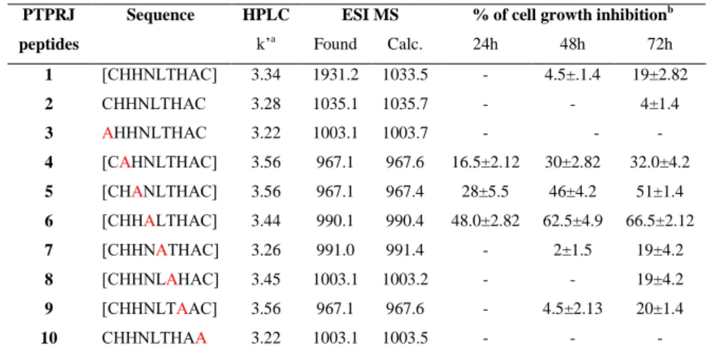
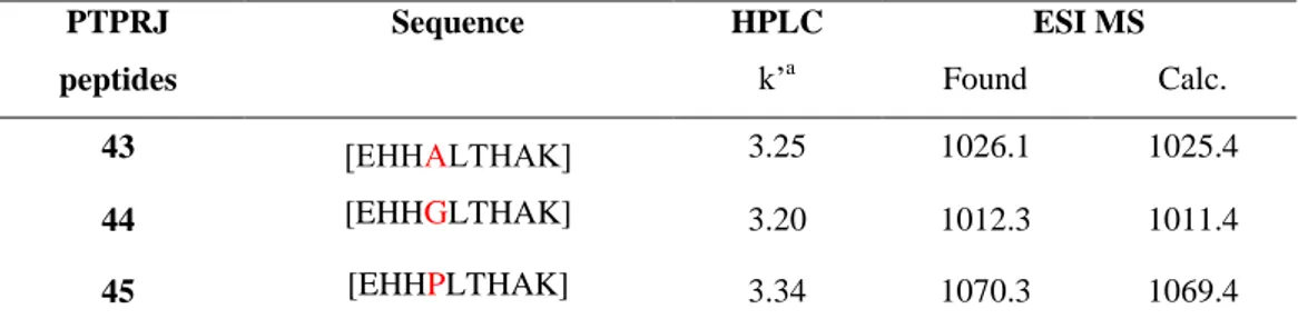
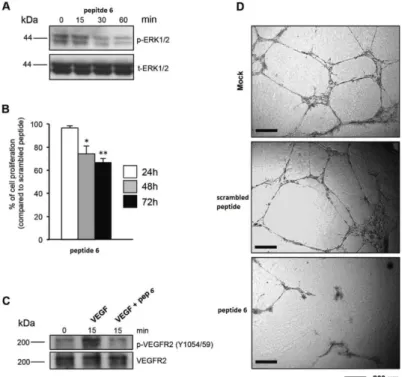
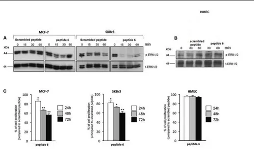
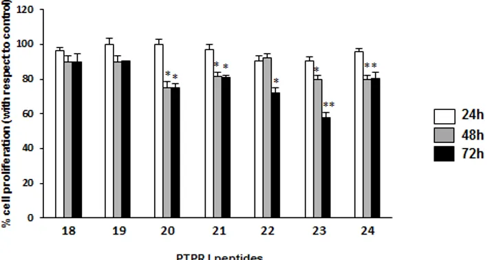
physiological and oncogenic pathways. Our work group previously reported that its expression is strongly reduced in the majority of explored cancer cell lines and tumor samples; moreover, its restoration blocks in vitro cancer cell proliferation and in vivo tumor formation. By means of a phage display library screening, we recently identified two peptides able to bind and activate PTPRJ, resulting in cell growth inhibition and apoptosis of both cancer and endothelial cells. Here, on a previously discovered PTPRJ agonist peptide, [CHHNLTHAC], we synthetized and assayed a panel of nonapeptide analogues with the aim to identify specific amino acid residues responsible for peptide activity. These second-generation nonapeptides were tested on both cancer and primary endothelial cells (HeLa and HUVEC, respectively). Interestingly, one of them ([CHHALTHAC]) was able to both dramatically reduce cell proliferation and effectively trigger apoptosis of both HeLa and HUVECs compared to its first-generation counterpart. Moreover this peptide significantly inhibited in vitro tube formation on Matrigel. Our compound inhibited ERK1/2 phosphorylation and cell proliferation in breast cancer cells (MCF-7 and SKBr3), while no effects were observed on primary normal human mammary endothelial cells (HMEC). Molecular modeling and NMR studies on these peptides reporting the possibility of self-aggregation states and highlighting new hints of structure-activity relationship. Thus, our results indicate that this nonapeptide might represent a great potential lead for the development of novel targeted anticancer drugs.
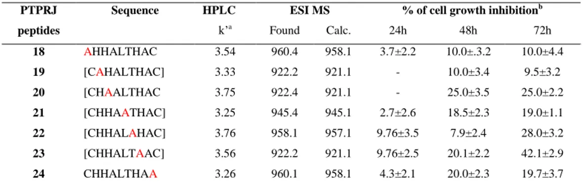
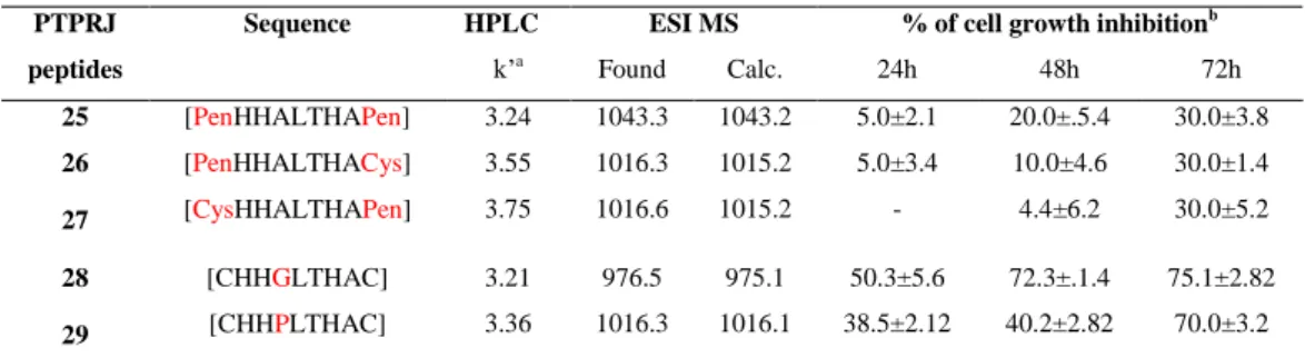
Furthermore and in order to enhance the potency of this peptide, we performed different modification on this hit, including changes to disulfide bridge with lactam bridge, changes in specific position and changes to Cysteine with its analog (Penicillamine).
Keywords PTPRJ, Ala scan peptide, disulfide bridge, lactam bridge,
microwave, HeLa cells, HUVEC cells, Molecular Modeling.
Abbreviations Abbreviations used for amino acids and designation of
peptides follow the rules of the IUPACIUB Commission of Biochemical Nomenclature in J Biol Chem 1972, 247, 977-983. Amino acid symbols denote L-configuration unless indicated otherwise.
- 29 -
The following additional abbreviations are used:
DCM, dichloromethane; DIPEA, diisopropylethyl-amine; DMF, N,N-dimethylformamide; iPr3SiH, or TIS triisopropylsilane; TFA, trifluoroacetic
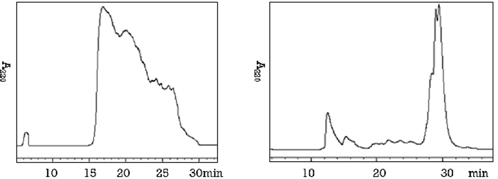
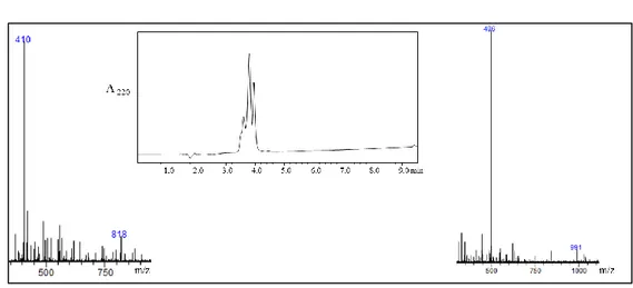
acid; Fmoc, 9-fluorenyl-methoxycarbonyl; HOBt, Nhydroxy- benzotriazole; HBTU, 2(1Hbenzotriazole1yl)1,1,3,3tetramethyluronium hexafluoro -phosphate; Trt, trityl; Pbf, 2,2,4,6,7–pentamethyldihydro benzofuran-5-sulfonyl; RP HPLC, reversed-phase high performance liquid chromatography; ESI, electrospray ionization; LCQ, liquid chromatography quadrupole mass spectrometry; HUVEC, Human umbilical vein endothelial cells.
1. Introduction
Reversible tyrosine phosphorylation, which is governed by the balanced action of protein tyrosine kinases (PTKs) and protein-tyrosine phosphatases (PTPs), regulates important signaling pathways that are involved in the control of cell proliferation, adhesion and migration.
The distinct but complementary function of these enzymes is emphasized by recent studies, in which kinases have been implicated in controlling the amplitude of signaling responses, whereas phosphatases are thought to have an important role in controlling the rate and duration of the response.1,2
The first PTP was purified in 1988, approximately 10 years after the discovery of tyrosine kinases.3 It is now known that PTPs constitute a large, structurally diverse family of tightly regulated, 4 highly specific enzymes with important regulatory roles.5,6
It is also clear that PTPs have both inhibitory and stimulatory effects on cancer associated signaling processes, and that deregulation of PTP function is associated with tumorigenesis in different types of human cancer.
The PTP-superfamily includes 109 genes, compared to 90 human PTK genes, suggesting similar levels of complexity between the two families.
- 30 - 1.1 Protein Tyrosine Phosphatases
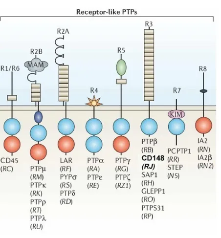
PTPs are broadly divided into receptor-like forms and non-receptor forms.7 The receptor-like PTPs have a single transmembrane domain and variable extracellular domains.
The intracellular parts of most of the receptor-like PTPs contain two tandem PTP domains (D1 and D2) with most, if not all, of the catalytic activity residing in D1. In many cases, the extracellular domains include immunoglobulin like domains and fibronectin type III domains, similar to the extracellular domains of cellular adhesion molecules. Non-receptor PTPs have striking structural diversity and often contain sequences that target them to specific subcellular locations or enable their binding to specific proteins (Figure 1). The catalytic PTP domain spans approximately 280 amino acids and contains a highly conserved active site with a Cysteine residue that is required for catalytic activity. Dephosphorylation of substrates occurs through a two-step mechanism consisting of the formation of a covalent PTP-phosphate intermediate that is subsequently hydrolysed.