UNIVERSITÀ DEGLI STUDI DELLA TUSCIA DI VITERBO
DIPARTIMENTO DI SCIENZE AGRARIE E FORESTALI
Corso di Dottorato di Ricerca in
Protezione delle piante - XXVIII Ciclo
SURVEY OF BACTERIAL DISEASES ON STONE FRUITS IN LEBANON AND INVESTIGATION OF PHENOTYPIC AND GENETIC DIVERSITY OF THE ISOLATED
PSEUDOMONAS SYRINGAE PATHOVARS
AGR/12 PhD thesis of:
Dr. Peter Moubarak
Coordinator: Supervisor:
Prof. Leonardo Varvaro Prof. Leonardo Varvaro
Supervisor:
Dr. Anna Maria D’Onghia
Co-advisor
Dr. Franco Valentini
Co-advisor
Dr. Blanca Landa
i
Summary
Stone fruits are of highly importance in Lebanon covering 17% of the total agricultural lands cultivated with permanent crops. These crops suffer from a diversity of diseases causing losses in production, including many of bacterial origin. Previously, two studies reported bacterial diseases of stone fruits in Lebanon but both of them were incomplete considering the number of samples collected and the identification protocols used at that time.
In order to accomplish this work and evaluate the incidence of bacterial diseases of stone fruits in Lebanon, we carried out a survey in 2013 when we collected 303 samples from all stone fruit growing areas and all commercial species. Results showed that bacterial canker is the main bacterial disease of stone fruits in this country where it appeared to be spread in all regions and on all cultivated species. In fact, preliminary identification of the isolated bacteria using physiological and biochemical tests allowed the identification of 102 Pseudomonas
syringae pv. syringae (Pss), 30 Pseudomonas syringae pv. morsprunorum race1 (Psm1) and 3 Pseudomonas syringae isolates. None of the other common bacterial diseases of stone fruits
including Pseudomonas syringae pv. avii, Pseudomonas syringae pv. morsprunorum race 2,
Pseudomonas syringae pv. persicae, Pseudomonas amygdali, and Xanthomonas arboricola
pv. pruni were found in the sampled orchards. Two gall symptoms suspected to be induced by
Agrobacterium tumefaciens were observed on peach and plum, and the isolates obtained were
conserved for further identification.
Pathogenicity of the collected isolates was assessed by inoculation on immature cherry fruits. With the exception of one Pseudomonas syringae isolate, all the others were able to produce disease symptoms. Interestingly, this technique clearly differentiate between isolates of the pathovar syringae that produced black necrotic lesions while isolates of the pathovar
morsprunorum produced brown, water soaked superficial lesions.
Molecular tools were also used in this study to confirm results of classical identification techniques and to evaluate the genetic diversity within the Lebanese isolates of Pseudomonas
syringae. In this context, we first conducted specific PCR for the detection of the gene coding
for hopAP1 protein reported to be present on the pathovar syringae. Results showed that the majority of Pss isolates (94/102 isolates) possess this effector gene. Later on, BOX-PCR was used as a molecular fingerprinting technique to assess the genetic diversity of the collected isolates. UPGMA analysis of the fingerprint patterns divided the Pseudomonas syringae isolates into three major groups: A, B and C. Pss isolates showed a high genetic diversity producing 17 different patterns distributed according to their similarity level between the group C (87 Pss and 1 Pseudomonas syringae) and the group B (15 Pss and 2 Pseudomonas
ii
syringae). Psm 1 isolates were very homogenous producing the same fingerprinting pattern
forming together the group A.
In order to classify our isolates and to compare them with others from all over the world, 58 representatives of the collected Pseudomonas syringae isolates were analyzed by MLST. The selection of the isolates was based on all the techniques used before in addition to the host plant and the region of origin. MLST was performed by sequencing part of four housekeeping genes (cts, gap A, rpo D and gyr B) that were concatenated to produce a single sequence of 1859 bp. Concatenated sequences were used together with public sequences extracted from Genbank to construct a maximum likelihood phylogenetic tree. The topology of the phylogenetic tree obtained was similar to the one presented by Berge et al. (2014) with correct allocation of phylogroups and clades. MLST divided the Lebanese Pseudomonas
syringae isolates in 2 phylogroups, named PG02 and PG03 according to Berge et al. (2014).
All isolates of the pathovar morsprunorum race 1 analyzed in this study were placed in the PG03 showing to be genetically closely related to each other and to Psm 1 strains from Genbank. Many other pathogens of woody and herbaceous plants were also enclosed in this phylogroup while the pathotype strain of the pathovar morsprunorum (M302280PT=CFBP 2351) that is supposed to belong to the race 2 was allocated in the PG01b. In fact, many previous studies suggested the possibility that the 2 races of the pathovar morsprunorum can be separated into two species considering them as genetically distant and distinct pathogens adapted to the same hosts. The additional sequences of isolates of the race 1 we have added in this study strengthen this hypothesis, proofing the high genetic distance between the 2 races (>8.8 %). Moreover, we found that the PG03 can be divided into at least 2 clades following a threshold of genetic difference of 2.3% that was used for delineation of clades: PG03a (Pseudomonas syringae. pv. lachrymans, pv. mori, pv. phaseolicola race 6 and some unclassified Pseudomonas syringae strains) and PG03b (Pseudomonas syringae. pv.
morsprunorum race 1 and pv. miricae).
Regarding the pathovar syringae, the Lebanese isolates were divided into 2 closely related clades within the PG02 (clades 2b and 2d). This phylogroup is considered to be the most ubiquitous group of Pseudomonas syringae found in all habitats analyzed to date. Nine Pss and one unclassified Pseudomonas syringae appear to belong to the clade 2b together with the type strain (CFBP 1392T) and many other pathovars. To note here that isolates of this clade were isolated from all the stone fruit species surveyed, except apricot trees. The clade 2d includes the largest part of the Lebanese isolates with 37 Pss and one unclassified
Pseudomonas syringae. This clade groups isolates from all stone fruit species and many Pss
iii Results of the two typing techniques used, BOX-PCR and MLST, were analogous to each other. The groups A, B and C of BOX-PCR were equivalent to phylogroups 3, 2b and 2d of MLST, respectively. Interestingly, we found also that isolates that do not possess hopAP1 protein belong to the PG02b while all isolates of the PG02d possess this effector gene.
We also started a preliminary investigation to detect other effector genes in the genome of the collected Pseudomonas syringae isolates. Specific primers were designed for the detection of hopAE1 and hopI1 that appeared to be present in the majority of the tested isolates with the exception of one Pss isolate and one unclassified Pseudomonas syringae isolate. Later on, the amplified regions were sequenced for some isolates and the obtained sequences were used to construct a phylogenetic tree with maximum likelihood method. Congruence was found between the Hop gene and the MLST phylogenies in the case of both hopAE1 and hopI1. This indicates a similar phylogenetic resolution between the core genome (housekeeping genes) and those effector genes that needs to be investigated better in the future.
This study supports solid preliminary conclusions for any future studies dealing with bacterial diseases on stone fruits in Lebanon and Pseudomonas syringae in general. We conducted the first survey dedicated specifically to such kind of diseases and we characterized for the first time isolates of Pseudomonas syringae from Lebanon using different techniques. The data provided will help in investigating the epidemiology, ecology, population genetics, and molecular evolution of this multifunctional group of bacteria.
iv
Thesis committee
- Prof. Stefania Tegli – Università degli Studi di Firenze (DISPAA), Italy.
- Prof. Giorgio M. Balestra - Università degli Studi della Tuscia (DAFNE), Italy.
- Prof. Marta Wilton de Vasconcelos – Universidade de Catolica Portuguesa (Escola Superior de Biotecnologia), Portugal.
v
Acknowledgments
I still remember the first day when I arrived to Italy, it has been five years already. In my mind it was a complete mess. From one side, a lot of confusion, fear, sadness and worries for what would be waiting for me in my new life. Thinking about the beloved ones I left behind in my adorable country Lebanon. But from the other side, a great sense of joy for how much I am lucky to have the opportunity to come here and pursue my studies. Now, I am at the last step before the target that I would not have been able to achieve without the presence of many special people in my life.
First of all, I want to thanks the Mediterranean Agronomic Institute of Bari (CIHEAM-MAIB) that accepted me as a member and opened the door for a better future and a better life. My gratitude goes to the Director Dr. C. Lacirignola, the Deputy Director Dr. M. Raeli and every person working in this institution.
My sincere thanks and appreciations go to the coordinator of “Integrated Pest Management” department in CIHEAM-MAIB, Dr. Anna Maria D’Onghia for her continuous support throughout my studies.
A special thanks to my tutor Dr. Franco Valentini who I consider as a friend with his unique kindness. I also appreciate the help of Dr. Toufic El Beaino to accomplish part of my research study.
My deepest appreciation to my supervisor Prof. Leonardo Varvaro. I will always be thankful for your generous support and continuous interaction in the supervision of this work. Here also I would like to thank Tuscia University for the opportunity to do a PhD and all the great people working there especially Dr. Giorgio Balestra, Dr. Angelo Mazzaglia, Dr. Anna Maria Vettraino and my friend Dr. Alfredo Fabi for their help, each in a different manner.
I am particularly grateful to the enormous availability of Dr. Blanca Landa with whom I accomplished a big part of my thesis. I will always be thankful to you and your institution, the Institute for Sustainable Agriculture-CSIC (Córdoba/Spain).
vi It was also a pleasure for me to do the first part of my thesis in the laboratories of the Lebanese Agricultural Research Institute (LARI-Fanar) under the supervision of Dr. Claudine Sebaaly who I greatly admire.
My dearest friends in Lebanon, Georgio, Abdo and Michelle, thank you for being always present when I needed you even if thousands of kilometres apart.
My big family in Bari, those friends that encouraged me to continue forward, thank you. You know yourself one by one and I am sure that we will always be a family despite in which corner of this world each of us would eventually be. Among you I want to say a special thanks to my smart friend Bachir who helped me in my thesis and supported me in every detail of my daily life, Ramy my big brother who was always available since the first day I have got to know him and finally, Ali and Ali my housemates and my brothers from Iraq.
My friends in Viterbo, you made my life there very enjoyable. I arrived one day to Viterbo not knowing anyone and now I have many real friends. Francesco, Rocco, Matteo, Elisa, Valentina, Claudia, Giacomo, Silvio, Giulia, Anna, Diana, Davide and many others, you will always be in my heart.
I cannot forget to thank my dear friend Dr. Yousseph Rouphael who was the reason behind my coming to Italy. I will be always grateful to you.
Finally, I would like to thank every member of my family, especially my parents, my sweet sister, my brave brother and my dear aunt Marie-Therese, for their support and their prayers that helped me to continue forward and accomplish this title. I hope you are proud of me.
vii
Table of Contents
Summary ... i
Thesis committee ... iv
Acknowledgments ... v
Table of Contents ... vii
List of Figures ... ix
List of Tables ... x
List of abbreviations ... xi
Chapter 1: Literature review ... 1
1.1 Lebanon overview ... 1
1.1.1 Main constraints facing the agricultural sector ... 1
1.1.2 The occupation of the soil and distribution of agricultural lands ... 2
1.1.3 Stone fruits cultivation ... 3
1.1.4 Main Diseases of stone fruits in Lebanon ... 4
1.2 Bacterial diseases of stone fruits with emphasis on bacterial canker ... 6
1.2.1 Bacterial canker of stone fruits ... 6
1.2.2 Bacterial spot ... 18
1.2.3 Crown and root gall disease ... 24
1.3 Management of stone fruit bacterial diseases ... 29
Thesis objectives ... 32
Chapter 2: Materials and Methods ... 33
2.1 Field survey and sampling ... 33
2.2 Isolation and purification ... 33
2.3 Conservation of the isolates ... 34
2.4 Identification of the isolated bacteria ... 34
2.4.1 Physiological and biochemical tests ... 34
2.5 Pathogenicity test on immature cherry fruits ... 38
2.6 Molecular characterization ... 39
2.6.1 DNA extraction ... 40
2.6.2 Detection of Psyr_1890 gene specific to Pss ... 40
2.6.3 BOX-PCR ... 40
2.6.4 MultiLocus Sequence Typing (MLST) ... 41
2.6.5 Preliminary trials on the type three secretion system effectors present in the Lebanese Pseudomonas syringae isolates ... 43
viii
Chapter 3: Results ... 46
3.1 Orchards survey ... 46
3.2 Strain isolation and identification ... 48
3.2.1 Physiological and biochemical tests ... 48
3.3 Infection by stone fruit species and governorates ... 53
3.4 Pathogenicity test on immature cherry fruits ... 55
3.5 HopAP1 detection: ... 59
3.6 BOX-PCR ... 60
3.7 Multilocus Sequence Typing ... 64
3.8 Preliminary detection of some type three secretion system effectors: ... 75
Chapter 4: Discussion ... 78
4.1 Current status of bacterial diseases in stone fruits orchards in Lebanon ... 79
4.2 Molecular characterization of Pseudomonas syringae isolated from stone fruits in Lebanon ... 81
4.2.1 Pathovar morsprunorum ... 84
4.2.2 Pathovar syringae ... 86
4.2.3 Allocation of strains using single gene phylogeny and division of the PG03 into 2 clades 90 4.2.4 Type three secretion system preliminary analysis ... 91
Conclusions and future perspectives ... 94
References ... 96
Annexes ... 120
Annex 1: Information about strains used in MLST analysis ... 120
Annex 2: Strains’ information used in hopI1 phylogeny ... 125
Annex 3: Strains’ information used in hopAE1 phylogeny ... 127
Annex 4: Media used in this study ... 128
ix
List of Figures
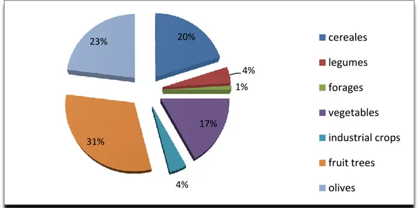
Figure 1: Importance of different crops cultivated in Lebanon (MoA, 2010) ... 3
Figure 2: Distribution of agricultural lands according to Lebanese regions (MoA, 2010) ... 3
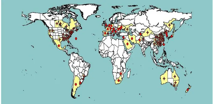
Figure 3: Distribution of stone fruits in Lebanon according to the governorates (MoA, 2010) 4 Figure 4: Distribution map of Xap (EPPO, 2015) ... 19
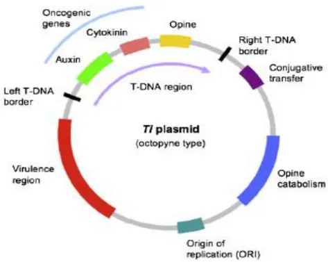
Figure 5: Ti plasmid of Agrobacterium tumefaciens (Pacurar et al., 2011) ... 27
Figure 6: Symptoms of bacterial canker disease of stone fruits observed in the field during the survey. ... 47
Figure 7: Crown gall symptoms observed in the field during the survey. ... 47
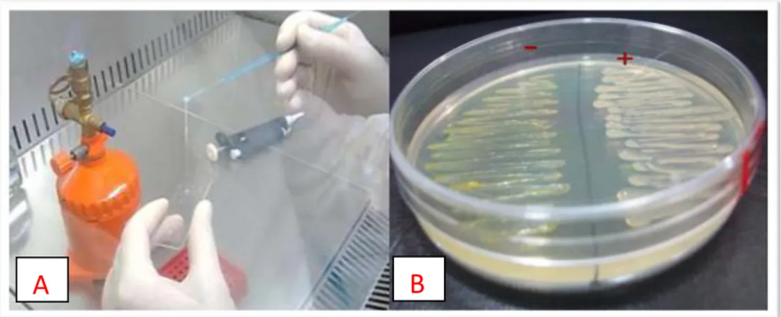
Figure 8: A: KOH test (Gram negative bacteria); B Levan production on NAS medium. ... 49
Figure 9: A: Oxidase production test; B: Potato rotting test. ... 49
Figure 10: A: Arginine dehydrolase test; B: Tobacco hypersensitivity test. ... 49
Figure 11: A: Gelatine hydrolysis test; B: Aesculin hydrolysis test; C: Tyrosinase activity; D: L(+) Tartrate test. ... 51
Figure 12: Incidence of Pseudomonas syringae pathovars and genotypes according to stone fruit species. ... 54
Figure 13: Incidence of Pseudomonas syringae pathovars and genotypes according to Lebanese governorates. ... 55
Figure 14: Symptoms obtained on cherry fruitlets cv. ‘Ferrovia’ inoculated with Pseudomonas syringae. ... 56
Figure 15: Representative gel for the detection of the effector hopAP1. ... 59
Figure 16: BOX fingerprints of the Lebanese Pseudomonas syringae isolates showing variability among strains of the pathovar syringae and homogeneity among strains of the pathovar morsprunorum race 1. ... 60
Figure 17: Dendrogram of genetic similarity of BOX fingerprint patterns generated by 135 Pseudomonas syringae isolates from stone fruit orchards in Lebanon. ... 63
Figure 18: Phylogenetic tree constructed on concatenated sequences (cts, gyr B, gap A and rpo D) of 70 P. syringae strains and 58 Lebanese Pseudomonas syringae isolates ... 65
Figure 19: Contracted phylogenetic tree constructed on concatenated sequences (cts, gyr B, gap A and rpo D). ... 69
Figure 20: Phylogenetic trees constructed based on rpo D partial sequences of P. syringae strains. ... 71
Figure 21: Phylogenetic trees constructed based on cts partial sequences of P. syringae strains. ... 72
Figure 22: Phylogenetic trees constructed based on gap A partial sequences of P. syringae strains. ... 73
Figure 23: Phylogenetic trees constructed based on gyr B partial sequences of P. syringae strains. ... 74
Figure 24: Maximum likelihood phylogenetic tree constructed using amino acid sequence of HopI1 effector. ... 76
Figure 25: Maximum likelihood phylogenetic tree constructed using amino acid sequence of HopAE1 effector. ... 77
x
List of Tables
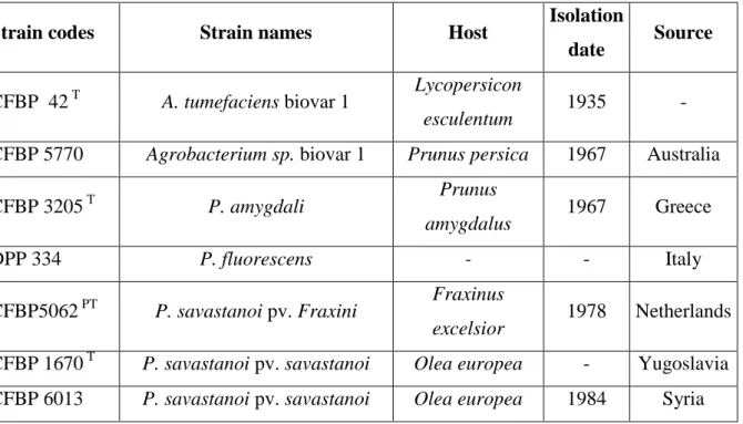
Table 1: Reference strains of Agrobacterium spp., Erwinia spp., Pseudomonas spp. and
Rhodococcus spp. used in this study. ... 37
Table 2: Primers used in this study. ... 44 Table 3: Regions of origin and plant species of the collected samples during the survey conducted on stone fruits in Lebanon in 2013. ... 46 Table 4: Original source, host plant and biochemical characteristics of the Lebanese
Pseudomonas syringae isolates. ... 51
Table 5: Pathogenicity test performed on immature cherry fruits cv. Ferrovia with the
Lebanese Pseudomonas syringae isolates ... 57 Table 6: Number of strains of Pseudomonas syringae generating 1 of 18 BOX genomic fingerprinting patterns. ... 62 Table 7: List of the Lebanese Pseudomonas syringae isolates evaluated by MLST ... 67
xi
List of abbreviations
% Percent
°C Degree Celsius
BOX BOX element of Streptococcus pneumonia
bp Base pairs
cv.(s) Cultivar(s)
DNA Deoxyribonucleic acid
dNTP Deoxynucleotide Triphosphate ELISA Enzyme-linked immunosorbent assay
ERIC Enterobacterial Repetitive Intergenic Consensus
g Gram
GATTa Gelatin, Aesculin, Tyrosinase and Tartrate tests
h Hour
ha Hectare
KB King’s B medium
L Litre
LARI Lebanese Agricultural Research Institute
LB Luria Bertani broth
LOPAT Levan, Oxidase, Potato, Arginine and Tobacco tests
mg Milligram
min Minute
ml Millilitre
MLST Multilocus sequence typing
mM Millimolar
MoA Lebanese Ministry of Agriculture
NA Nutrient agar
xii
NAS Nutrient agar sucrose
PCR Polymerase Chain Reaction
pH Potential Hydrogen
Psm1 Pseudomonas syringae pv. morsprunorum race 1
Pss Pseudomonas syringae pv. syringae
REP Repetitive Extragenic Palindromic sequence Rep-PCR Repetitive extragenic palindromic - PCR
rpm Revolutions per minute
SDW Sterile distilled water
sec. Second
spp. Species
TAE Tris Acetate-EDTA
Taq Thermophilus aquaticus
TTSS Type three secretion system
UPGMA Unweighted Pair Group Method with Arithmetic mean
UV Ultra violet
Xap Xanthomonas arboricola pv. pruni
YDC Yeast extract-Dextrose-calcium Carbonate agar YPGA Yeast-Peptone-Glucose agar
μl Microliter
1
Chapter 1: Literature review
1.1 Lebanon overview
Lebanon is a small country of the Middle East, bordered by Syria to the north and east, Palestine to the south and the Mediterranean Sea to the west with a cost line of 225 km. Its total surface is 10,452 km2 divided into four distinct physiographic regions: the coastal plain, the western mountain range, the Bekaa valley and the eastern mountain range. Having a moderate Mediterranean climate, Lebanon receives a relatively large amount of rainfall but varying in space and time according to the geographical position of each area. In fact, along the coastal part, winters are cool and rainy while summers are hot and humid. Passing to the mountainous areas, temperatures usually drop below freezing during the winter with heavy snow cover that remains until early summer on the higher mountaintops. The Bekaa valley sits between the two mountain ranges and is known by its fertile soil with dry summer and semi-arid winter where the annual average of precipitations can vary from about 700 mm in the South to 250 mm in the North (FAO, 2007).
1.1.1 Main constraints facing the agricultural sector
In 2004, the agricultural sector was estimated to contribute by less than 10 % of the Lebanese economy with 73 % attributed to crops and 23 % to livestock production (FAO, 2007). In fact, this contribution has decreased a lot since the 1960’s especially during the years of conflicts when the majority of the rural population was displaced.
In 2010, the total number of registered farmers was 169.512 showing a decrease of 2% comparing to 1998 with unequal distribution among regions. In marginal areas such as the south, the north and Baalbeck in Bekaa valley, the populations rely mainly on agricultural activities while in big cities and costal part, agriculture has a very low contribution in the economic cycle. A main problem of the remained farmers is the small surface of their lands. According to the Lebanese Ministry of Agriculture (MoA) (2010) 94% of farmers have less than 4 ha covering 49% of the total agriculture land, while those having more than 10 ha represent 2% of the total number of farmers although covering 33% of the arable lands. Another difficulty for the agricultural sector is the low national budget allocated to the MoA that never exceeds 0.5 % of the national budget. This issue affects largely the development of this sector with only few investments in research and new technology implementation. This
2 sector suffer also from inefficient management of land and water use accompanied with poor breeding programs thus conventional techniques of production and cultivation of old cultivars are still widely present.
In the last few years, even with the increased function of the MoA, a lot of work still needs to be done for better agriculture management. Here comes the role of extension services by controlling the quality of the products and by supporting farmers especially regarding plant protection and implementation of new innovative techniques. Another factor to be taken into consideration is the absence of a clear market strategy and regional market agreements. This issue became worst in the last few years because of the conflicts in all surrounding countries which makes impossible the land transportation routes and no enough effort have been placed to find other ways. Here comes the problem of high levels of pesticide residues in the Lebanese fruits and vegetables which is a limiting factor for exportation of agricultural goods to many countries. Finally, an old-new problem is the competitive price that neighboring countries can offer compared to high agricultural production costs in Lebanon.
1.1.2 The occupation of the soil and distribution of agricultural lands
The total cultivated land surface is approximately 230,000 ha covering around 23% of the total surface of the country. Those lands resources are thus very limited, considering that Lebanon has a population around 4.6 million (MoA, 2010). Despite this issue, the topographical and landscape diversity in this country create diverse agro-ecosystems that enable a large variety of agricultural products ranging from tropical to temperate cultivation. Depending on the region, some crops are cultivated under rain fed conditions but nearly 49% of the lands are under irrigation. To mention here that only one third of the surface water available is used for irrigation (FAO, 2007).
As we can see in figure 1, fruit trees occupy 31% of the total surface of cultivated lands followed by olives, cereals and vegetables covering 23%, 20% and 17%, respectively. The remaining 9% of agricultural land is occupied by forages and industrial crops, mainly tobacco and sugar beet (Fig. 1). Accordingly, lands cultivated with permanent crops constitute 54% of the total agricultural lands of Lebanon where the most important crops are olives (43%), stone fruits (17%), pome fruits (11%), citrus (8%), grapes (8%), followed by banana, avocado, anona (cherimoya), pomegranate, kiwi and kaki (MoA, 2010).
3 Figure 1: Importance of different crops cultivated in Lebanon (MoA, 2010)
The distribution of agriculture lands can vary from an area to the other ranking first Baalbek– Hermel region with 23% followed by the rest of the governorate of Bekaa with 19%. Aakar which belongs to North Lebanon comes third with 17 % and the rest of this governorate with 10 %. In the south, the two governorates South Lebanon and Nabtiyeh hold together 22 % of the total agricultural lands of the country and finally Mount Lebanon hold the least surface of agricultural lands with only 10 % that are about narrow terraces in the valleys of this mountainous region (Fig. 2).
Figure 2: Distribution of agricultural lands according to Lebanese regions (MoA, 2010)
1.1.3 Stone fruits cultivation
The total surface cultivated with stone fruits in Lebanon is 21,715 ha equivalent to 17% of the total land covered with permanent crops. The main commercial species cultivated are cherry,
20% 4% 1% 17% 4% 31% 23% cereales legumes forages vegetables industrial crops fruit trees olives 17% 10% 9% 19% 23% 11% 11% Aakkar
rest of North Lebanon Mount Lebanon rest of Bekaa Baalbeck-Hermel South Lebanon Nabatiyeh
4 almond, apricot, peach, nectarine and plum. Their distribution among the Lebanese regions is presented in figure 3, showing that Bekaa valley holds 71% of the total lands cultivated by stone fruits where they are concentrated mainly in the northern part (Baalbek-El Hermel). Second ranks North Lebanon with 18% followed by Mount Lebanon 7% and finally South Lebanon and Nabatiye together with only 4% of the total stone fruits in Lebanon (MoA, 2010).
Regarding the importance of each of the present species, cherry comes first covering 6,172 ha in 2010, a percentage of 28% of the total stone fruit cultivation followed by almond with 5,427 ha (25 %). After those 2 species ranks apricot, peach and nectarine, and plum covering 21, 16 and 9% respectively of the total lands cultivated by stone fruits (MoA, 2010).
Figure 3: Distribution of stone fruits in Lebanon according to the governorates (MoA, 2010)
1.1.4 Main Diseases of stone fruits in Lebanon
Many fungal and bacterial diseases affect stone fruits in Lebanon as in other regions of the world where those species are present. Fungal diseases are considered of major importance
4%
18%
55%
7%
5 and are periodically treated with fungicides by farmers in order to reduce as much as possible economical losses. According to the survey conducted by the Lebanese ministry of agriculture in 2010 and to personal communications with farmers that we have made in 2013, the main fungal diseases of stone fruits in Lebanon are the following: leaf curl (Taphrina deformans), powdery mildew (Sphaerotheca pannosa), shot hole (Wilsonomyces carpophilus) and brown rot (Monilia fructigena and Monilia laxa). Those diseases are usually treated based on a calendar using in the majority of the cases wide spectrum preventive fungicides such Ziram, Tetraconazole and Cupper. Other diseases can also be found but their importance can vary in space and time that are treated when it is necessary (Phytophtora root and crown rot, Armillaria root rot, Verticilium wilt …).
Regarding bacterial diseases, bacterial canker caused by Pseudomonas syringae pv. syringae (Pss) and Pseudomonas syringae pv. morsprunorum (Psm) have been reported in Lebanon. Bacterial spot disease caused by the quarantine bacterium Xanthomonas arboricola pv. pruni (Xap) and, crown and root gall disease caused by Agrobacterium tumefaciens, were also reported previously (EPPO, 2012). In the literature, the only published report on the occurrence of bacterial diseases on stone fruits in Lebanon goes back to 1969’s. This study was done by Saad and Nienhaus when they wrote a general report about plant diseases in Lebanon, including bacterial diseases of stone fruits. At that time they described that canker symptoms on almond trees that were observed in one location on the coast, were induced by
Xanthomonas pruni (new name Xap). Also on peach, the same bacterium was isolated from
Mount Lebanon and the symptoms were described as leaf-twig spots. Furthermore,
Pseudomonas morsprunorum (new name Psm), causing cankers and gummosis, was isolated
on cherry in Bekaa and on peach in Mont Lebanon. Agrobacterium tumefaciens was reported only on plum in Bekaa valley (Saad and Nienhaus, 1969).
Recently, the Lebanese Ministry of Agriculture conducted a survey dedicated to report different kind of diseases in stone fruit orchards. Regarding bacterial diseases, only few samples showing symptoms that may be of bacterial origin were collected, and it was reported that bacterial canker on cherry, almond and plum was caused by Pss while on one sample of peach it was caused by Psm race 1. None of the other bacterial diseases known to induce diseases on stone fruits, including Xap and Agrobacterium tumefaciens, were isolated during this survey (MoA, 2011, 2012).
6
1.2 Bacterial diseases of stone fruits with emphasis on bacterial canker
Many bacterial diseases affect stone fruit orchards in many regions of the world. In this thesis, we mainly discussed the most widely distributed diseases such as bacterial canker caused by different pathovars of Pseudomonas syringae, bacterial spot caused by Xanthomonas
arboricola pv. pruni and, crown and root gall caused by Agrobacterium tumefaciens. Special
emphasis was given to bacterial canker that is the main bacterial disease of stone fruits in Lebanon. Bacterial decline of peach caused by the quarantine bacterium Pseudomonas
syringae pv. persicae is also of highly importance but its distribution is limited to few
countries. Some other Pseudomonas spp. can also induce diseases on stone fruits but they are considered of less importance. The quarantine bacterium Xylella fastidiosa was also reported on peach, plum and almond from some countries but it will not be discussed in this thesis.
1.2.1 Bacterial canker of stone fruits
Pseudomonas spp. is a polyphagous bacterium causing diseases on both annual and perennial
plants, including fruit trees, ornamentals and vegetables (Agrios, 2005). It is one of the most adaptive plant pathogenic bacteria able to produce a variety of symptoms such as leaf spot, leaf blight, leaf speck or bacterial canker of wide range of plant species all over the world (Vinatzer et al., 2006). It is known to live part of its life as epiphytic on plant surface than later on, under convenient conditions, it is able to infect the plant and reach the apoplast (intercellular space) as a pathogenic endophyte (Hirano and Upper, 2000).
Diseases of different fruit tree species caused by Pseudomonas spp. are of major concern in fruit producing areas worldwide. They are extremely difficult to control a reason that gives them the ability to cause significant economical losses. Those pathogens have a complicated genetic diversity and consistent methods of identification and discrimination between different pathovars and strains do not exist yet (Vicente et al., 2004; Donmez et al., 2010). On stone fruits, 2 pathovars of Pseudomonas syringae, pathovar syringae and pathovar
morsprunorum, cause a disease called ‘bacterial canker of stone fruits’. Both causal agents are
spread almost everywhere where stone fruits are cultivated. A third pathovar, pv. persicae
(Psp), classified as a quarantine bacterium in Europe and it is included on the European Plant
Protection Organization A2 list, is also able to cause ‘bacterial decline of peach’ disease. This one still has a limited distribution in the world and it was reported so far in France, New Zealand and UK (EPPO, 2005, 2006). Other two species of less importance are Pseudomonas
7
amygdali the causal agent of ‘hyperplastic bacterial canker of almond’ and Pseudomonas syringae pv. avii newly reported to cause disease on wild cherry in France.
1.2.1.1 History and current geographical distribution
For a number of years, Pseudomonas syringae causing bacterial canker of stone fruits was known under different names according to the system of classification followed at the time and the wide range of hosts that this pathogen can infect. The name Pseudomonas syringae refers to Van Hall (1902) of the University of Amsterdam, when he proved the pathogenicity of this bacterium causing blight on lilac. At the same time in Poland, Brzezinski (1902) who was working on bacterial canker of stone fruits determined that gummosis and dieback of peach, plum, apricot and sweet cherry trees were of bacterial origin. In 1907, Aderhold and Ruhland described the pathogen causing death of sweet cherry trees in Germany as Bacillus
spongiosus. Few years later, Griffin (1911) reported that Pseudomonas cerasi was the causal
agent of gummosis and cankers on sweet cherries in USA (Bultreys and Kaluzna, 2010).
Pseudomonas morsprunorum was characterized for the first time in England by Wormald
(1932) as the causal agent of bacterial canker of plum trees. Wormald noticed also that another bacterium, Pseudomonas prunicola, was frequently accompanied with bacterial canker of stone fruits and blossom blight of pear (Wormald, 1932, 1937). Later on, researches figure out that B. spongiosus, Pseudomonas prunicola and Pseudomonas cerasi are all the same bacterium and they had to be considered as Pseudomonas syringae other than
Pseudomonas morsprunorum was retained as different species (Crosse and Garrett, 1963;
Garrett et al., 1966). Few years later, Pseudomonas morsprunorum race 2 was described, differing from the one known before in some biochemical and pathological characteristics (Freigoun and Crosse, 1975).
Pseudomonas syringae pv. persicae was described for the first time in 1967 on nectarine and
peach in France and almost simultaneously on nectarine, peach and Japanese plum in New Zealand (Young, 1988). It was also reported that the same pathogen was isolated once in the UK in 1966 from Prunus cerasifera (EPPO, 2005). Some unreliable data were also published about the presence of this bacterium in other countries but those reports cannot be taken into consideration (EPPO-PQR, 2015).
Pseudomonas syringae pv. avii isolated from wild cherries was described in 2003 by Menard
and his colleagues. This finding explained the identity of many isolates obtained from cherry trees during subsequent years. Those were considered before as intermediate non typical forms, physiotypes, or ecotypes because they are different in some biochemical, physiological
8 and genetic properties from typical Pseudomonas syringae or Pseudomonas morsprunorum (race 1) (Dowler and Weaver, 1975; Sobiczewski, 1984; Bultreys and Gheysen, 2003; Renick
et al., 2008).
At the moment, the taxonomic position of Pseudomonas syringae and Pseudomonas
morsprunorum has been changed to Pseudomonas syringae pv. syringae and Pseudomonas syringae pv. morsprunorum, respectively (Young et al., 1978; Young et al., 1992). This
classification of pathovars showed that Psm race 1 and Psm race 2 are clearly distinct organisms because they belong to the Genomospecies 2 and 3, respectively, whereas Pss belongs to the Genomospecies 1 (Gardan et al., 1999; Ménard et al., 2003).
Currently, bacterial canker of stone fruit occurs in all regions of stone fruit production in the world (Hattingh and Roos 1995; Agrios, 1997; Kennelly et al., 2007). The two main pathovars causing bacterial canker disease, Pss and Psm, are well adapted to different climatic conditions and cause severe damages in many countries.
1.2.1.2 Damages and economical importance
Pseudomonas syringae can induce serious diseases on stone fruits causing often high
economical losses (Scortichini et al., 2003; Vicente and Roberts, 2007; Renick et al., 2008; Gilbert et al., 2009; Kaluzna et al., 2010a). However, since the aggressiveness of the disease is not stable from season to season and varies between orchards and growing areas, only few quantitative data are available regarding this aspect. Damages can occur in nurseries, in wild cherry fields for wood production as well as in commercial orchards of stone fruits where reduction in fruit yield, quality and orchard life time can be dramatic (Vicente et al., 2004; Agrios, 2005; Janse, 2006; Kennelly et al., 2007). Losses can be the result of tree decline and death due to the development of cankers that girdle branches and main trunk or as direct reduction of the productivity due to cold induced by those bacteria leading to death of buds and flowers (Ogawa and English, 1991). In Germany, up to 30% of trees of plum orchards are uprooted every year because of bacterial canker, even with intensive copper sprays. Same losses were reported from Italy in one year old apricot orchards damaged by the same disease (Scortichini, 2006). In Turkey, almost 80% of apricot trees suffer from bacterial canker in Erzurum, Erzincan and Artvin (Kotan and Sahin, 2002) and 20% in Malatya (Donmez et al., 2010). Other countries such as Poland, Iran, France, USA, UK, Germany, New Zealand and Lithuania are also facing serious losses due to this disease (EPPO, 2005; Hinrichs-Berger, 2004; Vicente et al., 2004; Vicente and Roberts, 2007; Janse et al., 2008; Karimi-Kurdistani and Harighi, 2008; Vasinauskiene et al., 2008; Kaluzna et al., 2010a).
9
1.2.1.3 The causal agents
Pseudomonas syringae is a gram negative bacterium belongs to the genus Pseudomonas sensu stricto, included in the γ subclass of the Proteobacteria (Kersters et al., 1996). It is an aerobic,
motile, straight or slightly curved rod shape bacterium with one or several polar flagella (Holt
et al., 1994; Palleroni, 2005). They also produce the pigmented iron chelating siderophores
pyoverdins that are fluorescent under UV light. As many plant pathogenic bacteria they produce phytotoxins such as the two toxic lipodepsipeptides (TLP) syringomycins and syringopeptins produced by the pathovar syringae which play an important role in the virulence of the bacterium (Young et al., 1992; Sorensen et al., 1998; Bultreys and Gheysen, 1999; Gilbert et al., 2009).
Pseudomonas syringae includes saprophytic and pathogenic species harmful to human,
mushrooms and one of the most important plant pathogenic Pseudomonas species. Some strains were also isolated from environmental habitats including those closely linked to the water cycle outside of agricultural contexts (Berge et al., 2014).
This complex group is known to be very heterogeneous, causing diseases to more than 180 plant species including fruit trees, vegetables, ornamentals, and other annual and perennial plants (Bradbury, 1986; Young et al., 1996). This diversity was the reason why this species was divided into at least 57 pathovars (Gardan et al., 1997; Young, 2010) and nine Genomospecies (Gardan et al., 1999).
The pathovar syringae is maybe the most heterogeneous pathovar having the ability to cause diseases to a large number of unrelated plant genera, including Prunus species (Bradbury 1986; Young 1991; Weingart and Völksch 1997; Little et al., 1998; Vicente and Roberts 2007). Strains of Pss were isolated and identified from symptomatic plant tissues of woody and herbaceous hosts based on biochemical and physiological characteristics, and pathogenicity tests on different plant species (Little et al., 1998; Scortichini et al., 2003). In fact, because of the high diversity of strains of this pathovar, it is always recommended for an accurate identification to conduct pathogenicity tests on susceptible host plants since classical methods of identification are not enough (Little et al., 1998; Vicente and Roberts, 2007). Some studies reported that Pss strains isolated from a specific host are able to cause disease on a diversity of plant species (Scortichini et al., 2003; Vicente et al., 2004; Gilbert et al., 2009) while others are specific to the host of isolation such in the case of strains infecting grasses (Gross and De Vay, 1977) and beans (Cheng et al., 1989). Moreover, in a study conducted by Little et al., (1998), results showed that Pss isolated from stone fruit formed a distinct cluster separate from most of strains isolated from other hosts. Also strains from
10 different zones showed a genetic diversity among each other’s (Gonzalez et al., 2000) but it was not always the case that genetic diversity is related to host plant and/or region. According to Martín-Sanz et al. (2013), Pss strains isolated from peas were sometimes less virulent when artificially inoculated on the host of isolation itself than strains isolated from other plant species. They conclude that there is genetically and pathogenically distinct Pss strain groups from pea, a factor to be taken into consideration for the diagnostic and epidemiology of this pathogen and for disease resistance breeding.
This inconsistency makes the classification of this group of bacteria and the designation of pathovar a very complicated work. A standard protocol, either for identification or characterization must be followed in order to assess the host specificity and the virulence of different genetic and pathogenic groups of strains of the pathovar syringae.
The second pathovar, Pseudomonas syringae pv. morsprunorum, has a much narrower host range (Bradbury, 1986). It is more homogeneous than Pss but heterogeneity is known to occur within this pathovar since two genetically different races were described based on physiological and pathological characteristics (Freigoun and Crosse, 1975; Ménard et al., 2003; Vicente et al., 2004; Vicente and Roberts, 2007). Psm race 1 (Wormald, 1932) belongs to the Genomospecies 2 and it is pathogenic to cherry, plum and apricot, while Psm race 2 (Freigoun and Crosse, 1975) belongs to the Genomospecies 3 and it is pathogenic mainly to cherry (Bultreys and Kaluzna, 2010). Regarding isolates from cherry trees, it was noticed in Belgian orchards that Psm race 1 were more frequently isolated from sweet cherry and Psm race 2 mostly from sour cherry (Bultreys et al., 2007; Gilbert et al., 2009).
Pseudomonas syringae pv. avii is a newly described pathovar that infects wild cherry
cultivated for wood production in France. It belongs to the Genomospecies 3 and strains of this pathovar show high genetic homogeneity among each other’s (Ménard et al., 2003). Furthermore, the pathovar persicae belongs also to the Genomospecies 3 and it is pathogenic to peach, nectarine and Japanase plum (Young, 1988; EPPO, 2005). This is the only
Pseudomonas syringae pathovar classified as a quarantine bacterium A2 list by the European
Plant Protection Organization (EPPO, 2005).
Here we have to mention also Pseudomonas amygdali the causal agent of hyperplastic bacterial canker of almond. This bacterium is of less importance and it was reported only from Afghanistan, Greece and Turkey (EPPO-PQR, 2015).
As in the case of the pathovar syringae, different phytotoxins are produced by the other pathovars, a characteristic used in identification either by direct detection of the secondary metabolite itself or by detection of the genes involved in its production or secretion.
11 Coronatine is produced by Psm race 1 strains and the siderophore yersiniabactin is produced by the pathovars morsprunorum race 2, avii and persicae (Bereswill et al., 1994; Sorensen et
al., 1998; Bultreys and Gheysen, 1999; Bultreys et al., 2006). Moreover, Pseudomonas syringae pv. persicae secretes several substances named persicomycins representing a new
family among the phytobacterial toxins (Barzic and Guittet, 1996). All of them cause necrosis of peach tree tissues and they are involved in the die back disease of peach trees.
An important characteristic of some species of the genus Pseudomonas, including
Pseudomonas syringae, is the ice nucleation active (INA). In fact, those bacteria are able to
induce ice formation from water supercooled below 0°C (Lindow, 1983; Hirano and Upper, 2000). In Pseudomonas syringae, this ability is common for epiphytic strains not assigned to a pathovar and for the pv. syringae; but it was never reported for strains of the pathovar
morsprunorum race 1 (Lindow, 1983; Mittelstädt and Rudolph, 1998). It is known that frost
injury predisposes stone fruit to disease caused by Pss but it is not answered yet whether INA
Pss could induce frost injury on fruit species (Sobiczewski and Jones, 1992; Bultreys and
Kaluzna, 2010). However, according to Andrews et al., (1986) flowers have a lower water super cooling temperature than stems which makes them vulnerable to freezing injury during spring frost with the presence of INA bacteria even under very mild and transient frost. This characteristic causes a direct loss in production and unfortunately the attempts to limit freeze injury in stone fruit and pear orchards by controlling INA Pss are not yet successful (Cody et
al., 1987; Mittelstädt and Rudolph, 1998).
1.2.1.4 Ecology and biology of the bacterium
Pseudomonas syringae pathovars are able to colonize leaf surfaces of host trees and weeds in
the orchard, as epiphytic stage of the bacterium life. The epiphytism of plant pathogenic bacteria was first described on Psm by Crosse (1957, 1959). He found that the population of
Psm race 1 is able to survive and multiplicate on sweet cherry leaves without causing
symptoms. During hot dry summer, this population decreases considerably and reaches its lowest levels. Later on, an increase in bacterial population is observed during autumn when the temperature decrease and first rains start. In general, the bacteria responsible of bacterial canker have a facultative summer leaf spot stage, and an obligate spring, summer and autumn leaf epiphytic stage, with an overwintering stage within dormant buds and cankers (Crosse, 1955, 1956, 1957; Bultreys and Kaluzna, 2010; Scortichini, 2010). The natural drop of the leaves in autumn allows the pathogen transported by rain and wind or present already as epiphytic on plant surface to enter through leaf scars. Bacterial multiplication in cortical
12 tissues start from late autumn to early spring, but no important cankers develop before spring. Infected buds can remain symptomless or they can be killed by the pathogen before their opening in spring (Hatting et al., 1989). At that time, the pathogen colonizes leaves, blossoms and young fruits which under favorable wet conditions show spot symptoms. Colonization of leaf stomata without symptoms formation (Roos and Hattingh, 1983) and systemic invasions from leaves through the veins to other tissues of the plant have also been reported (Roos and Hattingh, 1987; Sundin et al., 1989). However, when the weather becomes more and more dry during late spring and summer, the overwintered cankers of the previous year become dry and the bacterial population decrease dramatically (Bultreys and Kaluzna, 2010).
All events affecting the health of the tree or causing wounds can be used by the pathogens to increase the infection level. Consequently, pruning wounds, poor nutrition, plant parasitic nematodes, frost injury, hails injuries, favor the penetration and the spreading of the bacteria inside the tree tissues and among trees (Hinrichs-Berger, 2004; Bultreys and Kaluzna, 2010). For example, cultivar and rootstock sensitivity seems to be a main predisposing factor enhancing the virulence of Pseudomonas syringae to stone fruits (Scortichini, 2006). As well, low calcium content, sandy or very clayey can enhance the sensitivity of apricot and peach trees to Pss infection. Moreover, a period of freezing followed by thawing, promote the displacement of endophytic bacteria within the stem tissues (Weaver, 1978; Vigouroux, 1989). However, this appeared to favor the multiplication and longitudinal progression of Pss while Psm race 1 was more efficient in lateral infection of cortical tissues and it is not favored by frost (Sobiczewski and Jones, 1992; Bultreys and Gheysen, 1999; Gilbert et al., 2010). It was also reported that Pss infection is more efficient through wounds while leaf scars are the main infection point of Psm race 1. Another difference between those 2 pathovars is that Pss population die out in cherry cankers earlier in dry summer than Psm race 1 (Garrett et al., 1966).
Knowing the behavior of the pathogen and the factors that can enhance or slow down the disease remain indispensable. This is the basic to determine the optimal method and time to apply control measures. To understand better this behavior there is need to have more knowledge about genetic characteristics of each pathovar and even strains within a specific pathovar that surely have a direct impact on epidemiology and pathogenicity of those bacteria.
13
1.2.1.5 Symptoms
Bacterial canker affects many parts of the tree and symptoms include blossom blast, spur dieback, leaf and fruit lesions, cankers associated with gummosis of woody tissue and overall tree decline (Hattingh and Roos, 1995; Renick et al., 2008; Bultreys and Kaluzna, 2010). The type and severity of the disease symptoms depends on many factors regarding cultivar, age of the infected tree, plant tissue invaded, strain of the pathogen and environmental factors (Gašić
et al., 2012). Anyhow, the most characteristic and destructive symptoms are cankers on trunk,
limb and branches that are often located around spurs, pruning wounds, on twigs at the base of flowers, leaf buds and at branch junctions. At those points the pathogen enters and makes circular to elongated, brown, water-soaked lesions in the bark. Cankers may expand upward rapidly early in the spring accompanied with gum exudation with diebacks that appear on terminal shoots or twigs because of the girdling of the main trunk or branches. Infected buds may fail to grow during the next spring or if few leaves develop, they will wilt soon during first days of hot summer (Goto, 1992; Hattingh and Roos, 1995; Bultreys and Kaluzna, 2010; Gašić et al., 2012).
When they are expressed, symptoms on the leaves are rounded to irregular lesions of different sizes, water-soaked, light brown color in early spring that turn darker with time surrounded by a yellow halo. Those spots become necrotic and rapidly drop out to produce a shot hole effect. Blossom blast is also common, showing brown, shriveled flowers often fall down before full opening. Immature fruit lesions are small, brown-black necrotic spots becoming sunken with dark center as the fruit matures. An important aspect is that leaves and fruits symptoms are not always formed depending on the susceptibility of cultivars and the amount of rainfall (Bultreys and Kaluzna, 2010; Kaluzna et al., 2012). Anyhow, when they are numerous on fruit surface they prevent the normal development of fruit epidermis, resulting in rusted areas or deformed fruits (Young, 1987).
Regarding bacterial decline caused by P. s. pv. persicae symptoms can be different on each of its hosts (EPPO, 2005). On Japanese plum, infection is mainly on nodes and a characteristic symptom is tip dieback with some occasional death of laterals and fruiting arms. On nectarine and peach, symptoms include shoot dieback, limb and root injury, tree death, leaf spots and fruit lesions. Dissection of the lesions show brown necrosis and water soaked areas developing along the vascular bundles (Young, 1987). They may extend more than one meter and girdle a big part of the tree with no distinct margin between healthy and necrotic tissue in lower parts of the tree. This is a distinctive aspect of bacterial decline from bacterial canker
14 (Hattingh and Roos, 1995). Moreover, in the case of bacterial decline the rootstock can also be infected, showing symptoms similar to those on woody shoots (EPPO, 2005).
In general, regarding host range and symptoms caused by Pss, Psm, Psa and Psp, it is difficult to distinguish between them in the field. This may have significant consequences because Psp is the only one considered as quarantine bacterium in many countries (EPPO, 2005; Gašić et
al., 2012).
1.2.1.6 Identification and characterization
Visual inspection of symptoms in the field would be very useful as first inspection to check the presence of diseases caused by Pseudomonas spp.. Cankers, necrotic twigs, leaf or fruit spots, gummosis or other symptoms should be taken into consideration. The problem is that those symptoms are not specific to diseases caused by this group of bacteria and they can be induced by other bacterial species or fungal pathogens and even by abiotic stress.
Accordingly, attention should be paid in order to accurately identify the causal agent of the disease to implement the most convenient control strategies. Traditionally, diagnostic and detection techniques used for plant pathogenic bacteria are based on microscopic observation, isolation on culture media, serological testing, bioassays and molecular assays. Isolation on culture media is time and labor consuming and confirmation of results may take several weeks (López et al., 2010). Anyway, it still indispensable in many cases to isolate the causal agent and to fulfill Koch postulates as it is mentioned in EPPO protocols.
Regarding phenotypic identification and characterization of Pseudomonas syringae, it is recommended to make isolation on King’s B medium (King et al., 1954) since isolates of this group of bacteria are known to produce fluorescent pyoverdin observed under UV light. To be aware that some pathovars isolated from stone fruits are not fluorescent such as some strains of Psm and all strains of the pathovars persicae and avii (Bultreys and Kaluzna, 2010).
After this, LOPAT tests are generally used to discriminate between Pseudomonas spp. (Lelliot et al., 1966). Those 5 tests are able to divide species of this genus into different groups where Pseudomonas syringae are classified in the group Ia. Bacterial species of this group are able to produce levan and to induce hypersensitivity reaction on tobacco leaves but they are not able to produce cytochrome C oxydase, pectinase neither arginine dehydrolase. Continuing with classical identification tools, GATTa tests are used to differentiate between
Pseudomonas syringae pathovars isolated from stone fruits (Latorre and Jones, 1979). Those
4 tests are very helpful with the exception of their incapability to clearly separate strains of the pathovar morsprunorum race 2 that show variable results (Gilbert et al., 2009).
15 Serological techniques are usually used as first screening for massive detection and the accuracy of data is related mainly to the quality of the antibodies. The main techniques used are indirect immunofluorescence (IF), enzyme-linked immunosorbent assay (ELISA) and recently lateral flow devices. In any case, serological techniques to detect Pseudomonas spp. are not commonly used because of the lack of specific polyclonal or monoclonal antibodies, so scientists do not advise their use for diagnosis or detection in the case of this group of bacteria (López et al., 2010).
In the last years, the use of molecular techniques has increased rapidly. Conventional Polymerase Chain Reaction (PCR), repetitive extragenic palindromic PCR (rep-PCR) and recently real-time PCR are more frequently used since they are highly sensitive techniques and relatively easy to perform. Many specific primers were developed to detect genes coding for the production of toxins or siderophores that are produced by Pseudomonas spp. such as
syrB and syrD genes, involved in the synthesis and export of these lipodepsipeptides among Pss strains (Bultreys and Gheysen, 1999; Sorensen et al., 1998). A limit of this technique is
that many toxins are not specific to one pathovar or even one species and results can be inaccurate (Bultreys and Gheysen, 1999). According to this fact, using PCR for the detection of secondary metabolites is not enough for the detection of all bacterial species and it is recommended to be combined with other techniques. Moreover, many specific primers were designed to detect different regions in the genome of Pseudomonas syringae pathovars such as Psyr_1890 primer pair used for the detection of Pss (Vieira et al., 2007). It was reported that those primers are able to detect the presence of hopAP1 effector that is specific to the pathovar syringae enabling the discrimination of strains of this pathovars from others.
For molecular characterization of Pseudomonas syringae isolated from stone fruits, rep-PCR using REP, BOX and ERIC, may be the most used technique. It is able to discriminate between different pathovars and to illustrate the genetic diversity that exists among strains of each of them. The only difficulty is the case of strains of the pathovar syringae that according to rep-PCR showed to be very diverse while it is very easy to identify strains of the other pathovars from stone fruits using this technique (Ménard et al., 2003; Vicente and Roberts, 2007; Gilbert et al., 2009; Kaluzna et al., 2010b; EPPO, 2005). In fact, this technique divided
Pss strains in many genetic groups according to pattern produced. In some studies, it was
reported that it does not exist a clear relationship between host plant and bacterial genomic fingerprint (Schortichini et al., 2013) while according to other references it was reported the presence of host-pathogen relationships within Pss and possible specializations of clonal populations on different hosts (Little et al., 1998; Gilbert et al., 2009). Variability was also
16 observed in pathogenicity and virulence tests on a variety of plant species between strains from different host of isolation (Schortichini et al., 2013). For this reason, it was recommended that pathogenicity test on susceptible host should be always conducted in the case of Pss strains (Gašić et al., 2012, Latorre and Jones, 1979; Vicente et al., 2004).
Molecular techniques based on sequencing of specific genes are more frequently used in the last years. This is due to the decrease in the cost of sequencing comparing to the past, the assured results obtained and the possibility to compare results with a large data base. Rapid determination of 16S ribosomal RNA sequences for phylogenetic analyses (Lane et al., 1985) and newly Multilocus Sequence Typing (MLST) were often used. This latter technique is based on housekeeping gene analysis revealing high discrimination among Pseudomonas
syringae strains. Many studies were published during the latest years in which MLST was
used to analyze the diversity and to classify strains of the complex Pseudomonas syringae group (Sarkar and Guttman, 2004; Hwang et al., 2005; Kaluzna et al., 2010b; Bull et al., 2011; Clarke et al., 2010). One of the most valuable studies was that of Berge et al. (2014) performing MLST on 216 Pseudomonas syringae isolates by partially sequencing four housekeeping genes (cts, gap A, rpo D, gyr B). They figure out that the Pseudomonas
syringae complex is divided into 23 clades within 13 phylogroups. They also analyzed the
phenotypic characteristics of strains that belong to each of these phylogroups and clades. The identified phylogroups have shown to be equivalent to the Genomospecies described by Gardan et al., (1999) based on DNA/DNA hybridization. In fact, Gardan et al. (1999) described 9 Genomospecies among the pathovars of Pseudomonas syringae where four of these represented the majority of species and pathovars of the complex. According to this classification, Pseudomonas spp. causing diseases on stone fruits were allocated in a way that the pathovar syringae belongs to the Gsp1 (=PG02 of Berge et al., 2014), the pathotype strain of the pathovar morsprunorum, the pathovar avii and the pathovar persicae belong to the Gsp 3 (PG01), and finally Pseudomonas amygdali belongs to the Gsp2 (PG03). To mention that it was reported that the pathotype strain of the pathovar morsprunorum is not representative of the pathovar (Young et al., 1996) and Gardan et al. (1999) showed that another strain of the pathovar morsprunorum (CFBP 2116) is a member of Genomospecies 2 (=PG03) and this strain is the proposed as neopathotype strain (Young, 2010).
1.2.1.7 Type three secretion system (TTSS)
Understanding the strategies used by microbial pathogens to infect and cause diseases in their host cells is taking more and more attention during the last years. It is known that many of
17 those pathogens transport effector proteins into the cytoplasm of the host cells in order to alter their metabolism, destabilize their immunity system and encourage their multiplication (Lindeberg et al., 2012).
Pseudomonas syringae is a model bacterium to study effector repertoires as dynamic systems.
It possesses the type three secretion system (TTSS), as many groups of bacteria, which is the main factor that enables them to cause disease. This system secretes and/or translocates a group of effector proteins that alter host cellular processes and promote disease development (Jin et al., 2003). The TTSS is only expressed after the bacterium comes into direct contact with the host. After that, the TTSS pilus is produced forming a direct channel between the pathogen and its host. TTSS effector proteins are directly inject through this pilus into the cytoplasm of the host, where they target host proteins and modulate the host defense response. TTSS effectors are known to suppress the defense response by interfering with signal transduction, causing cytoskeletal changes, or by having direct cytotoxic effects (Collmer et
al., 2002; Guttman et al., 2006).
Effectors are encoded by the hypersensitive reaction and pathogenicity genes hrp/hrc and in
Pseudomonas syringae they are usually designated as Hop proteins (hrp outer protein)
because they are able to move through the TTSS (Lindeberg, et al., 2005). Another set of effectors known as avirulence genes (avr), reduce the ability to cause disease if they get to be recognized by resistance proteins (R) of the plant. This reduction in pathogenicity is done by activation of the plant defense system (Lindeberg et al., 2005; Vinatzer et al., 2006).
According to this, the role of effectors can be divided into 2 functions. First of all, they suppress pathogen-associated molecular pattern (PAMP)-triggered immunity (PTI) which is evolved by common bacterial features such as flagellin, lipopolysaccharide (LPS), peptidoglycan and elongation factor Tu (Boller and Felix, 2009). Furthermore, the immunity system of plants is able to detect the effectors injected by the pathogen by resistance (R) proteins which results in localized programmed cell death known as the hypersensitive response (HR) (Lindeberg et al., 2012). As definition, hypersensitivity reaction (HR), is a phenomena that is produced when a high concentration of avirulent strain of bacteria is infiltrated in the apoplast of a non-host plant species or a resistant cultivar, that will cause a rapid programmed cell death of the infiltrated tissue (Lindgren et al., 1986; Greenberg and Yao, 2004).
Genomic sequence data of Pseudomonas syringae strains (Buell et al., 2003; Feil et al., 2005) in combination with in vitro and in vivo screening and bioinformatics analysis have revealed that strains of Pseudomonas syringae contain dozens of effectors that vary in size and
18 composition among strains (Guttman et al., 2002; Petnicki-Ocwieja et al., 2002; Greenberg and Vinatzer, 2003; Chang et al., 2005). According to the ‘Pseudomonas syringae genome resources’ website (http://www.Pseudomonas-syringae.org), Pseudomonas syringae pv.
tomato (DC3000), Pseudomonas syringae pv. syringae (B728a) and Pseudomonas syringae
pv. phaseolicola (1448a) share only 13 effectors and the remaining effectors are either unique to one of these strains or only shared between two of them. It is believed that these differences in effector repertoires among strains are the main determinants of host range in Pseudomonas
syringae (Alfano and Collmer, 2004; Baltrus et al., 2011).
Approximately 60 different TTSS gene families have been identified in gram-negative bacteria of both plants and animals, including Pseudomonas syringae (O’Brien et al., 2011). This number is supposed to increase with the increased number of genome sequences that have been done lately, allowing the identification of new members of these families as well as the identification of novel families.
1.2.2 Bacterial spot
Xanthomonas arboricola pv. pruni (Xap) is a plant pathogenic bacterium that causes bacterial
spot disease on a wide range of commercial, ornamental and forest Prunus species (Ritchie, 1995). It is regulated as a quarantine pathogen in many countries as in the European Union phytosanitary legislation (Anonymous, 2000 and amendments) and the European Plant Protection Organization EPPO A2 List (Anonymous, 2003). This disease is rated as of little economic importance by the EPPO countries where it currently occurs in many of them but its behavior elsewhere in the world suggests that it would be likely to establish more widely in this area with no important threat to arid regions (EPPO, 2005). However the most severe epidemics have been reported on the Sino-Japanese plum group (P. salicina and P. japonica) and their hybrids, peach (P. persicae) and its hybrids, and nectarine (P. persica var.
nectarina) (Ritchie, 1995; Stefani, 2010).
1.2.2.1 History and current geographical distribution
Since its first description on Japanese plum in the United States in 1903, Xap has been observed all over the world (Smith, 1903; Balestra and Varvaro, 1997; Battilani et al., 1999; Jami et al., 2005).
Smith (1903) named this bacterium Pseudomonas pruni, and described that symptoms appear on the foliage and green fruits with some notes about the growth of this organism on culture media. Two years later, Clinton identified the bacterial disease of peach leaves as that caused