Università degli Studi di Ferrara
DOTTORATO DI RICERCA IN
BIOCHIMICA, BIOLOGIA MOLECOLARE E BIOTECNOLOGIE
CICLOXXIV
COORDINATORE Prof. Francesco Bernardi
From cell signalling to cell death: endoplasmic reticulum-mitochondria calcium transfer and its remodelling for cancer cell survival
Settore Scientifico Disciplinare MED/04
Dottorando Tutore
Dott. Bononi Angela Prof. Pinton Paolo
1
A mia sorella Martina,
e a chi verrà...
…perchè anche se ancora non so dove tutto questo mi porterà,
ci sono legami che resistono a tutto, per sempre.
2
Contents
ABSTRACT _____________________________________________________________________ 4 ABSTRACT (Italiano): _____________________________________________________________ 5 1. INTRODUCTION: ______________________________________________________________ 7
1.1 The Ca2+-signalling toolkit ____________________________________________________________ 8 1.2 ER-mitochondria crosstalk: local microdomains support mitochondrial Ca2+ uptake ____________ 10 1.3 Calcium release from cellular store: structure and function of the IP3R ______________________ 13 1.4 Mitochondria: cell physiology and molecular nature of the mitochondrial Ca2+ uptake and release machinery __________________________________________________________________________ 15
Mitochondria: the basics ______________________________________________________________________ 15 Ca2+ transfer across the OMM __________________________________________________________________ 18 Ca2+ transfer across the IMM ___________________________________________________________________ 19
1.5 Mitochondrial Ca2+ function _________________________________________________________ 21
Physiological functions of Ca2+ uptake in the mitochondria ___________________________________________ 21 Mitochondrial Ca2+ overload ___________________________________________________________________ 23
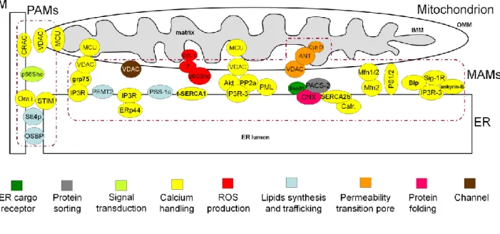
1.6 Remodelling ER-mitochondria Ca2+ transfer in cell survival and death _______________________ 24 1.7 Mitochondria-associated membranes: role of structural and regulatory proteins in the control of Ca2+ transfer between ER and mitochondria _______________________________________________ 27
2.AIMS: _______________________________________________________________________ 37 3.RESULTS: ____________________________________________________________________ 38
3.1 VDAC1 selectively transfers apoptotic Ca2+ signals to mitochondria _________________________ 38
Introduction ________________________________________________________________________________ 38 Results ____________________________________________________________________________________ 39 Silencing of the three VDAC isoforms differentially regulate cellular sensitivity to apoptotic stimuli _________ 39 All VDAC isoforms enhance mitochondrial Ca2+ __________________________________________________ 41 All VDACs do not affect ER Ca2+ content and cytosolic Ca2+ transients _________________________________ 42 VDAC1 specific coupling to ER Ca2+ releasing channels _____________________________________________ 43 Apoptotic treatment enhances VDAC1 specific coupling to IP3Rs ____________________________________ 43 VDAC1 selectively transfers apoptotic Ca2+ signals to mitochondria. __________________________________ 45 Discussion __________________________________________________________________________________ 45
3.2 PML regulates apoptosis at endoplasmic reticulum by modulating calcium release ____________ 48
Introduction ________________________________________________________________________________ 48 Results ____________________________________________________________________________________ 50 PML localizes at ER and MAMs regions and mediates Ca2+-dependent apoptotic cell death _______________ 50 PML absence induces a smaller release of Ca2+ from ER, leading to reduced mitochondrial Ca2+ uptake after agonist or apoptotic stimulation ______________________________________________________________ 50 The erPML chimera rescues Ca2+ homeostasis after physiological and apoptotic stimuli in Pml−/− MEFs ______ 53 PML is essential for Akt- and PP2a-dependent modulation of IP3R phosphorylation and in turn for IP3R-mediated Ca2+ release from ER _______________________________________________________________ 53 Discussion __________________________________________________________________________________ 55
3.3 PTEN localization at the ER and MAMs regulates calcium signalling and apoptosis _____________ 59
Introduction ________________________________________________________________________________ 59 Results ____________________________________________________________________________________ 60 PTEN is localized in different intracellular compartments including ER and MAMs_______________________ 60
3
PTEN silencing reduces ER Ca2+ release, thus impairing cytosolic and mitochondrial Ca2+ transients elicited by agonist stimulation ________________________________________________________________________ 61 ER-localized PTEN, but not wild-type PTEN, enhances the agonist-dependent mitochondrial Ca2+ response __ 63 Ca2+ mobilization from intracellular stores evoked by arachidonic acid is impaired when PTEN is silenced and increased through targeting of PTEN to the ER ___________________________________________________ 65 Ca2+-mediated apoptosis is prevented by PTEN silencing and enhanced through overexpression of ER-PTEN _ 67 Discussion __________________________________________________________________________________ 69 4.MATERIALS AND METHODS: ____________________________________________________ 73
Cells culture and Transfection___________________________________________________________ 73 Plasmid cloning ______________________________________________________________________ 73 Subcellular Fractionation ______________________________________________________________ 74 Co-immunoprecipitation _______________________________________________________________ 74 Immunoblot _________________________________________________________________________ 75 Immunofluorescence __________________________________________________________________ 75 Immunoelectron microscopy ___________________________________________________________ 76 Aequorin measurements _______________________________________________________________ 76 Fura-2/AM measurements _____________________________________________________________ 77 Induction of Apoptosis ________________________________________________________________ 77 Statistical analyses ____________________________________________________________________ 78 REFERENCES: __________________________________________________________________ 79
4
ABSTRACT
The tight interplay between endoplasmic reticulum (ER) and mitochondria is a key determinant of cell function and survival through the control of intracellular calcium (Ca2+) signalling. The physical platform for the association between the ER and mitochondria is a domain of the ER called the “mitochondria-associated membranes” (MAMs). MAMs are crucial for highly efficient transmission of Ca2+ from the ER to mitochondria, thus controlling fundamental processes involved in energy production and also determining cell fate by triggering or preventing apoptosis.
In particular, we show that: i) despite different roles in cell survival, all three isoforms of the outer mitochondrial membrane protein voltage-dependent anion channels (VDAC) are equivalent in allowing mitochondrial Ca2+ loading upon agonist stimulation, vice versa VDAC1, by selectively interacting with the inositol trisphosphate receptors (IP3Rs) - an interaction that is further strengthened by apoptotic stimuli - is preferentially involved in the transmission of the low-amplitude apoptotic Ca2+ signals to mitochondria, highlighting a non-redundant molecular route for transferring Ca2+ signals to mitochondria in apoptosis; ii) the promyelocytic leukemia (PML) tumor suppressor exerts its extranuclear proapoptotic action by its unexpected and fundamental role at MAMs, where PML was found in protein complexes with the type 3 IP3R, the protein kinase Akt and the phosphatase PP2a, which are essential for Akt- and PP2a-dependent modulation of IP3R phosphorylation and in turn for IP3R-mediated Ca2+ release from ER; iii) the PTEN (phosphatase and tensin homolog deleted on chromosome 10) tumor suppressor localizes at the ER and MAMs, and ER-localized PTEN is specifically involved in increasing both Ca2+ transfer from the ER to mitochondria and cell sensitivity to Ca2+-mediated apoptosis.
The improved knowledge of the functioning of proteins involved in regulating Ca2+ signalling may reveal novel unexplored pharmacological targets, and help in treating cancer as well as other pathologies.
5
ABSTRACT (Italiano):
L‟accoppiamento funzionale tra reticolo endoplasmatico (ER) e mitocondri è un fattore determinate per la funzionalità e la sopravvivenza cellulare, in quanto determina il controllo del segale calcio (Ca2+) intracellulare. Dal punto di vista fisico, la base per l‟associazione tra ER e mitocondri risiede in un dominio dell‟ER definito “membrane associate ai mitocondri” (MAMs). Le MAMs sono fondamentali per una trasmissione altamente efficiente degli ioni Ca2+ dall‟ER ai mitocondri, e per questo controllano processi indispensabili coinvolti nella produzione di energia, ed inoltre determinano il destino della cellula facilitando o ostacolando l‟apoptosi.
Specificamente, abbiamo dimostrato che: i) nonostante svolgano diversi ruoli nella sopravvivenza cellulare, tutte e tre le isoforme del canale anionico voltaggio dipendente (VDAC, “voltage-dependent anion channels”, una proteina della membrana mitocondriale esterna) hanno un ruolo equivalente nell‟accumulo mitocondriale di Ca2+
indotto da stimolazione con agonista, viceversa VDAC1, attraverso l‟intrerazione selettiva con i recettori dell‟inositolo trifosfato (IP3Rs) – un‟interazione ulteriormente rafforzata dagli stimoli apoptotici – è preferenzialmente coinvolto nella trasmissione ai mitocondri di stimoli apoptotici Ca2+ mediati che hanno entità inferiore, evidenziando un‟esclusiva via molecolare per il trasferimento del segnale Ca2+
ai mitocondri durante l‟apoptosi; ii) l‟oncosoppressore PML (leucemia promielocitica), quando localizzato al di fuori del nucleo, è comunque in grado di esercitare una funzione proapoptotica mediante la sua inaspettata localizzazione alle MAMs, dove PML è stato trovato in complessi proteici con i recettoti IP3R di tipo 3, la proteina chinasi Akt e la proteina fosfatasi PP2a, che sono essenziali per la modulazione dello stato di fosforilazione dell‟IP3R mediata da Akt e PP2a, e di conseguenza del rilascio di Ca2+ dall‟ER attraverso l‟IP3R; iii) l‟oncosoppressore PTEN (“phosphatase and tensin homolog deleted on chromosome 10”) localizza all‟ER e alle MAMs, e la quota di PTEN presente al reticolo è quella specificamente coinvolta nell‟aumento sia del trasferimento di Ca2+
dall‟ER ai mitocondri che nella suscettibilità a stimoli apoptotici mediati da Ca2+.
L‟avanzamento nella conoscenza del funzionamento di proteine coinvolte nel segnale Ca2+
potrà rivelare nuovi inesplorati bersagli farmacologici ed aiutare nel trattamento del cancro ed altre patologie.
6
Abbreviations:
m, mitochondrial membrane potential difference;
AEQ, aequorin; ArA, arachidonic acid;
ANT, adenine nucleotide translocase; Bap31, B-cell receptor-associated protein 31; BiP, Binding immunoglobulin Protein; Ca2+, calcium ions;
[Ca2+], Ca2+ concentration;
[Ca2+]c, cytosolic Ca2+ concentration;
[Ca2+]m, mitochondrial Ca2+ concentration;
CABPs, intraluminal Ca2+-binding proteins; CaMKII, calmodulin-dependent protein kinase II; Cyp D, cyclophilin D;
Drp1, dynamin-related protein 1; ER, endoplasmic reticulum;
ERp44, endoplasmic reticulum resident protein 44; ETO, etoposide;
FACL4, long-chain fatty acid-CoA ligase type 4; FAD, familial Alzheimer‟s disease;
Fis1, Fission 1 homologue;
FRET, fluorescence resonance energy transfer; GFP, green fluorescent protein;
GM1, GM1-ganglioside;
grp75, glucose-regulated protein 75; HK, hexokinase;
IMM, inner mitochondrial membrane; IMS, intermembrane space;
IP3, inositol 1,4,5-trisphosphate;
IP3R, inositol 1,4,5-trisphosphate receptor;
Letm1, leucine zipper-EF-hand containing transmembrane protein 1;
MAMs, mitochondria-associated membranes; MCU, mitochondrial Ca2+ uniporter;
MICU1, mitochondrial calcium uptake 1; Mfn, mitofusin;
VDAC, voltage-dependent anion channel; UCP, uncoupling protein;
MEN, menadione;
mHCX, mitochondrial H+/Ca2+ exchanger; mNCX, mitochondrial Na2+/Ca2+ exchanger;
MOMP, mitochondrial outer membrane permeabilization;
MPT, mitochondrial permeability transition; NCX, Na2+/Ca2+ exchanger;
OA, okadaic acid;
OMM, outer mitochondrial membrane; OPA1, optic atrophy 1;
OXPHOS, oxidative phosphorylation;
p66shc, 66-kDa isoform of the growth factor adapter shc;
PACS-2, phosphofurin acidic cluster sorting protein 2; PAMs, plasma membrane associated membranes; PDH, pyruvate dehydrogenase;
PI3K, phosphatidylinositol 3-kinase; PIP2, phosphatidylinositol 4,5-bisphosphate; PIP3, phosphatidylinositol 3,4,5-trisphosphate; PKA, protein kinase A;
PKC, protein kinase C; PLC, phospholipase C;
PMCA, plasma membrane Ca2+ ATPase; PML, promyelocytic leukemia protein; PP2a, protein phosphatase 2a;
PS1, presenilin;
PSS-1, phosphatidylserine synthase-1;
PTEN, phosphatase and tensin homolog deleted on chromosome 10;
PTP, permeability transition pore; ROS, reactive oxygen species; RyR, ryanodine receptor;
SERCA, sarco-endoplasmic reticulum Ca2+ ATPase; Sig-1R, Sigma-1 receptor;
SOCE, store-operated Ca2+ entry; SR, sarcoplasmic reticulum;
TIRF, total internal reflection fluorescence; TG, thapsigargin;
7
1. INTRODUCTION:
Changes in the levels of intracellular calcium ions (Ca2+) provide dynamic and highly versatile signals that regulates several processes as diverse as energy transduction, fertilization, secretions, muscle contraction, chemotaxis and neuronal synaptic plasticity in learning and memory (1). However, under certain conditions increases in intracellular Ca2+ are cytotoxic and lead to apoptosis (programmed cell death). Consequently, Ca2+ needs to be used in an appropriate manner to determine cell fate; if this balancing act is compromised, pathology may ensue (2).
Ca2+ signalling proteins and organelles are emerging as additional cellular targets of oncogenes and tumour suppressors. The Ca2+ signal has major roles in the regulation of processes relevant to tumorigenesis, including migration, invasion, proliferation, and apoptotic sensitivity (3). Intracellular Ca2+ homeostasis has been the focus of researchers characterizing changes in Ca2+ signalling in cancer cells. In order for the cancer cells to proliferate at higher rates and still protect themselves from apoptosis, many cancer cells remodel the expression or activity of their Ca2+ signalling machinery. Spatially restricted Ca2+ signalling within specific cellular compartments or discrete cytosolic domains provides an additional layer of complexity in the regulation of cellular processes important in tumorigenesis. . In normal cells, the Ca2+ signalling is highly regulated spatially such as between endoplasmic reticulum (ER) and mitochondria, two intracellular organelles which play crucial roles in Ca2+ signalling and may decide the ultimate fate of the cell. Indeed, by adjusting the load of Ca2+ imposed upon the mitochondrion, the same Ca2+ efflux from ER (the main intracellular Ca2+ store) that is responsible for regulating processes for maintaining life could also act as a death-inducing signal.
Since ER and mitochondria play significant roles in the regulation of cell proliferation and apoptosis, the remodelling of Ca2+ signalling machinery in ER and mitochondria in cancer cells seems imminent during oncogenic transformation. Therefore, targeting of the Ca2+ signalling apparatus in cancer cells could specifically disrupt their Ca2+ homeostasis, and so decrease cancer cell proliferation and increase cancer cell apoptosis. Such novel and highly innovative strategies can provide rationale and approaches for the design and development of novel technologies based on Ca2+ waves for the diagnosis and treatment of cancer, as well as other disease.
8
1.1 The Ca
2+-signalling toolkit
At the beginning of life, Ca2+ mediates the process of fertilization and regulates the cell cycle events during the early developmental processes. Once the cells differentiate to perform specific functions, changes in the levels of intracellular Ca2+ provide dynamic and highly versatile signals that control a plethora of cellular processes, yet under certain conditions increases in intracellular Ca2+ are cytotoxic (4). For this reason, the intracellular concentration of Ca2+, [Ca2+]i, in resting cells is
usually maintained very low, at ~100 nM.
In cells, due to the presence of several charged molecules, the Ca2+ diffusion rates are slow. In order to utilize Ca2+ as a second messenger, cells have devised an ingenious mechanism of signalling that has overcome the inherent problems associated with lower diffusion rates and potential cytotoxicity of Ca2+, by presenting changes in Ca2+ concentration as brief spikes which are often organized as regenerative waves (1). The universality of Ca2+-based signalling depends on its enormous versatility in terms of amplitude, duration, frequency and localization. The formation of the correct spatio-temporal Ca2+ signals is dependent on an extensive cellular machinery named the Ca2+ toolkit, which includes the various cellular Ca2+-binding and Ca2+-transporting proteins, present mainly in the cytosol, plasma membrane, ER and mitochondria (5).
To provide for a very fast and effective Ca2+-signaling, the cells use a great amount of energy to maintain an almost 20 000-fold Ca2+-gradient between their intracellular (~100 nM free) and extracellular (~1 mM) Ca2+ concentrations. To maintain this Ca2+ gradient, the cells chelate, compartmentalize, or remove Ca2+ from the cytoplasm through its active extrusion by the plasma membrane Ca2+ ATPase (PMCA) and the Na+/Ca2+ exchanger (NCX) (6, 7)
The increase of intracellular [Ca2+] can be elicited by two fundamental mechanisms (or a combination of both). The first involves Ca2+ entry from the extracellular milieu, through the opening of plasma membrane Ca2+ channels (traditionally grouped into three classes: voltage operated Ca2+ channels (VOCs) (8), receptor operated Ca2+ channels (ROCs) (9) and second messenger operated Ca2+ channels (SMOCs) (10)). The second universal mechanism for Ca2+ signaling is the release of Ca2+ from intracellular Ca2+ stores, mainly the ER and its specialized form in muscle, the sarcoplasmic reticulum (SR). In these intracellular stores, two main Ca2+ -release channels exist that, upon stimulation, -release Ca2+ into the cytosol, thus triggering Ca2+ signalling: the inositol 1,4,5-trisphosphate (IP3) receptors (IP3Rs) and the ryanodine receptors (RyRs) (11, 12). IP3Rs are ligand-gated channels that function in releasing Ca2+ from ER Ca2+ stores in response to IP3 generation. G protein coupled receptors (GPCRs) can activate phospholipase C β (PLCβ), and tyrosine-kinase receptors (TKR) can activate PLCγ, which then
9
cleave PIP2 into IP3 and diacylglycerol (DAG). IP3 binding to the IP3Rs that are present in the ER, causes efflux of Ca2+ from the ER to the cytoplasm resulting in increase in cytosolic Ca2+ concentration ([Ca2+]c) from ~100 nM to ~1 M for several seconds (13, 14). This rise in [Ca2+]c
results in various Ca2+-dependent intracellular events (Figure 1). A variety of cellular proteins with Ca2+-binding affinities ranging between nM to mM are utilized by the cells to buffer the cellular Ca2+ increase as well as to regulate cellular processes via Ca2+-signaling. The exact cellular outcome depends on the spatiotemporal characteristics of the generated Ca2+ signal (15).
Figure 1. Regulation of multiple cellular processes by the IP3/Ca2+ signalling pathway. (figure from (16))
Once its downstream targets are activated, basal [Ca2+]c levels are regained by the combined
activity of Ca2+ extrusion mechanisms, such as PMCA and NCX, and mechanisms that refill the intracellular stores, like sarco-endoplasmic reticulum Ca2+ ATPases (SERCAs) (6). Due to SERCA activity and intraluminal Ca2+-binding proteins (CABPs), i.e., calnexin and calreticulin (17), the ER can accumulate Ca2+ more than a thousand-fold excess as compared to the cytosol. Given that
10
PMCAs pump Ca2+ out of the cell faster than it can be repleted, IP3R mediated efflux of Ca2+ from the ER in response to receptor activation empties the ER, thus a Ca2+ entry mechanism is activated. This mechanism is called “Store-operated Ca2+
entry” (SOCE). The molecular determinants of SOCE have been identified in the very last few years and include the ER Ca2+ sensors STIM (stromal interaction molecule) 1 and 2, and the specialized plasma-membrane channels Orai1, Orai2 and Orai3 (for a recent review (18)).
Although the ER (and its specialized form in muscle, the SR) is generally considered the main intracellular Ca2+ store, almost all other organelles play a role in Ca2+ signalling: mitochondria (see below) (19), the Golgi apparatus (20), secretory vesicles (21), lysosomes (22), endosomes (23) and peroxisomes (24, 25).
Specificity in decoding Ca2+ signals can be provided by the affinity of Ca2+ sensor as well as its duration, amplitude and intracellular location: in this way a particular Ca2+ signal can specifically regulate many different cell functions (26).
1.2 ER-mitochondria crosstalk: local microdomains support mitochondrial Ca
2+uptake
While the role of the ER as a physiologically important Ca2+ store has long been recognized, a similar role for mitochondria have seen a reappraisal only in the past two decades (27). The uptake of the Ca2+ ions into the mitochondrial matrix implies different transport systems responsible for the transfer of Ca2+ across the outer and the inner mitochondrial membrane (OMM and IMM respectively). It has long been known that mitochondria can rapidly accumulate Ca2+ down the large electrochemical gradient (mitochondrial membrane potential difference, m = -180 mV, negative
inside) generated by the respiratory chain (28). Indeed, based on the chemiosmotic theory, the translocation by protein complexes of H+ across an ion-impermeable inner membrane generates a very large H+ electrochemical gradient and mitochondria employ the dissipation of this proton gradient not only to run the endoergonic reaction of ATP synthesis by the H+-ATPase, but also to accumulate cations into the matrix.
For a long time, however, due to the low affinity of the mitochondrial Ca2+ uptake system under physiological conditions (an apparent Kd of 20 to 30 M under conditions thought to mimic the
cytoplasm, estimated in the earlier work with isolated organelles) and the submicromolar global [Ca2+]c briefly reached after physiological stimulation (which rarely exceed 2-3 M), this process
11
in the Ca2+ overload that is observed in various pathological conditions (such as, for example, excitotoxic glutamate stimulation of neurons) (19). Mitochondrial Ca2+ returned to the limelight in 1992 when Rizzuto, Pozzan and colleagues generated a novel, genetically encoded chemiluminescent indicator, aequorin. This probe, specifically targeted to the mitochondrial matrix, allowed dynamic, accurate and specific monitoring of the [Ca2+] within the matrix of mitochondria in living cells (29). With this new tool they could show that mitochondria in living cells undergo very fast and large increases in their matrix Ca2+ levels (mitochondrial Ca2+ concentration, [Ca2+]m)
upon cell stimulation, reaching peaks similar or even larger than those in the cytoplasm, even for normal physiological cytoplasmic Ca2+ rises (30). Similar conclusions could be reached also with fluorescent indicators, such as the positively charged Ca2+ indicator rhod-2 (that accumulates within the organelle) (31) and the more recently developed GFP-based fluorescent indicators (32).
While enlivening the interest in mitochondrial Ca2+ homeostasis, these data raised an apparent contradiction between the prompt response of the organelle (where [Ca2+]m rise, in a few seconds, to
values above 10 M, and in some cell types up to 500 M) and the low affinity of the Ca2+ uptake system together with the low concentration of global Ca2+ signals observed in cytoplasm. Based on a large body of experimental evidence, it is now generally accepted that the key to the rapid Ca2+ accumulation rests in the strategic location of a subset of mitochondria, close to the opening ER or plasma membrane Ca2+ channels (30, 31, 33). The hypothesis, called “microdomain hypothesis” (26), proposes that microdomains of high [Ca2+] (10-20 M) can be transiently formed in regions of close apposition between mitochondria and Ca2+ channels of the ER/SR or of the plasma membrane (33). These high Ca2+ microdomains rapidly dissipate (due to diffusion) insuring that mitochondria do not overload with Ca2+ (Figure 2).
The “microdomain hypothesis” received a number of indirect confirmations in the last 20 years by different groups. More recently, such microdomains in selected regions of contact between ER and mitochondria were finally measured directly, by two complementary studies that demonstrated the existence and amplitude of high Ca2+ microdomains on the surface of mitochondria. Giacomello et
al. (34) targeted a new generation of FRET-based Ca2+ sensors (35) to the OMM and, through a sophisticated statistical analysis of the images, revealed the existence of small OMM regions where [Ca2+] reaches values as high as 15-20 μM. The probe detected Ca2+ hotspots on about 10% of the OMM surface that were not observed in other parts of the cell. The Ca2+ hotspots were not uniform, and their frequency varied among mitochondria of the same cell. Moreover, classical epifluorescence and total internal reflection fluorescence (TIRF) microscopy experiments were combined in order to monitor the generation of high Ca2+ microdomains in mitochondria located near the plasma membrane. With this approach, it could be shown that Ca2+ hotspots on the surface
12
of mitochondria occur upon opening of VOCs, but not upon SOCE. Csordás et al. (36) used a complementary approach in which they generated genetically encoded bifunctional linkers consisting of OMM and ER targeting sequences connected through a fluorescent protein, including a low-Ca2+-affinity pericam, and coupled with the two components of the FKBP-FRB heterodimerization system (37), respectively. Using rapamycin-assembled heterodimerization of the FKBP-FRB-based linker, they detected ER/OMM and plasma membrane/OMM junctions (the latter at a much lower frequency). In addition, the recruited low-Ca2+-affinity pericam reported Ca2+ concentrations as high as 25 M at the ER/OMM junctions in response to IP3-mediated Ca2+ release, which is in excellent agreement with the values obtained by Giacomello et al..
Figure 2. Intracellular Ca2+ signalling. Schematic model of intracellular Ca2+ homeostasis. Plasma membrane
G-protein coupled receptors activate phospholipase C-β (PLC-β) to promote the generation of inositol 1,4,5-trisphosphate (IP3) and the release of Ca2+ from the endoplasmic reticulum (ER) into the cytosol. Mitochondrial surface directly interacts with the ER through contact sites defining hotspots Ca2+ signalling units. Ca2+ import across the outer mitochondrial membrane (OMM) occurs by the voltage-dependent anion channel (VDAC), and then enters the matrix through the mitochondrial Ca2++ uniporter (MCU), the main inner mitochondrial membrane (IMM) Ca2+-transport system (Ca2+ levels reached upon stimulation are indicated in square brackets). Mitochondrial Ca2+ exchangers present in the IMM export Ca2+ from the matrix once mitochondrial Ca2+ has carried its function; another mechanism for Ca2+ efflux from mitochondria is the permeability transition pore (PTP). Ca2+ levels return to resting conditions (indicated in round brackets) through the concerted action of cytosolic Ca2+ buffers, plasma membrane Ca2+-ATPase (PMCA) and the Na+/ Ca2+ exchanger (NCX) that permit the ion extrusion in the extracellular milieu. Sarco-endoplasmic reticulum Ca2+ ATPase (SERCA) restablishes basal Ca2+ levels in intracellular stores. ANT adenine nucleotide translocase, Cyp D cyclophilin D, DAG diacylglycerol, PIP2 phosphatidylinositol 4,5-bisphosphate.
13
While based on cell morphology the close proximity between the mitochondria and the ER is expected and indeed often observed, i.e. in neuronal prolongings, a close interaction between ER-resident Ca2+ channels and mitochondria in non-excitable cells implies the assembly of a dedicated signaling unit at the organelle interphase (see section 1.7).
1.3 Calcium release from cellular store: structure and function of the IP3R
The ER is possibly the largest individual intracellular organelle comprising a three dimensional network of endomembranes arranged in a complex grid of microtubules and cisternae. It is made up of functionally and structurally distinct domains (reviewed extensively by a number of authors (38-41), in relation to the variety of cellular functions played by the organelle, primarily concerning protein synthesis, maturation and delivery to their destination (42, 43). Moreover, the ER is a dynamic reservoir of Ca2+ ions, which can be activated by both electrical and chemical cell stimulation (44, 45) making this organelle an indispensable component of Ca2+ signalling (46-48). Modern analysis methods enabled the determination of the molecular profile of the ER. This profile reflects the ER‟s role in signalling, as it comprises a number of components constituting the Ca2+
signalling pathway. It contains IP3Rs, RyRs, SERCAs, and in addition to these release channels and pumps, there are buffers (calnexin, calreticulin) and a number of ancillary proteins (FK 506-binding proteins, sorcin, triadin, phosholamban) that contribute to the ER Ca2+ signalling system (49). Many extracellular stimuli, such as hormones, growth factors, neurotransmitters, neutrophins, odorants, and light, function generating IP3 through the phospholipase C isoforms, activated in different manners: G-protein coupled receptors (acting via PLC-β), tyrosine-kinase coupled receptors (PLC-γ), an increase in Ca2+ concentration (PLC-δ) or activated by Ras (PLC-ɛ) (50, 51). The final effector are the IP3Rs, nonselective cationic channels that conduct Ca2+.
A functional IP3R Ca2+ channel is composed of tetramers with six transmembrane domains (of ~3000 amino acids) that can be either homotetramers or, to a lesser extent, heterotetramers of different isoforms. From the structural point of view, several domains are recognized in the protein sequence, with different functions. These include the IP3-binding domain (IP3-BD), i.e. the minimal sequence sufficient for IP3 binding, located near the N-terminus of the protein (aa 226-578). Interestingly, this protein domain contains armadillo-repeat protein structures that are engaged in protein-protein interactions, and mediates intramolecular interactions with other IP3R domains as well as the association with other regulatory proteins. N-terminally to the IP3-BD, i.e. within aa 1-222, a suppressor region is located that inhibits ligand binding and thus lowers the global receptor IP3 affinity in the physiological range. The six transmembrane-spanning domain is at the very
C-14
terminal end of each subunit, and, between them, an internal coupling domain assures the signal of IP3 binding is transferred to the channel-forming region, hence triggering its opening (52).
Three isoforms of IP3R encoded by different genes have been identified with different agonist affinities and tissue distribution (53). Given that the affinity of the IP3-binding core to its ligand is similar for the three isoforms, the tuning of the whole receptor's affinity appears to be due to the isotype-specific properties of the N-terminal suppressor domain (54).
The release of Ca2+ from the ER is a nonlinear, cooperative process wherein IP3 binds to four receptor sites on the IP3R, one on each subunit of the tetramer (52). IP3Rs are at first potentiated, then inhibited by Ca2+. Small perturbations in conditions, such as basal [Ca2+]i, [IP3], and various
regulators can cause uncoordinated bursts of local release across a cell. The brief opening of IP3R channels gives rise to localized Ca2+ pulses, called “sparks” or “blips” and “puffs” (1). The smallest Ca2+ release events, “blips”, probably reflect random openings of single IP3R. Spontaneous clustering of IP3Rs (in particular of IP3R2, due to its higher IP3 affinity) have been proposed to be the underlying mechanism responsible for Ca2+ “puffs” observed in the cytoplasm (55). Recruitment of neighboring IP3Rs and combination of Ca2+ “puffs” results in Ca2+ waves, ensuring that the Ca2+ signal propagates to the entire cell (56), or remains confined to specific subcellular regions (57).
Ca2+ oscillations, depend upon both the spatial organization of IP3Rs and their regulation by Ca2+, although the links between IP3R activities and Ca2+ oscillations are not fully understood. Ca2+ regulates channel activity in a biphasic manner. Early studies demonstrated inhibition of IP3-mediated Ca2+ mobilization by micromolar concentrations of Ca2+ (58). Lower concentrations were subsequently found to potentiate the effects of IP3 (59). In addition, also the ER Ca2+ content retains the capability to regulate the channel opening: in permeabilized hepatocytes, an increase in [Ca2+]er enhances the sensitivity of IP3R for its ligand, promoting also spontaneous Ca2+ release, but
the nature of this direct regulation and the protein involved are still a matter of debate (60). In this context, the tight spatial relationship between ER and mitochondria, and the capacity of the latter to rapidly clear the high [Ca2+] microdomain generated at the mouth of the IP3R, makes mitochondria an active player in the control of IP3R function. The first clear demonstration of this concept came from the fine work of Lechleiter et al., who demonstrated that energized mitochondria, by regulating the kinetics of ER Ca2+ release, finely tune the spatio-temporal patterning of Ca2+ waves in Xenopus oocytes. Then, the observation that Ca2+ uptake by mitochondria controls the [Ca2+] microdomain at the ER/mitochondrial contacts and thus the kinetics of IP3R activation/inactivation was extended to a variety of mammalian cell lines, e.g. hepatocytes, astrocytes and BHK-21 cells, thus highlighting its general relevance (61).
15
Whereas IP3 and Ca2+ are essential for IP3R channel activation, other physiological ligands, such as ATP, are not necessary but can finely modulate the Ca2+-sensitivity of the channel (62). As for Ca2+, the modulation of IP3R by ATP is biphasic: at micromolar concentrations, ATP exerts a stimulatory effect, while inhibiting channel opening in the millimolar range (63, 64).
Finally, in their coupling/suppressor domains, the IP3Rs possess consensus sequences for phosphorylation by numerous kinases; currently, at least 12 different protein kinases are known to directly phosphorylate the IP3R (65), among them: Akt (66), protein kinase A (cAMP-dependent) (67), protein kinase G (cGMPdependent) (68), calmodulin-dependent protein kinase II (CaMKII) (69), protein kinase C (PKC) (70), and various protein tyrosine kinases (71).
1.4 Mitochondria: cell physiology and molecular nature of the mitochondrial
Ca
2+uptake and release machinery
Mitochondria: the basics
The mitochondrion represents a unique organelle within the complex endomembrane systems that characterize any eukaryotic cell. Complex life on earth has been made possible through the “acquisition” of mitochondria which provide an adequate supply of substrates for energy-expensive tasks. The mitochondrion is a double membrane-bounded organelle thought to be derived from an
-proteobacterium-like ancestor, presumably due to a single ancient invasion occurred more than 1.5 billion years ago. The basic evidence of this endosymbiont theory (72) is the existence of the mitochondrial DNA (mtDNA), a 16.6 Kb circular double-stranded DNA molecule with structural and functional analogies to bacterial genomes (gene structure, ribosome). This mitochondrial genome encodes only 13 proteins (in addition to 22 tRNAs and 2 rRNAs necessary for their translation), all of which are components of the electron transport chain (mETC) complexes (I, III and IV), while the whole mitochondrial proteome consists of more than 1000 gene products. Thus, one critical step in the transition from autonomous endosymbiont to organelle has been the transfer of genes from the mtDNA to the nuclear genome. At the same time, eukaryotes had to evolve an efficient transport system to deliver nuclear-encoded peptides inside mitochondria: TIM (Transporters of the Inner Membrane), TOM (Transporters of the Outer Membrane) and mitochondrial chaperones (such as hsp60 and mthsp70) build up the molecular machinery that allows the newly-synthesized unfolded proteins to enter mitochondrial matrix (73).
16
Mitochondria are defined by two structurally and functionally different membranes: the plain outer membrane, mostly soluble to ions and metabolites up to 5000 Da, and the highly selective inner membrane, characterized by invaginations called cristae which enclose the mitochondria matrix. The space between these two structures is traditionally called intermembrane space (IMS), but recent advances in electron microscopy techniques shed new light on the complex topology of the inner membrane. Cristae indeed are not simply random folds but rather internal compartments formed by profound invaginations originating from very tiny “point-like structures” in the inner membrane (74). These narrow tubular structures, called cristae junctions, can limit the diffusion of molecule from the intra-cristae space towards the IMS, thus creating a micro-environment where mETC complexes (as well as other proteins) are hosted and protected from random diffusion. Mitochondria were identified as the powerhouse for energy production in eukaryotic cells thanks to decades of extensive biochemical work on carbohydrate metabolism and organelle morpho-functional characterization, carried out in the first half of the 20th century by leading scientific figures such as Krebs, Corey, Claude, Palade and many others. Mitochondria are the main site of ATP production. When glucose is converted to pyruvate by glycolysis, only a small fraction of the available chemical energy has been stored in ATP molecules; the main enzymatic systems involved in this process are the tricarboxylic acid (TCA) cycle and the mETC. Products from glycolysis and fatty acid metabolism are converted to acetyl-CoA which enters the TCA cycle where it is fully degraded to CO2. More importantly, these enzymatic reactions generate NADH and FADH2 which
provide reducing equivalents and trigger the electron transport chain. mETC consists of five different protein complexes: complex I (NADH dehydrogenase), complex II (succinate dehydrogenase), complex III (ubiquinol cytochrome c reductase), complex IV (cytochrome c oxidase) and complex V that constitutes the F1F0-ATP synthase. Electrons are transferred from
NADH and FADH2 through these complexes in a stepwise fashion: as electrons move along the
respiratory chain, energy is stored as an electrochemical H+ gradient across the inner membrane, thus creating a negative mitochondrial membrane potential (estimated around -180 mV against the cytosol). H+ are forced to reenter the matrix mainly through complex V which couples this proton driving force to the phosphorylation of ADP into ATP, according to the chemiosmotic principle. ATP is then released to IMS through the electrogenic Adenine Nucleotide Translocase (ANT) which exchange ATP with ADP to provide new substrate for ATP synthesis. Finally, ATP can easily escape the IMS thanks to the mitochondrial porin of the outer membrane, VDAC (voltage-dependent anion channel).
With the general acceptance of the chemiosmotic hypothesis, it has became clear that the across the mitochondrial inner membrane is the driving force for mitochondrial Ca2+ accumulation (75).
17
Thus, Ca2+ enters the mitochondrial matrix down its electrochemical gradient, that can be generated either by the electron flow in the mETC or by reversal of the ATP synthase. Mitochondrial Ca2+ accumulation plays a key role in the regulation of many cell functions, ranging from ATP production to cell death. Mitochondrial Ca2+ uptake and release is central not only for the regulation of cellular Ca2+ homeostasis, but is vital also for the regulation of intramitochondrial enzymes concerned with the utilization of oxidizable substrates. However, excess Ca2+ accumulation by mitochondria is a common event in the process of cell death, by both necrosis and apoptosis (76) (see , sections 1.5 and 1.6).
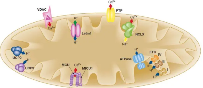
Despite the basic mechanisms of mitochondrial Ca2+ homeostasis have been firmly established for decades, the molecular identities of the channels and transporters responsible for Ca2+ uptake and release (schematized in Figure 3) have remained mysterious until very recently.
Figure 3. Schematic representation of the mitochondrial Ca2+, Na+ and H+ handling machinery. Ion fluxes are indicated by arrows. Red arrow, Ca2+; blue arrow, H+; green arrow, K+; yellow arrow, Na+. ETC, electron transport chain; Letm1, Leucine-zipper EF-hand containing transmembrane protein 1; MCU, mitochondrial Ca2+ uniporter; MICU1, mitochondrial calcium uptake 1; NCLX, Na+/Ca2+ exchanger; PTP, permeability transition pore; UCP2/3, uncoupling protein 2/3; VDAC, voltage-dependent anion channel (figure from (77)). See text for details.
We and other groups extensively worked on this topic and what emerged was that the outer mitochondrial membrane (OMM, although traditionally considered freely permeable) is a critical determinant of the mitochondrial Ca2+ accumulation (78). Thus, the mitochondrial Ca2+ uptake machinery will be discussed, starting from the channels of the OMM, to the last identified components of the IMM.
18
Ca2+ transfer across the OMM
The OMM was previously thought to be freely permeable for ions and small molecules, but now it is clear that the so-called voltage-dependent anion channels, VDAC, also reffered to as the mitochondrial porin, are major regulators of the various ion, nucleotide and molecule fluxes across this membrane, including the Ca2+ fluxes (79).
Yeast possesses only one VDAC isoform (but has also another VDAC gene that correctly inserts into OMM showing no channel activity), while higher multicellular organisms and mammals have three distinct VDAC genes (VDAC1, VDAC2 and VDAC3), with VDAC1 representing the best characterized one. These three isoforms show a substantial sequence homology (from 65 to 75% in identity) and similar structure, with the only exception of VDAC2 that has a longer (11 aminoacids) N-terminal tail (80). Yeasts lacking VDAC gene cannot grow on non-fermentable medium, thus highlighting the relevance of this channel in mitochondrial function: reintroduction of any of the mammalian VDAC genes in this yeast strain can promptly restore growth defects (81, 82).
VDAC can exist in multiple conformational states with different selectivity and permeability. This 30-35 kDa protein, is traditionally considered as a large,high-conductance, weakly anion-selective channel, fully opened (pore diameter about 2.5 nm) at low potential (<20-30 mV), but switching to cation selectivity and lower conductance (the so-called “closed” state, with a smaller pore diameter of about 1.8 nm) at higher potentials (both positive and negative). When reconstituted into liposomes, each isoform induced a permeability with a similar molecular weight cut-off (between 3400 and 6800 Da based on permeability to polyethylene glycol). Its structure, as determined by NMR and X-ray crystallography, consists of a 19-stranded β-barrel forming a pore with an inner diameter of about 1.5×1 nm and an N-terminal α-helix domain residing inside the pore: this segment most likely represents the voltage sensor since it is ideally positioned to regulate the conductance of ions and metabolites passing through the VDAC pore (83-85). As the main function of VDAC is assumed to be the gateway for ATP and metabolites, its “open” or “closed” states are defined with respect to those molecules (80, 86). However, the physiological relevance of the voltage gating properties of VDAC is still obscure and a matter of debate, since it requires the existence of a potential across the OMM. The existence of any membrane potential across the OMM has never been directly demonstrated (although some have assumed such a potential is not possible, others have proposed several clues in support of this hypothesis, as discussed in (87)). Despite this, a number of reports show that numerous cytosolic components can significantly modulate VDAC gating properties, including NADH (88), members of Bcl-2 protein family (89), metabolic enzymes (90), chaperones (91) and cytoskeletal elements (92).
19
A recent work by Tan and Colombini describes the higher permeability of VDAC to Ca2+ in the closed states (with low permeability to anionic metabolites), rather than the opened state. So VDAC closure seems to promote Ca2+ flux into mitochondria, with consequent permeability transition and cell death (see section 1.5), accordingly with previous observations that VDAC closure is a pro-apoptotic signal (93, 94). These notions have a direct impact on mitochondrial Ca2+ transport, as variations in OMM permeability to Ca2+ can represent a bottleneck for the efficient ion transfer from the high [Ca2+] microdomain generated by the opening of the IP3R to the intermembrane space. Indeed, transient expression of VDAC in various cell types enhanced the amplitude of the agonist-dependent increases in mitochondrial matrix Ca2+ concentration by allowing the fast diffusion of Ca2+ from ER release sites to the inner mitochondrial membrane (78). As to the functional consequences, VDAC overexpressing cells are more susceptible to ceramide-induced cell death, thus confirming that mitochondrial Ca2+ uptake has a key function in the process of apoptosis.
VDAC has been considered a master regulator of the apoptotic process: on one hand it was thought to be one of the main component of the permeability transition pore (PTP), the megachannel mediating the collapse of mitochondrial membrane potential during apoptosis; on the other side it has long been believed a key mediator of Bax-mediated release of cytochrome c (see sections 1.5 and 1.6). However, despite the huge amount of work carried out on this protein, several recent papers (95-97) have raised serious doubt about our functional understandings of this channel. Indeed, new approaches mainly based on mice knockout models failed to clearly confirm any of the above mentioned functions and rather suggest that a substantial rethinking of VDAC roles is needed.
Ca2+ transfer across the IMM
Many attempts were made to identify the molecular nature of the mitochondrial Ca2+ uniporter (MCU), starting in the early 1970s, that is, soon after the discovery of mitochondrial Ca2+ function. MCU has always been described as an highly selective ion channel located in the IMM, with a dissociation constant 2 nM over monovalent cations, reaching saturation only at supraphysiological [Ca2+]c. Also Sr2+ and Mn2+ are conducted by MCU and the relative ion
conductance is: Ca2+Sr2+Mn2+Ba2+. Studies performed on isolated mitochondria allowed the identification of some regulatory molecules acting on MCU, in particular the most effective inhibitors are the hexavalent cation Rutenium Red (RuR) and its related compound RuR360; MCU is also modulated by aliphatic polyamines, such as spermine and aminoglycosides, and by the adenine nucleotides, in the order of effectiveness ATP>ADP>AMP (whereas the nucleoside
20
adenosine is ineffective) (98) as well as several plant-derived flavonoids (99). Another important regulator of MCU is Ca2+ itself. The apparent affinity of the MCU for Ca2+, under physiological conditions (i.e. 1 mM Mg2+), is very low (apparent Kd of 20-30 M) and the influx rate only becomes substantial when the extramitochondrial [Ca2+] reaches values above 5-10 M. As demonstrated by Moreau and its group (99), in fact, MCU has a biphasic dependence on [Ca2+]c
increase, that can both activate or inactivate mitochondrial Ca2+ uptake. This mechanism allows the mitochondrial Ca2+ oscillation, but it prevents an excessive mitochondrial Ca2+ accumulation when intracellular Ca2+ elevation is prolonged.
The MCU has been molecularly identified only very recently, preceded by the discovery of mitochondrial calcium uptake 1 (MICU1), an uniporter regulator which appears essential for mitochondrial Ca2+ uptake (100).
The identification of MICU1 came from the establishment of the so-called MitoCarta database in which about 1000 proteins, specifically present in mitochondria, have been identified (many of them with unknown functions) (101). MICU1 is a 54-kDa protein, with only one putative transmembrane domain, which makes it unlikely that it can function as a Ca2+ channel, so it is not known whether it actually forms (part of) a Ca2+ channel, or functions as Ca2+ buffer, or as a Ca2+ -dependent regulatory protein acting as a Ca2+ sensor (it has a pair of Ca2+-binding EF-hand domains, the mutation of which eliminates the mitochondrial Ca2+ uptake). Taken together the above-mentioned characteristics suggest that MICU1 is not the channel-forming subunit of MCU itself, but rather an associated key subunit.
Finally, last year, two indipendent papers identified the same protein, termed CCDC109A (coiled-coil domaincontaining protein 109A) and renamed MCU, that possesses all the characteristics expected by the elusive Ca2+ uniporter of the IMM (102, 103). MCU is a 40-kDa protein ubiquitously expressed in all mammalian tissues and in most eukaryotes, but missing a yeast orthologue. MCU possesses two transmembrane domains and this characteristic makes it reasonable that it forms (through oligomerization) a gated ion channel. Downregulation of MCU drastically reduces mitochondrial Ca2+ uptake whereas transfection with the native channel rescues the phenotype of the specific siRNA-treated cells. Moreover, the other classical properties of mitochondria (that is, organelle shape and ER-mitochondrial interactions, O2 consumption, ATP
synthesis and ) are not affected by MCU down-regulation. Just the protein‟s orientation is the mainly discrepancy between the two papers, one affirming a C-terminus localization in the intermembrane space (102), the other in the matrix (103). Importantly, thanks to the molecular
21
identification of the MCU, we can now expect a strong acceleration in the search for the functional role of this property of mitochondria, in both physiology and physiopathology.
In the IMM are also present the mitochondrial Na+/Ca2+ exchanger (mNCX) and the H+/Ca2+ exchanger (mHCX). Their main function is probably to export Ca2+ from the matrix once mitochondrial Ca2+ has carried out its function, to reestablish resting conditions (104). In spite of a few remarkable reports identifying the stoichiometry of the Na+/ Ca2+ exchanger (3 or 4 Na+ ions per Ca2+) (105), their molecular identity remained, until very recently, completely mysterious. They have yet to be identified, although recently strong evidence has been provided that the Na+/Ca2+ exchanger isoform NCLX (until then considered an isoform of the PM Na+/ Ca2+ exchanger family) fulfils the criteria to be the elusive mitochondrial Na+-dependent Ca2+ efflux (106). They showed that practically all endogenous NCLX localizes in the mitochondrial fraction and knockout of NCLX drastically reduced Na+-dependent Ca2+ efflux in isolated mitochondria; moreover it is sensitive to the classical mitochondrial Na+/ Ca2+ exchanger inhibitor CGP-37157.
Finally, the low conductance mode of the PTP, a channel of still debated nature localized in the IMM (107), can be also considered as a non-saturating mechanism for Ca2+ efflux from mitochondria. When open, PTP allows the passage of ions and molecules with a molecular weight up to 1.5 kDa, including Ca2+. Short-time openings may have a physiological function but its long-time activation leads to the demise of the cell, either by apoptosis or by necrosis, depending on whether PTP opening occurs in only a small fraction of the mitochondria or in all of them (see the following section and references (108, 109)).
1.5 Mitochondrial Ca
2+function
Physiological functions of Ca2+ uptake in the mitochondria
The first role assigned to the Ca2+ ions taken up into the mitochondrial matrix was the stimulation of the mitochondrial ATP production since important metabolic enzymes localized in the matrix, the pyruvate-, α-ketoglutarate- and isocitrate-dehydrogenases are activated by Ca2+, with different mechanisms: the first through a Ca2+-dependent dephosphorylation step, the others via direct binding to a regulatory site (110, 111). Those three enzymes represent rate-limiting steps of the Krebs cycle thus controlling the feeding of electrons into the respiratory chain and the generation of the proton gradient across the inner membrane, in turn necessary for ATP production through oxidative phosphorylation (OXPHOS). These events were directly visualized in intact, living cells using a molecularly engineered luciferase probe, which revealed an increase in the [ATP] of the
22
mitochondrial matrix following agonist stimulation and mitochondrial Ca2+ uptake (112). As the ATP produced by mitochondria is subsequently transferred to the cytosol, mechanisms that control ATP production will not only affect overall cell life but, more specifically, will regulate the activity of ATP-sensitive proteins localized in the close vicinity of mitochondria, such as IP3Rs and SERCA, which are stimulated by ATP (113, 114). The bidirectional relation between Ca2+ release and ATP production allows for a positive feedback regulation between ER and mitochondria during increased energetic demand (115).
The uptake of Ca2+ in mitochondria will also affect Ca2+ signalling at both the local and the global level. Assuming the microdomain concept (30, 33), the local [Ca2+] will depend on both the amount of Ca2+ released by IP3Rs and that taken up by mitochondria. Since both SERCA pumps and IP3Rs are also regulated by Ca2+, the local [Ca2+] in the vicinity of mitochondria will determine the refilling of the ER and eventually the spatiotemporal characteristics of the subsequent Ca2+ signals (116). This will in turn depend on the exact subcellular localization of mitochondria, as well as the efficiency of the coupling between the ER and the mitochondrial network (117). In some conditions, the presence of mitochondria can completely block the further propagation of a Ca2+ signal through the cytoplasm. In pancreatic acinar cells, the mitochondria serve as efficient firewalls, absorbing cytosolic Ca2+ signals. As a result, the propagation of the Ca2+ signal will be limited to the apical pole of the cell and will be prohibited from entering the nucleus (117). The local Ca2+ concentration can also affect mitochondrial motility and ER-mitochondria associations in various ways, hence the connection between mitochondria and the ER can be highly dynamic (118). Proteins involved in mitochondrial movement along microtubules, dynein and kinesin, are prone to high [Ca2+]c mediated by a Ca2+ sensor. As the mitochondrial motility is inhibited by Ca2+ levels in
the low micromolar range, it means that mitochondria will be trapped in the neighbourhood of active Ca2+-release sites allowing for a more efficient Ca2+ uptake (119, 120). Apart from organelles movement, mitochondria also continuously remodel their shape. Many of the gene products mediating the fission and fusion processes have been identified in yeast screens, and most are conserved in mammals, including the fission mediators dynamin-related protein 1 (Drp1, Dnm1 in yeast) and Fis1 (Fission 1 homologue), as well as the fusion mediators mitofusins (Mfn) 1 and 2 (Fzo1 in yeast) and optic atrophy 1 (OPA1, Mgm1 in yeast) (121). Several previous studies have indicated that elevation of [Ca2+]c perturbs mitochondrial dynamics (122), and more recent works
have clearly demonstrated that mitochondrial shape can be controlled by an ER-dependent signalling pathway (123, 124). Mitochondria also undergo a more „macroscopic‟ remodelling of their shape during programmed cell death: after apoptosis induction, mitochondria become largely fragmented, resulting in small, rounded and numerous organelles. However, the relationship
23
between mitochondrial fusion/fission and apoptosis is complex and mitochondrial fragmentation is not necessarily related to apoptosis (125).
Finally, mitochondria may play an even more active part in Ca2+ signaling since the ions can propagate through the mitochondrial network, allowing for mitochondrial release of Ca2+ at a distance of the original uptake site (126).
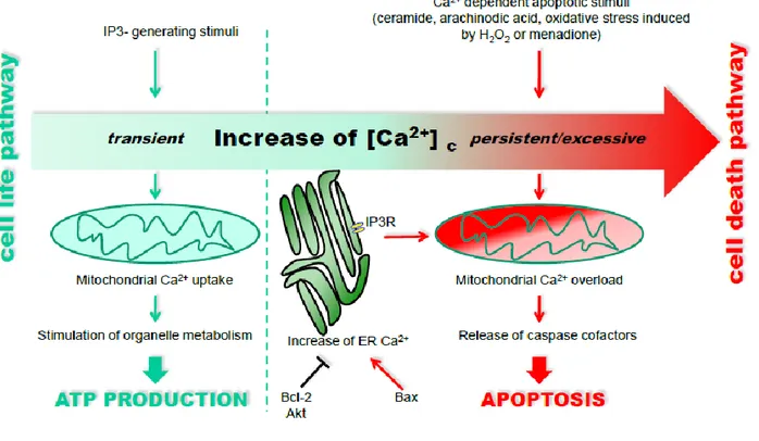
Mitochondrial Ca2+ overload
Although Ca2+ uptake in the mitochondria is crucial for vital cell functions, there exists a risk of mitochondrial Ca2+ overload, which may result in the induction of cell death (Figure 4). There are two pathways that can lead to apoptosis, the death receptor pathway (extrinsic apoptotic pathways) and the mitochondrial pathway (intrinsic apoptotic pathways), both converging on the activation of the executioner caspases (127).
The mitochondrial IMS contains many pro-apoptotic factors such as cytochrome c, apoptosis-inducing factor (AIF), Smac/Diablo, HtrA2/Omi and endonuclease G (EndoG). These are released from mitochondria to the cytosol in response to apoptotic signals (for a review see (128)). Released pro-apoptotic proteins can initiate three signalling cascades leading to apoptosis: i) released cytochrome c, together with pre-existing cytosolic apoptosis protease activating factor 1 (APAF-1) forms the “apoptosome”, which results in the activation of procaspase-9 and in turn activation of effector caspases (caspases-3, -6, and -7), the primarily responsible for the cleavage of cellular proteins leading to the biochemical and morphological characteristics of apoptosis; ii) released Smac/DIABLO and Omi/HtrA2 favour caspase activation by antagonizing the endogenous inhibitor of apoptosis (IAP) proteins in the cytosol; and iii) released AIF and EndoG favour DNA fragmentation and chromatin condensation.
The release of pro-apoptotic factors is preceded by the OMM permeabilization, a crucial step in apoptosis. However, the exact mechanism of mitochondrial OMM permeabilization is not yet clear (129). Ca2+ is a critical sensitizing signal in the pro-apoptotic transition of mitochondria, that plays a key role in the regulation of cell death. At a high concentration, mitochondrial Ca2+ stimulates drastic changes in mitochondrial morphology and functional activity due to the opening of a non-specific pore, commonly known as the PTP, a mitochondrial megachannel likely to be located in the inner-outer contact sites of the mitochondrial membranes (108). This event, also known as mitochondrial permeability transition (MPT), leads to osmotic swelling of the mitochondria, loss of their membrane potential, and rupture of the OMM, causing the release of IMS proteins, including cytochrome c, into the cytosol (129, 130). This process can be facilitated by inorganic phosphate, oxidation of pyridine nucleotides, ATP depletion, low pH, and ROS. The PTP is generally believed
24
to be a multimeric complex, composed of VDAC in the OMM, ANT in the IMM, and a matrix protein, cyclophilin D (CypD). Ca2+ binding to cyclophilin D positively regulates PTP opening and in turn cell death (131). However, the molecular nature of the PTP is still unresolved (108). An important point hereby was the demonstration that the MPT was not affected by the genetic ablation of any or all of the 3 VDAC isoforms (95). PTP opening may ultimately also lead to necrosis, if MPT and subsequent uncoupling of mitochondria occur in a large subpopulation of these organelles; indeed the border between apoptotic and necrotic cell death is quite diffuse.
Mitochondrial membrane permeabilization can also result from a distinct, yet partially overlapping process known as mitochondrial outer membrane permeabilization (MOMP) (128). In MOMP, pro-apoptotic members of the B-cell CLL/lymphoma-2 (Bcl-2)-protein family may form protein-permeable pores in the OMM (for example, by binding to the VDAC channels and regulating their properties or by forming multimeric channel complexes (132)), causing the release of IMS proteins into the cytosol. Moreover, Bcl-2 family members function as regulators of Ca2+ signalling; this important aspect will be discussed in the following section (the interested reader should also refer to (133)).
1.6 Remodelling ER-mitochondria Ca
2+transfer in cell survival and death
ER and mitochondria functions are intimately connected. A major area of functional interaction between the ER and mitochondria is the control of Ca2+ signalling, that is a topic of major interest in physiology and pathology. These two organelles form a highly dynamic interconnected network within which they cooperate to generate Ca2+ signals. The mitochondria play an important role in shaping the Ca2+ signal released from the ER. During normal signalling, there is a continuous flow of Ca2+ between these two organelles. The normal situation is for most of the Ca2+ to reside within the lumen of the ER except during Ca2+ signalling when a small bolus is periodically released to the cytoplasm and is then re-sequestered with a proportion passing through the mitochondria. At equilibrium, therefore, the bulk of internal Ca2+ is in the ER where it not only functions as a reservoir of signal Ca2+ but it also plays an essential role in maintaining the activity of the chaperones responsible for protein processing (26). However, despite controlling many processes essential for life, Ca2+ arising from the ER can be a potent death-inducing signal (134, 135).
The release of Ca2+ from ER stores by IP3Rs has been implicated in multiple models of apoptosis as being directly responsible for massive and/or a prolonged mitochondrial Ca2+ overload. The requirement of IP3Rs for Ca2+-dependent cell death is exemplified by the resistance to apoptosis of cellsin which InsP3R expression has been ablated or reduced (136, 137). Mitochondria seem to be
25
the downstream effectors of this pathway, as KO of IP3R3 significantly decreased agonist-induced mitochondrial Ca2+ uptake (138). In this picture, the three isoforms of the IP3R appear to play distinct roles. IP3R3 seems to play a selective role in the induction of apoptosis by preferentially transmitting apoptotic Ca2+ signals into mitochondria, whereas IP3R1 predominantly mediates cytosolic Ca2+ mobilization (139, 140). However, other studies have shown that the type 1 isoform can also mediate apoptosis (141).
Several observations underline the significance of the role of the ER-mitochondrial Ca2+ flux in stimulating apoptosis. Indeed, a wide number of apoptotic stimuli, such as ceramide, arachinodic acid, and oxidative stress induced by H2O2 or menadione, trigger both a progressive release of Ca2+
from the ER and an activation of the capacitative Ca2+ influx (142, 143). This sustained ERCa2+ release, in turn, induced a mitochondrial Ca2+ overload with a consequent release of mitochondrial proteins involved in the apoptotic process (Figure 4).
Figure 4. Differential decoding of Ca2+-linked stimuli evoking the activation of cell metabolism or apoptosis. (figure
modified from (135)
Since ER and mitochondria play significant roles in the regulation of cell proliferation and apoptosis, the remodeling of Ca2+ signaling machinery in ER and mitochondria of cancer cells seems imminent during oncogenic transformation, to limit death-inducing Ca2+ signals during
26
cancer. The first indication came from the observation that in cancer cells the increased expression of anti-apoptotic members of the Bcl-2 family of proteins (Bcl-2 and Bcl-XL), or decreased
expression of the pro-apoptotic BH3-only proteins (Bax or Bak) can protect these cells from apoptosis by modulating intracellular Ca2+ signals. These proteins reside in the ER, cytosol and mitochondria as homo o heterodimers. Of interest, the proapoptotic protein Bcl-2 affects ER-mitochondrial Ca2+ crosstalk, as the over-expression of Bcl-2 reduces the Ca2+ content of the ER (144) making the cells resistant to apoptosis. Similarly, genetic ablation of the proapoptotic proteins Bax and Bak that drastically increases the resistance to death signals also results in a dramatic reduction in ER Ca2+ content, and consequently in a reduction of the Ca2+ that can be transferred to mitochondria (143). The use of a Bax/Bak double-knockout model system demonstrated that Bcl-2 forms a macromolecular complex with the IP3Rs. The decreased level of Bax and Bak hereby correlated inversely with the amount of Bcl-2 bound to the IP3R, the phosphorylation status of the IP3R and the Ca2+ leak from the ER, leading to the conclusion that Bcl-2 regulated ER Ca2+-store content by regulating the phosphorylation status and the activity of the IP3R. The phosphorylation of IP3R1 was proposed to be due to protein kinase A, but the role of other kinases could not be dismissed (145).
IP3R phosphorylation appears to be a key common feature for modulation of channel function and, as consequence, apoptotic signalling. IP3Rs possess consensus sequences for phosphorylation by numerous kinases, including the pro-survival protein kinase Akt. The consensus site for phosphorylation by Akt has been identified at the carboxyl terminus (serine 2618) of all three mammalian IP3R isoforms and is conserved from mammals to flies (66). This phosphorylation event decreases IP3-stimulated Ca2+ release from the ER and so diminishes flux of Ca2+ to the mitochondria following stimulation with pro-apoptotic agonists, thereby reducing apoptosis (146, 147). This is an interesting observation, because in some cancer cells in which Akt is constitutively active (e.g. prostatic carcinoma cells), IP3Rs are hyper-phosphorylated (66). These data suggest that this functional interaction between Akt and IP3Rs is retained in tumour cells, endowing them with a significant survival advantage by limiting Ca2+-dependent death signalling.
ER-mitochondria Ca2+ transfer appears to be a key sensitizing in various apoptotic routes. Hence, therapeutic modulation of targets that regulate [Ca2+]er and/or ER-mitochondrial Ca2+ transfer may
be able to augment apoptosis in cancer cells without disrupting global Ca2+ homeostasis. However, the precise molecular definition of this process still awaits a fine clarification of the macromolecular complex assembled at the interphase between the two organelles. As will be discussed shortly, significant research efforts have been made to shed some light on this signalling pathway, and this was also the main aim of this thesis project.


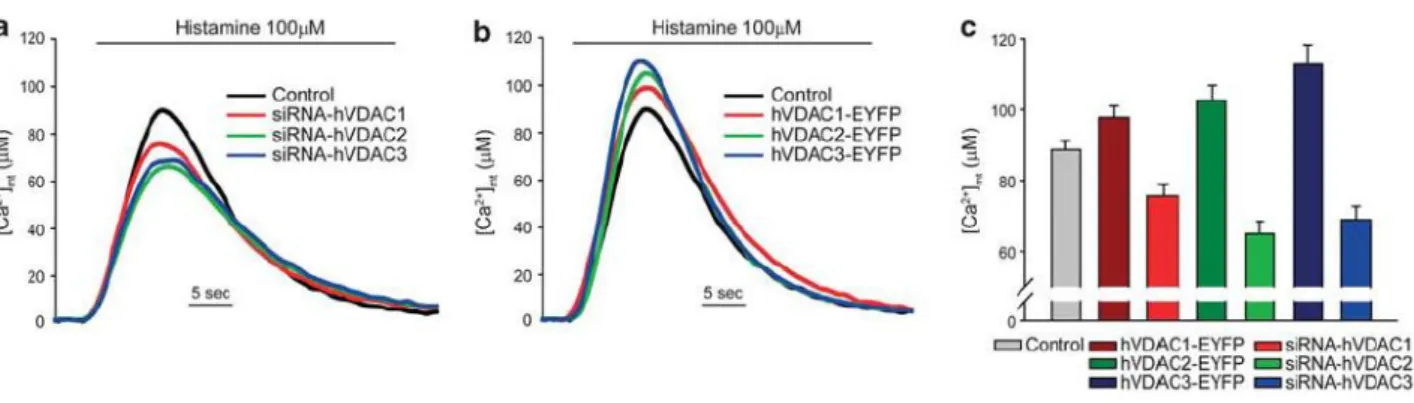
![Figure 10. Effect of VDAC isoform silencing or overexpression on ER and cytosolic [Ca 2+ ]](https://thumb-eu.123doks.com/thumbv2/123dokorg/4700120.44751/43.892.95.802.476.681/figure-effect-vdac-isoform-silencing-overexpression-er-cytosolic.webp)
