UNIVERSITY OF CATANIA
International Ph.D. Program in Neuroscience
XXX Cycle
GAIT DISORDERS IN PARKINSON'S DISEASE:
PHARMACOLOGICAL AND NON-PHARMACOLOGICAL
APPROACHES
PhD Thesis
Valeria Dibilio
Coordinator: Prof. Salvatore Salomone Tutor: Prof. Mario Zappia
2
TABLE OF CONTENTS
Chapter I. General Introduction 1. Parkinson's Disease
2. Gait Disorders in Parkinson's Disease 3. Gait Assessment
4. Treatment of Gait Disorders in Parkinson's Disease 4.1 Pharmacological approach
4.2 Non-pharmacological approach
Chapter II. Pharmacological approach: Dopaminergic and Non-Dopaminergic
Gait Components Assessed by Instrumented Timed Up and Go Test in Parkinson's Disease
Chapter III. Non-pharmacological approach: Computer-assisted cognitive
rehabilitation on freezing of gait in Parkinson’s disease: A pilot study
General Discussion
Closing Remarks
References
3
Chapter I. General Introduction
1. Parkinson's Disease
Parkinson's disease (PD) is the second most common progressive neurodegenerative disorder after Alzheimer's disease. Although its cause remains unknown, PD probably arises from an interaction between genetic and environmental factors that leads to progressive degeneration of neurons in susceptible regions of the brain
(Pringsheim et al. Mov Disord 2014). Reported standardized incidence rates of PD
are 8-18 per 100.000 person-years and its prevalence in industrialized countries is generally estimated at 0,3% of the entire population and about 1% in people over 60 years of age (de Lau et al. Lancet Neurol 2006).
Clinically, PD is characterized by a broad spectrum of motor and non-motor features. The diagnosis is typically based on a combination of cardinal motor features and the clinical response to L-dopa (LD) (Rao et al. JAMA 2003). Diagnostic criteria have been developed by the UK Parkinson’s Disease Society Brain Bank (Hughes et al J
Neurol Neurosurg Psychiatry 1992). Recently new diagnostic criteria have been
proposed by the Movement Disorders Society. Although motor abnormalities remain central, increasing recognition has been given to non motor manifestations. Two levels of certainty are delineated: clinically established PD (maximizing specificity at the expense of reduced sensitivity) and probable PD (which balances sensitivity and specificity) (Postuma et al. Mov. Disord 2015). Differential diagnosis of PD versus other forms of parkinsonism, including secondary and atypical parkinsonisms, is very important for prognosis, diagnosis and therapy since it usually responds to LD
4 therapy (Fahn et al. Principles and practice of movement disorders. 2nd ed. 2011) which still represents the main drug to treat the motor symptoms of PD.
2. Gait Disorders in Parkinson's Disease
Physiological gait is defined as a complex motor and mental ability that requires the interaction between different neuronal systems and it consists of three main mechanisms: locomotion, balance and ability to adapt to the environment (Snijders et
al. Lancet Neurol 2007). Virtually all levels of the nervous system are needed for
normal gait (Morris et al. Adv Neurol 2001).
The safety and efficacy of normal walking rely not only on sensorimotor systems, but also critically depend on the interaction between the executive control dimension with the cognitive dimension and the affective dimension (Snijders et al. Lancet
Neurol 2007). The interplay between cognitive functions and gait can be investigated
by evaluating a secondary task during gait, as it creates competition for attention and allocation of cognitive resources (Yogev-Seligmann et al. Gait Posture 2012;
Woollacott et al. Gait Posture 2002).
Gait disorders can be classified according to the system responsible for the abnormal locomotion, according to the underlying disease associated with the abnormal gait or by its phenomenology.
They are common in elderly people and their prevalence increases with age. The most severe consequence is falling with higher risk for head trauma, bone fractures, development of fear of falling and consequent loss of mobility and independence
5 Gait deficits are common and debilitating symptoms of PD and they can be classified as episodic and continuous (Schaafsma et al Eur J Neurol 2003). Continuous gait deficits include stopped posture, reduced arm swing, reduced speed, reduced stride length and increased stride-to-stride variability. In the more advanced stages, reduced stride length and exponentially-increased cadence can lead to festination and freezing of gait (FOG), which are considered episodic deficits (Nieuwboer et al. Mov Disord
2001, Chee et al. Brain 2009).
FOG is a gait disorder characterized by sudden, relatively brief episodes of inability to step, or by extremely short steps (Nutt et al. Lancet Neurol 2011; Nonnekes et al.
Lancet Neurol 2015). FOG is frequently evoked by challenging walking tasks such
as walking with short steps or by turning as rapidly as possible (Chee et al. Brain
2009; Snijders et al. Parkinsonism Relat Disord 2012; Spildooren et al. Mov Disord 2010) but it can appear also during start walking and during walking through narrow
spaces. FOG correlates with disease duration and it usually appears in “OFF” state but i can occur also in “ON” phases (Schaafsma et al. Eur J Neurol 2003).
It has been hypothesized that PD patients increase their attention during walking to compensate for their gait deficits (Yogev-Seligmann et al. Gait Posture 2012; Yogev
et al. Eur J Neurosci 2005; Rochester et al. Neuroscience 2014). When cognitive
compensation becomes insufficient, particularly when challenging walking tasks further increase attentional and executive demands, FOG might emerge (Giladi et al.
J Neurol Sci 2006; Vandenbossche et al. Neuropsychology 2013).
In PD, cognition has shown to be affected, even in the early stage of disease (Amboni
6 abilities are common fields involved (Amboni et al. Mov Disord 2013; Bloem,et al.
Mov Disord 2004). It could be hypothesized that the cognitive decline seen in
patients with PD might exacerbate subjects difficulties in maintaining gait steadiness and rhythmicity, leading to FOG and increased risk of falling (Giladi et al. J Neurol
Sci 2006).
3. Gait assessment
Adequate and timely recognition of balance and gait disorders is important to identify patients at risk of falling. Also, quantifying gait and balance deficits is relevant for monitoring patients over time (Bloem et al. 2016 Mov Disord; Shulman
Mov Disord 2010). Assessment of gait disorders includes first a neurological
examination and a systematic gait assessment. Simple undisturbed gait can be informative, but additional abnormalities can be cleared when gait is challenged
(Snijders et al. Lancet Neurol 2007) asking patient to turn or to perform two tasks at
the same time. Moreover it is common the use of standard rating scales investigating gait and balance deficits, as UPDRS motor section, which includes some items regarding gait and balance (Postural Instability and Gait Disability score), the Tinetti mobility index, Berg Balance scale, The Freezing of Gait Questionnaire (FOG-Q). All these tests help to score different aspects of gait and balance.
Timed tests can be also used to quantify gait velocity and to assess the effect of treatment. The Timed Up and Go test (TUG) is a widely used clinical test which has proved to be useful for the evaluation of balance and mobility. It is quick and easy to administer: it requires subjects to stand up from a chair, walk three meters, turn 180
7 degrees, walk back to the chair, and sit down. The traditional clinical outcome of this test is the total duration, which is usually measured with a stopwatch (Podsiadlo et
al. J Am Geriatr Soc 1991). The TUG showed consistent reliability and validity as
marker for increased fall risk in the general elderly population (Shumway-Cook et al.
Phys Ther 2000).
In neurological conditions, such PD, it may be useful to discriminate PD subjects from healthy controls as well as “OFF” from “ON” motor phases (Morris et al. Phys
Ther 2001). However, TUG presents also some limitations: it measures only the total
time to perform a series of complex activities without focusing on the separate parts of the performance (Salarian et al. IEEE Trans Neural Syst Rehabil Eng 2010). The attempt to address some of these issues, such as employing an Extended Timed Get Up and Go (ETGUG) test, (Wall et al. J Rehabil Res Dev 2000) or simply using a stopwatch to measure each of the four components of the tests, is somewhat difficult and subjective, and it does not overcome the main limitation of TUG, i.e. focusing only on time.
To obtain more quantitative and objective measures, gait evaluation can be completed with different methods. A standard gait analysis with the full analysis of the motion of all body segments produces well-quantified and accurate results over short distances. A gait laboratory is usually equipped with an optoelectronic motion-capture system, a video camera, a surface EMG system and force plates used to measure ground-reaction force. The system provide the 3D kinematic and kinetic data and the time–distance parameters. The second method is newly developed and uses wearable sensors with a long-term monitoring system; the equipment involved is light, small, and inexpensive and can be carried for long periods and distances to
8 quantify gait parameters (Pei-Hao Chen et al. International Journal of Gerontology
2013).
Recently, an instrumented version of the TUG test (instrumented Timed Up and Go or “iTUG”) has been proposed, providing quantitative information about the TUG performance in different clinical backgrounds (Mirelman et al. J Am Geriatr Soc
2014, Greene et al. IEEE Trans Biomed Eng 2010, Zampieri et al. Parkinsonism Relat Disord 2011). The iTUG appears to be useful to differentiate gait performances
in older adults with mild cognitive impairment from those with normal cognitive function,10 to estimate fall risk in elderly people (Greene et al. IEEE Trans Biomed
Eng 2010) and to evaluate motor function in PD subjects (Zampieri et al. Parkinsonism Relat Disord 2011). iTUG has also proved to be sensitive in detecting
clinically subtle balance or gait deficits in early PD, such as reduced cadence, range and velocity of trunk rotation, arm swing and turning velocity in untreated PD
(Zampieri et al. J Neurol Neurosurg Psychiatry 2010).
4. Treatment of gait disorders in Parkinson's Disease
4.1 Pharmacological approach
Pharmacological treatment aiming to increase the dopamine neurotransmitter is most effective for bradykinesia, rigidity and tremor. Although less effective for postural instability and falls, gait can partly be improved by levodopa, dopamine agonists, or inhibitors of dopamine metabolism (Smulders et al. Parkinsonism and Related
Disorders 2016).
9 the effect of LD, or to increase the dose in patients already treated with LD
(Nonnekes et al. Lancet Neurology 2015). Usually gait disorders occur when
medication has worn off, so it could be useful to treat the response fluctuations in order to reduce the “OFF” state. Main results showed that bradykinetic and hypometric spatial characteristics of gait and turning improve with dopaminergic medication (Suppa et al. Front Neurol 2017; Curtze et al. Mov Disord 2015; Sterling
et al. J Parkinsons Dis 2015; Doan et al. J Neurodegener Dis 2013; Rochester et al. Mov Disord 2011; Bryant et al. Neurol Res 2011; Franzen et al. Exp Neurol 2009; Fregni et al. Eur Neurol 2006; Rocchi et al. Neurosci Lett 2006; Burleigh-Jacobs et al. Mov Disord 1997; Weller et al. Br J Clin Pharmacol 1993; Blin et al. J Neurol Sci 1991; Bowes et al. Br J Clin Pharmacol 1990). However, it is unclear whether
more challenging tasks, such as gait initiation and gait adjustments, improve with dopaminergic treatment (Pieruccini-Faria et al. J Mot Behav 2013; Jacobs et al. Exp
Neurol 2009). Dopaminergic treatment effects on stability measures of gait, such as
spatial or temporal variability, are thus far inconclusive (Smulders et al.
Parkinsonism Relat Disord 2016) (See Supplement Material).
Levodopa is a double-edged sword for treating mobility dysfunction in people with PD. When ON, subjects with PD walk and turn more quickly but became less stable during quiet standing and probably turning. Dyskinesia rather than disease severity accounted for these negative effects of levodopa (Curtze et al. Mov Disord 2015). Indeed LD treatment is often complicated by dose limiting side-effects (Nonnekes et
al. Lancet Neurology 2015).
Dopamine agonists, MAOB and COMT inhibitors as adjunctive therapy to levodopa are also used most likely to improve or prolong the effects of levodopa, hence
10 improving bradykinesia and hypometria in gait (Smulders et al. Parkinsonism Relat
Disord. 2016).
4.2 Non-pharmacological approach
The effect of dopaminergic treatment on gait disorders and FOG can result inconsistent in some cases, so different approaches have been used to treat postural instability and gait disorders in PD, principally including physiotherapy protocols
(Sto˙zek et al. Aging Clin Exp Res 2016) and deep brain stimulation.
Rehabilitative approaches to PD have been characterized by a large heterogeneity. General physiotherapy (stretching, muscle strengthening, balance and postural exercises), occupational therapy, and treadmill training, are frequently adopted to improve specific aspects of mobility (Tomlinson et al. Cochrane Database Syst Rev
2012; Tomlinson et al. Cochrane Database Syst Rev 2014; Abbruzzese et al. Parkinsonism and Related Disorders 2016). Physiotherapy includes both dedicated
strategies (cues) that can assist patients to overcome freezing of gait episodes (eg, conscious movement strategies to increase step amplitude, retaining stepping rhythm, making lateral weight shifts, directing attention to gait, and making wide arcs when turning) and the recommendation to maintain sufficient exercise levels (Nonnekes et
al. Lancet Neurol 2015; Morris et al. Phys Ther 2010; Nieuwboer Mov Disord 2008).
Some innovations have been recently proposed and concern motor imagery and action observation, virtual reality and exergaming, and robot-assisted training
(Abbruzzese et al. Parkinsonism and Related Disorders 2016).
11 demonstrated to improve neural plasticity and attentional functions in treated PD patients (Cerasa et al. Neurol. Sci. 2014). Current clinical recommendations for a cognitive rehabilitation protocol are sufficient for limited neurological conditions such astraumatic brain injury and stroke (Cicerone et al. Arch. Phys. Med. Rehabil.
2011). For PD, effects of a cognitive training have been systematically evaluated
only for cognitive and behavioral outcome measures, indicating an overall modest effect on cognition in patients with mild to moderate PD (Leung et al. Neurology
2015). Despite the growing interest on gait and cognition, to date there are no studies
demonstrating the effect of a cognitive rehabilitation protocol on gait disorders in PD patients. In this pilot study, we hypothesized that a computer-assisted cognitive rehabilitation protocol may influence some gait parameters in people with PD and FOG.
In all cases, reliable tools are required to determine the severity of gait disorders and evaluate the efficacy of interventions (Silva de Lima et al. J Neurology 2017).
12
Supplement Material. Summary of the main studies regarding the L-dopa effects on
different gait parameters.
Authors N.
subjects Parameters significantly improved Parameters not significantly improved
Suppa et al. 2017 44 Step velocity Stride lenght, stride time, cadence Curtze et al. 2015 104 Gait velocity, stride/step
lenght, stride/step duration, cadence, arm swing, trunk movement, APA, turning during walking (180°)
Double support time, step execution
Sterling et al. 2015 16 Arm swing Doan et al. 2013 10 Gait adaptability
Pieruccini et al. 2013 12 Gait adaptability
Rochester et al. 2011 50 Gait velocity, stride/step length
Stride/step duration, cadence, temporal variability
Bryant et al. 2011a 33 Gait velocity, stride/step lenght, spatial and temporal variability Bryant et al. 2011b 21 Gait velocity, stride/step
lenght, double support time
Stride/step duration, cadence
Jacobs et al. 2009 10 APA
Franzen and Horak 2009 15 Turning during walking (180°)
Fregni et al 2006 14 Gait velocity
Rocchi et al. 2006 21 APA, step execution, Burleigh-Jacobs et al. 1997 6 APA, step execution Weller et al. 1993 9 Stride/step lenght Blin et al. 1991 20 Gait velocity, stride/step
length
Stride/step duration, temporal variability Bowes et al. 1990 14 Gait velocity, stride/step
length
Double support time
13
Chapter II: Pharmacological approach
Dopaminergic and Non-Dopaminergic Gait Components Assessed by Instrumented Timed Up and Go Test in Parkinson's Disease.
1. Aim of the study
We hypothesized that L-dopa may have different effects on gait parameters recorded by a portable inertial sensor. To test this idea, we evaluated iTUG test in PD patients in OFF and ON-state.
2. Materials and methods
2.1 Participants
Twenty-eight patients with PD participated. All patients were diagnosed according to the UK Brain Bank diagnostic criteria (Hughes et al. J Neurol Neurosurg Psychiatry
1992). Exclusion criteria were: a) any other neurological or orthopedic disorder
affecting gait; b) severe cognitive impairment and medication negatively affecting gait or balance. The study was approved by the local medical ethics committee (Comitato etico Catania1). All subjects gave their written informed consent prior to the experiment.
2.2 Clinical assessment
PD patients were assessed clinically with the Motor Examination subsection of the Unified Parkinson’s Disease Rating Scale (UPDRS-ME, score/108), (Fahn et al.
14
Healthcare Information, 1987) and the Abnormal Involuntary Movement Scale
(AIMS) for L-dopa induced dyskinesia (Munetz et al. Hosp Community Psychiatry
1988). The Postural Instability/Gait Difficulties (PIGD) subscore was also computed
using the UPDRS-ME (Jankovic et al. Neurology 1990). MDS UPDRS motor scores were calculated using the proposed conversion method (Goetz et al. Mov Disord
2008). Daily doses of L-dopa and other antiparkinsonian agents at the time of the
study entry were also recorded and the LED (L-dopa Equivalent Dose) was calculated for each study patient (Tomlinson et al. Mov Disord 2010). Patients clinical evaluations were performed during their own “practical-off” motor state or “OFF-state”, thus before taking the first daily dose of the dopaminergic drug after an overnight fast, as well as during their “ON-state” at the peak-of-dose after assuming the first daily dose of the dopaminergic medication. Patients with FOG were classified based on the presence of at least one observed episode of freezing evoked during clinical examination at the time of the assessment.
2.3 Experimental set-up and protocol
Patients performed the TUG test wearing the inertial sensor BTS G-WALK (BTS Bioengineering S.p.A., Italy) on a waist belt covering the L4–L5 inter-vertebral space. The portable system consists in a wireless network of inertial sensors for human movement analysis. The sensors are controlled by a data logger unit. Each sensor is sized 62 mm × 36 mm × 16 mm, weighs 60 g, and is composed of a 3-axis accelerometer (max range ± 6 g), a axis gyroscope (full scale ± 300 deg/s), and a 3-axis magnetometer (full scale ± 6 gauss). The system is connected to a computer via Bluetooth. At the end of the measurement data are automatically processed by a
15 dedicated software (BTS G-STUDIO), which automatically provides information about fluency of sitting and rising movements, acceleration, speed and angles analysis during turning and walking pattern analysis. Subjects were instructed to perform the task both in OFF and in ON-state. The system differentiates the test in six phases: a) stand-to-sit phase, b) forward phase; c) mid-turning phase; d) backward phase; e) turning-before-sitting phase; f) stand-to-sit phase. The following parameters were recorder during the different test phases: duration of phases, accelerations in antero-posterior (AP), medio-lateral (ML) and vertical axes, average and peak angular speeds during turning. All sensor measures were obtained by two trials for each participant and then averaged to be analyzed. Algorithm for the automatic segmentation of TUG into the six phases and the extraction of the related measures have been tested in previous published works (Kleiner et al. Parkinsons
Dis 2015).
2.4 Statistical analysis
Scalar measures are described using mean ± standard deviation. Categorical variables are expressed as frequency (percent). Differences in means within groups were tested by the paired-samples t test (2-tailed). Differences in means between groups were tested by the independent-samples t test (2-tailed). A repeated measures ANOVA was also performed to test the differences between OFF and ON-state in freezers and non freezers.
16
3.1 Clinical assessment
Clinical characteristics of the participants were the following: age 66.3 ± 8.5 years, age of onset 58.5 ± 8.8 years, average LED 625.6 ± 333.8 mg, Hoehn and Yahr stage 2.2 ± 0.4, UPDRS-ME OFF score 28.1 ± 10 [equivalent to MDS UPDRS OFF motor score 35.7 ± 11.92], UPDRS-ME ON score 22.1 ± 10.1 [equivalent to MDS UPDRS ON motor score 28.4 ± 12], PIGD OFF subscore 2.8 ± 1.7, PIGD ON subscore 2 ± 1.4, AIMS score 3.5 ± 4.1. No significant differences were evident between OFF and ON conditions for axial scores of UPDRS-ME (items 27, 28 and 30).
3.2 TUG parameters
All participants were able to complete the measurement without stopping during the tasks. The TUG parameters recorded during OFF and ON-states are shown in Table 1. Overall the temporal parameters improved in ON-state. The total test duration and both forward and backward phases duration significantly decreased as well as the mid-turning and turning-before-sitting duration. No significant changes were observed for both sit-to-stand and stand-to-sit durations. During the sit-to-stand phase, both ML and vertical accelerations increased after taking L-dopa therapy, while no statistical differences were observed in the AP acceleration. Mid-turning average and peak angular speed, as well as turning-before-sitting average and peak angular speed significantly increased. No significant changes were observed between OFF and ON-states for all acceleration parameters in the stand-to-sit phase.
3.3 Differences between freezers and non freezers
17 did not. Freezers presented FOG during the OFF-state, which was responsive to L-dopa. Clinical data for freezers and non freezers are shown in Table 2. Patients with freezing were significantly older and had higher PIGD scores in OFF condition than patients without freezing. In OFF-state, freezers had significant longer duration in the total test and forward phase than non freezers.
Indeed, when tested in OFF-state, freezers had worse results than non freezers especially in turning-related parameters (Table 3). Comparing OFF-state and ON-state freezers showed statistically significant variations in mid-turning average and peak angular speed, as well as in turning-before-sitting duration, average and peak angular speed. Non-freezers showed statistically significant differences only in mid-turning duration, average and peak angular speed.
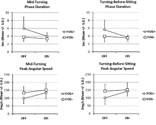
A great OFF-ON state improvement of kinematic parameters in freezers was generally related to a worse condition in the OFF-state compared to non-freezers. Indeed, the gain of performance due to dopaminergic efficacy was greater for freezers, thus making possible the values in ON-state of patients with FOG almost similar to values of non freezers. Significant OFF-ON state changes on turning in kinematic parameters of PD patients with or without FOG are shown in Figure 1. A repeated measures ANOVA was executed to detect the impact of the categorical variable “FOG” on differences between OFF and ON pharmacological states for all kinematic parameters. According to these results, the presence of FOG had significant effect in determining changes between OFF and ON-state on turning, particularly in duration of mid-turning phase (F = 6.88; p = 0.01) and turning-before sitting phase (F = 6.77; p = 0.02), and peak angular speed for mid-turning phase (F = 4.69; p = 0.04) and turning-before-sitting phase (F = 7.39; p = 0.01) as well.
18 Also using the simpler analytic approach already reported in table 3, when tested in OFF-state, freezers had worse results than non freezers especially in turning-related parameters. Comparing OFF-state and ON-state, freezers showed statistically significant variations in mid-turning average and peak angular speed, as well as in turning-before-sitting duration, average and peak angular speed, confirming results obtained by the repeated measures ANOVA.
19
Table 1
Tab.1. iTUG parameters in OFF and ON-state in 28 PD subjects
OFF-state ON-state p-valuesa
Test duration (s) 20.2 ± 12.6 15.4 ± 5.2 0.021 *
Forward phase duration (s) 5 ± 4.5 3.5 ± 1.8 0.037 *
Backward phase duration (s) 3.2 ± 2.3 2.4 ± 1.4 0.01 *
Sit-to-stand phase
Sit-to-stand phase duration (s) 1.8 ± 0.8 1.6 ± 0.5 0.126
Sit-to-stand phase AP acceleration (m/s2) 7.8 ± 2.1 7.9 ± 2.1 0.647
Sit-to-stand phase ML acceleration (m/s2) 2.9 ± 1.5 3.5 ± 1.9 0.013 *
Sit-to-stand phase Vertical acceleration (m/s2) 6.1 ± 2.7 7 ± 3.1 0.009 *
Mid-turning-phase
Mid-turning phase duration (s) 4.4 ± 1.9 3.5 ± 0.9 0.008 *
Mid-turning average angular speed (deg/s) 48.4 ± 15.9 55.8 ± 13.2 0.001 * Mid-turning peak angular speed (deg/s) 123.8 ± 39.8 146.9 ± 42.1 < 0.001 *
Turning-before-sitting phase
Turning-before-sitting phase duration (s) 4.4 ± 1.9 3.6 ± 0.8 0.008 * Turning-before-sitting average angular speed (deg/s) 44.3 ± 13.1 50.3 ± 12.9 0.004 * Turning-before-sitting peak angular speed (deg/s) 130.8 ± 47.6 149.7 ± 39.6 0.004 *
Stand-to-sit phase
Stand-to-sit phase duration (s) 2.4 ± 0.9 2.3 ± 0.8 0.828
Stand-to-sit phase AP acceleration (m/s2) 7.7 ± 1.9 8.1 ± 1.8 0.133
Stand-to-sit phase ML acceleration (m/s2) 5.2 ± 1.9 5.1 ± 1.9 0.802
Stand-to-sit phase Vertical acceleration (m/s2) 6.6 ± 2.1 6.4 ± 2.3 0.533
Notes: Values are means ± standard deviations.a paired-samples t-test; * significant difference (p < 0.05). Legend: iTUG = instrumental Timed Up and Go Test; AP = antero-posterior; ML = medio-lateral.
20
Table 2
Tab.2. Clinical characteristics and iTUG duration parameters in OFF-state of PD patients with or without freezing of gait.
Freezers (N = 8) Non-Freezers(N = 20)
p-valuea
Age (years) 70 ± 3.9 64.8 ± 9.4 0.049 *
Age of onset (years) 59.7 ± 4.7 58 ± 10 0.538
Average LED (mg) 649.1 ± 366.7 625.6 ± 333.8 0.692
Average L-dopa test dose (mg) 163.7 ± 57.3 163.8 ± 57.5 0.997
Hoehn & Yahr stage 2.4 ± 0.5 2.1 ± 0.4 0.121
UPDRS-ME OFF score 28.9 ± 6.9 27.8 ± 11.2 0.812
UPDRS-ME ON score 23.6 ± 6.3 21.4 ± 11.4 0.617
PIGD OFF subscore 4.2 ± 1.3 2.2 ± 1.5 0.002 *
PIGD ON subscore 2.4 ± 1.3 1.9 ± 1.5 0.482
AIMS score 4.4 ± 4.4 3.1 ± 4.1 0.490
iTUG Total duration OFF-state (s) 29.4 ± 18.5 16.5 ± 7.1 0.012 *
iTUG Forward phase duration OFF-state (s) 8.2 ± 7 3.8 ± 2.4 0.018 *
iTUG Backward phase duration OFF-state (s) 4.2 ± 2.9 2.8 ± 2 0.239
Notes: Values are means ± standard deviations; a independent-samples t-test; * significant difference (p < 0.05). Legend: LED = L-dopa Equivalent Dose; UPDRS-ME = Unified Parkinson’s Disease Rating Scale - Motor Examination; PIGD = Postural Instability/Gait Difficulties; AIMS = Abnormal Involuntary Movement Scale.
21
Table 3
Tab.3. iTUG parameters on turning in OFF- and ON-state of PD patients with or without freezing of gait.
Freezers (N = 8) Non-Freezers (N = 20)
OFF-State ON-State
p-valuea Gain (%)b OFF-State ON-State p-valuea Gain (%)b
Mid-turning phase
Mid-turning phase duration (s) 6 ± 2.9 4 ± 1 0.073 1.9 ± 2.7 3.8 ± 1 3.3 ± 0.8 < 0.001* 0.4 ± 0.4 Mid-turning average angular
speed (deg/s)
35.2 ± 15.8 48.1 ± 11.8 0.025 * -12.9 ± 12.7 53.7 ± 12.8 58.9 ± 12.7 0.009 * -5.2 ± 8 Mid-turning peak angular
speed (deg/s) 97.8 ± 32.6 134.2 ± 37.4 0.005 * -36.4 ± 25.9 † 134.2 ± 38.3 152.1 ± 43.7 < 0.001* -17.9 ± 18 † Turning-before-sitting phase Turning-before-sitting phase duration (s) 5.6 ± 2.4 3.7 ± 0.6 0.041 * 1.9 ± 2.1 3.9 ± 1.4 3.5 ± 0.9 0.075 0.4 ± 0.9 Turning-before-sitting average
angular speed (deg/s)
33.8 ± 10.6 45.3 ± 9.1 0.031 * -11.5 ± 12 48.5 ± 11.7 52.3 ± 13.9 0.062 -3.8 ± 8.7 Turning-before-sitting peak
angular speed (deg/s)
100.4 ± 40.3 142.7 ± 30.3 0.025 * -42.3 ± 42.1 ‡ 143 ± 45.6 152.5 ± 43.2 0.069 -9.5 ± 22 ‡
Notes: Values are means ± standard deviations; a paired-samples t-test; b percent change between OFF- and ON-state computed as (OFF-state – ON-state) x 100 / OFF- state; * significant difference (p < 0.05). Legend: iTUG = instrumental Timed Up and Go Test. † independent-samples t test p = 0.04; ‡ independent-samples t test p = 0.012.
22
Figure 1. Significant OFF-ON state changes in kinematic parameters on turning of PD patients with (FOG+) or without
23
Chapter III. Non-pharmacological approach
Computer-assisted cognitive rehabilitation on freezing of gait in Parkinson’s Disease: A pilot study
1. Aim of the study
Despite the growing interest on gait and cognition, to date there are no studies demonstrating the effect of a cognitive rehabilitation protocol on gait disorders in PD patients. In this pilot study, we hypothesized that a computer-assisted cognitive rehabilitation protocol may influence some gait parameters in people with PD and FOG.
2. Materials and methods
2.1. Participants
Patients with PD participated to the study. All patients were diagnosed according to the UK Brain Bank criteria (Huges et al. J Neurol Neurosurg Psychiatry 1992). Inclusion criteria were: Mini Mental State Examination (MMSE) score >26, Hoehn and Yahr stage ≤3, disease duration ≥5 years, presence of FOG evaluated clinically the day of the assessment as well as historically by The Freezing of Gait Questionnaire (FOG-Q) (Nieuwboer et al. Gait Posture 2009). Patients who were treated with antiparkinsonian medications or antidepressants maintained stable doses of the drugs during the study period. Eight patients were selected based on the
24 inclusion criteria (3 women; age 64.3 ± 8.03 years). The study was approved by the local medical ethics committee and was conducted in accordance with the Declaration of Helsinki and with local ethical guidelines. All subjects gave their written informed consent prior to the experiment.
2.2. Clinical assessment
PD patients were assessed clinically with the Unified Parkinson’s Disease Rating Scale - Motor Examination (UPDRS-ME) section (Fahn et al. Recent developments
in Parkinson's disease. Florham Park, NJ: Macmillan Healthcare Information, 1987) and the Abnormal Involuntary Movements Scale (AIMS) (Munetz et al. Hosp Community Psychiatry 1988). Daily doses of L-dopa and other antiparkinsonian
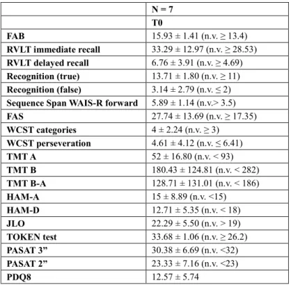
drugs at the time of the study entry were also recorded. All patients completed an extensive battery of neuropsychological tests (Cerasa et al. Neurol. Sci. 2014), including: Frontal Assessment Battery (FAB), Rey Auditory Verbal Learning Test (RVLT), Sequence Span WAIS-R,Verbal fluency: Controlled Oral Word Association Test (COWAT),Modified Wisconsin Card Sorting Test (WCST), Trail Making Test(TMT), Hamilton Anxiety Scale A), Hamilton Depression Scale (HAM-D), Judgment Line Orientation Test (JLO), TOKEN Test, Paced Auditory Serial Addition Task (PASAT), Parkinson’s Disease Questionnaire 8 (PDQ8). Motor and cognitive performances were recorded at baseline (T0). All PD patients were measured in ON-state, when they experienced the peak-of-dose effect after the intake of their medication dose.
25 The gait analysis was conducted using the following equipment: a six-camera optoelectronic system with passive markers (BTS SMART-DX, Milan, Italy), working at frequency acquisition up to 2000 Hz, and six force platforms (BTS P-6000, Milan, Italy) equipped with twelve sensors (transducers) for each platform to provide the 3D kinematic and kinetic data and the time–distance parameters; a TV camera video system (BTS VIXTA, Milan, Italy) synchronized with the optoelectronic and force platform systems for video recording. Patients performed ten trials walking at their self-selected speed along a 6-m walkway.
2.4. Experimental set-up and protocol
Patients were treated twice a week for 1-h sessions for six consecutive weeks. Sessions consisted of computer-assisted training of several attention ability and information processing tasks. Cognitive training was performed using the package RehaCom (http://www.Schuhfried.at) (Cerasa et al. Neurol Sci 2014). The rehabilitation training consisted of following modules: attention and concentration; vigilance; visual-motor coordination; logical reasoning; divided attention. All the training sessions were performed in ON state.
2.5. Statistical analysis
Quantitative variables are described using mean ± standard deviation. Statistical inference was performed using non-parametric tests based on the small sample size adopted. Differences in means within groups were tested by Wilcoxon signed-rank test or by the Friedman’s ANOVA test. As post-hoc test a Wilcoxon signed-rank test was run on the different combinations of related groups when significant differences
26 were detected.
3. Results
3.1. Clinical assessment
A total of seven patients completed the assessments at T1 (3 women, 42.9%; age 63.6 ± 8.4 years) with one patient dropped out because of a worsening of his clinical systemic conditions which was independent from the study protocol. Six patients completed the assessments at T2, with another one patient dropped-out who refused to complete the program at T2. Clinical characteristics of included patients at T0 and T1 (N = 7) were: women 3 (42.9%); age63.6 ± 8.4 years; disease duration 9.1 ± 4.7 years; Hoehn-Yahr stage2.1 ± 0.2; MMSE score 27.5 ± 0.9; FOG-Q score 12.4 ± 5.5; UPDRS-ME score 25 ± 6.1; AIMS score 5.6 ± 4.9. At T1 with respect to T0 no significant changes were detected in UPDRS-ME (24.1 ± 6.5; p = 0.1) and AIMS (6.6 ± 3.4; p = 0.2) scores. All patients were in L-dopa therapy (cumulative daily dosage: 610.7 ± 308.8 mg). Concerning other antiparkinsonian medications, two patients were treated with pramipexole (cumulative daily dosage: 1.44 ± 1.66 mg), one with ropinirole (cumulative daily dosage: 2 mg), two with rotigotine (cumulative daily dosage: 5 ± 4.24 mg), three with ICOMT (cumulative daily dosage: 433.3 ± 152.75 mg), four with IMAO-B (cumulative daily dosage: rasagiline 1 mg). All patients had normal results at neuropsychological assessment (Table 1).
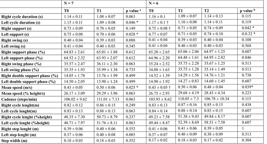
3.2. Gait parameters
27 (T1), gait cycle duration was significantly improved on the left and it had a statistical trend on the right. Also mean velocity significantly improved at T1 compared to T0.Although not significantly, other parameters improved compared to the baseline, such as the cadence and left cycle length. The six patients who completed three months of follow-up (T2) did not differ with respect to the baseline for gait parameters.
28
Table 1 Neuropsychological evaluations. N = 7 T0 FAB 15.93 ± 1.41 (n.v. ≥ 13.4) RVLT immediate recall 33.29 ± 12.97 (n.v. ≥ 28.53) RVLT delayed recall 6.76 ± 3.91 (n.v. ≥ 4.69) Recognition (true) 13.71 ± 1.80 (n.v. ≥ 11) Recognition (false) 3.14 ± 2.79 (n.v. ≤ 2)
Sequence Span WAIS-R forward 5.89 ± 1.14 (n.v.> 3.5)
FAS 27.74 ± 13.69 (n.v. ≥ 17.35) WCST categories 4 ± 2.24 (n.v. ≥ 3) WCST perseveration 4.61 ± 4.12 (n.v. ≤ 6.41) TMT A 52 ± 16.80 (n.v. < 93) TMT B 180.43 ± 124.81 (n.v. < 282) TMT B-A 128.71 ± 131.01 (n.v. < 186) HAM-A 15 ± 8.89 (n.v. <15) HAM-D 12.71 ± 5.35 (n.v. < 18) JLO 22.29 ± 5.50 (n.v. > 19) TOKEN test 33.68 ± 1.06 (n.v. ≥ 26.2) PASAT 3” 30.38 ± 6.69 (n.v. <32) PASAT 2” 23.33 ± 7.16 (n.v. <23) PDQ8 12.57 ± 5.74
29
Table 2 Gait parameters in single-task at self-selected speed
N = 7 N = 6
T0 T1 p value a T0 T1 T2 p value b
Right cycle duration (s) 1.14 ± 0.11 1.08 ± 0.07 0.063 1.16 ± 0.1 1.09 ± 0.07 1.14 ± 0.13 0.115
Left cycle duration (s) 1.15 ± 0.11 1.09 ± 0.08 0.046 * 1.17 ± 0.1 † 1.10 ± 0.08 1.14 ± 0.11 0.119
Right support (s) 0.73 ± 0.09 0.70 ± 0.05 0.149 0.75 ± 0.08 † 0.71 ± 0.05 0.74 ± 0.09 0.042 *
Left support (s) 0.75 ± 0.08 0.70 ± 0.06 0.028 * 0.77 ± 0.07 0.71 ± 0.05 0.74 ± 0.10 0.0.32 *
Right swing (s) 0.40 ± 0.04 0.39 ± 0.03 0.088 0.41 ± 0.04 0.39 ± 0.03 0.40 ± 0.04 0.108
Left swing (s) 0.41 ± 0.04 0.40 ± 0.03 0.345 0.41 ± 0.04 0.40 ± 0.03 0.40 ± 0.03 0.568
Right support phase (%) 64.83 ± 2.61 65.01 ± 1.88 0.612 65.20 ± 2.65 65.06 ± 2.06 64.97 ± 1.25 0.607
Left support phase (%) 64.52 ± 2.32 63.93 ± 2.07 0.612 64.96 ± 2.20 64.48 ± 1.61 64.95 ± 2.82 0.846
Right swing phase (%) 35.57 ± 2.47 36.11 ± 2.30 0.063 35.24 ± 2.52 35.75 ± 2.28 35.67 ± 1.25 0.513
Left swing phase (%) 35.35 ± 1.93 35.99 ± 1.38 0.735 34.88 ± 1.63 35.75 ± 1.28 35.14 ± 1.49 0.513
Right double support phase (%) 14.05 ± 1.78 13.76 ± 1.99 0.499 14.52 ± 1.39 14.29 ± 1.56 14.76 ± 1.21 0.738
Left double support phase (%) 14.50 ± 2.05 13.90 ± 1.24 0.499 14.90 ± 1.92 14.27 ± 0.83 14.60 ± 1.45 0.607
Mean speed (m/s) 0.43 ± 0.05 0.50 ± 0.06 0.025 * 0.43 ± 0.05 † 0.50 ± 0.06 0.48 ± 0.04 0.039*
Mean speed (% height/s) 26.17 ± 3.09 29.29 ± 3.96 0.063 26.75 ± 2.93 29.68 ± 4.19 28.43 ± 4.34 0.223
Cadence (steps/min) 106.02 ± 9.62 111.01 ± 7.13 0.063 103.93 ± 8.62 110.05 ± 7.3 106.78 ± 10.34 0.115
Right cycle lenght(m) 0.82 ± 0.12 0.86 ± 0.15 0.249 0.83 ± 0.13 0.87 ± 0.16 0.85 ± 0.15 0.438
Left cycle lenght(m) 0.83 ± 0.13 0.88 ± 0.13 0.063 0.84 ± 0.14 0.88 ± 0.14 0.83 ± 0.15 0.607
Right cycle lenght (%height) 48.35 ± 7.30 50.73 ± 8.79 0.237 49.23 ± 7.58 51.38 ± 9.43 49.84 ± 8.17 0.607
Left cycle lenght (%height) 48.71 ± 7.97 51.76 ± 8.11 0.063 49.44 ± 8.47 52.39 ± 8.69 58.51 ± 7.58 0.607
Right step lenght (m) 0.39 ± 0.06 0.40 ± 0.06 0.553 0.41 ± 0.06 0.41 ± 0.06 0.39 ± 0.05 1
Left step lenght (m) 0.37 ± 0.06 0.40 ± 0.08 0.085 0.37 ± 0.07 0.40 ± 0.09 0.38 ± 0.09 0.513
Step width (m) 0.18 ± 0.03 0.18 ± 0.03 0.552 0.17 ± 0.02 0.18 ± 0.03 0.17 ± 0.02 0.304
Legend: data are mean ± S.D. T0, T1, and T2 are respectively baseline, 6-weeks and 3-months follow-up visits. a) Wilcoxon signed-rank
test p values between T0 and T1; b) Friedman's ANOVA test p values between T0, T1 and T2. * p < 0.05. † Significant difference between groups at the post-hoc analysis using Wilcoxon signed-rank test (p < 0.05).
30
General Discussion
1. Pharmacogical approach
The timed up and go test investigated by an inertial sensor showed that all the measures except those related to sit-to-stand (duration and AP acceleration) and stand-to-sit (all the parameters) phases were significantly improved by Ldopa.
These results support the hypothesis that gait components are differently modulated by dopamine replacement in PD (Chastan et al. Brain 2009). To the best of our knowledge, this is the first study which tested L-dopa effect on all kinematic parameters during the execution of the TUG test, focusing on the entire sequence of complex actions, as standing up from a chair, walking, turning and sitting. We have used a wearable inertial sensor that, compared to other quantitative methods, is lighter, smaller and can be carried for long periods and distances to evaluate
gait parameters. Testing our patients in OFF and then in ON pharmacological
state, a common trend of improvement was showed for most parameters. In particular, we found that sit-to-stand and turning phases proved to be more sensitive to L-dopa acute effect, while stand-to-sit phase was less responsive. Moreover, AP acceleration seems to be less responsive to L-dopa therapy than ML and vertical accelerations. This could mean that dopaminergic transmission at basal ganglia, which is essential to maintain the cortically selected motor pattern, could influence gait subcomponents differently and play major role just for some of
31
L-dopa acute effect on kinematic parameters of TUG phases
Sit-to-stand phase in PD subjects is known to be influenced by inadequate lower
extremity forces, especially at the hip, bradykinesia (Duncan et al. Arch Phys Med
Rehabil 2011), and above all impaired anticipatory postural control, resulting in a
failure to bring the center of mass (COM) adequately forward over the feet prior to the lift-off of the buttocks from the chair (Inkster et al. Exp Brain Res 2004).
We evaluated the time needed to complete the action, the accelerations in AP, ML and vertical planes. All these parameters improved in ON: sit-to-stand duration reduced, even if not critically, and acceleration increased especially in ML and in vertical planes more than in AP. This is consistent with Burleigh-Jacobs et al.,
(Burleigh-Jacobs et al. Mov Disord 1997) since L-dopa increases force production
and velocity of movement and accelerates the execution of the anticipatory postural adjustments prior to step initiation. Considering that sit-to-stand movement is a rapid transition from a large base of support in a stable position to a smaller one in a less stable position, with the shift of the COM in the forward and upward directions, a movement of the COM that goes beyond the base of support may lead to imbalance and falling (Siriphorn et al. J Phys Ther Sci 2015). AP acceleration could not increase further because the ON pharmacological state improves balance and allows a major control of the COM shift.
Both forward and backward phases needed less time to be completed, as the whole TUG test, since speed increased meaningfully.
According to our results, stand-to-sit phase was instead not particularly influenced by L-dopa. It could be influenced by other factors than simply L-dopa transmission in basal ganglia. It has been demonstrated that multiple neural circuits, such as
32 cholinergic circuits involving brainstem peduncolopontine nucleus neurons, are implicated in control of balance and gait, with varying sensitivity to L-dopa (Curtze
et al. Mov Disord 2015). On the other hand, it could speculate that this peculiar
aspect of TUG may be not specific of the disease differently from the other phases and, thus, L-dopa could not further improve the stand-to-sit phase in PD.
It is interesting to note that symmetrical phases of TUG, i.e., sit-to-stand and stand-to-sit phases, were differently modulated by L-dopa administration. The mechanisms underlying the motor control of this apparently ‘‘symmetric’’ phases of TUG are probably different, thus justifying the different dopaminergic responsiveness between sit-to-stand and stand-to-sit phases. During the stand-to-sit phase, probably PD patients presented reduced postural control stability and this could be related to altered trunk control (Fernandes et al. Med Eng Phys 2015; van der Burg et al.
Parkinsonism Relat Disord 2006). This aspect is a characteristic of conditions
strongly associated with postural instability and high risk of falls, such as Progressive Supranuclear Palsy, with poor L-dopa responsiveness.
Turning is often difficult for PD subjects, since they require more steps and time to
turn in-place or turn while walking than healthy people, especially those who present FOG. Individuals with PD also employ different muscle activation strategies, with a simultaneous rotation of the head, trunk, and pelvis body segments rather than the cranio-caudal rotation sequence present in normal people (McNeely et al. J Park Dis
2011; Curtze et al. Phys Ther 2016). Our study considered the time needed to turn,
the average and the peak angular speed during mid-turning and turning-before-sitting. All these kinematic parameters improved after L-dopa administration. It has been demonstrated that dopaminergic drugs improve turning in L-dopa responsive
33 PD patients (McNeely et al. J Park Dis 2011). However, some turning impairments may also remain in ON-state since not all the aspects of turning are sufficiently addressed by L-dopa. This may suggest that turning impairments in PD could be only partially mediated by dopaminergic systems and degeneration of other non-dopaminergic systems may be also involved (McNeely et al. J Park Dis 2011).
Differences between freezers and non freezers on turning during L-dopa OFF and ON-state
Further information derived from separating the study population in two groups, based on the presence of FOG. As we expected, freezers got worse results compared to non-freezers. Mid-turning and turning-before-sitting durations, peak and average angular speeds were worse in freezers but they displayed greater improvements with medication, which was probably due to a worse condition in the OFF-state compared to non-freezers. Indeed, turning values of freezers in ON-state approached the values of patients without freezing. Results are in agreement with those of a previous study, in which the TUG test was used to evaluate the effects of medication on turning in PD patients compared to healthy older adults (controls), and in freezers compared to non-freezers. The authors showed that medication partially improved turning in PD patients, freezers turned worse than non-freezers, but they improved more with medication. They concluded that probably further treatment options may be needed to address ON turning deficits in PD (McNeely et al. J Park Dis 2011). Our data support the hypothesis that motor performance during turning are acutely modulated by L-dopa in responsive freezers.
34 can be employed in clinical practice to evaluate the effects of a pharmacological or probably even physical therapy in PD subjects. Data are consistent with the assumption that the various components of the TUG show a different sensitivity to dopaminergic stimulation.
35
Non-pharmacological approach
Many pharmacological and non-pharmacological therapies have been proposed to improve FOG in PD patients (Nonnekes et al. Lancet Neurol 2015). In the present pilot study, we evaluated the effect of a cognitive rehabilitation protocol, focused on executive functions, on gait parameters in patients with PD and FOG. We observed a significant reduction in cycle duration, mainly on the left, with an increment in mean velocity. The cadence also improved, although not significantly. The increasing in cadence was not linked to a stride length reduction as observed in festination and FOG (Nieuwboer et al. Mov Disord 2001), while it was associated with increased average speed. We evaluated the patients at follow up after three months from the end of the cognitive rehabilitation program and we observed no significant differences with respect to the baseline. The lack of a significant effect at three months should require further solutions to achieve a more prolonged clinical effect, in order to implement the proposed rehabilitation protocol for clinical purposes. Despite the growing interest on gait and cognition, to date there are very few evidences on the possible effects of a cognitive rehabilitation protocol on gait disorders in PD patients. The improvement of some gait parameters we observed at the end of the cognitive rehabilitation treatment in patients without apparent deficits of executive functions addresses some questions. We could speculate a potentiation of executive functions induced by the treatment, and influencing gait, but this hypothesis could not be appreciated considering that in our patients the baseline performances at the neuropsychological evaluation were normal. It could be more interesting to evaluate in further studies the effect on gait of a cognitive rehabilitation
36 program in patients with some impairment of executive functions. Our study presents some limitations as the small sample size affecting statistical power and the lack of a control group. Despite these limitations, results from this pilot study suggest that a rehabilitation protocol based on training in executive functions could improve some gait parameters in PD. Patients were trained and tested in ON-state and medications were maintained stable during the entire study period to avoid confounding effects. Furthermore, we assessed changes in gait parameters using the gait analysis to objectively evaluate the rehabilitative intervention. More effective and prolonged results could be obtained in the context of a multidisciplinary rehabilitation program. Moreover, based on the preliminary results obtained by the current pilot study, it could be useful to plan a larger and controlled study to compare a single cycle of cognitive rehabilitation versus reinforcement of another cycle of rehabilitation after a specific period of time, to confirm if a cognitive rehabilitation protocol may improve walking performances in people with PD and FOG.
37
Closing Remarks
Pharmacological treatment and non-pharmacological treatment of gait disorders in PD have been investigated by quantitative methods. L-dopa seems to modulate gait parameters in different ways, mostly improving the turning phases and less acting on postural controls during the sit-to-stand and stand-to-sit phases. This information is relevant to define an effective therapy for those aspects which are not improved by conventional pharmacological treatments. Furthermore a computer-assisted rehabilitation based on executive functions training has shown to improve walking in PD patients with FOG. Further studies are needed to confirm the results.
38
References
Pringsheim T, Jette N, Frolkis A, Steeves TD. The prevalence of Parkinson's disease: a systematic review and meta-analysis. Mov Disord. 2014 Nov;29(13):1583-90.
de Lau LM, Breteler MM. Epidemiology of Parkinson's disease. Lancet Neurol. 2006 Jun;5(6):525-35.
Rao G, Fisch L, Srinivasan S, D’Amico F, Okada T, Eaton C, et al. Does this patient have Parkinson disease? JAMA. 15 2003;289(3):347–53
Hughes AJ, Daniel SE, Kilford L, Lees AJ. Accuracy of clinical diagnosis of idiopathic Parkinson’s disease: a clinico-pathological study of 100 cases. J Neurol Neurosurg Psychiatry. 1992;55(3):181–4
Postuma RB, Berg D, Stern M, Poewe W, Olanow CW, Oertel W, Obeso J, Marek K, Litvan I, Lang AE, Halliday G, Goetz CG, Gasser T, Dubois B, Chan P, Bloem BR, Adler CH, Deuschl G. MDS clinical diagnostic criteria for Parkinson's disease. Mov Disord. 2015 Oct;30(12):1591-601.
Fahn S, Jankovic J, Hallett M. Principles and practice of movement disorders. 2nd ed. 2011)
Snijders AH, van de Warrenburg BP, Giladi N, Bloem BR. Neurological gait disorders in elderly people: clinical approach and classification. Lancet Neurol. 2007 Jan;6(1):63-74.
Morris ME, Huxham FE, McGinley J, Iansek R. Gait disorders and gait rehabilitation in Parkinson’s disease. Adv Neurol 2001; 87: 347–6.
39 Effects of explicit prioritization on dual task walking in patients with Parkinson's disease. Gait Posture. 2012 Apr;35(4):641-6.
Woollacott M, Shumway-Cook A. Attention and the control of posture and gait: a review of an emerging area of research. Gait Posture. 2002 Aug;16(1):1-14.
Schaafsma JD, Balash Y, Gurevich T, Bartels AL, Hausdorff JM, Giladi N. Characterization of freezing of gait subtypes and the response of each to levodopa in Parkinson's disease. Eur J Neurol. 2003 Jul;10(4):391-8.
Nieuwboer A, Dom R, De Weerdt W, Desloovere K, Fieuws S, Broens-Kaucsik E. Abnormalities of the spatiotemporal characteristics of gait at the onset of freezing in Parkinson's disease. Mov Disord. 2001 Nov;16(6):1066-75.R.
Chee R, Murphy A, Danoudis M, Georgiou-Karistianis N, Iansek R. Gait freezing in Parkinson's disease and the stride length sequence effect interaction. Brain. 2009 Aug;132(Pt 8):2151-60.
Nutt JG, Bloem BR, Giladi N, Hallett M, Horak FB, Nieuwboer A. Freezing of gait: moving forward on a mysterious clinical phenomenon. Lancet Neurol. 2011 Aug;10(8):734-44.
Nonnekes J, Snijders AH, Nutt JG, Deuschl G, Giladi N, Bloem BR. Freezing of gait: a practical approach to management. Lancet Neurol. 2015 Jul;14(7):768-78.
Spildooren J, Vercruysse S, Desloovere K, Vandenberghe W, Kerckhofs E, Nieuwboer A. Freezing of gait in Parkinson's disease: the impact of dual tasking and turning. Mov Disord. 2010 Nov 15;25(15):2563-70.
40 Yogev G, Giladi N, Peretz C, Springer S, Simon ES, Hausdorff JM. Dual tasking, gait rhythmicity, and Parkinson's disease: which aspects of gait are attention demanding? Eur J Neurosci. 2005 Sep;22(5):1248-56.
Rochester L, Galna B, Lord S, Burn D. The nature of dual-task interference during gait in incident Parkinson's disease. Neuroscience. 2014 Apr 18;265:83-94.
Giladi N, Hausdorff JM. The role of mental function in the pathogenesis of freezing of gait in Parkinson's disease. J Neurol Sci. 2006 Oct 25;248(1-2):173-6.
Vandenbossche J, Deroost N, Soetens E, Coomans D, Spildooren J, Vercruysse S, Nieuwboer A, Kerckhofs E. Impaired implicit sequence learning in Parkinson's disease patients with freezing of gait. Neuropsychology. 2013 Jan;27(1):28-36.
Amboni M, Cozzolino A, Longo K, Picillo M, Barone P. Freezing of gait and executive functions in patients with Parkinson's disease. Mov Disord. 2008 Feb 15;23(3):395-400.
Amboni M, Barone P, Hausdorff JM. Cognitive contributions to gait and falls: evidence and implications. Mov Disord. 2013 Sep 15;28(11):1520-33. Bloem BR, Hausdorff JM, Visser JE, Giladi N. Falls and freezing of gait in
Parkinson's disease: a review of two interconnected, episodic phenomena. Mov Disord. 2004 Aug;19(8):871-84.
Bloem BR, Marinus J, Almeida Q, Dibble L, Nieuwboer A, Post B, Ruzicka E, Goetz C, Stebbins G, Martinez-Martin P, Schrag A; Movement Disorders Society Rating Scales Committee. Measurement instruments to assess
41 posture, gait, and balance in Parkinson's disease: Critique and recommendations. Mov Disord. 2016 Sep;31(9):1342-55.
Shulman LM. Understanding disability in Parkinson's disease. Mov Disord. 2010;25 Suppl 1:S131-5.
Podsiadlo D, Richardson S. The timed "Up & Go": a test of basic functional mobility for frail elderly persons. J Am Geriatr Soc. 1991 Feb;39(2):142-8. Shumway-Cook A, Brauer S, Woollacott M. Predicting the probability for
falls in community-dwelling older adults using the Timed Up & Go Test. Phys Ther 2000;80:896-903.
Morris S, Morris ME, Iansek R. Reliability of measurements obtained with the Timed "Up & Go" test in people with Parkinson disease. Phys Ther 2001;81:810-818.
Salarian A, Horak FB, Zampieri C, et al. iTUG, a sensitive and reliable measure of mobility. IEEE Trans Neural Syst Rehabil Eng 2010;18:303-310. Wall JC, Bell C, Campbell S, et al. The Timed Get-up-and-Go test revisited:
measurement of the component tasks. J Rehabil Res Dev 2000;37:109-113. Pei-Hao Chen, Rong-Long Wang, De-Jyun Liou, Jin-Siang Shaw. Gait
Disorders in Parkinson’s Disease: Assessment and Management. International Journal of Gerontology 7 (2013) 189-193.
Mirelman A, Weiss A, Buchman AS, Bennett DA, Giladi N, Hausdorff JM. Association between performance on Timed Up and Go subtasks and mild cognitive impairment: further insights into the links between cognitive and motor function. J Am Geriatr Soc. 2014 Apr;62(4):673-8.
42 RA. Quantitative falls risk assessment using the timed up and go test. IEEE Trans Biomed Eng. 2010 Dec;57(12):2918-26.
Zampieri C, Salarian A, Carlson-Kuhta P, Nutt JG, Horak FB. Assessing mobility at home in people with early Parkinson's disease using an instrumented Timed Up and Go test. Parkinsonism Relat Disord. 2011 May;17(4):277-80.
Zampieri C, Salarian A, Carlson-Kuhta P, Aminian K, Nutt JG, Horak FB. The instrumented timed up and go test: potential outcome measure for disease modifying therapies in Parkinson's disease. J Neurol Neurosurg Psychiatry. 2010 Feb;81(2):171-6.
Smulders K, Dale ML, Carlson-Kuhta P, Nutt JG, Horak FB. Pharmacological treatment in Parkinson's disease: Effects on gait. Parkinsonism Relat Disord. 2016 Oct;31:3-13.
Suppa A, Kita A, Leodori G, Zampogna A, Nicolini E, Lorenzi P, Rao R, Irrera F. l-DOPA and Freezing of Gait in Parkinson's Disease: Objective Assessment through a Wearable Wireless System. Front Neurol. 2017 Aug 14;8:406.
Curtze C, Nutt JG, Carlson-Kuhta P, Mancini M, Horak FB. Levodopa Is a Double-Edged Sword for Balance and Gait in People With Parkinson's Disease. Mov Disord. 2015 Sep;30(10):1361-70.
Sterling NW, Cusumano JP, Shaham N, Piazza SJ, Liu G, Kong L, Du G, Lewis MM, Huang X. Dopaminergic modulation of arm swing during gait among Parkinson's disease patients. J Parkinsons Dis. 2015;5(1):141-50. Doan JB, de Bruin N, Pellis SM, Suchowersky O, Whishaw IQ, Brown LA.
43 Obstacle Avoidance amongst Parkinson Disease Patients Is Challenged in a Threatening Context. J Neurodegener Dis. 2013;2013:787861.
Rochester L, Baker K, Nieuwboer A, Burn D. Targeting dopa-sensitive and dopa-resistant gait dysfunction in Parkinson's disease: selective responses to internal and external cues. Mov Disord. 2011 Feb 15;26(3):430-5.
Bryant MS, Rintala DH, Hou JG, Charness AL, Fernandez AL, Collins RL, Baker J, Lai EC, Protas EJ. Gait variability in Parkinson's disease: influence of walking speed and dopaminergic treatment. Neurol Res. 2011 Nov;33(9):959-64.
Franzén E, Paquette C, Gurfinkel VS, Cordo PJ, Nutt JG, Horak FB. Reduced performance in balance, walking and turning tasks is associated with increased neck tone in Parkinson's disease. Exp Neurol. 2009 Oct;219(2):430-8.
Fregni F, Boggio PS, Bermpohl F, Maia F, Rigonatti SP, Barbosa ER, Pascual-Leone A. Immediate placebo effect in Parkinson's disease--is the subjective relief accompanied by objective improvement? Eur Neurol. 2006;56(4):222-9.
Rocchi L, Chiari L, Mancini M, Carlson-Kuhta P, Gross A, Horak FB. Step initiation in Parkinson's disease: influence of initial stance conditions. Neurosci Lett. 2006 Oct 2;406(1-2):128-32.
Burleigh-Jacobs A, Horak FB, Nutt JG, Obeso JA. Step initiation in Parkinson's disease: influence of levodopa and external sensory triggers. Mov Disord. 1997 Mar;12(2):206-15.
44 Dobbs RJ, Dobbs SM. Defining small differences in efficacy between anti-parkinsonian agents using gait analysis: a comparison of two controlled release formulations of levodopa/decarboxylase inhibitor. Br J Clin Pharmacol. 1993 Apr;35(4):379-85.
Blin O, Ferrandez AM, Pailhous J, Serratrice G. Dopa-sensitive and dopa-resistant gait parameters in Parkinson's disease. J Neurol Sci. 1991 May;103(1):51-4.
Bowes SG, Clark PK, Leeman AL, O'Neill CJ, Weller C, Nicholson PW, Deshmukh AA, Dobbs SM, Dobbs RJ. Determinants of gait in the elderly parkinsonian on maintenance levodopa/carbidopa therapy. Br J Clin Pharmacol. 1990 Jul;30(1):13-24.
Pieruccini-Faria F, Vitório R, Almeida QJ, Silveira CR, Caetano MJ, Stella F, Gobbi S, Gobbi LT. Evaluating the acute contributions of dopaminergic replacement to gait with obstacles in Parkinson's disease. J Mot Behav. 2013;45(5):369-80.
Jacobs JV, Nutt JG, Carlson-Kuhta P, Stephens M, Horak FB. Knee trembling during freezing of gait represents multiple anticipatory postural adjustments. Exp Neurol. 2009 Feb;215(2):334-41.
Stożek J, Rudzińska M, Pustułka-Piwnik U, Szczudlik A. The effect of the rehabilitation program on balance, gait, physical performance and trunk rotation in Parkinson's disease. Aging Clin Exp Res. 2016 Dec;28(6):1169-1177.
Tomlinson CL, Patel S, Meek C, Clarke CE, Stowe R, Shah L, Sackley CM, Deane KH, Herd CP, Wheatley K, Ives N. Physiotherapy versus placebo or
45 no intervention in Parkinson's disease. Cochrane Database Syst Rev. 2012 Aug 15;(8):CD002817.
Tomlinson CL, Herd CP, Clarke CE, Meek C, Patel S, Stowe R, Deane KH, Shah L, Sackley CM, Wheatley K, Ives N. Physiotherapy for Parkinson's disease: a comparison of techniques. Cochrane Database Syst Rev. 2014 Jun 17;(6):CD002815.
Abbruzzese G, Marchese R, Avanzino L, Pelosin E. Rehabilitation for Parkinson's disease: Current outlook and future challenges. Parkinsonism Relat Disord. 2016 Jan;22 Suppl 1:S60-4.
Morris ME, Martin CL, Schenkman ML. Striding out with Parkinson disease: evidence-based physical therapy for gait disorders. Phys Ther 2010; 90: 280– 88.
Nieuwboer A. Cueing for freezing of gait in patients with Parkinson’s disease: a rehabilitation perspective. Mov Disord 2008; 23 (suppl 2): S475– 81.
Cerasa A, Gioia MC, Salsone M, Donzuso G, Chiriaco C, Realmuto S, Nicoletti A, Bellavia G, Banco A, D'amelio M, Zappia M, Quattrone A. Neurofunctional correlates of attention rehabilitation in Parkinson's disease: an explorative study. Neurol Sci. 2014 Aug;35(8):1173-80.
Cicerone KD, Langenbahn DM, Braden C, Malec JF, Kalmar K, Fraas M, Felicetti T, Laatsch L, Harley JP, Bergquist T, Azulay J, Cantor J, Ashman T. Evidence-based cognitive rehabilitation: updated review of the literature from 2003 through 2008. Arch Phys Med Rehabil. 2011 Apr;92(4):519-30.
46 Cognitive training in Parkinson disease: A systematic review and meta-analysis. Neurology. 2015 Nov 24;85(21):1843-51.
Silva de Lima AL, Evers LJW, Hahn T, Bataille L, Hamilton JL, Little MA, Okuma Y, Bloem BR, Faber MJ. Freezing of gait and fall detection in Parkinson's disease using wearable sensors: a systematic review. J Neurol. 2017 Aug;264(8):1642-1654.
Fahn S, Elton RL, and the members of the UPDRS development committee. Unified Parkinson's Disease Rating Scale. In: Fahn S, Marsden CD, Calne DB, Goldstein M, eds. Recent developments in Parkinson's disease. Florham Park, NJ: Macmillan Healthcare Information, 1987:153-163.
Munetz MR, Benjamin S. How to examine patients using the Abnormal Involuntary Movement Scale. Hosp Community Psychiatry 1988;39:1172-1177.
Jankovic J, McDermott M, Carter J, et al. Variable expression of Parkinson's disease: a base-line analysis of the DATATOP cohort. The Parkinson Study Group. Neurology 1990;40:1529-1534.
Goetz CG, Tilley BC, Shaftman SR et al. Movement Disorder Society UPDRS Revision Task Force. Movement Disorder Society-sponsored revision of the Unified Parkinson’s Disease Rating Scale (MDS-UPDRS): scale presentation and clinimetric testing results. Mov Disord. 2008 Nov 15;23(15):2129-70.
Tomlinson CL, Stowe R, Patel S, Rick C, Gray R, Clarke CE. Systematic review of levodopa dose equivalency reporting in Parkinson's disease. Mov Disord. 2010 Nov 15;25(15):2649-53.
47 Kleiner A, Galli M, Gaglione M, Hildebrand D, Sale P, Albertini G, Stocchi F, De Pandis MF. The Parkinsonian Gait Spatiotemporal Parameters Quantified by a Single Inertial Sensor before and after Automated Mechanical Peripheral Stimulation Treatment. Parkinsons Dis. 2015;2015:390512.
Nieuwboer A, Rochester L, Herman T, Vandenberghe W, Emil GE, Thomaes T, Giladi N. Reliability of the new freezing of gait questionnaire: agreement between patients with Parkinson's disease and their carers. Gait Posture. 2009 Nov;30(4):459-63.
Chastan N, Westby GW, Yelnik J, Bardinet E, Do MC, Agid Y, Welter ML. Effects of nigral stimulation on locomotion and postural stability in patients with Parkinson's disease. Brain. 2009 Jan;132(Pt 1):172-84.
Duncan RP, Leddy AL, Earhart GM. Five times sit-to-stand test performance in Parkinson's disease. Arch Phys Med Rehabil 2011;92:1431-1436.
Inkster LM, Eng JJ. Postural control during a sit-to-stand task in individuals with mild Parkinson's disease. Exp Brain Res 2004;154:33-38.
Siriphorn A, Chamonchant D, Boonyong S. The effects of vision on sit-to-stand movement. J Phys Ther Sci 2015;27:83-86.
Fernandes Â, Sousa AS, Couras J, Rocha N, Tavares JM. Influence of dual-task on sit-to-stand-to-sit postural control in Parkinson's disease. Med Eng Phys. 2015 Nov;37(11):1070-5.
van der Burg JC, van Wegen EE, Rietberg MB, Kwakkel G, van Dieën JH. Postural control of the trunk during unstable sitting in Parkinson's disease. Parkinsonism Relat Disord. 2006 Dec;12(8):492-8.
48 with Parkinson disease with and without freezing of gait. J Park Dis 2011;1:259-270.
Curtze C, Nutt JG, Carlson-Kuhta P, Mancini M, Horak FB. Objective Gait and Balance Impairments Relate to Balance Confidence and Perceived Mobility in People With Parkinson Disease. Phys Ther. 2016 Nov;96(11):1734-1743.