DEPARTMENT OF AGROBIOLOGY AND AGROCHEMISTRY
PhD Course of Study
BIOLOGICAL AND BIOCHEMICAL EVOLUTION – XX CYCLE
“Role of Apoptosis in the final outcome of DNA damage” BIO/18
Coordinator: Prof. Federico Federici ………..
Tutor: Dr. Roberta Meschini ………
PhD student: Paola Belloni ………..
Abstract
Damage to DNA is intrinsic to life, as the cell continuosly suffers from numerous exogenous agents, including radiation and chemicals, and from endogenous sources, such as free radicals generated during essential metabolic processes. The broad spectrum of DNA lesions induced by these agents includes damage to nucleotide bases, DNA protein cross-links and DNA single- and double-strand breaks (DBSs). Because of their high cytotoxicity (unrepaired or misrepaired DNA DSBs can kill a cell) and their ability to induce chromosomal aberrations (that may ultimately lead to carcinogenesis) cell survival and mantenaince of genome integrity are critically dependent on efficient repair of DNA DSBs. When the burden of genomic insult is too large to be effectively repaired, cells are able to initiate apoptosis (programmed cell death). On the basis of these considerations, the aim of this thesis has been to investigate the role of apoptosis in the final outcome of DNA damage.
Since G0 lymphocytes are the most common tissue used in biodosimetry studies, and the amount of chromosomal damage detected depends on the time between exposure and sampling, it was of interest to investigate the relationship between the frequencies of radiation induced chromosomal aberrations and the extent of apoptosis in G0 human lymphocytes (Paper I). In human lymphocytes irradiated in G0 without subsequent stimulation to proliferate, a p53-dependent apoptotic pathway preferentially eliminates cells bearing unstable aberrations.
Studies in Paper II demonstrated that in human lymphocytes the type of chromosome damage influences the induction of programmed cell death and provided direct evidence that cells bearing dicentric chromosomes are eliminated by apoptosis.
The lymphocytes undergo apoptosis during storage, and this loss of viability is accelerated by increasing both temperature and storage time, was demonstrated in Paper III. Studies on the effects of storage conditions of whole blood on the viability and proliferation of lymphocytes revealed optimal the storage at 4°C for 96 h in the presence of phytohaemagglutinin.
The influence of genetic background on the induction of apoptosis in cells of different origin, treated with a potential anticancer drug “STDS2323”, was studied in Paper IV.
In conclusion, this thesis suggests that apoptosis has an important role in the final outcome of DNA damage. The thesis further suggests that these results may have relevance
for biodosimetrical and molecular epidemiological studies and in evaluations of the efficacy of radio- and chemotherapy.
This Thesis is dedicated to all Blood Donors
Gabbiani
Non so dove i gabbiani abbiano il nido, ove trovino pace.
Io son come loro, in perpetuo volo.
La vita la sfioro com'essi l'acqua ad acciuffare il cibo.
E come forse anch'essi amo la quiete, la gran quiete marina,
ma il mio destino è vivere balenando in burrasca.
List of original publications
This thesis is based on the following publications, which will be referred to by their Roman numerals:
I. Belloni P, Meschini R, Czene S, Harms-Ringdahl M, Palitti F. (2005). Studies on radiation-induced apoptosis in G0 human lymphocytes. Int. J. Radiat. Biol. 81 (8): 587-599.
II. Belloni P, Meschini R, Lewinska D, Palitti F. (2008). Apoptosis preferentially eliminates irradiated G0 human lymphocytes bearing dicentric chromosomes. Radiat. Res. 169 (2): 181-187.
III. Belloni P, Meschini R, Palitti F. (2008). Effects of storage conditions of human whole blood on the viability of lymphocytes. Int. J. Radiat. Biol. Accepted.
IV. Meschini R, Lorenti Garcia C, Latini P, Belloni P, Palitti F. (2008). Higher induction of apoptosis by a potential chemotherapeutic agent “STDS2323” in human bladder cancer cell lines. Manuscript
TABLE OF CONTENTS
INTRODUCTION………. ………….6
Radiation………..6
Ionizing and non-ionizing radiation………. 6
Important definitions, terms and units in radiation biology………..7
Cellular damage……….. 8
DNA lesions of biological importance……….. 9
Cell cycle and its checkpoints……….. ………….10
Repair mechanisms………11
Radiation induced chromosomal alterations……….. …………12
Chromosome types aberrations: unstable aberrations………13
Chromosome types aberrations: stable aberrations………14
Apoptosis……… 14
Biological dosimetry……….. 20 Radiation sensitivity………..…………20
Radiation and Radiotherapy……… 21
THE AIM………... …………23
PRESENT INVESTIGATION………. ………...24
Cell types employed………...24
Assays used……….26 Agents used………. ………...30 INDIVIDUAL PAPERS………32 Paper I……….32 Paper II………...33 Paper III………. 33 Paper IV………. 34 ACKNOWLEDGEMENTS………. 37 REFERENCES………...38
INTRODUCTION
The maintenance and replication of genetic information in DNA are of primary importance for living organisms. Damage to DNA is intrinsic to life, as the cell continuously suffers from numerous exogenous agents, including radiation and chemicals, and from endogenous sources, such as free radicals generated during essential metabolic processes (Friedberg et al. 1995).
Radiation
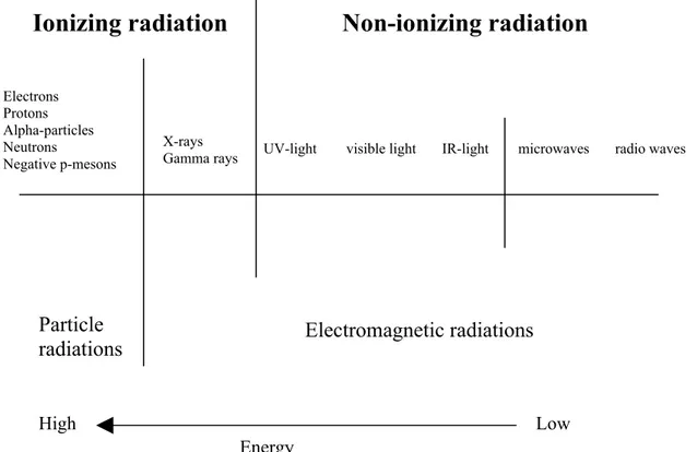
Radiation can be divided into two main categories: electromagnetic and particle radiation. Electromagnetic radiation may be defined as waves and or/photons. The electromagnetic spectrum is the range of wavelengths and frequencies that electromagnetic radiation can assume. This is a very broad range, and these waves exhibit a variety of properties associated with wavelength and frequency. It includes, in order of increasing frequency, radio waves, microwaves, infrared waves, visible light, ultraviolet rays, X- rays, and gamma rays. X-rays are divided into two categories: soft and hard X-rays. Soft X-rays have longer wavelengths and are closer to the ultraviolet band of the spectrum. Hard X-rays are closer to the gamma ray band of the spectrum and have much shorter wavelengths. X-rays are produced when material objects hit high-velocity electrons. Gamma X-rays are the shortest-wavelength, highest-frequency type of electromagnetic radiation. They are essentially identical to X-rays in their effect, but are the most penetrating of all electromagnetic radiation being produced by excited nuclei instead of inner electrons. X-rays are generated by instruments (X-rays machines), while gamma rays are produced by radioactive decay.
Particle radiation can be emitted from unstable atomic nuclei (radioactive decay) in the form of positively charged alpha particles (helium nuclei), positively and negatively charged beta particles (electrons and positrons) and uncharged neutrons. They can also artificially be produced, for example by cyclotrons.
When the energy of radiation is high enough it will produce ionizations and excitations in the target. Radiation that causes ionizations (ejecting one or more orbital electrons) is called “ionizing radiation”, and corresponds to radiation with energy higher than a few electron volts (eV) to 24.5 eV (for helium), and is related to the ionizing potential of the atom (Podgorsak 2003). This energy is the minimum energy to ionize an atom (removing an electron from its atoms orbit). For biological effects 124 eV is the more general norm where radiation is considered causing biological effects (Hall 1994). Furthermore, excitation (raising one of the electrons in an atom or molecule to a higher energy level without ejection of the electron) in the target through which the radiation traverses can also occur. Both particle radiation (electrons, protons, alpha particles, or neutrons) and electromagnetic radiation (high energetic photons such as X- and gamma rays) belong to the category of ionizing radiation.
The type of radiation called “non-ionizing radiation”, e.g. UV and visible light, infrared radiation and micro- and radio waves, does not produce ionizations and excitations when interacting with matter but will interact with many cellular components through photochemical reactions. An example of damage caused by non-ionizing radiation is the redness of the skin caused by exposure of UV radiation, which induces pyrimidine dimers in the DNA- molecule of the cell.
Important definitions, terms and units in radiation biology
Cell damage can be related to the amount of energy absorbed in the cell and is called the dose of radiation (quantity of absorbed energy in matter (J/Kg) = absorbed dose), measured in gray (Gy). The dose rate is the absorbed dose divided by the time of its delivery. Due to limitations of the repair capacity in a living cell per unit of time, high dose rates are usually more damaging.
When ionizing radiation is absorbed in a biological material there is a random distribution of ionizations and excitations along the tracks of the charged particle in a pattern that is dependent on the type of radiation concerned, where e.g. X-rays are considered as sparsely ionizing radiation and 500 keV protons are densely ionizing radiation. There are large differences in ionizations between different types of radiations. In other words, the linear energy transfer (LET) values for different types of radiation illustrate the mean energy released in keV per micrometer (μm) of the tissue traversed (keV/μm). Example of low LET (sparsely ionizing) and high LET (densely ionizing) radiation are X- and gamma rays and energetic neutrons, protons and heavy charged particles respectively. The
demarcation between low and high LET is 10 keV/μm. The equivalent dose relates the absorbed dose (Gy) in tissue to the biological effects of a specific radiation quality. The unit for equivalent dose is sievert (Sv). To determine equivalent dose (Sv), an absorbed dose (Gy) is multiplied by a radiation quality factor (Q). For example 1 Gy equals 1 Sv for gamma radiation (Q for gamma is 1), 1 Gy equals 20 Sv for alpha particles (Q for alpha is 20). Typical energy from gamma ray decay is several million eV. In biological tissue, the mean energy needed to create one ionization is about 16 eV. Alpha particles belong to high-LET and they can penetrate a tissue of paper (or skin) and travel only a few centimeters in air but generates a dense track of ionizations, which results in greater damage to the cell. High-LET radiation is therefore considered to be far more harmful than low-LET radiation (Figure 1).
Figure 1. An illustration of different types of radiations (from: Meijer AE. 1999. ISBN 91-7153-844-4).
Cellular damage
It has long been known that DNA is the major target for biological effects of radiation (Hall 1994). DNA damage is generated through direct and indirect effects. For direct effects, ionizing radiation may deposit its energy directly to a DNA molecule in a cell, where the target itself can be ionized or excited. Direct effects are most frequent in cells exposed to high LET radiation. The direct effect contributes 30% of the radiation induced DNA damage. (Reuvers et al.1973). Alternately, the radiation may also induce damage by ionizing other
Ionizing radiation Non-ionizing radiation
High Low Energy Particle radiations Electrons Protons Alpha-particles Neutrons Negative p-mesons X-rays
Gamma rays UV-light visible light IR-light microwaves radio waves
molecules in close association with DNA in the cell, which is referred to as the indirect effect. This indirect effect of DNA damage is dominant, since a cell consists of about 70% water, and hence mostly water molecules are ionized. This process is called radiolysis of water, in which extremely reactive and short-lived water products are made, free radicals. The main products of radiolysis are hydroxyl radicals (OH•), hydrogen radicals (H•) and solvated electrons (e-aq). Free radicals have unpaired electrons and they react rapidly with everything close by such as other water molecules, cellular proteins, membranes, organelles, and the most critical target, DNA molecules by breaking chemical bonds. A lot of the cell damage caused by low LET radiation is of this indirect type.
The damage of the cell can be lethal (the cell dies) or sub-lethal (the cell can repair itself). The effects on cells can be, according to the International Commission on Radiological Protection (ICRP), deterministic and stochastic. Deterministic effects have a threshold of irradiation under which they do not appear and are the necessary consequence of irradiation. The damage they cause depends on the dose: they are sub lethal from 0,25 to 2 Sv (a less pronounced form of disease), lethal from 2 to 5 Sv (a certain percent of population dies in 60 days), above 5 Sv the majority of people die in 60 days and above 6 to 7 Sv all people die. Of course, these effects depend also on many other factors, like age, sex, health etc. Stochastic effects are coincidental and can not be avoided. They have not a threshold. These can be divided into somatic and genetic. Among the somatic ones, the cancer is the most important. It develops because radiation causes DNA mutations directly and indirectly. The genetic confer the predisposition to cancer to the offspring. They are not very well understood.
DNA lesions of biological importance
Radiation induced DNA damage can be due to both indirect and direct effects of radiation. The hits can occur on either the phosphate backbone, sugar group or on the base, resulting in single strand breaks (SSBs, 1000 breaks/cell per Gy), double strand breaks (DSBs, 20-40 breaks/cell per Gy), and base modifications (1000 modifications/cell per Gy) (Czene and Ringdahl 1995, Goodhead 1994, Pouget et al. 1999, Svoboda and Harms-Ringdahl 2005). A particular feature of radiation induced chemical alterations is the production of unique types of multiple damages sites (MDS), consisting of clusters of strand breaks and base damages within one or two turns of the DNA (10 to 20 base pairs) (Goodhead et al. 1993, Sutherland et al. 2001), they are suggested to be a specific signature of ionizing radiation.
The most dangerous lesions are the DNA DSBs induced by X-rays, chemicals or during replication of SSBs. DSBs are generated when the two complementary strands of the DNA double helix are broken simultaneously at sites that are sufficiently close to one another that base-pairing and chromatin structure are insufficient to keep the two DNA ends juxtaposed. As a consequence, the two DNA ends generated by a DSB are liable to become physically dissociated from one another, making ensuing repair difficult to perform and providing the opportunity for inappropriate recombination with other sites in the genome.
Two SSBs can theoretically induce a DSB depending on the distance between them. DSBs may also create during replication due to an SSB encountered at the replication fork with the fatal consequence of a collapse of the replication fork. DSBs are also important for normal biological functions and are created in a controlled way in cells during meiosis, DNA replication and recombination processes. Probably the best characterized example of this in higher eukaryotes is the V(D)J recombination pathway in the maturation process of developing B- and T-lymphocytes to provide the basis for the antigen-binding diversity of the immunoglobulin and T-cell receptor proteins
.
(Weinert 1998, Zhou and Elledge 2000).Cell cycle and its checkpoints
As cells progress through the cell cycle, they undergo several transitions that are coordinated in time and order. The cycle is divided into four major parts; the G1 phase, the gap before DNA replication which determine the readiness of the cell and thereby preventing the cell from progressing too early into the next step; the S phase, the period of DNA synthesis; the G2 phase, the gap between S and M, and M phase, the mitotic phase. There is also a G0 phase were the cell stays in a quiescent state. The G1-, S- and G2 phases form the interphase. While cells are in the S phase of the cell cycle they duplicate their genome. Several biochemical processes in the cytoplasm determines timing and completion of the cell cycle. Following the S and G2 phases, division starts within the M phase that includes a separation of the duplicated chromosomes and the formation of two daughter cells.
DSBs are the most critical lesions due to their high cytotoxicity and genotoxicity and, if they occur during the replication of the genome and during the segregation of duplicated chromosomes into daughter cells, broken chromosomes are carried through mitosis, and the acentric chromosome fragments will not be segregated between daughter cells. Therefore, eukaryotes have developed several checkpoints to prevent cells from starting DNA replication (the G1/S checkpoint), from progressing with replication (the intra S checkpoint) or from going into mitosis (the G2/M check-point), if they contain damaged DNA (Dasika et
al 1999, Zhou and Elledge 2000). However, it has become clear that DNA damage-induced cell cycle checkpoint pathways actually regulate several other events and, moreover, that various aspects of these responses may even take place in cells that are not actively dividing (Jackson, 2002).
The actions initiated by the Ataxia Telangiectasia (ATM) kinase on activation by DSBs are multiple and complex (Rotman and Shiloh 1998). Part of the signal- transduction cascade is mediated by the p53 protein, which is phosphorylated in response to ionizing radiation in an ATM-dependent fashion. In humans and mice, deletion of the gene that encodes p53 results in a defective G1/S cell cycle checkpoint and a reduced apoptotic response upon irradiation. Mice harbouring a homozygous Trp53 deletion develop T-cell-derived tumours at a young age (Donehower et al. 1992). Furthermore, most human tumours also contain somatically acquired mutations in the TP53 gene, which encodes p53, and the Li-Fraumeni syndrome (which is characterized by a high tumour incidence) can be caused by a heterozygous TP53 mutation, underlining the importance of this gene for tumour prevention (Dasika et al. 1999).
Another phosphorylation target of ATM is the chromatin component H2AX. It is a variant of histone H2A and its phosphorylation is an early response to DSBs (Rogakou et al. 1998). Phosphorylated H2AX is found in foci at which other DSB-repair-proteins, including RAD50 accumulate over time (Paull et al. 2000). At least one other phosphatidylinositol-3-OH kinase (PI (3) K) family member, DNA dependent protein Kinase (DNA-PK, also known as PRKDC), has been implicated in H2AX phosphorilation (Rogakou et al. 1998). This redundancy suggests that H2AX phosphorilation might be crucial for the cellular response to DSBs. Finally, under conditions where the extent of DNA damage is too great; cells can instead enter an apoptotic programme. Although the details of how this solution is reached are not yet clear, it appears that this pathway involves the actions of proteins that also function in other aspects of the DNA-damage response (Rich et al. 2000, Hirao et al. 2000, Herzog et al. 1998)
.
Repair mechanisms
Among the various stress responses induced by damage to DNA perhaps the most understood are the various forms of the DNA repair (Barzilay et al. 1995). The significance of DNA repair is illustrated by the phenotypes of xeroderma pigmentosum, Cockayne´s syndrome, trichothiodystrophy and hereditary non polyposis colorectal cancer patients (de la
Chapelle and Peltomaki, 1995). These disorders are caused by mutations in DNA repair genes that predispose the patients to cancer, neurological abnormalities or both.
All eukaryotes have evolved several mechanisms to deal with DSBs, which indicates the importance and difficulty to repairing this type of DNA injury, such as: homologous recombination (HR) and non-homologous end joining (NHEJ) both involved in repair of DSBs; base excision repair (BER) acting on base damages and SSBs; nucleotide excision repair (NER) mainly involved in removal of bulky adducts (induced by chemicals) and thymidine dimmers (induced by UV light); and the pathway of nucleotide incision repair (NIR) that is involved in repair of base damages (Wood et al. 2005).
Radiation induced chromosomal alterations
Ionizing radiation is a strong clastogen, causing chromosome breakage and resulting in cytogenetic aberrations in exposed cells (Natarajan and Obe 1978, 1984, Goodhead 1994, Ward 1995). When high LET radiation (particle) or low LET radiation (photon) passes through the nucleus, in the case of high LET radiation, the ionizations occur close together. Ionization within a chromosome may result in a break in the DNA. If such breaks are improperly repaired, they may be resolved as either stable (pericentric inversion) or unstable chromosomal aberrations (centric ring and acentric fragment) as illustrated in Figure 2. The same dose of low LET radiation will create more diffuse damage from a larger number of photon tracks with ionization events occurring further apart, resulting in fewer multiple lesions within the same chromosome. When two or more different damaged chromosomes interact to resolve breaks, different types of stable (translocation) and unstable (dicentric and acentric fragment) aberrations result.
Figure 2. Representation of radiations of different quality interacting with chromosomes in a cell nucleus. The dashed arrows represent the track of a particle or photon passing through the nucleus. (From: Amundson et al. 2001. Expert Rev Mol Diagn 1: 89-97).
Chromosome types aberrations: unstable aberrations Dicentrics
The dicentric is the main aberration used for biodosimetry. It is an exchange between the centromeric pieces of two broken chromosomes, which in its complete form is accompanied by a fragment composed of the acentric pieces of these chromosomes. Particularly after high doses, multicentric configurations can be formed. Tricentrics are accompanied by two fragments, quadricentrics by three fragments, etc.
Centric rings
In human lymphocytes, centric rings are much rarer than the dicentrics. Some researchers combine them with dicentrics while others choose to ignore them for dose estimation. The ring chromosome is an exchange between two breaks on separate arms of the same chromosome and is also accompanied by an acentric fragment.
Acentrics
Acentric aberrations can be formed independently of the exchanges described above and as such are usually referred to as excess acentrics. They can be terminal or interstitial deletions of varying sizes but it is not always possible to determine their origin and so they are combined. Minutes, which appear as double dots and acentric rings where clear spaces may be seen within the dots, are normally considered to be interstitial deletions; this has been confirmed by telomere probing (Fomina et al. 2000).
Chromosome types aberrations: stable aberrations Reciprocal translocations
The reciprocal (or complete or two-way) translocation is the exchange of terminal portions of two separate chromosomes. Using the G-banding technique and karyotyping originally described the various types of translocation, but this procedure is too laborious for routine biodosimetry. With solid Giemsa staining, translocations are not observed reliably. Their application to dosimetry is possible with the fluorescence in situ hybridization (FISH) method; by the FISH method these are visualized as bicoloured monocentric chromosomes.
Non-reciprocal translocations
When only one bicoloured chromosome can be seen, this has often been called a terminal, or incomplete, or one-way translocation. However, using a combination of chromosome painting, centromeres and telomere probes, a number of translocations designated as terminal or incomplete was found to be in reality reciprocal. It is very likely that the signal of the missing counterpart is below the limit of visual resolution, and it has therefore been suggested to designate such patterns as one-way exchanges or translocations. The current view is that true terminal translocations do exist but they form a small percentage of the total, e.g. at 4 Gy they are about 5% (Fomina et al. 2000).
Interstitial translocations (insertions)
It is a bicoloured chromosome where an acentric piece of one chromosome has been inserted within an arm of another chromosome.
Apoptosis
When the burden of genomic insult is too large to be effectively repaired, cells are able to initiate programmed cell death, thereby eliminating themselves from a population that otherwise might suffer serious pathological consequences (Harms-Ringdahl et al. 1996, Cory and Adams, 2002).
As aberrant regulation of apoptosis is often observed in cancer cells, the mechanism of apoptosis response elicited by chemotherapeutic drugs and ionizing radiation are of interest for the design of new strategies for cancer treatment (Martin et al. 1994). Intensive chemotherapy regimens using citotoxic agents do effectively kill certain malignancies, especially of hematopoietic origin and some solid cancers. However, malignant tumours are often resistant to chemotherapy and even develop acquired chemo-resistance or show multi-drug resistance as a consequence of the previous treatment. Thus development of chemo-resistance remains a major obstacle for curable cancer treatment.
Apoptosis, a genetically programmed form of cell death, is a physiological event that is involved in development, elimination of unwanted cells and homeostasis of multicellular organisms, and is distinct from necrosis, the consequence of accidental cell death inducing substantial inflammatory responses (Wyllie et al. 1980).
Apoptosis is characterized by distinct morphological changes, e.g., condensation of chromatin at the nuclear periphery, surface membrane blebbing, and cell shrinkage (Falcieri et al. 1994, Gomez-Angelats et al. 2000). These changes are accompanied by a number of biochemical alterations, where activation of caspases (cysteine aspartate-specific proteases) plays a central role.
Activation of caspases, a family of cysteine proteases which specifically cleave at aspartic acid residues, is a central event to the execution of apoptosis (Thornberry and Lazebnik 1998) and occurs by two major pathways: one involves dysfunction of mitochondria in response to a variety of stresses such as UV irradiation, withdrawal of growth factors, and genotoxic stimuli including treatment with chemotherapeutic agents; this pathway involves initiator caspase 9 and cytochrome c released to cytoplasm (Green and Reed, 1998). A second pathway is mediated directly by death-inducing membrane receptors (death receptors) upon engagement with their cognate ligands (death ligands) through activation of caspases 8 or 10 (Ashkenazi and Dixit 1998).
The notion that the caspases are involved in apoptosis emerged from the use of the nematode Caenorhabditis elegans. Genetic studies revealed that 131 cells out of 1,093 die during the development of the worm and that a specific set of genes are required for this process to occur. Cell death is induced by ced-3, ced-4, and egl-1 and inhibited by ced-9 (Horvitz 1999). The products of these four genes have counterparts in mammals. Thus, ced-3 is homologous to mammalian caspases. Caspases are synthesized as inactive precursors and undergo proteolytic maturation upon apoptosis induction. Ced-4 is an adaptor protein that is necessary for Ced-3 activation. The mammalian homolog of Ced-4 in mammals is Apaf-1
(apoptotic protease activating factor-1), which together with cytochrome c and dATP recruits pro-caspase-9 to form the so-called “apoptosome”, the pro-caspase-9 activation complex. Ced-9 is an anti-apoptotic protein homologous to the human oncoprotein Bcl-2. In mammals, several proteins similar to Bcl-2 have been discovered, all carrying at least one “Bcl-2 Homology” (BH) domain. 2 family members are both anti-apoptotic (2, XL, Bcl-w) and pro-apoptotic. Some of the pro-apoptotic members have three BH domains in their sequence (Bax, Bak, etc.), whereas others like Bim and Bid carry only one domain and for this reason are called “BH3-only”. The homologs of C. elegans pro-apoptotic protein Egl-1 stand in the “BH3-only”category. In C. elegans, Egl-1 interacts with Ced-9, thereby dissociating the interaction between Ced-9 and Ced-4. Ced-9 is a mitochondrial protein, which normally tethers Ced-4 to mitochondrial membranes. Upon expression of Egl-1, Ced-4 dissociates from mitochondria and associates with the nuclear envelope. Moreover, once released, Ced-4 interacts with Ced-3 to cause its activation. It would be incorrect, however, to assume that caspase activation in mammals follows the same rules as those established in
C. elegans. Thus, the mammalian Ced-4 and Ced-9 equivalents, Apaf-1 and Bcl-2 like
proteins, do not interact physically, and Apaf-1 is actually a cytosolic (not a mitochondria-associated) protein (Haraguchi et al. 2001). Moreover, Apaf-1 possesses WD domains which are missing in Ced-4 and which need to interact with cytocrome c so that Apaf-1 can trigger caspase activation. Apoptosis is often referred to as a caspase-dependent process. However, certain forms of apoptosis have been shown to be caspase-independent (Leist and Jaattela 2001). Nevertheless, recent work has provided evidence that mitochondria play an important role in both caspase-dependent and independent forms of apoptosis.
DNA damage, Kinase inhibition, trophic factor deprivation, ischemia, ultraviolet radiation and oxidative stress are some of apoptotic signals channelled through the mitochondria. There are several factors relaying death stimuli to the mitochondria, the most studied of these are the BH3-only Bcl-2 family members and the pro-apoptotic protein Bax.
Upon reception of apoptotic stimuli, these proteins translocate from the cytosol to the mitochondria. Interesting examples of such translocating proteins are Bid and Bim (which mediate the cell surface death receptor and trophic factor deprivation mitochondrial apoptotic responses, respectively and PUMA or Noxa (which mediates the p53-dependent death response to DNA damage, (Oda et al. 2000, Nakano and Wousden 2001), which, when activated, translocate to the mitochondria, where they interact with the proteins of the Bcl-2/Bcl-XL or Bax/Bak subfamilies to induce to release of apoptogenic mitochondrial proteins. Studies have also shown that two transcriptional activators, p53 and TR3, target
mitochondria in response to DNA damage in the nucleus. TR3, an orphan receptor from the steroid/thyroid receptor family, has been shown to relocate from the nucleus to the surface of the mitochondria, where it triggers the release of cytochrome c (Li et al. 2000). In contrast, p53 increases the expression of pro-apoptotic Bcl-2 family proteins. Potential transcription-independent functions of p53 co-operate with the Bcl-2 protein to induce the release of apotogenetic factors from the mitochondrial outer membrane. Released apoptogenic factors facilitate the activation of the effectors caspases through the Apaf-1-caspase 9-apoptosome. In addition, alternative pathways of p53-dependent caspase activation such as by generation of reactive oxygen species (ROS) have been postulated (Polyak et al. 1997, Li et al. 1999). However, using experimental systems, ROS inhibitors failed to protect against p53-mediated apoptosis (Shuler et al. 2000), and the generation of ROS appeared to occur downstream of p53-induced caspase activation. In conclusion, the intrinsic apoptotic pathway (Figure 3.) is both necessary and sufficient for stress-induced and p53-dependent caspase activation in
vitro and in vivo and it appears that a wide range of cell death stimuli are relayed to the
mitochondria by proteins that translocate to the organelle and trigger the apoptotic mitochondrial response.
Figure 3. Summary of the pathways of p53-dependent apoptosis (from: Schuler and Green. 2001. Biochemical Society Transactions 29: 684-687).
Other important features of apoptotic cells are fragmentation of DNA progressing from high molecular weight (300-700 kbp) fragments (Bicknell et al. 1994) to fragments of
size around 50 kpb (Zhivotovsky et al. 1994) and variations in Mitochondrial Membrane Potential (∆ψ) generated by mitochondrial electron transport chain which drives a proton flow from matrix through inner mitochondria membrane to cytoplasm, thus creating an electrochemical gradient. This gradient is in turn responsible for the formation of ATP molecules by Fo-F1 ATP synthase. For this reason ∆ψ is an important parameter for mitochondrial functionality and an indirect evidence of energy status of the cells.
Most recently has been described the endoplasmic reticulum (ER) pathway of apoptosis which may be interconnected with the others. The ER regulates protein synthesis, protein folding, and calcium homeostasis (Rao et al. 2004). Cellular Ca2+ import through the plasma membrane occurs largely by receptor-operated, voltage-sensitive and store-operated channels (SOCS). Once inside the cell, Ca2+ can either interact with Ca2+ binding protein or become sequestrated into the ER or mitochondria. The largest Ca2+ store in the cell is found in the ER. Ca2+ levels in the ER are affected by the relative distribution of sarco (endo) plasmic reticulum Ca2+- ATPase (SERCA) pumps and of inositol 1,4,5-trisphosphate (ins (1,4,5) P3) receptor (IP3R) and rhyanodine receptors (RYRs), as well as by the relative abundance of Ca2+ binding proteins (calreticulin an calsequestrin) in the ER. The cytosolic Ca2+ concentration in unstimulated cells is kept at ~100 nM by both uptake in the ER and efflux by plasma membrane Ca+-ATPase (PMCA) pump. The mitochondria take-up Ca2+ through a uniporter channel, which has high affinity for Ca2+. Bcl-2 proteins are key regulators of apoptosis.
Several ER member proteins interact with Bcl-2 family members and influence apoptosis (Orrenius et al. 2003). Bcl-2-associated protein-31 (BAP31), a 28-kDa integral protein of the ER, contains a cytosolic domain that interacts preferentially with pro-caspase-8, Bcl-2 and Bcl-xL. Active caspase-8 can cleave BAP31, with amino terminal fragment remaining integrated in the ER and involved in induction of apoptosis. There is now convincing evidence that the release of Ca2+ from the ER stores, followed by its translocation into the mitochondria, is an important signal for the activation of apoptosis process. Several studies indicate that depletion of ER Ca2+ stores correlate better with induction of apoptosis than the resultant capacitative influx of extracellular Ca2+ or the overall increase in intracellular Ca2+. Bcl-2 targeted to ER is capable of blocking most, although not all, types of apoptosis. Furthermore, there is an existence of apoptotic cross talk between the ER and mitochondria, controlled by the ER-localized Bcl-2. Prolonged ER stress stimulates the activation of procaspase- 12 (Orrenius et al. 2003). Pro-caspase 12 is localized in the ER membrane and is activated and cleaved by m-calpain during ER stress, or in response to mobilization of
intracellular Ca2+ stores. Once activated caspase-12 acts on effector caspases to induce apoptosis. Therefore, these findings indicate that ER stress, as caused by calcium depletion or alterations in the Ca2+ transport system, the sarcoendoplasmic reticulum Ca2+-ATPase (SERCA) pump and the IP3R, can be linked directly to caspase activation. Alterations in Ca2+ homeostasis and accumulation of misfolded protein in the ER cause ER stress, which ultimately lead to apoptosis. A role of intracellular Ca2+ in the apoptosis of thymocytes is well established (Cohen and Duke 1984, Wyllie 1980); however, its role in T cell subsets has not been explored.
Induction of apoptosis appears an important determinant of cancer cell sensitivity to chemotherapeutic agents. Since chemotherapy-induced tumour cell killing is associated with mitochondrial damage, activation of death ligand/receptor pathways could provide a therapeutic strategy to combat malignant tumours, perhaps even those with high levels of chemoresistence.
Even thought apoptosis is a very straightforward and effective mechanism of cell disposal; several reports indicate its reversibility up to certain point of progression. For instance, radiation-induced apoptosis of human peripheral blood lymphocytes (HPBL) can be counteracted by the addition of mitogens (Stewart et al. 1988, Carloni et al. 2001), or growth factors (Meijer et al. 1999). Lymphocyte homeostasis is a balance between lymphocytes proliferation and lymphocytes death. Tight control of apoptosis is essential for immune function, because its altered regulation can result in cancer and autoimmunity. Signals from members of the tumour-necrosis-factor receptor (TNF-R) family, such as Fas and TNF-R1, activate the caspase cascade and result in lymphocytes death by apoptosis. Anti-apoptotic proteins, such as FLIP (also known as FLICE/caspase-8 inhibitory protein) have been identified. FLIP expression is tightly regulated in T cells and might be involved in the control of both T-cell activation and death. Abnormal expression of FLIP might have a role not only in autoimmune diseases, but also in tumour development and cardiovascular disorders (Thome and Tschopp 2001).
It can be hypothesized that residual DNA strand breaks from abortive apoptosis might interact and give rise to chromosomal aberrations. Apoptosis is a physiological process of cell death that occurs during embryog enesis, metam orphosis and tissue atrophy and tumour regression.
Biological dosimetry
Biological dosimetry, based on the analysis of dicentric chromosomes, has been used since the mid 1960s. The intervening years have seen great improvements bringing the technique to a point where dicentric analysis has become a routine component of the radiological protection programmes (IAEA 1986). Experience of its application in thousands of cases of actual or suspected overexposures has proved the worth of the method and also helped to define its limitations.
Chromosomal aberrations in lymphocytes are used as a dosimeter and provide a very important input into the compendium of information that needs to be collected and considered when a radiological accident is investigated (Turai 2000).
In the investigation of radiation accidents it is important to estimate the dose to exposed persons for several reasons. In the case of high exposures (>1 Gy acute), information on doses assists in the planning of therapy and in alerting physicians to likely deterministic health consequences that could arise in the following weeks and months. For exposures below the level where treatment is needed, dosimetric information is important for the physician in counselling irradiated persons on the risk of their developing late stochastic diseases — i.e. cancer.
For persons whose exposure is very low (<50 mGy), the knowledge that no significant elevation in chromosomal damage could be found is frequently very reassuring. This is particularly the case where details of events are poorly known and no physical dose measurements or calculations are available. Then biological dosimetry may be the only means of quantifying dose, although there is quantification problems associated with factors such as non-uniform exposures, intake of radionuclides and delayed blood sampling.
Biological dosimetry also has a valuable role to contribute in the early period after an accident where many persons may have been exposed. At this time, triage of casualties using biological and clinical endpoints that initially and rapidly can give just an approximate estimate of dose is needed.
Radiation sensitivity
Radiosensitivity is the relative susceptibility of cells, tissues, organs or organisms to the harmful effect of ionizing radiation. Cells are least sensitive when in the S phase, then the G1 phase, then G2 phase and the most sensitive in the M phase of the cell cycle as described by the law of Bergonie and Tribondeau, formulated in 1906: "X-rays are more effective on
cells which have a greater reproductive activity." From their observation, they concluded that quickly dividing tumor cells are generally more sensitive than the majority of body cells. This is not always true. Tumor cells can be hypoxic and therefore less sensitive to X-rays that mediate most of their effects through free radicals produced by ionizing oxygen. Later it has been shown that the most sensitive cells are those that are undifferentiated, well nourished, divide quickly and are highly metabolically active. Amongst the body cells, the most sensitive are spermatogonia and erythroblasts, epidermal stem cells, gastrointestinal stem cells. Very sensitive cells are also oocytes and lymphocytes, although they are resting cells and do not meet the criteria described above. The reasons for their sensitivity are not clear. Radioresistant tissues are brain, muscle, liver, lung and kidney (Gudkov and Komarova 2003).
Different genes are involved in the different DNA repair mechanisms and inactivation of these genes result in changes in the radiation sensitivity of the cell (Hall 1994). Furthermore, some causes of radiosensitivity are inherited genetic disorders. The best described disorder is ataxia telangiectasia (AT). Those patients have a mutated AT gene (ATM) and suffer from neuromuscular degeneration, dilated ocular blood vessels, immunodeficiency, chromosomal instability, and a substantially increased incidence of some cancers (Hecht and Hecht 1990). Others are Nijmegen breakage syndrome, characterized by variable immune deficiencies, developmental delay, chromosomal instability, and a strong predisposition to lymphoid malignancy (Weemaes et al. 1981, Seemanova et al 1985, Barbi et al. 1991, van der Burgt et al. 1996), Bloom’s syndrome, a rare autosomal recessive disorder with genetic instability (Kuhn 1980, Aurias et al. 1985), and another autosomal recessive disorder, Fanconi’anemia, most commonly presenting with acute myeloid leukemia (15,000-increased risk ) (Joenje et al. 1995).
Radiation and Radiotherapy
Radiation therapy is defined as the treatment of diseases (mostly malignant tumours) with ionizing radiation. In 1898 and 1899, just a few years after the discovery of X-rays in 1895 by Wilhelm Roentgen, two Swedish scientists treated three patients with skin tumour by X-rays. One of the patients was treated once a day under three months and became the first cancer patient who was cured by radiotherapy. After World War II new radiation equipments with higher energies became available allowing the treatment of deep-seated tumours. The new 60Co sources allowed shorter treatment times and almost all radiotherapy
clinics in the world used these sources from 1950 until 1960. At the end of 1950-ties radiation machines with energy outputs in the MeV ranges (linear accelerators) were introduced and are still the most common radiation equipments in clinical praxis.
The use of new radiotherapeutic modalities and improvement of diagnostic techniques such as computer tomography (CT), positron emission tomography (PET), magnet resonance imaging (MRI) and ultra sound increased the possibilities for the oncologists to distinguish a tumour from a normal tissue and thus optimize the dose planning and treatment regimes. Radiotherapy can be used alone as a curative, adjuvant (in combination with surgery and chemotherapy) or palliative (pain relieving) treatment. The aim of modern radiotherapy is to cure patients from cancer with limited adverse effects to the normal tissue surrounding the cancer tumour. This can be achieved by carefully planning of the dose delivery to the tumour (dose planning system) and more detailed understanding of the tumour and healthy tissue responses to radiation.
For many years it has been recognized by clinicians that some patients show hypersensitivity to the radiotherapy. During the radiotherapy both cancer and normal cells are being exposed to the radiation. Radiation-induced damage in tissues may lead to the cell death, and toxic components from dead cells may affect the whole body causing symptoms. Therefore, early and late adverse effects in exposed healthy tissues are the major factors limiting dose escalation in radiotherapy of cancer patients. The Radiation Therapy Oncology Group (RTOG) is the most commonly used system, based on the intensity of the deterministic effects (acute as well late effects) on tissues, for evaluation of radiosensitivity in a patient. At the present time, doses used in radiotherapy are adapted to what can be tolerated by the most radiosensitive patients (with severe persistent normal tissue damage below 5-10%); despite the fact that many patients could tolerate a larger dose without severe tissue reactions. Therefore, information on individual radiosensitivity would greatly benefit radiotherapy.
THE AIM
The aim of this thesis was to understand the relationships between apoptosis and the final outcome of DNA damage in human cells.
In particular:
• The relationships between the frequencies of radiation-induced chromosomal alterations and the extent of apoptosis in G0 human lymphocytes (paper I);
• A direct role of apoptosis in eliminating irradiated G0 human lymphocytes, once prematurely condensed, bearing dicentric chromosomes (paper II);
• To investigate the effect of storage conditions of human whole blood to retain viability of lymphocytes (paper III);
• The role of apoptosis in human bladder cancer cell lines treated with a potential anticancer drug “STDS2323” (paper IV).
PRESENT INVESTIGATION
Cell types employed
Whole Blood (Paper III)
Human peripheral blood samples were collected by venipuncture from healthy individuals into multiple vacutainers (greiner bio-one GmbH, Austria) containing lithium heparin. Donors gave their informed consent and the ethical committee of the Belcolle Hospital approved the experiments. The whole blood was diluited 1:3 with fresh Roswell Park Memorial Institute 1640 (RPMI 1640) culture medium supplemented with 2mM L-glutamine, 100 IU/ml of penicillin, 100 µg/ml streptomycin, and 10% of heat-inactivated fetal bovine serum and 1% of Hepes (4-(2-hydroxyethyl)-1-piperazineethanesulfonic acid) (all from Gibco RL, Life Technologies, Europe) and aseptically transferred to test tubes. When appropriate, the cultures were stimulated to grow with 2% phytohaemagglutinin (PHA) (Murex Biotech Ltd, Dartford, UK). Stimulated (G1) and unstimulated (G0) whole blood samples were stored at 4°C or at 20°C or in a humidified atmosphere of 95% air and 5% CO2 (37°C).
G0 lymphocytes (Paper I, II, III, IV)
Freshly obtained “buffy coats” from healthy human donors were obtained at the Belcolle Hospital, Viterbo, Italy, and at Blood Donor Centre of the Karolinska Hospital, Stockholm, Sweden. Donors gave their informed consent and the ethical committee of both Hospitals approved the experiments. G0 lymphocytes were isolated by separation in Histopaque-1077 (Sigma, Italy), or Ficoll-Paque (Amersham Pharmacia Biotech AB, Sweden) according to manufacturer´s instructions, and were cultured at a concentration of 3 x 10⁶ cells/ml in RPMI 1640 culture medium supplemented with 2mM L-glutamine, 100 IU/ml of penicillin, 100 µg/ml streptomycin, and 10 % of heat-inactivated fetal bovine serum, (all from Gibco RL, Life Technologies, Europe) and incubated at 37ºC in a humidified atmosphere of 95% air and 5% CO2.
G1 lymphocytes (Paper I, III, IV)
When appropriate, G0 lymphocytes were stimulated to grow with 2% PHA and cultured at a concentration of 3 x 10⁶ cells/ml in RPMI 1640 culture medium supplemented with 1% L-glutamine, 100 IU/ml of penicillin, 100 µg/ml streptomycin, and 10 % of
heat-inactivated fetal bovine serum, (all from Gibco RL, Life Technologies, Europe) and incubated at 37ºC in a humidified atmosphere of 95% air and 5% CO2.
Mitotic Chinese hamster ovary (CHO) cells (Paper II)
CHO cells were routinely grown in Ham’s F-10 medium supplemented with 1% L-glutamine, 100 IU/ml of penicillin, 100 µg/ml streptomycin, and 10% of heat-fetal bovine serum, (all from Gibco RL, Life Technologies, Europe). Log-phase cultures were arrested in mitosis with 0.2 μg/ml Colcemid (Gibco RL, Life technologies, Europe). Mitotic cells were collected by shake-off and frozen in culture medium supplemented with 10% of Dimethyl sulfoxide (DMSO) (Sigma, Italy) and 0.2 μg/ml Colcemid.
TK6 and WTK1 (Paper IV)
These cell lines are derived from the same donor and differ in p53 activity, being p53 gene mutated in WTK1 but not in TK6 cells (Amundson et al. 1993, Little et al.1995, Zhen et al. 1995, Xia et al. 1995). The TK6 and WTK1 human B lymphoblast cell lines have been described in detail elsewhere (Liber and Thilly 1982, Amundson et al. 1993). These cell lines were maintained under conditions of exponential growth in RPMI-1640 medium supplemented with 10% heat-inactevated horse serum (Bio-Whittaker), 2mM L-glutamine, 100 μg/ml streptomycin and 100 units/ml penicillin. Cells were grown in suspension at 37°C in a humidified atmosphere of 95% air and 5% CO2 and sub cultured every 3 days to mantein cell density between 103 and 105 cells/ml.
RT112, MGH-U1 and U1-S40b (Paper IV)
Three bladder cancer cell lines were used: 1) the RT112, radioresistant originally established from a transitional cell carcinoma of the bladder at the Institute of Oncology of the University of London (Masters et al. 1986) characterized as homozygous for p53 mutations (Warenius et al. 2000); 2) the MGH-U1 radio-resistant cell line from a transitional bladder carcinoma (Kato et al. 1997), classified as expressing wild type p53 (Warenius et al. 2000); and 3) the U1-S40b, radiosensitive clone of MGH-U1 (Mc Millan and Holmes 1991). Cells were grown as an attached monolayer in Dulbecco’s Modified Essential Medium (DMEM) supplemented with 10% fetal bovine serum, 2mM L-glutamine, penicillin (50U/ml), and streptomycin (50μg/ml) (all from Gibco RL, Life Technologies, Europe). Cultures were kept at 37ºC in a humidified atmosphere of 95% air and 5% CO2.
Assays used
Trypan Blue Exclusion (TBE) assay (Paper I, II, III, IV)
The TBE method is based on the differential uptake of the dye by living and non-viable cells. Equal volume of 0.4% (w/v) trypan blue was added to the cells suspension, mixed well, and scored in a Bürker chamber. Viability was calculated as follows: cell viability (%) = total viable cells (unstained) x 100/total cells (stained and unstained).
Fluorescence staining (Paper IV)
The apoptotic cells were detected by fluorescence microscopy. A combination of fluorescein di-acetate (FDA; 5 μg/ml), propidium iodide (PI, 0.5 μg/ml) and Hoechst (HO, 1 μg/ml) dyes were used to detect the apoptotic and necrotic cells from viable cells. FDA and HO are vital dyes that stain the cytoplasm and the nucleus of the viable cells, respectively. The necrotic and the late stage of apoptotic cells are readily identified by PI staining. Cells in the early-phase (viable-HO stained) and late-phase (dead-PI stained) of apoptosis displayed the characteristic pattern of chromatin fragmentation. Approximately 2000 randomly chosen cells were microscopically analysed for apoptosis.
Comet Assay (Paper IV)
The standard alkaline (pH > 13) single cell gel electrophoresis (SCGE), or comet assay, was performed according to the method developded by Singh and co-workers (1988). SCGE analyses were carried out under non-UV fluorescent light as described by Tice et al. (2000). Immediately after the treatment, cells were collected and processed for the assay. Briefly, 20 μl of cell suspension (2 x 10⁶ cells) was mixed with 180 µl of 0.75% low melting point agarose in phosphate buffer solution (PBS) at 37°C and was immediately pipetted on to
a frosted glass microscope slide precoated with a layer of 1% normal melting point agarose, similarly prepared in PBS. Slides were then incubated in a lysis solution (2.5 M NaCl, 10mM Tris-HCl, 100 mM EDTA, pH 10, with 1% Triton and 10% DMSO added fresh) for 50 min.
After lysis, slides were placed on a horizontal electrophoresis unit containing fresh buffer (1 mM EDTA and 300 mM NaOH, pH 13) and incubated for 15 min to allow unwinding of DNA. Elecrophoresis was then conducted in fresh electrophoresis buffer for 20 min at 25 V and 300 mA at 4°C. Subsequently, the slides were gently washed in neutralization solution (0.4 M Tris-HCl, pH 7.5) for 5 min and fixed in 100% fresh methanol for 3 min. Slides were stained with 50 μl ethidium bromide (20 μg/ml) and covered with a
coverslip. Stained nucleoids were examinated at x400 magnification with an automatic image analyser (Comet Assay III; Perceptive Instruments, UK) connected to a fluorescence microscope (Eclipse E400; Nikon). To evaluate the amount of DNA damage, computer generated tail moment values and percentage of DNA damage were used. For each sample and for each treatment 100 cells were evaluated from two different coded slides.
JC-1 staining (Paper I, II, III)
The technique of JC-1 staining has been developed to detect variations in mitochondrial membrane potential at the single cell or at the single organelle level. JC-1 exhibits potential-dependent accumulation in mitochondria, indicated by a fluorescent emission shift from green to red (Reers et al. 1991, Smiley et al. 1991). Cells were stained with 1 µg/ml solution of JC-1 in RPMI 1640 culture medium for 15-20 min in the dark at room temperature, washed twice in phosphate buffered saline (PBS) and resuspended again in RPMI 1640 culture medium. Then, 40 µl of cell suspension was spread onto a microscope slide. The slides were coded and at least 200 cells/sample were counted under a fluorescence microscope using an appropriate filter. The cells were divided into 2 categories: normal cells- red; apoptotic cells- green.
The Premature Chromosome Condensation (PCC) and the centromere-banding (C-banding) techniques (Paper II)
The PCC assay is the most sensitive method to detect radiation damage in interphase cells (Pantelias and Maillie 1984, Hittelman and Rao 1974, Cornforth and Bedford 1983) using fusion of the test cells with mitotic cells, which transmit a signal for dissolution of the nuclear membrane and condensation of interphase chromosomes. In G0 prematurely condensed chromosomes the distinction of dicentrics is difficult due to their morphology where the centromere is not visible and only one chromatid is present. The C-banding technique produces selective staining of constitutive heterochromatin located mostly at the centromeric regions of chromosomes, and hence the PCC method combined with the C-banding assay allows examining chromosome-specific damage in the interphase scoring of dicentrics, centric rings and excess acentrics.
The technique used was essentially that described by Pantelias and Maillie (1983). Briefly, interphase lymphocytes (7 x 10⁶) were fused immediately and 48 h after X-rays exposure (3 Gy) with mitotic Chinese Hamster Ovary (CHO) cells (2 x 10⁶). Control samples were processed simultaneously. Polyethylene-glycol (PEG molecular weight 1450, 55% w/v
in PBS) (Sigma, Italy) was used as a fusing agent. After fusion, the cells were incubated for 1 h in a complete growth medium containing Colcemid (0.2 μg/ml) at 37°C. Air-dried preparations were made after hypotonic treatment (0.075 M KCl) and fixation with acetic acid and methanol (1:3). Slides were C-banded according to the method described by Sumner (1972) and stained with 5% Giemsa. The chromosomal aberrations were classified according to Savage (1975). Every polycentric PCC was considered as (number of centromeres – 1) dicentric. The excess in the frequencies of fragments with respect to the number of dicentrics was reported as excess acentrics. For measurements, 75 well-spread C-banded cells were analysed for each point. An automated search and relocation system (Metafer and Relosys, MetaSystems Hard & Software GmbH, Altlussheim, Germany) was used for scoring.
Isolation of lymphocytes (Paper I, II, III, IV)
G0 lymphocytes were separated from the concomitant erythrocytes and thrombocytes by centrifugation in Histopaque-1077 (Sigma, Italy) or Ficoll-Paque (Amersham Pharmacia Biotech AB, Sweden), according to manufacturer´s instructions. Monocytes and other surface-active cells were removed by incubation in low-serum RPMI-1640 culture medium supplemented with 1% L-glutamine, 100 IU/ml of penicillin, 100 µg/ml streptomycin, and 1% of heat-inactivated fetal bovine serum, (all from Gibco RL, Life Technologies, Europe) for 2 h at 37º C in a humidified atmosphere of 95% air and 5% CO2. Afterwards, the medium with the free-floating G0 lymphocytes was decanted into Falcon tubes and collected by 15 min centrifugation at 100 g at room temperature. The cells were resuspended in fresh normal-serum medium (the same as low-normal-serum medium but with 10% of the inactivated fetal bovine serum) and incubated over night at the conditions indicated above.
Ficoll separation technique (Paper II)
This is a simple and reproducible technique for separating apoptotic (high-density) and normal (normal density) cells on the base of their sedimentation property reflecting their different cellular morphology (Czene et al.2002). Cells were divided by Ficoll gradient into high-density fraction termed as HD (the cell pellet at the bottom of the centrifugation tube) and normal density fraction termed as ND (the cells in the interphase layer).
Chromosomal aberrations (Paper I)
Cells were arrested in metaphase with a 3 h treatment with Colcemid (0.2 μg/ml). Chromosome preparations were made by standard techniques after incubation in a hypotonic
solution (0.075 M KCl) and 3:1 methanol/acetic acid fixation. Air-dried slides were processed according to the Fluorochrome plus Giemsa (FPG) method by Perry and Wolff (1974) in order to score only first cell cycle mitosis. Chromosomal aberrations were classified according to Savage (1975). Every polycentric chromosome was considered as (number of centromeres –1) dicentrics and extra fragments as B”.
Mitotic index (Paper III)
Cells stimulated to proliferate with 2% PHA were placed for 56 h in a humidified atmosphere of 95% air and 5% CO2 (37°C) and supplemented with 6 µg/ml 5-bromodeoxyuridine (BrdUrd) (Sigma, Italy). Colcemid (0,2 µg/ml) was added 3 h before harvesting. Chromosome preparations were made by standard techniques after incubation in a hypotonic solution (0,075 M KCl) and 3:1 methanol / acetic acid fixation. Air-dried slides were processed according to FPG (Fluorochrome plus Giemsa) method by Perry and Wolf (1974) in order to evaluate the cell proliferation. The proliferation index (PI) is the average number of replications completed by metaphase cells and is calculated as follows:
PI = [(% first division metaphases + 2 x (% second division metaphases) + 3 x (% subsequent division metaphases)] / 100.
For each experimental point, the mitotic index was analysed on a total of 3000 cells (1000 cells per donor).
Flow cytometric assay for p53 expression (Paper I)
The expression of p53 protein was evaluated according to Kastan et al. (1991), who demonstrated that the amount of p53 protein detected by flow cytometry correlates well with that assessed by Western blotting or immunoprecipitation. For each experimental point, 2 x 10⁶ cells were fixed and permeabilized by the drop wise addition of 50% methanol/PBS and overnight incubation at –20°C. The cells were then washed twice in PBS and incubated for 15 min in a blocking solution (0.2% Tween 20 plus 1% bovine serum albumin, BSA, in PBS) (PTA). After centrifugation, the cells were incubated for 1 h at +4°C with mouse monoclonal Ig-G2a anti-p53-antibody (DO-1) (Santa Cruz Bio Technology, Santa Cruz, CA, USA) (diluted 1:6 in PTA).
The cells were washed three times in PTA, incubated for 1 h at + 4°C with fluorescein (FITC) anti-mouse-Ig-G (H + L) (Vector Laboratories, CA, USA) (diluted 1:200 in PTA), washed three times in PTA, stained with propidium iodide (PI) (5 μg /ml) (both from Sigma) in PBS and analysed on a fluorescence-activated cell sorter (FACS) Calibur
flow cytometer (Becton-Dickinson, San Jose, CA, USA) equipped with a Class 1 Laser operating at 488 nm wavelength and 15 mW of power. Data on relative changes in FITC (FL1H) and PI fluorescence (FL2H) (indicative of changes in p53 protein and DNA content, respectively) were obtained and analysed using Cell Quest software, v.3.0 (Becton Dickinson, San Jose, CA, USA). p53 positive cells were calculated by gating the populations with FL1H > 101. Negative control for p53 protein expression was performed with untreated cells processed only with the secondary fluorescein anti-mouse antibody.
Agents used
Radiation (Paper I, II)
Irradiation with gamma rays was carried out in a 137Cs source (Scanditronix, Sweden) at a dose rate of 0.68 Gy/min; while irradiation with X-rays was carried out in a 250 KV and 6mA Gilardoni (Italy) MGL 200/8 D X-ray apparatus at a dose rate of 0.6 Gy/min.
Cytosine arabinoside (Ara-C) (Paper I)
Inhibition of the repair process by Ara-C (Dunn and Regan 1979, Preston 1980, Collins et al.1982, Ahnstrom 1989, Zittoun et al. 1991) is known to specifically enhance X-ray induced unstable aberrations, but not the stable ones (Natarajan et al. 1994, Bassi et al. 2003). Ara-C is one of a group of chemotherapy drugs called anti-metabolites. Anti-metabolites are similar to normal body molecules but they are slightly different in structure. Anti-metabolites often stop cells making and repairing DNA. Cancer cells need to make and repair DNA to grow and multiply. This drug is used to treat acute leukemias, some head and neck cancers and non-Hodgkin’s lymphoma.
Ara-C was purchased from Sigma (Italy) and dissolved in RPMI-1640 medium at final concentration of 5 x 10⁻³ M and used at a concentration of 5 x 10⁻⁵ M for treatment.
[R]-N-[tetradecylcarbamoyl]-aminocarnitine (STDS2323) (Paper IV)
The generic carnitine palmitoyltransferase (CPT) inhibitor STDS2323, a substrate analog mimicking palmitoylcarnitine and currently in clinical trials for diabetes mellitus treatment, was purchased from Sigma-Tau (Italy) and dissolved in RPMI-1640 medium at a final concentration of 15 μM and kept frozen at -20°C until use at appropriate concentrations for treatment.
Bromodeoxyuridine (5-bromo-2-deoxyuridine, BrdUrd) (Paper I, III)
BrdUrd is a synthetic nucleoside which is an analogue of thymidine. It is commonly used in the detection of proliferating cells in living tissues. It can be incorporated into the newly synthesized DNA of replicating cells (during the S phase of the cell cycle), substituting during DNA replication. Scoring the metaphases, the BrdUrd-DNA labeling allows distinguishing the different cell division cycles.
BrdUrd was purchased from Sigma (Italy) and dissolved in PBS at a concentration of 10-3 M and kept frozen at -20°C until use at appropriate concentration (6 μg/ml) for treatment.
2’-deoxycitidine (dCTD) (Paper I)
dCTD is like cytidine (nucleoside) but with one oxigen atom removed. dCTD (Sigma, Italy) was dissolved in RPMI-1640 medium as a 10–2 M stock solution and used at a concentration of 10–4 M for treatment.
Phytohaemagglutinin (PHA) (Paper I, III, IV)
PHA is a lectin found in plants. Medically it is used as a mitogen to trigger cell division in T-lymphocytes. It is known that addition of mitogen can enhance the radio resistance of human lymphocytes (Meijer et al. 1999).
PHA was obtained from Murex Biotech Ltd (Dartford, UK), dissolved in PBS and used at a final concentration of 300 μg/ml.
INDIVIDUAL PAPERS
Paper I
Since peripheral lymphocytes represent the most common target cells for biological dosimetric studies, it was of interest to understand to what extent the frequency of cells bearing radiation-induced unstable chromosomal aberrations, depending on the interval between exposure and sampling, is modulated by apoptosis. In particular, the present study was designed to determine the relationship between the frequencies of radiation-induced chromosomal alterations and the extent of apoptosis in G0 human lymphocytes.
The study has been conducted with human peripheral blood lymphocytes (HPBL) from Swedish and Italian healthy donors. HPBL were X- or gamma irradiated, in the presence or absence of the repair inhibitor cytosine arabinoside (Ara-C), known to enhance the formation of unstable chromosome rearrangements. After irradiation, some lymphocytes were kept in G0 and analysed at various recovery times for cell viability and apoptosis.
To elucidate further the relationship between DNA damage and the induction of programmed cell death of X-irradiated G0 human lymphocytes, the status of the p53 protein was investigated. In order to relate radiation-induced dicentrics and apoptosis, the G0 lymphocytes were stimulated to proliferate immediately or 48 h after irradiation. Then the lymphocytes were analysed for the frequencies of chromosomal aberrations at various recovery times.
Our results showed that the fraction of cells bearing dicentrics was reduced in lymphocytes stimulated to grow 48 h post irradiation as compared to lymphocytes stimulated immediately after irradiation. The decrease in the frequency of dicentrics correlated with the increase in the number of apoptotic cells. The operative apoptotic pathway in irradiated G0 lymphocytes was dependent on the expression of p53.
In conclusion, the data presented in this paper indicated that the radiation-induced apoptotic response of G0 lymphocytes is p53 dependent and increases with the time they are held in G0. When mitogen was added 48 h after irradiation, cells with dicentrics were either preferentially eliminated or did not enter mitosis. Thus the radiation-induced damage can be under evaluated depending on the time between radiation exposure and the induction of proliferation. These results may have relevance for biodosimetry studies or for evaluations of the efficacy of radiotherapy, which are based on the frequencies of chromosomal aberrations.
Paper II
The question whether there is a direct relationship between radiation-induced dicentric chromosomes and the triggering of apoptosis motivated us to investigate these effects in G0 human peripheral blood lymphocytes (PBLs). After 48 h of liquid holding in G0, i.e., in normal-serum medium without phytohaemagglutinin, irradiated lymphocytes were divided by Ficoll gradient into a high-density fraction (the cell pellet at the bottom of the centrifugation tube) and a normal density fraction (the cells in the interphase layer). This is a simple and reproducible technique for separating apoptotic (high-density) and normal (normal density) cells on the basis of their sedimentation properties, which reflect their different morphologies (Czene et al. 2002). All lymphocytes were analysed 0 and 48 h after irradiation for viability by trypan blue exclusion, for apoptosis using JC-1 assay, and for chromosome damage by centromere-banding (C-banding) of prematurely condensed chromosomes.
Our results showed that the majority of apoptotic cells, once prematurely condensed, contained dicentric chromosomes. These results demonstrated that in human lymphocytes, the type of chromosome damage influence the induction of programmed cell death and provide direct evidence that cells bearing dicentrics were eliminated by apoptosis.
We concluded that apoptosis removes cells bearing dicentric chromosomes, under our experimental conditions.
Paper III
Very little has been published on selection and validation of standard operating procedures for quality of the samples and validity of the results and how they can affect the outcome of molecular epidemiological (Gunter 1997, Landi and Caporaso 1997) and biodosimetrical studies (IAEA 2001, Ramalho et al. 1995, Rao and Natarajan 2001). The loss of cellular viability that occurs during blood storage ex vivo is a major limitation and may have an impact on the reliable use of blood samples in biodosimetrical studies.
As reported in the method devised by M.S. Sasaki in the Manual of Cytogenetic Analysis for Radiation Dose Assessment, published by International Atomic Energy Agency (IAEA) (2001), the problem can be overcome by stimulating the lymphocytes with phytohaemagglutinin (PHA) immediately after venipuncture and keeping them below 20°C so that the lymphocytes do not transform and progress through the cell cycle until the cells are warmed up to 37°C.