www.nature.com/scientificreports
Physico‑chemical properties
and toxicological effects on plant
and algal models of carbon
nanosheets from a nettle fibre
clone
Syed Shaheen Shah
1, Mohammed Ameen Ahmed Qasem
1, Roberto Berni
2,3,
Cecilia Del Casino
2, Giampiero Cai
2, Servane Contal
4, Irshad Ahmad
5,
Khawar Sohail Siddiqui
6, Edoardo Gatti
7, Stefano Predieri
7, Jean‑Francois Hausman
8,
Sébastien Cambier
4, Gea Guerriero
8*& Md.Abdul Aziz
1*Carbon nanosheets are two‑dimensional nanostructured materials that have applications as energy storage devices, electrochemical sensors, sample supports, filtration membranes, thanks to their high porosity and surface area. Here, for the first time, carbon nanosheets have been prepared from the stems and leaves of a nettle fibre clone, by using a cheap and straight‑forward procedure that can be easily scaled up. The nanomaterial shows interesting physical parameters, namely interconnectivity of pores, graphitization, surface area and pore width. These characteristics are similar to those described for the nanomaterials obtained from other fibre crops. However, the advantage of nettle over other plants is its fast growth and easy propagation of homogeneous material using stem cuttings. This last aspect guarantees homogeneity of the starting raw material, a feature that is sought‑after to get a nanomaterial with homogeneous and reproducible properties. To evaluate the potential toxic effects if released in the environment, an assessment of the impact on plant reproduction performance and microalgal growth has been carried out by using tobacco pollen cells and the green microalga Pseudokirchneriella subcapitata. No inhibitory effects on pollen germination are recorded, while algal growth inhibition is observed at higher concentrations of leaf carbon nanosheets with lower graphitization degree.
Carbon (C) nanomaterials obtained from renewable resources are attracting much interest in light of their sustainability. Several papers have indeed reported the manufacture of porous C nanomaterials (like hierarchi-cal porous C nanosheets-CNS) from several types of plant biomass, such as waste coffee grounds1, leaves of tal
palm2, hemp fibres3, cornstalk4, pomelo peels5, bamboo shoots6, ginkgo leaves7 and even flower petals presenting
a layered structure8. Plant biomass is cheap and renewable, two important factors that contribute to comply with
the requirements of a sustainable economy. Additionally, the anisotropy and porosity of plant components, such as wood, is an interesting feature to consider when preparing CNS, since ion transfer is promoted and electrode material tortuosity alleviated9.
OPEN
1Center of Research Excellence in Nanotechnology, King Fahd University of Petroleum and Minerals, Dhahran 31261, Saudi Arabia. 2Department of Life Sciences, University of Siena, via P.A. Mattioli 4, 53100 Siena, Italy. 3TERRA Teaching and Research Center, Gembloux Agro-Bio Tech, University of Liège, 5030 Gembloux, Belgium. 4Environmental Research and Innovation (ERIN) Department, Luxembourg Institute of Science and Technology (LIST), 5, avenue des Hauts-Fourneaux, Esch-sur-Alzette, Luxembourg. 5Life Sciences Department, King Fahd University of Petroleum and Minerals (KFUPM), Dhahran 31261, Saudi Arabia. 6School of Biotechnology and Biomolecular Sciences (BABS), The University of New South Wales, Sydney, NSW 2052, Australia. 7Institute of Bioeconomy (IBE), National Research Council, Via P. Gobetti, 101-I, I-40129, Bologna, Italy. 8Environmental Research and Innovation (ERIN) Department, Luxembourg Institute of Science and Technology, 5, rue Bommel, Z.A.E. Robert Steichen, 4940 Hautcharage, Luxembourg. *email: [email protected]; [email protected]
nodes of a fibre clone of nettle, clone 1317,18, grown under controlled conditions. The presence of mature bast
fibres with a thick gelatinous cell wall was observed in older stem internodes18.
In this study, for the first time, highly porous activated CNS have been synthesized from the leaves and stems of a stinging nettle (U. dioica) fibre clone via pyrolysis at 650 °C and by using NaHCO3 as an activating agent. The detailed structure and morphology of the resulting nettle-derived CNS have been here characterized by X-ray dif-fraction (XRD) and field emission scanning electron microscopy (FESEM), whereas their elemental compositions monitored by energy dispersive X-ray spectroscopy (EDS). The choice of a clone bred for increased fibre yield is motivated by the willingness to use a homogeneous starting material in terms of morphology and composition.
Large-scale production of nanomaterials could, however, lead to a significant release and accumulation of these compounds in the environment, as experienced with other synthetic materials. Due to their nano- and/ or micrometric size, such materials could be dispersed into the air and, once aero-dispersed, they could be transported over long distances and come into contact with both terrestrial and aquatic organisms. This raises important concerns about possible negative impacts caused by their release. At present, different (positive and negative) effects of nanocomposites on terrestrial plants and algae have been reported due to different experi-mental conditions and species tested19–22. To evaluate the impact of nettle CNS if released in the environment,
the effects on plant cells and microalgae have been studied by using tobacco (Nicotiana tabacum L.) pollen as a model of terrestrial plant cells and Pseudokirchneriella subcapitata as a model of aquatic organism.
Results and discussion
Surface morphology and porosity of CNS.
Plant biomass is chiefly composed of cellulose. This biopol-ymer is composed for approximately 44% of C; however, the C content can increase to around 80% with pyrolysis at high temperatures and reach 95% using various activating agents23,24. The preparation of activated C involvestwo steps: (i) the pyrolysis of biomass in an inert atmosphere and (ii) the activation of carbonized C with acti-vating agents. The former step produces C from raw materials and the latter is used to enlarge the diameters of fine pores and develop new pores after the carbonization step25,26. The morphology, porous nature and specific
surface area of C nanomaterials derived from plant biomass mainly depend on the activating agents, e.g. ZnCl2, KOH, NaOH, H3PO4 and carbonates2,27,28. Among them, sodium bicarbonate (NaHCO3) is a well-known acti-vating agent used to generate pores in C nanomaterials and increase their specific surface area2,28. Pyrolysis at
650 °C and NaHCO3 were used to prepare CNS from nettle biomass. The surface morphology and porous nature of the nettle-derived CNS were investigated through FESEM measurements.
The FESEM micrographs of the CNS obtained with 1:1 and 1:2 nettle powder: NaHCO3 ratio show an inter-connected nanosheet-like structure containing a network of pores (Fig. 1). Figure 1(panels a–c) shows the FESEM images at different magnification of CNS1:1 prepared from nettle stems, while Fig. 1(d–f and g–i) those relative to the nettle leaf-derived CNS1:1 and CNS1:2, respectively. The chemical activation of CNS using NaHCO3 treatment at a high temperature forms pores in the C skeleton, thus resulting in the formation of interconnected highly porous CNS. The ImageJ software (ImageJ bundled with 64-bit Java 1.8.0_172, https:// imagej. nih. gov/ ij/ downl oad. html) was used to measure the thickness of the prepared CNS: the average thicknesses were 36, 32 and 27 nm for the nettle stem-derived CNS1:1 (Fig. 1b) and leaf-derived CNS1:1 (Fig. 1e) and CNS1:2 (Fig. 1h), respectively. It was observed that changing the ratio of activating agent and nettle powder did not alter the mor-phology of the CNS. The above results clearly indicate that NaHCO3 activation can increase the active surface area of the prepared C by producing pores. The porous structure of the C is useful for several applications, such as supercapacitor, electrochemical sensing29 (fast diffusion of the electrolyte2) and to increase the surface area
for biological applications30.
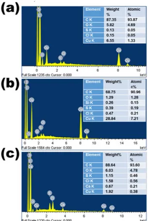
Elemental analysis of CNS.
EDS was used to detect the presence of elements in the prepared C samples. Both qualitative and quantitative EDS results are shown in Fig. 2. These results indicate the presence of very high C content (> 90% atomic weight) as a major element and low O content (between 1.29 and 6.03% atomic weight) as a minor element in all the prepared CNS. Traces of Ca, Si and Cl are also present, which generally occur in plant tissues31 and, consequently, in plant-derived C nanomaterials32. Additionally, nettle is known as a silicifier,with its trichomes typically containing silica at the tip33.
Some trace amounts of Cu and S can also be seen in all the prepared samples: the presence of Cu is due to the substrate, because of the drop drying of CNS on the surface of Cu tape used as conductive support. S is a fake peak around 2.3 eV and derives from the combination of O and Si; more specifically, it is a sum of O Kα and Si Kα peaks.
www.nature.com/scientificreports/
Elemental analysis was also carried via X-ray photoelectron spectroscopy (XPS) on a representative nettle CNS sample (nettle leaves 1:1), which confirmed the existence of C as major element with O and N as minor elements and Si as a trace element in the prepared CNS (Fig. 3).
In conclusion, the EDS and XPS analyses reveal that C is the major element in the nettle-derived CNS. The presence of O is also confirmed, which is common in biomass-derived C34. These analyses further confirm the
absence of any metal impurities in the nettle-derived CNS.
TGA of CNS.
The thermogravimetric analysis (TGA) analysis was performed in air to investigate the thermal decomposition of the prepared CNS. The TGA curves shown in Fig. 4 demonstrate that the mass loss occurred in two steps. In the first step, the mass loss occurred in the temperature range of 40 °C to 100 °C, which is mainly due to the evaporation of the moisture and residual water molecules. In the second step, the major mass loss was observed between 400 °C and 700 °C in all the three samples, showing the start of thermal decomposition at around 400 °C and stabilizing at around 700 °C. Another aspect worth being highlighted is the final mass: above 700 °C, all the prepared CNS resulted in complete decomposition (i.e. zero mass) which indicates the prepara-tion of metal-free CNS by this process35.Diffraction pattern of CNS.
XRD was used to investigate the diffraction nature and phase formation of the prepared CNS. Figure 5 shows the XRD patterns of nettle-derived CNS prepared with different activation processes, i.e. (a) CNS1:1 (nettle stems: NaHCO3), (b) CNS1:1 (nettle leaves: NaHCO3) and (c) CNS1:2 (nettle leaves: NaHCO3). All the samples show identical diffraction patterns and all the peaks are well consistent with the pure graphitic C. The XRD spectra of the activated CNS show two peaks: the first characteristic broad peak at around 2θ = 24° is related to C(002) diffraction peak, which corresponds to the reflection of graphitic C and a second less intense, broader peak at around 2θ = 42° is assigned to C(100) diffraction peak, which accounts for amorphous C (JCPDF: 41–1487)36,37. No additional peak from the minor trace elements could be observed in theXRD spectra, due to their relatively low concentration, suggesting the formation of pure CNS.
Figure 1. FESEM images at different magnification of CNS prepared at 650 °C from nettle stems (a–c) and leaves (d–i) using NaHCO3 as an activating agent. The ratio of nettle powder and NaHCO3 is 1:1 (a–f) and 1:2 (g–i).
Figure 2. EDS spectra of CNS prepared at 650 °C from nettle stems (a) and leaves (b,c) using NaHCO3 as an activating agent. The ratio of nettle powder and NaHCO3 is 1:1 (a,b) and 1:2 (c).
Figure 3. XPS analysis: (a) survey scan, high resolution deconvoluted (b) C1 & (c) O1s spectra for nettle leaves 1:1 CNS.
www.nature.com/scientificreports/
Figure 4. The TGA curves of CNS prepared at 650 °C from nettle (a) stems and (b,c) leaves using NaHCO3 as activating agent. The ratio of nettle powder and NaHCO3 is 1:1 (a,b) and 1:2 (c).
Figure 5. XRD spectra of CNS prepared at 650 °C from nettle (a) stems and (b,c) leaves using NaHCO3 as activating agent. The ratio of nettle powder and NaHCO3 is 1:1 (a and b) and 1:2 (c).
Raman Spectroscopy.
Raman spectroscopy was used to investigate various disordered domains and the graphitization degree of the prepared nettle-derived CNS. The Raman spectra show two peaks, i.e. the G-band and D-band (Fig. 6), with the latter accounting for disorder and the former for perfect graphite crystals38.Typi-cally, the G band centered at 1570 cm−1 corresponds to the two dimensional in-plane motion of strongly bonded sp2 C atoms, highlighting a characteristic of graphitic C and the D band centered at 1340 cm−1 relates to the defect sites or disordered tetrahedral sp3-hybridized C atoms39,40. The ratio of integrated intensities of D band
and G band (ID/IG) represents the number of defects in the structure38. The ratios of areas under D-band and
G-band was used to calculate ID/IG41. The calculated ID/IG ratios are 1.2, 1.1 and 1.3 for nettle stem-derived
CNS1:1, nettle leaf-derived CNS1:1 and CNS 1:2, respectively. This high intensity ratios entail the existence of disordered C in the prepared porous CNS2. These results suggest that the ID/IG ratio of nettle leaf-derived CNS1:1
is lowest, i.e. this samples possess the lowest number of defects42. This means that the highest degree of
graphiti-zation is found in NaHCO3-activated nettle leaf-derived CNS1:1.
The high graphitization degree of the nettle stem-derived CNS is due to the structure of the tissues: just like hemp3, the stem of nettle also contains bast fibres (Supplementary Fig. 1), that are long elements with a thick
cellulosic wall (called gelatinous, or G-layer). Bast fibres are indeed characterized by crystalline cellulose and are hypolignified43. There are however differences between hemp and nettle bast fibres. The latter do not show
labelling when the LM10 antibody recognizing the hemicellulose xylan is used and display a different structure of the G-layer18 compared to hemp44. At the transmission electron microscope (TEM), the G-layer of nettle bast
fibres appears indeed less compact than that of hemp, with the formation of local loose regions where the cel-lulosic layer seems to be flaking off (Supplementary Fig. 1c)18. Such a loose structure may facilitate the process
of CNS preparation by favouring disassembly of the layers and penetration of the activating agent.
Nitrogen adsorption–desorption isotherms and corresponding BJH pore‑size distribu‑
tions.
The pore-network structure and Brunauer–Emmett–Teller (BET) specific surface area for the prepared nettle-derived CNS were investigated based on the physical nitrogen adsorption–desorption analysis. High sur-face area and combined pores containing C samples are very important for improved biological applications. NaHCO3 was used as an activating agent to improve the porous structure and enhance the BET specific surface area of the prepared CNS. The nitrogen adsorption isotherms of the prepared samples are shown in Fig. 7 and described according to the IUPAC report45. It can be clearly seen that all the three samples show similar type-IVisothermal sorption curves, due to the presence of hysteresis loops45–47. The initial isotherm curvatures of the
prepared C samples are assigned to micro- and mesopores, while the ascending curvature of the plateau (type H3 loop) in the high relative pressure range (P/P0 = 0.85 − 1.00) is inherent to macropores45–47. The wider and higher
ascending curvature in the CNS prepared from leaves indicates the presence of more macropores than the CNS prepared from the stems. This more pronounced adsorption property in the isotherms of CNS from leaves is assigned to an enhanced N2 uptake associated with micropore filling.
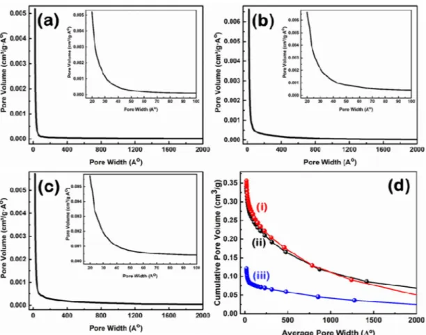
The corresponding Barrett-Joyner-Halenda (BJH) pore size distribution of the prepared samples is shown in Fig. 8(a–c), while the structure properties of the prepared CNS using different ratios of the nettle powder and NaHCO3 are summarized in Table 1. The corresponding cumulative pore size distribution based on the BJH method is shown in Fig. 8d.
The two samples of CNS prepared using nettle leaves have a relatively similar BET specific surface area, while the CNS prepared using nettle stems show significantly higher value than the leaf samples (Table 1). The average pore volume and pore diameter of the CNS prepared from nettle leaves are larger than the CNS prepared from stems. This observation can be explained by the presence of a high number of macropores in the CNS prepared from nettle leaves, while the additional micropores formation in the CNS prepared from stems contribute to the improvement of the BET specific surface area48. As shown in Table 1, CNS1:1 prepared from nettle stems
show the highest BET surface area of 718 m2⁄g, while CNS1:1 and CNS1:2 have BET surface areas of 482 m2/g and 478 m2/g, respectively. The BET specific surface area is in the same range as the one previously calculated for cornstalks-derived C nanomaterial (between 320–790 m2⁄g)4, while the thickness is in agreement with the values reported for CNS prepared from hemp (10–30 nm)3, tal palm (22 nm)2, waste coffee ground (ca. 20 nm)1. Hence, nettle leaves and stems are suitable renewable resources for the preparation of CNS with the advantage Figure 6. Raman spectra of CNS prepared at 650 °C from nettle leaves (a,c) and (b) stems using NaHCO3 as activating agent. The ratio of nettle powder and NaHCO3 is 1:1 (a,b) and 1:2 (c).
www.nature.com/scientificreports/
of providing very homogeneous material, since the plants used are clones, as well as a higher amount of fibres compared to wild nettle.
Effects of nettle‑derived CNS on the germination of tobacco pollen.
Evaluating the environmen-tal impact of either the production or utilization of a new material (such as CNS) implies a complex system of analysis requiring the contribution of different, but complementary, scientific disciplines. In plants, C nanoma-terials negatively impact germination in both monocots and dicots. For example, in rice, graphene at 50 µg/mL delayed by three days the germination of the seeds compared to the control group and also caused a decrease in moisture content, probably due to the physical blocking of the seed coat pores and consequent water uptake impairment49. Recently, a study focused on an economically-important crop, i.e. tomato, showed that while Cnanotubes (single-walled and multi-walled) in soil delayed early growth and flowering, they did not affect later developmental stages50. Single-walled nanotubes, however, caused an increase in salicylic acid, a finding
denot-ing the presence of a stress response in the plants which is not present in the case of multi-walled tubes50.
The germination of tomato seeds primed with C nanomaterials was not affected; however, parameters such as root length and hypocotyl biomass were impaired, while chlorophyll content, as well as vitamin C, β-carotene, phenols and flavonoids increased51. These results show that C nanomaterials can be used as biostimulants, but
their stimulating effects should be carefully determined beforehand to establish the right concentration for the crop examined.
As a first step towards the study of the effects of C nanomaterials on plants, it is possible to use a simplified system consisting in cell models that respond quickly to environmental stressors52,53. Obviously, such analyses
do not allow an overall assessment of the environmental impact of a material or technology, but they can provide an initial indication of their toxic potential. Many model cells can be used in such an assessment, including the pollen tube, because it is a critical structure during plant reproduction54,55. One of the most important reasons
for selecting the pollen tube as a model cell is that the evaluation of toxic effects can be quantified in a relatively simple way. In fact, the pollen tube is a cell that grows linearly and in which many cytological events follow one another in a precise spatial–temporal order. These characteristics make the pollen tube a model cell system suit-able for providing measursuit-able data56.
Pollen is very sensitive to environmental pollutants compared to other plant cells and this allows to assess the impact of a wide range of chemicals on plant metabolism57,58. Therefore, it is used as an indicator of air
pol-lution because the performance of pollen (described in terms of germination and elongation of pollen tubes) is affected by airborne gas pollutants. Atmospheric particulate matter also affects pollen performance, for example Ag and Pd nanoparticles impair in vitro pollen germination and pollen elongation in kiwi59,60. Graphene oxide
nanoparticles can also have a negative effect on the germination and growth of tobacco and hazel pollen tubes19.
In Supplementary Fig. 2, images from inverted microscopy and SEM are shown. CNS tend to form aggre-gates, especially at the highest concentration (Supplementary Fig. 2a–c), rather than evenly distribute in the Figure 7. BET Isotherms of CNS prepared at 650 °C from nettle (a) stems and (b,c) leaves using NaHCO3 as an activating agent. The ratio of nettle powder and NaHCO3 is 1:1 (a,b) and 1:2 (c).
medium. This is confirmed by SEM observations at low resolution, where the aggregates formed are well visible (Supplementary Fig. 2d–f).
In the presence of pollen grains, the CNS clusters tend to adhere to the pollen grains (Supplementary Fig. 3). It was previously suggested that the pollenkitt of N. tabacum pollen grains could act as a matrix favouring the adsorption of CNS19. The presence of CNS aggregates sometimes made it difficult to correctly visualize the
pollen and therefore to evaluate the germination percentage.
As shown in Fig. 9, the percentage of germinated pollen increases slightly from 1 to 3 h of growth. From personal experience, a value above 50% is normally considered as acceptable in the case of frozen pollen. Ger-mination data are thus consistent with what is usually observed. Data in Fig. 9 also show that the presence of stem- and leaf-derived CNS has no effect on pollen germination at any of the three concentrations tested for a total germination time of 3 h. Therefore, even if CNS form aggregates around pollen grains, this has no impact on pollen germination. As can be seen in Fig. 9, CNS do not affect pollen growth, even at the maximum concen-tration. Therefore, nettle-derived CNS have no inhibitory effects on tobacco pollen germination. After all, the germination of pollen grains is only one of the processes that take place during the reproduction of seed plants. Therefore, this result is not surprising because the possible negative effects of CNS could affect other steps, such as pollen activation, or pollen tube growth19, or specific molecular events22. Future studies, focused on the further
development of pollen tubes, will clarify the possible effects of CNS during other processes related to pollen tubes Figure 8. BJH pore size distribution of CNS prepared at 650 °C from nettle (a) stems and (b,c) leaves using NaHCO3 as an activating agent. The ratio of nettle powder and NaHCO3 is 1:1 (a and b) and 1:2 (c). Insets show the corresponding low pore width zone. (d) The corresponding cumulative pore size distribution of nettle leaf- (i and ii) and nettle stem- (iii) derived CNS with the ratio for nettle powder and NaHCO3 of 1:1 (ii and iii) and 1:2 (i).
Table 1. BET surface area, average pore volume and average pore diameter of the prepared nettle-derived CNS at 650 °C, using different NaHCO3: tissue powder ratios.
Samples Biomass sources BET surface area (m2/g) Average pore volume (cm3/g) Average pore diameter (Å)
CNS1:1 Nettle stems 718 0.12 20.1
CNS1:1 Nettle leaves 482 0.34 31.7
www.nature.com/scientificreports/
such as, for example, the transport of sperm cells or a possible inhibitory effect of nanocomposites in planta i.e. on the adhesion, activation and growth of the pollen tube on stigma and style.
Effects of the nettle‑derived CNS on the growth of P. subcapitata.
The evaluation of the ecotoxi-cological effects of nettle CNS was complemented by tests on the green microalgae P. subcapitata. This is a phyto-planktonic freshwater aquatic model for ecotoxicological investigations and is widely used for studies addressing the interaction of nanoparticles with microalgae cells in aquatic environments61,62.In the literature, several studies have investigated the effects of C nanomaterials on microalgae and shown a higher toxicity in the case of oxidized nanomaterials. Oxidation occurs as a spontaneous aging/weathering process after the nanomaterials are released in the environment. Chlorella pyrenoidosa increased the antioxi-dant response as a result of exposure to oxidized multi-walled C nanotubes, with enhanced pentose phosphate pathway, cell division and formation of polyphosphate bodies63. However, when these protective responses were
overwhelmed, the algae suffered from toxicity caused by membrane damage and denaturation of macromolecules. Oxidized C nanotubes adsorbed to the cell surface and penetrated via puncture and subsequent endocytosis63.
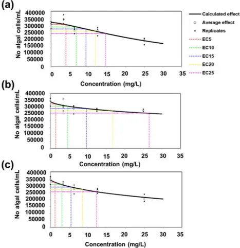
Different concentrations (3.4–6.25–12.5 and 25 µg/mL) were tested and the number of algal cells was counted after 72 h of growth. As it can be observed in Fig. 10, at higher concentrations, the nettle CNS cause a decrease in the number of P. subcapitata cells for the stem- and leaf-derived CNS prepared using a ratio NaHCO3: nettle powder equal to 1:2. The nettle CNS tend to form aggregates (Supplementary Fig. 2) and the accumulation of C nanomaterials around algal cells was reported to cause shading and, subsequently, to reduce light availability thus resulting in growth impairment64,65.
The toxicity of C nanomaterials is due to different physical parameters, i.e. size, surface charge and area66. A
higher surface area could adsorb more transition metals and ultimately trigger the formation of more reactive oxygen species (ROS) via Haber–Weiss and Fenton-like reactions67. The BET surface area for the leaf CNS is,
however, the same, irrespective of the ratio of activating agent used (Fig. 7 and Table 1).
The toxicity of C nanomaterials in algae can also be due to the presence of heavy metal impurities due to low-efficiency manufacturing process (which, for the lowest manufacturing processes, can constitute up to 99.9% of the total production output68). A recent study has shown that metal impurities in C nanotubes affect the
esterase activity of the algae Attheya ussuriensis, Chaetoceros muelleri, Heterosigma akashiwo and Porphyridium
purpureum (with the latter showing the lowest changes)69. It should however be noted that, in the present study,
no metal impurities were detected (Figs. 2 and 3).
The higher inhibitory impact on growth by the nettle leaf-derived CNS1:2 may also be ascribed to its high defective nature (higher ID/IG, lower graphitization) and smaller average sheet thickness with a higher adsorption average pore width. Such properties of leaf-derived CNS1:2 facilitate the easy absorption at higher concentra-tions and thus cause a decrease in the number of P. subcapitata cells. Although the EC25 value of leaf CNS1:2 is higher, it should however be noted that all the EC25 values reported for the nettle CNS lie within the same Figure 9. Effects of increasing concentrations (10–50-100 µg/mL of BKsuc) of CNS on the germination of pollen grains from N. tabacum. The measurements are relative to the control (CNTRL) and comprise a total treatment time of 3 h. The values are reported as means ± standard deviation (n = 150). A two-way ANOVA was conducted to examine the effect of time and different CNS concentration on pollen germination. There was a statistically significant interaction of time on germination, F (2, 60) = 252.460, p = 0.000. Different letters denote statistically significant changes among groups at the two-way ANOVA followed by Tukey’s post-hoc test.
order of magnitude; thereby, no statistically significant differences are visible between the CNS. The values in Table 2 report the various essential parameters that may contribute to the toxicity of nettle C nanomaterials on microalgal cells.
Conclusions
The preparation of highly porous activated CNS from nettle tissues with interesting properties in terms of porosity and active surface area was demonstrated in the present study by using pyrolysis at 650 °C. The stems and leaves of U. dioica clone 13 were used as raw material and NaHCO3 as activating agent. Various ratios of the nettle powder and NaHCO3 were mixed and carbonized together under N2 environment to get the porous CNS. The C obtained from this process showed high porosity and BET specific surface area. The highest BET specific surface area (718 m2/g) was obtained from the nettle stem-derived CNS, which is in the range of commercially available activated C.
The potential ecotoxicological effects of nettle CNS were evaluated on tobacco pollen and the green microalga
P. subcapitata. Tobacco pollen germinated both under control conditions and in the presence of progressively
increasing concentrations of CNS; under the experimental conditions used, CNS formed dark aggregates that Figure 10. Inhibitory effect of nettle CNS at different concentrations on algal cell number after 72 h. CNS from stem 1: 1 (a), CNS from leaves 1:1 (b), CNS from leaves 1:2 (c).
Table 2. Comparison of surface and structural parameters of the nettle-derived CNS for the effect toxicity using P. subcapitata. EC25 (mg/L, with the 95% confidence intervals between parentheses) are indicated for comparative purposes.
Parameter Stem CNS Leaf CNS1:1 Leaf CNS1:2
ID/IG 1.2 1.1 1.3
CNS average thickness 36 nm 32 nm 27 nm
Adsorption average pore width 20.1 Å 31.7 Å 32.2 Å
BJH adsorption average pore width 70.8 Å 98.7 Å 111.2 Å
www.nature.com/scientificreports/
were easily observable as their concentration increased. The presence of such dense aggregates forming a sheath around the algal cells and reducing light availability is probably the reason for the observed growth inhibition. The growth inhibition for algae was however associated to high concentrations of the CNS. The strongest inhibitory effects on the growth of P. subcapitata were observed for leaf CNS1:2, a finding suggesting that the low degree of graphitization has a negative effect on algal growth.
By using a type of biomass that can be propagated rapidly by stem cuttings and in large amounts, highly porous CNS can be produced at a large scale and low cost for useful industrial applications. Overall, the work here presented is novel because it shows an additional way to valorize a neglected species considered a weed. The advantage of nettle over other plants is its fast growth and propagation using stem cuttings, which ensures homogeneous starting raw material.
Materials and methods
Growth of plants and chemicals.
The whole stems and leaves of stinging nettle (U. dioica clone 13) were collected from 1.5 months-old plants grown under controlled conditions17. The clone used produces ahigher yield of bast fibres (16%) compared to wild nettle (ca. 4–5%) and it was bred by G. Bredemann between 1927–1950 at the Institute of Applied Botany in Hamburg70. Sodium bicarbonate (NaHCO
3) and hydrochloric acid (HCl) were obtained from Sigma-Aldrich. The nitrogen (N2) gas with a purity of 99.99% was purchased from SCG gas supply center, Jubail, Saudi Arabia. De-ionized (DI) water was obtained from a water purification system (Barnstead Nanopure, Thermo Scientific, USA).
Preparation of CNS.
CNS were prepared by pyrolysis of the nettle stems and leaves using NaHCO3 as an activating agent. The stems and leaves were collected, washed with DI water and then dried in an electric oven at 100 °C for 24 h. The dried stems and leaves were separately crushed by a high-precision grinding machine to obtain a very fine powder. Powder (particle size ≤ 100 µm) was collected after passing it through a 100 µm mesh. Three different samples were prepared by mixing the obtained powder with different ratios of NaHCO3. The ratio of nettle stem powder and NaHCO3 was 1:1 (referred to as CNS1:1), while for the leaves and NaHCO3 the mass ratios were 1:1 and 1:2 (referred to as CNS1:1 and CNS1:2, respectively). The final uniform mixture was then placed in an alumina crucible and carbonized at 650 °C for 5 h at a heating and cooling rate of 10 °C/min and 5 °C/min, respectively, in a tube furnace under the protection of nitrogen flow. For the last step, the samples were placed in glass beakers containing 0.5 M HCl and ultrasonicated for 2 h and then washed two times with DI water. Following this step, the obtained samples were then filtered through a filter paper. Finally, the filtered samples were dried in an electric oven at 60 °C for 24 h to obtain the nettle-derived CNS.Characterization of CNS.
The morphology of the prepared samples was analyzed using FESEM (TESCAN LYRA 3, Czech Republic). The FESEM was operated at 20 kV. EDS data were recorded with an Oxford Instru-ments Xmass detector, equipped with the FESEM and analyzed with the LINK INCA program system.The TGA of CNS were recorded by the thermal analyzer (TGA1 STAR e SYSTEM), having a temperature range from room temperature up to 1000 °C. An X-ray monochromator (ESCALAB 250Xi XPS Microprobe, Thermo Scientific, USA) was utilized to record the XPS of the prepared CNS. An X-ray diffractometer, Rigaku Ultima (high-resolution) equipped with Cu-K (alpha) radiation was used to obtain the XRD patterns of the prepared CNS. The graphite structure of the synthesized CNS was detected and studied by using a Raman spectrometer, iHR320 with CCD detector (HORIBA), equipped with green laser (300 mW) having an excitation wavelength λo = 532 nm. For the measurement of the surface area and pore size of the prepared CNS, BET and BJH analyses were performed by using Micromeritics ChemiSorb 2750. A high-precision grinding machine was used to make a fine powder and a Power Sonic 603 ultrasonic cleaner was used for sonication.
Optical and transmission electron microscopy (TEM) of nettle stem tissues.
The preparation of nettle stem tissue sections and Toluidine Blue O staining were performed as previously described17,71. Nettlebottom internodes (5 mm thickness, localized at the stem base) were fixed in glutaraldehyde/paraformalde-hyde/caffeine (1%/2%/1% v/v in Milli-Q water) under vacuum for 15 min and o/n at 4 °C, dehydrated in an ethanol series (70–95–100%), impregnated in resin containing PEG 400 (2% v/v) and dimethacrylate ethylene glycol (0.4% w/v) and finally included in Technovit 7100 resin (Technovit 7100, Heraeus Kulzer GmbH, Hanau, Germany). Cross sections of 10 μm thickness were prepared using a microtome and stained with 0.05% w/v Toluidine Blue O.
Sample processing for immunoTEM and detection of crystalline cellulose with CBM3a were as previously reported18. Briefly, the bottom internode was sampled by using a clean razor blade and thereafter fixed for 2 h
at room temperature (RT) and o/n at 4 °C in 2% v/v glutaraldehyde and 1.6% v/v paraformaldehyde in 0.1 M phosphate buffer pH 6.9. Samples were rinsed with phosphate buffer two times for 10 min each, then dehydrated in a graded series of ethanol. Samples were infiltrated with LR‐White resin at 1:1 ratio with ethanol to pure resin. After polymerization for 48 h at 40 °C, ultrafine sections were obtained with a diamond knife and the ultrami-crotome LKB NOVA. The sections were collected on gold grids and blocked for 20 min with normal goat serum diluted 1:30 in dilution buffer (0.05 M Tris‐HCl pH 7.6, 0.9% w/v NaCl and 0.2% w/v BSA). Sections were incu-bated with the CBM3a protein72 at 5 μg/mL at RT for 1.5 h in conjunction with the mouse monoclonal anti‐His
antibody diluted 1:100 in dilution buffer + 0.1% v/v Tween 20 for 1.5 h. Samples were washed and incubated with the anti‐mouse antibody conjugated with 10 nm gold particles for 45 min at RT. Sections were visualized with the Philips MORGAGNI 268 80 kV transmission electron microscope, equipped with MEGAview II camera and elaborated with the Analysis software.
P. subcapitata growth in the presence of nettle CNS.
The growth inhibition test was performed with the green algal species P. subcapitata (CCAP 278/4, Scotland, United Kingdom). The test is a modification of the OECD 201 standard74 in a 12-well plate format. A pre-culture of algae was started 3 days before thebegin-ning of the experiment and algae in exponential growth phase were used. The OECD medium74 (FUJIFILM
Wako Pure Chemical Corporation) was used in order to prepare the test concentrations, which were inoculated with exponentially growing algae in a concentration of 105 cells/mL. One mL of algae with or without the dif-ferent nettle CNS at increasing concentrations (3.4–25 mg/L) were placed in the wells of 12-well plates and were continuously shaken at 50 rpm (Gerhardt Analytical Systems, Germany) under a cool white fluorescent light (46.2 mmol m-2 s-1) and temperature 20 °C ± 1 °C. Algal suspension with the highest concentration of the carrier solvent (2.5% v/v DMSO) was used as negative control. Three replicates per concentration and for the control were used. After 72 h, the cell concentration was determined manually in a Bürker-Türk haemocytom-eter. Growth rates were calculated as indicated in the guideline and the results were expressed as % inhibition of growth rate relative to the untreated control.
Data availability
All data generated or analyzed during this study are included in this published article and the supplementary material.
Received: 23 November 2020; Accepted: 8 March 2021
References
1. Yun, Y. S. et al. Hierarchically Porous Carbon Nanosheets from Waste Coffee Grounds for Supercapacitors. ACS Appl. Mater.
Interfaces 7, 3684–3690 (2015).
2. Ahammad, A. J. S. et al. Porous tal palm carbon nanosheets: preparation, characterization and application for the simultaneous determination of dopamine and uric acid. Nanoscale Adv. 1, 613–626 (2019).
3. Wang, H. et al. Interconnected Carbon Nanosheets Derived from Hemp for Ultrafast Supercapacitors with High Energy. ACS Nano 7, 5131–5141 (2013).
4. Wang, L. et al. Porous graphitic carbon nanosheets derived from cornstalk biomass for advanced supercapacitors. Chemsuschem 6, 880–889 (2013).
5. Li, J., Liu, W., Xiao, D. & Wang, X. Oxygen-rich hierarchical porous carbon made from pomelo peel fiber as electrode material for supercapacitor. Appl. Surf. Sci. 416, 918–924 (2017).
6. Chen, X. et al. A novel hierarchical porous nitrogen-doped carbon derived from bamboo shoot for high performance supercapaci-tor. Sci. Rep. 7, 7362 (2017).
7. Hao, E. et al. Rich sulfur doped porous carbon materials derived from ginkgo leaves for multiple electrochemical energy storage devices. J. Mater. Chem. A 5, 2204–2214 (2017).
8. Chang, J. et al. Activated porous carbon prepared from paulownia flower for high performance supercapacitor electrodes.
Elec-trochim. Acta 157, 290–298 (2015).
9. Yang, H., Ye, S., Zhou, J. & Liang, T. Biomass-Derived Porous Carbon Materials for Supercapacitor. Front. Chem. 7, 1 (2019). 10. Kumar, S., Yadav, A., Yadav, M. & Yadav, J. P. Effect of climate change on phytochemical diversity, total phenolic content and in vitro
antioxidant activity of Aloe vera (L.) Burm.f.. BMC Res. Notes 10, 1 (2017).
11. Xu, J. et al. Response of Bioactive Phytochemicals in Vegetables and Fruits to Environmental Factors. Eur. J. Nut. Food Saf. 1, 233–247. https:// doi. org/ 10. 9734/ ejnfs/ 2019/ v9i33 0062 (2019).
12. Liu, M., Liu, G., Gong, L., Wang, D. & Sun, J. Relationships of Biomass with Environmental Factors in the Grassland Area of Hulunbuir China. . PLoS ONE 9, 1 (2014).
13. Joshi, R., Singla-Pareek, S. L. & Pareek, A. Engineering abiotic stress response in plants for biomass production. J. Biol. Chem. 293, 5035–5043 (2018).
14. Farag, M. A., Weigend, M., Luebert, F., Brokamp, G. & Wessjohann, L. A. Phytochemical, phylogenetic, and anti-inflammatory evaluation of 43 Urtica accessions (stinging nettle) based on UPLC-Q-TOF-MS metabolomic profiles. Phytochemistry 96, 170–183 (2013).
15. Xu, X. et al. Insights into Lignan Composition and Biosynthesis in Stinging Nettle (Urtica dioica L.). Molecules 24, 3863 (2019). 16. Guerriero, G. et al. Production of Plant Secondary Metabolites: Examples, Tips and Suggestions for Biotechnologists. . Genes
(Basel) 9, 1 (2018).
17. Backes, A. et al. Sucrose synthase gene expression analysis in the fibre nettle (Urtica dioica L.) cultivar “clone 13”. Ind. Crops Prod. 123, 315–322 (2018).
18. Xu, X. et al. Cell wall composition and transcriptomics in stem tissues of stinging nettle (Urtica dioica L): Spotlight on a neglected fibre crop. Plant Direct 3, e00151 (2019).
19. Carniel, F. C. et al. Graphene oxide impairs the pollen performance of Nicotiana tabacum and Corylus avellana suggesting potential negative effects on the sexual reproduction of seed plants. Environ. Sci. Nano 5, 1608–1617 (2018).
20. Begum, P., Ikhtiari, R. & Fugetsu, B. Graphene phytotoxicity in the seedling stage of cabbage, tomato, red spinach, and lettuce.
www.nature.com/scientificreports/
21. Anjum, N. A. et al. Single-bilayer graphene oxide sheet impacts and underlying potential mechanism assessment in germinating faba bean (Vicia faba L). Sci. Total Environ. 472, 834–841 (2014).
22. Candotto Carniel, F. et al. Beyond graphene oxide acidity: Novel insights into graphene related materials effects on the sexual reproduction of seed plants. J. Hazard. Mater. 393, 122380 (2020).
23. Wu, K. et al. Large and porous carbon sheets derived from water hyacinth for high-performance supercapacitors. RSC Adv. 6, 29996–30003 (2016).
24. Suhas, et al. Cellulose: A review as natural, modified and activated carbon adsorbent. Biores. Technol. 216, 1066–1076 (2016). 25. Yahya, M. A., Al-Qodah, Z. & Ngah, C. W. Z. Agricultural bio-waste materials as potential sustainable precursors used for activated
carbon production: A review. Renew. Sustain. Energy Rev. 46, 218–235 (2015).
26. Jain, A., Balasubramanian, R. & Srinivasan, M. P. Hydrothermal conversion of biomass waste to activated carbon with high poros-ity: A review. Chem. Eng. J. 283, 789–805 (2016).
27. Ahammad, A. J. S. et al. Activated jute carbon paste screen-printed FTO electrodes for nonenzymatic amperometric determination of nitrite. J. Electroanal. Chem. 832, 368–379 (2019).
28. Deb Nath, N. C., Shah, S. S., Qasem, M. A. A., Zahir, M. H. & Aziz, M. A. Defective Carbon Nanosheets Derived from Syzygium
cumini Leaves for Electrochemical Energy-Storage. ChemistrySelect 4, 9079–9083 (2019).
29. Wang, L. et al. Electrochemical Sensing and Biosensing Platform Based on Biomass-Derived Macroporous Carbon Materials. Anal.
Chem. 86, 1414–1421 (2014).
30. Hinks, N. J., McKinlay, A. C., Xiao, B., Wheatley, P. S. & Morris, R. E. Metal organic frameworks as NO delivery materials for biological applications. Microporous Mesoporous Mater. 129, 330–334 (2010).
31. field survey and in vivo X-ray analyses. Harada, E. et al. Assessment of willow (Salix sp.) as a woody heavy metal accumulator.
Metallomics 3, 1340–1346 (2011).
32. Shah, S. S. et al. Preparation and characterization of manganese oxide nanoparticles-coated Albizia procera derived carbon for electrochemical water oxidation. J. Mater. Sci: Mater. Electron. 30, 16087–16098 (2019).
33. Thurston, E. L. Morphology, Fine Structure, and Ontogeny of the Stinging Emergence of Urtica Dioica. Am. J. Bot. 61, 809–817 (1974).
34. Suman, S., Panwar, D. S. & Gautam, S. Surface morphology properties of biochars obtained from different biomass waste. Energy
Sources, Part A: Recovery, Utilization, and Environmental Effects 39, 1007–1012 (2017).
35. Mutyala, S. & Jayaraman, M. Synthesis of Nitrogen Doped Carbon and Its Enhanced Electrochemical Activity towards Ascorbic Acid Electrooxidation. Int. J. Electrochem. https:// www. hinda wi. com/ journ als/ ijelc/ 2014/ 246746/ (2014) https:// doi. org/ 10. 1155/ 2014/ 246746.
36. Zhang, Z. H., Zhao, T. S., Bai, B. F., Zeng, L. & Wei, L. A highly active biomass-derived electrode for all vanadium redox flow bat-teries. Electrochim. Acta 248, 197–205 (2017).
37. Shang, H. et al. Preparing high surface area porous carbon from biomass by carbonization in a molten salt medium. RSC Adv. 5, 75728–75734 (2015).
38. Sodtipinta, J. et al. Interconnected open-channel carbon nanosheets derived from pineapple leaf fiber as a sustainable active mate-rial for supercapacitors. Ind. Crops Prod. 104, 13–20 (2017).
39. Chu, M. et al. Novel biomass-derived smoke-like carbon as a supercapacitor electrode material. R. Soc. Open Sci. 6, 190132 (2019). 40. Rezma, S., Birot, M., Hafiane, A. & Deleuze, H. Physically activated microporous carbon from a new biomass source: Date palm
petioles. C. R. Chim. 20, 881–887 (2017).
41. Puech, P. et al. Analyzing the Raman Spectra of Graphenic Carbon Materials from Kerogens to Nanotubes: What Type of Informa-tion Can Be Extracted from Defect Bands?. C Journal of Carbon Research 5, 69 (2019).
42. Gu, W., Sevilla, M., Magasinski, A., Fuertes, A. B. & Yushin, G. Sulfur-containing activated carbons with greatly reduced content of bottle neck pores for double-layer capacitors: a case study for pseudocapacitance detection. Energy Environ. Sci. 6, 2465–2476 (2013).
43. Guerriero, G., Sergeant, K. & Hausman, J.-F. Integrated -omics: a powerful approach to understanding the heterogeneous lignifica-tion of fibre crops. Int J Mol Sci 14, 10958–10978 (2013).
44. Behr, M. et al. Distribution of cell-wall polysaccharides and proteins during growth of the hemp hypocotyl. Planta https:// doi. org/ 10. 1007/ s00425- 019- 03245-9 (2019).
45. Thommes, M. et al. Physisorption of gases, with special reference to the evaluation of surface area and pore size distribution (IUPAC Technical Report). Pure Appl. Chem. 87, 1051–1069 (2015).
46. Ponomarev, N. & Sillanpää, M. Combined chemical-templated activation of hydrolytic lignin for producing porous carbon. Ind.
Crops Prod. 135, 30–38 (2019).
47. Sing, K. S. W. et al. Reporting Physisorption Data for Gas/Solid Systems With Special Reference to the Determination of Surface Area and Porosity.
48. Morishita, T. et al. A review of the control of pore structure in MgO-templated nanoporous carbons. Carbon 48, 2690–2707 (2010). 49. Nair, R. et al. Effect of carbon nanomaterials on the germination and growth of rice plants. J Nanosci Nanotechnol 12, 2212–2220
(2012).
50. Jordan, J. T. et al. Carbon nanotubes affect early growth, flowering time and phytohormones in tomato. Chemosphere 256, 127042 (2020).
51. López-Vargas, E. R. et al. Seed Priming with Carbon Nanomaterials to Modify the Germination, Growth, and Antioxidant Status of Tomato Seedlings. Agronomy 10, 639 (2020).
52. Tardieu, F. & Tuberosa, R. Dissection and modelling of abiotic stress tolerance in plants. Curr. Opin. Plant Biol. 13, 206–212 (2010). 53. Bressan, R., Bohnert, H. & Zhu, J.-K. Abiotic stress tolerance: from gene discovery in model organisms to crop improvement. Mol
Plant 2, 1–2 (2009).
54. Hedhly, A., Hormaza, J. I. & Herrero, M. Global warming and sexual plant reproduction. Trends Plant Sci. 14, 30–36 (2009). 55. Mesihovic, A., Iannacone, R., Firon, N. & Fragkostefanakis, S. Heat stress regimes for the investigation of pollen thermotolerance
in crop plants. Plant Reprod 29, 93–105 (2016).
56. Kroeger, J. H. & Geitmann, A. Pollen tube growth: Getting a grip on cell biology through modeling. Mech. Res. Commun. 42, 32–39 (2012).
57. Sabrine, H., Afif, H., Mohamed, B., Hamadi, B. & Maria, H. Effects of cadmium and copper on pollen germination and fruit set in pea (Pisum sativum L.). (2010).
58. Wolters, J. H. B. & Martens, M. J. M. Effects of Air Pollutants on Pollen. Bot. Rev. 53, 372–414 (1987).
59. Speranza, A., Leopold, K., Maier, M., Taddei, A. R. & Scoccianti, V. Pd-nanoparticles cause increased toxicity to kiwifruit pollen compared to soluble Pd(II). Environ. Pollut. 158, 873–882 (2010).
60. Speranza, A. et al. In vitro toxicity of silver nanoparticles to kiwifruit pollen exhibits peculiar traits beyond the cause of silver ion release. Environ Pollut 179, 258–267 (2013).
61. Hoecke, K. V., Schamphelaere, K. A. C. D., der Meeren, P. V., Lcucas, S. & Janssen, C. R. Ecotoxicity of silica nanoparticles to the green alga Pseudokirchneriella subcapitata: Importance of surface area. Environ. Toxicol. Chem. 27, 1948–1957 (2008).
62. Katsumata, M., Koike, T., Nishikawa, M., Kazumura, K. & Tsuchiya, H. Rapid ecotoxicological bioassay using delayed fluorescence in the green alga Pseudokirchneriella subcapitata. Water Res. 40, 3393–3400 (2006).
72. Blake, A. W. et al. Understanding the biological rationale for the diversity of cellulose-directed carbohydrate-binding modules in prokaryotic enzymes. J. Biol. Chem. 281, 29321–29329 (2006).
73. Brewbaker, J. L. & Kwack, B. H. The Essential Role of Calcium Ion in Pollen Germination and Pollen Tube Growth. Am. J. Bot. 50, 859–865 (1963).
74. OECD. Test No. 201: Freshwater Alga and Cyanobacteria, Growth Inhibition Test in OECD Guidelines for the Testing of Chemicals, Section 2, (OECD Publishing, Paris, 2011).
Acknowledgments
The authors thank Xuan Xu and Laurent Solinhac for their help with growing and sampling the nettle biomass. Claudia Faleri is also acknowledged for providing the images in Supplementary Fig. 1. The support of CENT-KFUPM research facility utilization in C sample preparation and their characterizations is acknowledged.
Author contributions
G.G., K.S.S. and M.A.A. planned the work; S.S.S., M.A.A.Q., R.B., C.D.C., G.C., S.C., S.Ca., G.G., M.A.A. per-formed the experiments; S.S.S., M.A.A.Q., R.B., C.D.C., G.C., I.A., S.Ca., G.G., M.A.A. wrote the manuscript; all the authors edited the manuscript and contributed to the interpretation of the results.
Funding
This research was funded by the Fonds National de la Recherche, Luxembourg (FNR) (Project CABERNET C16/SR/11289002).
Competing interests
The authors declare no competing interests.
Additional information
Supplementary Information The online version contains supplementary material available at https:// doi. org/ 10. 1038/ s41598- 021- 86426-5.
Correspondence and requests for materials should be addressed to G.G. or M.A. Reprints and permissions information is available at www.nature.com/reprints.
Publisher’s note Springer Nature remains neutral with regard to jurisdictional claims in published maps and institutional affiliations.
Open Access This article is licensed under a Creative Commons Attribution 4.0 International License, which permits use, sharing, adaptation, distribution and reproduction in any medium or format, as long as you give appropriate credit to the original author(s) and the source, provide a link to the Creative Commons licence, and indicate if changes were made. The images or other third party material in this article are included in the article’s Creative Commons licence, unless indicated otherwise in a credit line to the material. If material is not included in the article’s Creative Commons licence and your intended use is not permitted by statutory regulation or exceeds the permitted use, you will need to obtain permission directly from the copyright holder. To view a copy of this licence, visit http:// creat iveco mmons. org/ licen ses/ by/4. 0/.