Histological effects of givinostat in boys with Duchenne
muscular dystrophy
Paolo Bettica
a,*
,1, Stefania Petrini
b,1, Valentina D’Oria
b, Adele D’Amico
c, Michela Catteruccia
c,
Marika Pane
d, Serena Sivo
d, Francesca Magri
e, Simona Brajkovic
e, Sonia Messina
f,g,
Gian Luca Vita
g, Barbara Gatti
a, Maurizio Moggio
h, Pier Lorenzo Puri
i,j, Maurizio Rocchetti
k,
Giuseppe De Nicolao
l, Giuseppe Vita
f,g, Giacomo P. Comi
e, Enrico Bertini
c, Eugenio Mercuri
daItalfarmaco S.p.A., Milan, Italy
bResearch Laboratories, Confocal Microscopy Core Facility, Bambino Gesù Children’s Hospital, IRCCS, Rome, Italy
cUnit of Neuromuscular and Neurodegenerative Disorders, Laboratory of Molecular Medicine, Department of Neurosciences, Bambino Gesù Children’s
Hospital, IRCCS, Rome, Italy
dDepartment of Paediatric Neurology, Catholic University, Rome, Italy
eDino Ferrari Centre, Neuroscience Section, Department of Pathophysiology and Transplantation, Neurology Unit, IRCCS Foundation Ca’Granda Ospedale
Maggiore Policlinico, University of Milan, Milan, Italy
fDepartment of Neurosciences, University of Messina, Messina, Italy gNEMO SUD Clinical Centre for Neuromuscular Disorders, Messina, Italy
hNeuromuscular and Rare Disease Unit, Department of Neuroscience, Foundation IRCCS Ca’ Granda Ospedale Maggiore Policlinico, Dino Ferrari Centre,
University of Milan, Milan, Italy
iSanford-Burnham medical Research Institute, Sanford Children’s Health Research Center, La Jolla, California, USA jIRCCS Fondazione Santa Lucia, Rome, Italy
kIndependent Consultant, Milan, Italy
lDepartment of Electrical, Computer and Biomedical Engineering, University of Pavia, Pavia, Italy
Received 16 February 2016; received in revised form 29 June 2016; accepted 7 July 2016
Abstract
Duchenne Muscular Dystrophy (DMD) is caused by mutations in the dystrophin gene leading to dystrophin deficiency, muscle fiber degeneration and progressive fibrotic replacement of muscles. Givinostat, a histone deacetylase (HDAC) inhibitor, significantly reduced fibrosis and promoted compensatory muscle regeneration in mdx mice. This study was conducted to evaluate whether the beneficial histological effects of Givinostat could be extended to DMD boys. Twenty ambulant DMD boys aged 7 to<11 years on stable corticosteroid treatment were enrolled in the study and treated for≥12 months with Givinostat. A muscle biopsy was collected at the beginning and at the end of treatment to evaluate the amount of muscle and fibrotic tissue. Histological effects were the primary objectives of the study. Treatment with Givinostat significantly increased the fraction of muscle tissue in the biopsies and reduced the amount of fibrotic tissue. It also substantially reduced tissue necrosis and fatty replacement. Overall the drug was safe and tolerated. Improvement in functional tests was not observed in this study, but the sample size of the study was not sufficient to draw definitive conclusions. This study showed that treatment with Givinostat for more than 1 year significantly counteracted histological disease progression in ambulant DMD boys aged 7 to 10 years.
© 2016 The Authors. Published by Elsevier B.V. This is an open access article under the CC BY-NC-ND license (http://creativecommons.org/ licenses/by-nc-nd/4.0/).
Keywords: Duchenne muscular dystrophy; Givinostat; Histone deacetylase inhibitor; Histology; Clinical trial
1. Introduction
Duchenne Muscular Dystrophy (DMD) is the most common
muscular dystrophy in childhood[1]. The disease is caused by
mutations in the dystrophin gene, leading to dystrophin
deficiency and subsequent cell membrane instability.
This instability determines uncontrolled calcium influx, inflammation, necrosis, and replacement of muscle with fibrotic tissue and fat, which leads to severe muscle wasting and weakness.
Pharmacological blockade of the histone deacetylase
activity, which is constitutively active in DMD muscles[2]by
* Corresponding author. Italfarmaco, Via dei Lavoratori 54, 20092 Cinisello Balsamo, MI, Italy. Fax:+39 02 6443 3554.
E-mail address:[email protected](P. Bettica).
1 Paolo Bettica and Stefania Petrini contributed equally to this article.
http://dx.doi.org/10.1016/j.nmd.2016.07.002
0960-8966/© 2016 The Authors. Published by Elsevier B.V. This is an open access article under the CC BY-NC-ND license (http://creativecommons.org/ licenses/by-nc-nd/4.0/).
Available online atwww.sciencedirect.com
Neuromuscular Disorders 26 (2016) 643–649
www.elsevier.com/locate/nmd
ScienceDirect
HDAC inhibitors (HDACi), prevents fibrosis and promotes compensatory regeneration in the mdx mouse, a model of DMD
[3,4].
Givinostat (aka ITF2357) is a potent HDACi currently being
developed for the treatment of DMD. In mdx mice [3],
Givinostat dose and concentration dependently increased the cross-sectional area of myofibers, decreased the cellular inflammatory infiltrate and prevented the formation of fibrotic scars. These findings strongly suggested that in this DMD animal model Givinostat was able to inhibit all the processes which determine muscle fibrotic substitution (inflammation, necrosis, fatty replacement and fibrosis) and to stimulate muscle regeneration with the formation of larger muscle fibers and overall more muscle tissue. Results also suggested that exposures of Givinostat of 300 ng*h/mL are required to exert the beneficial effect.
To evaluate the potential of Givinostat as a treatment for DMD, we conducted the study summarized in this manuscript. The primary objective of this study was to confirm also in humans that Givinostat can counteract the histological signs of the disease.
2. Methods
2.1. Patients
Twenty boys aged 7 to <11 years with an
immunohistochemical and molecular diagnosis of DMD were enrolled in this study after an informed consent form was signed by a parent/guardian and child had assented to be in the study (if applicable). Boys were on a stable dose of systemic corticosteroids for at least six months and were able to complete the two screening 6 minute walk tests (6MWT) with a minimal distance of at least 250 meters each with the
results of these tests within ± 30 meters of each other.
Exclusion criteria were aimed at avoiding confounding factors from other potentially active treatments, and at recruiting boys without significant co-morbidities and without clinical alterations that could be worsened by Givinostat, e.g. low platelet counts. Detailed inclusion/exclusion criteria are summarized in the Supplementary Information.
2.2. Design
This was an open label two-part, phase 2 study. The primary study objective was the evaluation of the histological effects of Givinostat comparing baseline and end of treatment muscle biopsies (brachial biceps). Secondary objectives of the study were safety and tolerability, and functional assessments (6MWT, North Star Ambulatory Assessment (NSAA) and Performance of Upper Limb (PUL)).
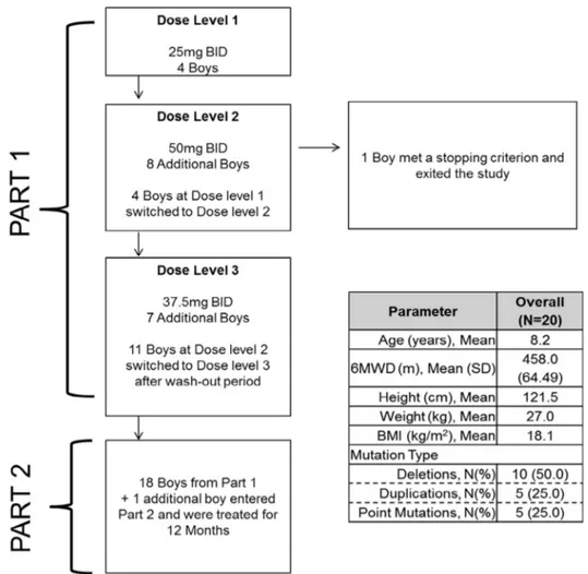
Fig. 1 summarizes the study design. Part 1 was a dose escalation study. Boys were asked to return to sites every week for physical examination, vital signs, ECG, laboratory tests, AEs collection, drug dispensing and PK sampling (only at week 2).
All the boys who completed Part 1 entered Part 2. One boy entered the study directly in Part 2. Boys visited their study site at months 1, 2, 3, 4.5, 6, 7.5, 9, 10.5 and 12 of Part 2 for
physical examination, vital signs, ECG, laboratory tests, AEs collection, drug dispensing and PK sampling (only at month 12). 6MWT, NSAA, and PUL were evaluated at screening, start of part 2, and at months 3, 6 and 12. Echocardiogram and spirometry were conducted at screening and at the end of part 2. 2.3. Standard protocol approvals, registrations and patient consents
The study was sponsored by Italfarmaco S.p.A. (Milan, Italy), performed in compliance with Good Clinical Practice and the Declaration of Helsinki and it was registered (Identifier
NCT01761292) at www.Clinicaltrials.gov. The study was
approved by the local Ethics Committees and authorized by the Competent Authority of Italy. A parent or guardian of the participants provided informed written consent and each subject provided written assent before participation.
2.4. Treatments
All boys were treated with Givinostat. During part 1 the dose was escalated from 25 mg BID to 50 mg BID and then reduced to 37.5 mg BID dose. During part 2 all boys were started on the 37.5 mg BID; seven completed the study on this dose and twelve reduced the dose to 25 mg BID (see below for details). All boys continued the steroid treatment regimen they were on at screening.
2.5. Endpoints
Histology: the primary endpoint was the change in histology comparing the brachial biceps biopsies before and after ≥12 months of treatment with Givinostat. The histological parameters assessed were: muscle fiber area fraction (MFAF), cross-sectional area (CSA), necrosis, hypercontracted (hyaline) fibers, fatty replacement and fibrosis (total, endomysial, perimysial). Details on muscle biopsy collection, preparation and histological assessments are provided in the Supplementary Information.
Muscle Function Tests: change in 6MWT, NSAA and PUL after treatment with Givinostat were secondary endpoints.
Details on these function tests were reported previously[5–7].
Safety and tolerability were assessed by AEs collection, laboratory tests, physical examination, vital signs, ECG, echocardiogram and spirometry. The following laboratory tests were performed: hematology, total bilirubin, alkaline phosphatase, amylase, ALT, AST, LDH, C-reactive protein, creatine kinase, total protein, albumin, uric acid, sodium, potassium, chloride, calcium, glucose, creatinine, BUN, and CPK, creatinine clearance, and urinalysis. Laboratory tests were conducted at local laboratories.
2.6. Statistical analysis
Based on the results reported in the Desguerre publication
[8], a sample size of 20 boys completing the study provided a
90% power (at a 2-sided alpha level of 5%) to detect at least a 25% relative increase in MFAF between pre- and post-treatment using a paired t-test and assuming a normal distribution.
The histological parameters were analyzed on all boys who completed the study, received at least 80% of the Givinostat dose in Part 2, had one baseline and one post-baseline assessment of biopsies, and had no major protocol violations. The ITT population, including all boys enrolled in Part 1 or Part 2 of the study, was used for all the other assessments.
The statistical significance of the change from baseline to the end of the study was tested by paired T-test. Normality assumptions were confirmed by the Shapiro–Wilk test of normality. All statistical tests were performed using a two-tailed 5% significance level.
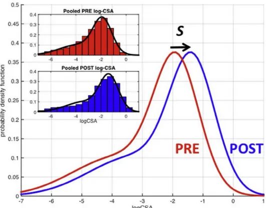
Givinostat effect on fiber size was evaluated considering the distribution of CSA before treatment (PRE) and at the end of the study (POST). For each individual, PRE and POST CSA values were normalized to the maximum PRE CSA value of that individual. The pooled PRE and POST log-CSA values histograms obtained after normalization and logarithmic transformation were inspected to ascertain a possible shift towards higher fiber size values in the POST distribution. In particular, a common shape underlying the PRE and POST distributions, differing just by a shift S in terms of fiber size, was considered. The common shape was described as a 2-component Gaussian mixture, whose parameters were estimated by the Expectation–Maximization method. The value
of the shift S quantifies the effect of Givinostat on a generic
muscle fiber, whose size, after treatment, results K= exp(S)
times larger than the baseline value.
3. Results
The baseline characteristics of the 20 boys are summarized inFig. 1. In Part 1, four boys started at the 25 mg BID dose, eight at the 50 mg BID dose and another seven boys at the 37.5 mg BID dose. Overall, Part 1 lasted 2 months. One of the 19 boys enrolled in Part 1 was discontinued from the study as he
reached a stopping rule (platelet counts<50 × 109/L) while on
treatment with Givinostat 50 mg BID. Another boy treated with Givinostat 50 mg BID reached a stopping rule of
temporary treatment suspension (platelet counts <75 × 109/L
but>50 × 109/L). As a result and according to the predefined
rules to declare the Maximum Tolerated Dose the dose of 50 mg BID was considered not tolerated. The remaining 18 boys and one other boy entered in Part 2. The dose of 37.5 mg BID was considered the Maximum Tolerated Dose and recommended for Part 2. At the beginning of Part 2 some boys treated at 37.5 mg BID had platelet reduction below the LLN but
never <75 × 109/L (temporary stopping criterion). Therefore,
the protocol was amended to require that platelet counts
be assessed≥every 2 weeks in the first 2 months of therapy
at 37.5 mg BID and that the dose be lowered to 25 mg BID if
persistent platelet counts ≤150 × 109
/L were observed.
Givinostat dose was maintained at 37.5 mg BID in 7 boys and reduced to 25 mg BID in 12. All 19 boys who entered Part 2 completed that part of the study. End of study biopsy could not be evaluated in one boy due to poor conditions of the tissue collected.
All biopsies showed a negative immunoreaction for
dystrophin at baseline and end of study. Table 1 and Tables
S1–S2 summarize the histological results (Fig. S2 provides examples of histological results). At baseline on average 46% of the tissue samples were occupied by fibrosis, and 51% by muscle. At the end of Part 2, there was a significant reduction of necrotic and hyper contracted fiber number, fat tissue replacement, and endomysial and perimysial fibrosis. MFAF increased due to a homogeneous increment of the CSA value of all fibers. Mean CSA increased by 77.7%. As shown in
Fig. 2, the distribution of log-transformed CSA had a similar shape before and after treatment with Givinostat. Treatment with Givinostat shifted the distribution to the right, indicating a multiplicative increase in fiber size by a factor
K= exp(S) = 1.70 (p ≤ 0.001). K values were significantly
larger in boys treated with 37.5 mg BID throughout Part 2 of the study compared to those who switched to the 25 mg BID dose
(Mean± SD 37.5 mg BID (N = 7) = 2.11 ± 0.52; Mean ± SD
25 mg BID (N= 12) = 1.61 ± 0.37, p < 0.05). Histological
parameters did not correlate with either age or steroid treatment duration either at baseline or at the end of Part 2
(age: R= −0.18 to 0.43, NS; steroid duration: R −0.16 to 0.34,
NS).
Three serious AEs were reported during the study (platelet reduction, rhabdomyolysis during muscle biopsy anesthesia, tibioperoneal fracture). Only the Platelet reduction was considered drug related (dose: 50 mg BID) and led to study
discontinuation. All AEs were mild to moderate in intensity except for the aforementioned serious AEs and a platelet and
neutrophil decrease (drug related) in Part 2.Table 2summarizes
the drug related AEs reported in Part 1 (Table 2A) and Part 2
(Table 2B) by more than 1 boy.
Platelet decrease and diarrhea were the most frequent AEs in the study. In Part 1, white blood cell decreased and most of the platelet decreased was reported only at the 50 mg BID dose. In Part 2, platelet decrease has been reported more frequently, with the 37.5 mg BID dose (63.2% vs 41.7%). Givinostat treatment was not associated to any other clinically significant laboratory abnormality. Similarly, vital signs, FEV1, FVC, FEV1/FVC, PEF and ECGs were not significantly altered.
Table 3 reports the results in the functional tests. One child could not complete the 6MWT at month 12 due to a tibioperoneal fracture at month 6. MFAF (directly) and total fibrosis (indirectly) correlated with the 6MWT at baseline and end of study. MFAF and Total Fibrosis correlated also with NSAA total score at baseline and with time to rise from floor at
the end of the study (Table S3).
Pharmacokinetic analyses indicated that following oral BID administration givinostat plasma exposures were dose-proportional. Cmax was observed at 2 to 4 hours post-dosing (median) at all doses administered. At all dose levels, the variability of plasma PK parameters was limited. Givinostat was at steady-state following 7 days of administration. Analysis of the accumulation ratios suggested a slight reduction (approximately 30%) at the end of the study compared to the results obtained following one week of treatment.
4. Discussion
While other treatments have previously shown to partially
restore dystrophin [9,10], this is the first time that a
pharmacological treatment was shown to produce beneficial
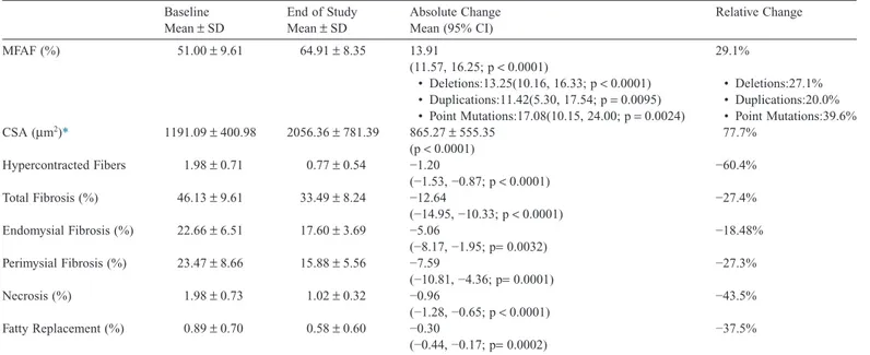
Table 1
Histological parameters at baseline and end of study and absolute and relative change. Baseline Mean± SD End of Study Mean± SD Absolute Change Mean (95% CI) Relative Change MFAF (%) 51.00± 9.61 64.91± 8.35 13.91 (11.57, 16.25; p< 0.0001) 29.1% • Deletions:13.25(10.16, 16.33; p< 0.0001) • Deletions:27.1% • Duplications:11.42(5.30, 17.54; p= 0.0095) • Duplications:20.0% • Point Mutations:17.08(10.15, 24.00; p= 0.0024) • Point Mutations:39.6% CSA (μm2)* 1191.09± 400.98 2056.36± 781.39 865.27± 555.35 (p< 0.0001) 77.7% Hypercontracted Fibers 1.98± 0.71 0.77± 0.54 −1.20 (−1.53, −0.87; p< 0.0001) −60.4% Total Fibrosis (%) 46.13± 9.61 33.49± 8.24 −12.64 (−14.95, −10.33; p< 0.0001) −27.4% Endomysial Fibrosis (%) 22.66± 6.51 17.60± 3.69 −5.06 (−8.17, −1.95; p= 0.0032) −18.48% Perimysial Fibrosis (%) 23.47± 8.66 15.88± 5.56 −7.59 (−10.81, −4.36; p= 0.0001) −27.3% Necrosis (%) 1.98± 0.73 1.02± 0.32 −0.96 (−1.28, −0.65; p< 0.0001) −43.5% Fatty Replacement (%) 0.89± 0.70 0.58± 0.60 −0.30 (−0.44, −0.17; p= 0.0002) −37.5% * Absolute change is mean± SD.
histological effects in muscles of DMD patients, though a functional benefit could not be definitively assessed due to the small sample size. Previous studies on muscle biopsies in DMD
[8,11]suggest that reductions in necrosis, fatty replacement and fibrosis and increase in muscle fiber area fraction do not occur with disease progression in boys seven to ten years of age. This is also supported by recent data with MRI and MRS that show a progressive tissue damage in boys with DMD five years and
older [12]. Thus these histological effects are attributable to
Givinostat. The role of Givinostat treatment is further supported by the lack of correlation between MFAF and total fibrosis and either age or steroid treatment duration. Since Givinostat acts on the pathogenetic events downstream of the genetic defects, Givinostat is potentially a treatment for the whole DMD population. The similar results in boys with deletions, duplications or point mutations support this conclusion.
Fig. 2. Comparison of the estimated distributions of normalized log-CSA values measured in the biopsies before treatment (PRE) and at the end of part 2 (POST). In the inset, the estimated distributions are superimposed on the pooled histograms.
Table 2
Drug related Treatment Emergent Adverse Events (TEAEs) that occurred in more than 1 patient during the Part 1 of the study (A) and during the Part 2 of the study (B). A 25.0 mg BID (N= 4) 37.5 mg BID (N= 7) 50.0 mg BID (N= 12) Overall (N= 19)
Number (%) of Patients with drug related TEAEs 3 (75.0%) 6 (85.7%) 12 (100%) 18 (94.7%)
Abdominal pain 0 (0.0%) 2 (28.6%) 1 (8.3%) 3 (15.8%)
Diarrhea 2 (50.0%) 3 (42.9%) 5 (41.7%) 8 (42.1%)
Feces soft 0 (0.0%) 1 (14.3%) 1 (8.3%) 2 (10.5%)
Platelet decreased 0 (0.0%) 1 (14.3%) 7 (58.3%) 8 (42.1%)
White blood cell count decreased 0 (0.0%) 0 (0.0%) 4 (33.3%) 4 (21.1%)
Rash 1 (25.0%) 0 (0.0%) 1 (8.3%) 2 (10.5%) B 25.0 mg BID (N= 12) 37.5 mg BID (N= 19) Overall (N= 19)
Number (%) of Patients with drug related TEAEs 9 (75.0%) 18 (94.7%) 18 (94.7%)
Abdominal pain 0 (0.0%) 5 (26.3%) 5 (26.3%) Diarrhea 6 (50.0%) 9 (47.4%) 11 (57.9%) Vomiting 1 (8.3%) 2 (10.5%) 3 (15.8%) Platelet decreased 5 (41.7%) 12 (63.2%) 12 (63.2%) Decreased appetite 2 (16.7%) 4 (21.1%) 6 (31.6%) Headache 1 (8.3%) 1 (5.3%) 2 (10.5%)
The mechanism of action at the basis of these histological changes induced by Givinostat is still not fully elucidated. However, it is already evident that Givinostat treatment is followed by a homogeneous increase in muscle fibers size (Fig. 2). Although follistatin and myostatin levels and/or their downstream effects could not be assessed in the muscle biopsies in this study, HDACi have been previously shown to
increase follistatin translation[4,13]. Follistatin down regulates
myostatin a major inhibitor of muscle fibers regeneration
[14–16] and in nature myostatin defects are accompanied by large muscle mass. Future studies will need to clarify the role of follistatin translation in the effects on muscle fiber size observed with Givinostat.
The other key objective of this study was to confirm that doses determining a significant histological effect were also tolerated in boys with DMD. Platelet reductions and gastrointestinal AEs have been the most frequent AEs in this study. Platelet reduction has been observed with all HDACi tested so far including Givinostat and it is considered related to
their pharmacological effect [17]. In this study, platelet
reduction, which met predefined stopping rules, were observed only at 50 mg BID, a dose considered not tolerable. In Part 2, platelet counts remained within the normal range after treatment with 37.5 mg BID in 7 boys, while in other 12 the dose was lowered to 25 mg BID to maintain platelet counts within the normal range. The decision to lower the dose was made even if no clinical manifestation of low platelet counts (e.g. hemorrhage, petechiae, etc.) was observed.
In this study we wanted to maintain exposures to Givinostat as high as possible to maximize the chances of seeing a histological benefit. Using a starting dose of 37.5 mg BID and applying the dose adjustment rules described in the Results section met the objective of maintaining high exposures without abnormal platelet counts. However, the dose was reduced to 25 mg BID in 2/3 of patients. Significant histological improvements were observed in all children in Part 2, regardless of their dose. However, CSA increased significantly more in boys treated at the highest dose throughout part 2. As CSA fibers enlargement appears to be a key event in the Givinostat effect, this result suggests that even if exposures
obtained after the 25 mg BID dose are efficacious, there is a potential benefit in trying to maintain Givinostat exposures as high as possible. Further studies will confirm if larger Givinostat exposures are beneficial and if the dose adjustment rules adopted in this study will allow maintaining platelet counts stable and within normal ranges also in larger cohorts of DMD patients.
Diarrhea was frequently reported in this study, but was mild or moderate, never required treatment adjustments and never led to drug discontinuation, suggesting that this AE is quite manageable.
This study was not designed to assess efficacy and functional tests were performed to evaluate possible negative effects on muscle function. The changes in the function tests were relatively small and similar to those expected in a DMD
population similar to the one in this study[18–20]. Because of
the small sample size and of the lack of a control group, no further considerations can be made on the Givinostat effects on muscle function. The significant correlation between the MFAF and total fibrosis and most of the functional tests, however, suggests that the histological improvements observed with Givinostat may eventually lead to a functional benefit.
This study has challenges and limitations. Muscle biopsies are always a challenge. A very tight control was maintained throughout the study on muscle biopsy collection, transport, processing and analysis. As a result only 1 biopsy was considered not suitable for analysis. In our study the histological and morphological analysis was performed to the best standards following well documented identification criteria
typical of DMD skeletal muscle alterations [21], and
morphological analysis was carried out using MetaMorph, a recognized reliable software. In fact, our baseline results are in
line with the results previously reported[8,22]. This study did
not include a control group, since it was not considered ethical to have a group of boys on placebo undergoing 2 biopsies without any chance of a potential treatment benefit. However, as no histological improvements are expected in a group of DMD boys aged seven to ten years, the significant histological results obtained in this study are not affected by the lack of a control group. Even if the patient number was small, the study was fully powered to show an increase in MFAF at least as large as the
one observed in the preclinical study[3].
In conclusion, this study shows that administration of Givinostat for more than one year significantly counteracts histological disease progression in ambulant DMD boys aged seven to ten years. These results support further development of Givinostat in DMD.
Acknowledgments
We gratefully acknowledge the boys and their families for the participation in this study.
We thank the staff members for their dedication: Giuseppe Pontrelli, M.D., Ph.D. and Susanna Livadiotti, M.D. from Clinical Trial Centre, Bambino Gesu’ Children’s Research Hospital, Rome; Giorgio Tasca, M.D., Ph.D., Adelina Carlesi, Phys, Giulia Colia, Phys, Anna M. Bonetti, Phys from Unit of Neuromuscular and Neurodegenerative Disorders, Department
Table 3
Results of Functional Tests at each study visit (Mean (SD, N)) during Part 2. 6MWT Distance
(meters)
NSAA Total Score
Rise from floor (seconds) PUL Total Score Baseline 453.0 28.1 4.43 71.7 (62.23, 19) (5.13, 19) (1.41, 18) (2.40, 19) Month 0 450.9 28.4 4.87 71.9 (58.00, 19) (4.46, 19) (1.57, 18) (2.61, 18) Month 3 416.6 27.6 6.75 72.2 (86.13, 19) (6.16, 19) (8.42, 19) (2.32, 19) Month 6 421.9 27.3 6.87 72.4 (73.96, 19) (5.58, 19) (8.22, 19) (1.57, 19) Month 12 432.2 26.2 5.04 71.6 (63.60, 18*) (6.10, 18*) (2.09, 17*) (2.81, 19) * A patient could not perform the 6MWT and was excluded from the NSAA and Rise from floor analysis relevant to month 12 because he fractured his tibia and fibula just after the 6-month functional assessments.
of Neurosciences Bambino Gesu’ Children’s Research Hospital, Rome; Flavia Bianco, M.D., Concetta Palermo, M.D., Lavinia Fanelli, M.D., Elena Mazzone, Phys from Department of Paediatric Neurology, Catholic University, Rome; Claudia Cinnante, M.D., Alessandra Govoni, M.D., Claudio Bellani, Phys, Clara Ceruti, Phys from Fondazione IRCCS Ca’ Granda Ospedale Maggiore Policlinico, Milan; Costanza Barcellona, M.D., Filippo Cavallaro, Phys, Matteo La Rosa, M.D., Maria Sframeli, M.D., Carmelo Rodolico, M.D. from Department of Neurosciences, University of Messina; NEMO SUD Clinical Centre for Neuromuscular Disorders, Messina; Silvia Consalvi, Ph.D., Valentina Saccone, Ph.D. from IRCCS Santa Lucia Foundation, Rome.
We also thank Telethon, Italy for help and suggestions and Filippo Buccella and the staff of Parent Project onlus, Italy for cooperation and support for the psycological and logistic aspects of the clinical trial; Carlo Bianchini, M.D., Paolo Mascagni, Ph.D., Christian Steinkühler, Ph.D., Flavio Leoni, M.Sc., Gialuca Fossati, M.Sc., Giuseppe Colombo, M.Sc., Roberta Artico, Silvia Puccianti, M.Sc., Sara Cazzaniga, M.Sc., Valeria Lovato, Ph.D. and all staff members from Italfarmaco S.p.A., Milan, for assistance and discussion.
Appendix: Supplementary material
Supplementary data to this article can be found online at
doi:10.1016/j.nmd.2016.07.002.
References
[1] Mendell JR, Shilling C, Leslie ND, et al. Evidence-based path to newborn screening for Duchenne muscular dystrophy. Ann Neurol 2012;71(3): 304–13.
[2] Colussi C, Mozzetta C, Gurtner A, et al. HDAC2 blockade by nitric oxide and histone deacetylase inhibitors reveals a common target in Duchenne muscular dystrophy treatment. Proc Natl Acad Sci USA 2008;105(49): 19183–7.
[3] Consalvi S, Mozzetta C, Bettica P, et al. Preclinical studies in the mdx mouse model of duchenne muscular dystrophy with the histone deacetylase inhibitor givinostat. Mol Med 2013;19:79–87.
[4] Minetti GC, Colussi C, Adami R, et al. Functional and morphological recovery of dystrophic muscles in mice treated with deacetylase inhibitors. Nat Med 2006;12(10):1147–50.
[5] Laboratories ATSCoPSfCPF. ATS statement: guidelines for the six-minute walk test. Am J Respir Crit Care Med 2002;166(1):111–17. [6] Mayhew A, Mazzone ES, Eagle M, et al. Development of the Performance
of the Upper Limb module for Duchenne muscular dystrophy. Dev Med Child Neurol 2013;55(11):1038–45.
[7] Mercuri E, McDonald C, Mayhew A, et al. International workshop on assessment of upper limb function in Duchenne Muscular Dystrophy: Rome, 15–16 February 2012. Neuromuscul Disord 2012;22(11):1025–8. [8] Desguerre I, Mayer M, Leturcq F, Barbet JP, Gherardi RK, Christov C. Endomysial fibrosis in Duchenne muscular dystrophy: a marker of poor outcome associated with macrophage alternative activation. J Neuropathol Exp Neurol 2009;68(7):762–73.
[9] Cirak S, Arechavala-Gomeza V, Guglieri M, et al. Exon skipping and dystrophin restoration in patients with Duchenne muscular dystrophy after systemic phosphorodiamidate morpholino oligomer treatment: an open-label, phase 2, dose-escalation study. Lancet 2011;378(9791): 595–605.
[10] Mendell JR, Rodino-Klapac LR, Sahenk Z, et al. Eteplirsen for the treatment of Duchenne muscular dystrophy. Ann Neurol 2013;74(5): 637–47.
[11] Peverelli L, Testolin S, Villa L, et al. Histological muscular history in Duchenne Dystrophy patients with and without steroid treatment. Neurology 2015;85(21):1886–93.
[12] Forbes SC, Willcocks RJ, Triplett WT, et al. Magnetic resonance imaging and spectroscopy assessment of lower extremity skeletal muscles in boys with Duchenne muscular dystrophy: a multicenter cross sectional study. PLoS ONE 2014;9(9):e106435.
[13] Iezzi S, Di Padova M, Serra C, et al. Deacetylase inhibitors increase muscle cell size by promoting myoblast recruitment and fusion through induction of follistatin. Dev Cell 2004;6(5):673–84.
[14] Mendell JR, Sahenk Z, Malik V, et al. A phase 1/2a follistatin gene therapy trial for becker muscular dystrophy. Mol Ther 2015;23(1): 192–201.
[15] Nakatani M, Takehara Y, Sugino H, et al. Transgenic expression of a myostatin inhibitor derived from follistatin increases skeletal muscle mass and ameliorates dystrophic pathology in mdx mice. FASEB J 2008;22(2): 477–87.
[16] Tsuchida K. Myostatin inhibition by a follistatin-derived peptide ameliorates the pathophysiology of muscular dystrophy model mice. Acta Myol 2008;27:14–18.
[17] Subramanian S, Bates SE, Wright JJ, Espinoza-Delgado I, Piekarz RL. Clinical toxicities of histone deacetylase inhibitors. Pharmaceuticals 2010;3(9):2751–67.
[18] Pane M, Mazzone ES, Sivo S, et al. Long term natural history data in ambulant boys with Duchenne muscular dystrophy: 36-month changes. PLoS ONE 2014;9(10):e108205.
[19] McDonald CM, Henricson EK, Abresch RT, et al. The 6-minute walk test and other endpoints in Duchenne muscular dystrophy: longitudinal natural history observations over 48 weeks from a multicenter study. Muscle Nerve 2013;48(3):343–56.
[20] Mazzone ES, Pane M, Sormani MP, et al. 24 month longitudinal data in ambulant boys with Duchenne muscular dystrophy. PLoS ONE 2013; 8(1):e52512.
[21] Dubowitz VS, Sewry CA, Oldfords A. Muscle biopsy: a practical approach. 4th ed. London: Saunders Ltd.; 2013.
[22] Wang JF, Forst J, Schroder S, Schroder JM. Correlation of muscle fiber type measurements with clinical and molecular genetic data in Duchenne muscular dystrophy. Neuromuscul Disord 1999;9(3):150–8.