Report
Loss of Centrobin Enables Daughter Centrioles to
Form Sensory Cilia in Drosophila
Highlights
d
Basal body fate is restricted to mother centrioles in
Drosophila sensory neurons
d
Depletion of Centrobin enables daughter centrioles to form
sensory cilia
d
Ectopic localization of Centrobin in mother centrioles inhibits
cilia formation
Authors
Marco Gottardo, Giulia Pollarolo, Salud
Llamazares, ..., Maria G. Riparbelli,
Giuliano Callaini, Cayetano Gonzalez
Correspondence
[email protected]
In Brief
Gottardo et al. show that, despite the lack
of centriole maturity traits, basal body
fate is reserved to mother centrioles in
Drosophila sensory neurons. Moreover,
depletion of the daughter centriole
protein Centrobin enables daughter
centrioles to form cilia, whereas ectopic
localization of Centrobin in mother
centrioles inhibits cilia formation.
Gottardo et al., 2015, Current Biology25, 2319–2324 August 31, 2015ª2015 Elsevier Ltd All rights reserved http://dx.doi.org/10.1016/j.cub.2015.07.038
Current Biology
Report
Loss of Centrobin Enables Daughter Centrioles
to Form Sensory Cilia in
Drosophila
Marco Gottardo,1,4Giulia Pollarolo,2,4Salud Llamazares,2,4Jose Reina,2Maria G. Riparbelli,1Giuliano Callaini,1
and Cayetano Gonzalez2,3,*
1Department of Life Sciences, University of Siena, Via Aldo Moro 2, Siena 53100, Italy
2Institute for Research in Biomedicine (IRB Barcelona), Baldiri Reixac 10, Barcelona 08028, Spain
3Institucio´ Catalana de Recerca i Estudis Avanc¸ats (ICREA), Passeig Lluı´s Companys 23, Barcelona 08010, Spain 4Co-first author
*Correspondence:[email protected] http://dx.doi.org/10.1016/j.cub.2015.07.038
SUMMARY
Sensory cilia are organelles that convey information
to the cell from the extracellular environment. In
ver-tebrates, ciliary dysfunction results in ciliopathies
that in humans comprise a wide spectrum of
devel-opmental disorders [
1–3
]. In
Drosophila, sensory cilia
are found only in the neurons of type I sensory
or-gans, but ciliary dysfunction also has dramatic
con-sequences in this organism because it impairs the
mechanosensory properties of bristles and chaetae
and leads to uncoordination, a crippling condition
that causes lethality shortly after eclosion [
4–7
]. The
cilium is defined by the ciliary membrane, a
protru-sion of the cell membrane that envelops the core
structure known as the axoneme, a microtubule array
that extends along the cilium from the basal body. In
vertebrates, basal body function requires centriolar
distal and subdistal appendages and satellites.
Because these structures are acquired through
centriole maturation, only mother centrioles can
serve as basal bodies. Here, we show that although
centriole maturity traits are lacking in
Drosophila,
basal body fate is reserved to mother centrioles in
Drosophila type I neurons. Moreover, we show that
depletion of the daughter-centriole-specific protein
Centrobin (CNB) enables daughter centrioles to
dock on the cell membrane and to template an
ectopic axoneme that, although structurally
defec-tive, protrudes out of the cell and is enveloped by a
ciliary membrane. Conversely, basal body capability
is inhibited in mother centrioles modified to carry
CNB. These results reveal the crucial role of CNB in
regulating basal body function in
Drosophila ciliated
sensory organs.
RESULTS
Basal Body Fate Is Reserved to Mother Centrioles in Drosophila Type I Neurons
Mother centrioles in Drosophila present neither appendages— distal or subdistal—nor satellites, and, therefore, as far as these
ultrastructural features are concerned, they are indistinguishable from daughter centrioles [8, 9]. This lack of ultrastructural dimorphism opens the question of whether in Drosophila either of the two centrioles of a diplosome can serve as basal body or whether, on the contrary, basal body fate is also centriole age dependent, as it is often assumed [10, 11].
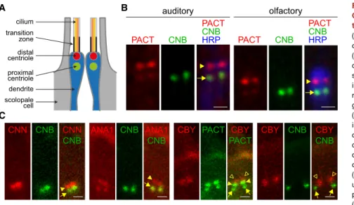
We have addressed this question by examining pancentriolar, daughter-centriole-specific, and transition zone reporters in the sensory neurons of both chordotonal auditory and external ol-factory sensilla. Each neuron within these sensilla presents two tandemly arranged centrioles located at the apical tip of the sen-sory dendrite. The most distal of each centriole pair serves as basal body from which the axoneme projects (Figure 1A) [12].
Upon coexpression of PACT-RFP and YFP-CNB (Centrobin), in agreement with previous reports [11], we found a strong PACT-RFP fluorescence signal on the distal centrioles ( Fig-ure 1B, red, arrowhead). Moreover, we also detected a weak PACT-RFP proximal signal (Figure 1B, red, arrow) that colocal-izes with YFP-CNB (green). These results are consistent with published data on Drosophila syncytial embryos and larval neu-roblasts (NBs) where PACT is found in both centrioles but is more abundant on the mother, while YFP-CNB labels only the daughter centriole [11, 13–15]. To further determine the identity of the proximal and distal centrioles in Drosophila sensory neurons, we performed double fluorescence labeling of CNN (Centrosomin)/CNB, ANA1/CNB, CBY (Chibby)/PACT, and CNB/CBY (Figures 1C and S1). We found that CNB, ANA1, and the weak PACT signal colocalize in the proximal centriole; ANA1, CNN, and the strong PACT signal colocalize on the distal; and the transition zone marker CBY is further distal to both cen-trioles (Figures 1C andS1, arrow, arrowhead, and empty arrow-head, respectively). These observations are fully consistent with the hypothesis that, despite the lack of centriole maturation fea-tures, basal body fate is reserved to mother centrioles in Drosophila auditory and olfactory neurons.
Ectopic Localization of CNB in Mother Centrioles Inhibits Cilia Formation in Drosophila Type I Neurons Ultrastructural centriole dimorphism is also lacking in Drosophila NBs where mother and daughter centrioles, which disengage from each other soon after mitosis, are also functionally unequal [16–19]. NBs maintain a potent microtubule organizing center (MTOC) that is localized near the presumptive apical cortex throughout interphase and is organized by the daughter centriole alone. The mother centriole organizes little, if any, pericentriolar
material and has essentially no MTOC activity during interphase. The daughter centriole protein CNB appears to be necessary and sufficient to trigger centriole asymmetry in NBs because CNB depletion impedes daughter centrioles to assemble a func-tional MTOC, and mother centrioles carrying ectopic CNB become active MTOCs [20]. Polo, CNN, Pericentrin-Like Protein (PLP), Partner of Inscuteable (PINS), and BLD10/CEP135 are also known to be essential for such a mother-daughter centriole asymmetry in NBs [11, 18, 20–23].
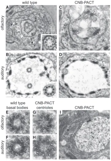
Because CNB localization exerts such a determining role in establishing functional differences between mother and daughter centrioles in Drosophila NBs, we tested whether CNB could play a similar role in centriole function in sensory neurons. To this end, we first examined the effect of localizing CNB ectop-ically on mother centrioles. Transverse sections through the cilia of wild-type olfactory and auditory sensilla containing three and two sensory neurons, respectively, show the corresponding number of axonemes (Figures 2A, 2B, andS2A). In contrast, ax-onemes were missing in most (23 out of 24) olfactory and (25 out of 27) auditory sensilla expressing YFP-CNB-PACT, henceforth referred to as CNB-PACT (Figures 2C, 2D, andS2B). Unlike in wild-type sensilla where electron microscopy (EM) sections through centrioles are relatively easy to obtain following the axo-nemes, it is very difficult to identify centrioles in non-ciliated CNB-PACT sensilla. We have, however, identified two centrioles in two different CNB-PACT-expressing auditory neurons and one in an olfactory neuron, strongly suggesting that failed ciliogenesis in these cells is not due to the lack of centrioles. Indeed, expression of CNB-PACT does not affect centriole number in other cell line-ages in Drosophila [20]. Unlike wild-type basal bodies, which are made of doublets (Figures 2E and 2F), the only two centrioles for which we have obtained transverse EM section in CNB-PACT-expressing neurons (one olfactory and one auditory) presented singlets or incomplete doublets (Figures 2G and 2H, respec-tively), as is often the case in wild-type proximal centrioles.
Inter-estingly, the centriole from the CNB-PACT olfactory neuron was found close to the nucleus (Figure 2I, arrow), many centriole diameters away from the tip of the dendrite where centrioles are located in wild-type cells. This result strongly suggests that upon CNB-PACT expression, centrioles may become scattered over the cell body, as they are in Plp mutant auditory neurons [11], thus rendering their identification extremely difficult. No rootlets were found in CNB-PACT-expressing neurons.
Consistent with the absence of cilia, we found that CNB-PACT-expressing flies are uncoordinated, cannot fly, get stuck to the food, and die shortly after eclosion. On the contrary, con-trol flies expressing either YFP-PACT or YFP-CNB hatch, feed, and mate normally, showing that overexpression of PACT (without CNB) or overexpression of CNB (without PACT) does not produce the phenotypic traits brought about by expression of YFP-CNB-PACT.
These results strongly suggest that ectopic CNB localization impedes mother centrioles to function as basal bodies in Drosophila type I sensory neurons.
Loss of CNB Enables Daughter Centrioles to Function as Basal Bodies in Drosophila Type I Neurons
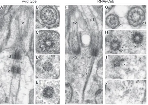
We then investigated cilia formation in sensilla depleted of CNB. As reported previously [12, 24], wild-type olfactory (Figures 3A– 3E) and auditory (Figure S3A) neurons present a pair of tandemly arranged centrioles, one distal (d) that serves as basal body from which the cilium (c) is assembled and one proximal (p). Rootlets (r) that emerge from the distal centriole enclose the proximal centriole and extend further proximally. Ciliary rootlets differ in different types of sensilla [12, 25]. In olfactory neurons ( Fig-ure 3A), the rootlet bundle is thinner than that of chordotonal auditory sensilla (Figure S3A), which shows a characteristic periodic cross-striation [12, 26–29].
We have found that CNB depletion causes a major disruption of the mono-axial arrangement of cilium and centrioles observed A C cilium dendrite transition zone distal centriole proximal centriole scolopale cell B auditory PACT CNB CNBHRP PACT olfactory PACT CNB CNBHRP PACT ANA1 CNB CNB ANA1 CNB CNN CNN CNB PACT CBY CBY PACT CNB CBY CBY CNB
Figure 1. Axonemes Assemble from Mother Centrioles inDrosophila Olfactory and Audi-tory Neurons
(A) Graphical summary of the arrangement of centrioles and cilia in sensilla with two neurons (blue). Axonemes (orange) project from the distal centrioles (red) that serve as basal bodies. Tran-sition zones are shown as thick black lines. Prox-imal centrioles are marked in green. Accessory non-neural cells within the sensilla are depicted in gray.
(B) Detail of the apical ends of the dendrites of individual auditory and olfactory sensilla, each containing two neurons. PACT-RFP (red) is pan-centriolar but significantly more abundant on the distal (arrowhead) than on the proximal (arrow) centriole. The daughter centriole marker YFP-CNB (green) is prominent on proximal centrioles and undetectable on distal centrioles. Anti-horseradish peroxidase (HRP) (blue) marks dendrite and cilium. (C) Apical ends of pairs of olfactory neurons showing fluorescence signals corresponding to YFP-CNB/RFP-CNN, YFP-CNB/ANA1-tdTomato, GFP-PACT/CBY-Tomato, and YFP-CNB/CBY-Tomato. ANA1 and PACT mark both the proximal (arrow) and distal (arrowhead) centrioles, while CNB and CNN are specifically located at the proximal and distal centrioles, respectively; CBY (empty arrowhead) localizes distally to all these markers.
in wild-type sensory neurons. Upon expression of RNAi-Cnb, most olfactory (13 out of 15;Figures 3F–3J) and auditory (11 out of 15;Figures S3B, S3C, and S2C) neurons are biciliated (c1 and c2) and present two centrioles that are arranged in par-allel, not in tandem, both serving as basal bodies (d1 and d2) from which axonemes assemble, and centrioles proximal to the basal bodies are lacking. The same result was observed in olfactory neurons that carry the hypomorph Cnb mutant allele PBac{RB}Cnbe00267over Def(3L)ED4284, which uncovers Cnb (Figure S3G).
In all 14 cells for which we have transverse section (8 olfactory; 6 auditory), we found that one of the two cilia present in RNAi-Cnb-expressing sensory neurons was shorter than normal and had defects in the 9-fold symmetry of the axoneme, with dou-blets that were incomplete or missing (Figure 3G, c2;Figure S3C, c2;Figures S3D and S3E;Figure S3G, c2). The two basal bodies, however, presented a rather normal ultrastructure (Figure 3H, d1 and d2;Figure S3F). Judged by both the abundance of doublets rather than singlets and the electron-dense stripes between dou-blets that could be observed at their distal ends, which are thought to be the Drosophila equivalent of Y linkers, the two basal bodies of CNB-depleted cells present the appearance of a wild-type distal centriole. Consistently, PACT is equally distrib-uted on each centriole in PBac{RB}Cnbe00267/Def(3L)ED4284
mutant olfactory neurons (Figures S3H and S3I). However, the two basal bodies can still be told apart by the distribution of ciliary rootlets that were always (n = 12; 7 olfactory and 5 audi-tory) attached to only one of the two basal bodies (Figure 3F, d1;Figure S3B, d1).
Neither flies expressing RNAi-Cnb nor PBac{RB}Cnbe00267/ Def(3L)ED4284 mutant flies are uncoordinated, and their perfor-mance in negative geotaxis tests (climbing assays) is not signif-icantly different from that of wild-type strains (data not shown).
The presence of a pair of cilia and basal bodies and the corre-sponding lack of proximal centrioles strongly suggest that CNB depletion enables daughter centrioles to function as basal bodies in Drosophila type I neurons.
DISCUSSION
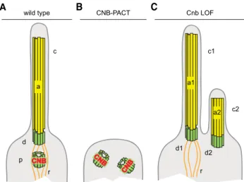
A graphical summary of our findings and working hypothesis on the role of CNB in ciliogenesis in Drosophila type I sensory neu-rons is shown in Figure 4. In wild-type sensory neurons ( Fig-ure 4A), axoneme (a), basal body (d), from which rootlets (r) emanate, and proximal centriole (p), where CNB localizes, are ar-ranged along one axis. This mono-axial arrangement is critically dependent on normal CNB function. CNB ectopic localization on mother centrioles eliminates this axis altogether; axonemes do not grow, and centrioles are not tandemly arranged (Figure 4B). Likely, these cells contain two centrioles, as it has been shown to be the case in other Drosophila cell lineages upon expression of the CNB-PACT fusion protein [20]. In turn, CNB depletion ( Fig-ure 4C) brings about a biaxial arrangement made of two axo-nemes (a1 and a2) with their corresponding basal bodies (d1 and d2), which are not associated to proximal centrioles. Only one of the two basal bodies observed in these cells presents rootlets (r). Therefore, CNB seems to be required for both keep-ing mother-daughter centriole alignment and preventing daughter centriole docking on the cell membrane. We do not know whether these two functions operate independently or depend on each other.
Often, one of the two cilia of CNB-depleted cells is shorter than normal and has defects in the axoneme’s 9-fold symmetry and in the ciliary membrane. Such defects may reflect yet unknown roles of CNB in ciliogenesis. Alternatively, they may arise from the dilution of one or more critical ciliogenesis factors whose concentration in normally monociliated cells like type I sensory neurons may be rate limiting for the assembly of only one fully formed cilium. A dilution phenotype that affects ciliary function
olfactory
auditory
wild type CNB-PACT
auditory wild type basal bodies CNB-PACT centrioles olfactory CNB-PACT A C B D E G F H I N
Figure 2. Pancentriolar CNB Localization Inhibits Axoneme Assem-bly in Type I Neurons
(A and B) Transverse sections through olfactory (A) and auditory (B) wild-type sensilla containing three and two neurons, respectively. Each neuron pro-trudes a cilium that contains a fully assembled axoneme (insets).
(C and D) Axonemes are missing in olfactory and auditory sensilla expressing the CNB-PACT fusion protein, which drives CNB to both mother and daughter centrioles.
(E and F) Basal bodies from wild-type olfactory (E) and auditory (F) sensilla contain doublets and a few triplets.
(G and H) Centrioles from olfactory (G) and auditory (H) neurons expressing CNB-PACT are mostly made of singlets.
(I) Low-magnification view of the section containing the centriole shown in (G) (arrow), which is located close to the nucleus (N), away from the apical dendrite.
Scale bars of (A)–(D) represent 250 nm; scale bars of (E)–(H) represent 100 nm; and the scale bar of (I) represents 500 nm. See alsoFigure S2.
has been reported in IMCD-3 cells and in cells mutant for the tuberous sclerosis gene TSC2 that carry supernumerary centri-oles. When these cells form more than one cilium, ciliary concen-tration of Smoothened in response to Sonic hedgehog (Shh) stimulation is reduced [30]. Nonetheless, despite the presence of malformed cilia present in CNB-depleted Drosophila type I sensory neurons, the fully formed cilia that are also present seem to be capable of sustaining normal ciliary function because adult flies bearing CNB-depleted sensilla perform just like wild-type flies in climbing assays.
It is remarkable that although centriolar functions in sensory neurons and NBs are notably different, in both cell types, daughter centrioles acquire mother centriole traits upon CNB depletion, and mother centrioles become daughter centriole like upon CNB binding [20]. Something similar applies to PLP. In NBs and in type I sensory neurons, PLP is enriched on the mother centriole ([11, 21] and our own results). Upon loss of PLP function, mother centrioles behave like daughters in NBs [21], and cilia are lacking in sensory neurons, which indeed causes uncoordination in adult flies [31]. However, despite these tantalizing similarities, the molecular pathways that make centri-oles functionally unequal in NBs and in type I sensory neurons present conspicuous differences. For instance, in NBs, where CNB is essential to recruit CNN [20], CNN specifically localizes on the daughter centrosome. In sensory neurons, on the con-trary, CNB and CNN do not colocalize: CNN is associated with the basal body (Figures 1C andS1; [32]), while CNB marks the proximal centriole. Moreover, in Bld10 mutant NBs, mother cen-trioles recruit CNN [23], which is a critical daughter centriole trait in wild-type cells, but Bld10 mutant adults display no obvious sign of uncoordination [22].
Biciliated cells have been described in nature in fish [33], frogs [34], rodents [35–37], and primates [38]. In rodents and primates,
biciliated cells account for more than 80% of the cells lining the central canal epithelium in the adult brain and are highly prolifer-ative. Notably, these biciliated cells present two basal bodies that are not associated with daughter centrioles [37, 38]. This obser-vation strongly suggests that biciliation in these cells does not result from an additional round of centriole duplication but from centriole splitting and maturation of the daughter centriole as basal body [37, 38]. The same applies to biciliated cells experi-mentally induced in Xenopus embryos by high Foxj1 misexpres-sion [34] or in the MCF10A cell line following an unscheduled pulse of PLK1 activity [39]; in both cases, daughter centrioles pre-cociously mature, and ultrastructural, normally age-dependent differences between centrioles are erased.
Our findings demonstrate that in Drosophila ciliated sensory organs, both internal and external, CNB localization plays a key role in the regulation of centriole conversion into basal bodies.
EXPERIMENTAL PROCEDURES
Details on experimental procedures are described inSupplemental Experi-mental Procedures.
SUPPLEMENTAL INFORMATION
Supplemental Information includes Supplemental Experimental Procedures and three figures and can be found with this article online athttp://dx.doi. org/10.1016/j.cub.2015.07.038.
AUTHORS CONTRIBUTIONS
M.G., G.P., S.L., J.R., M.G.R., G.C., and C.G. conceived and designed the ex-periments and analyzed the data. S.L. and J.R. generated transgenic fly strains. S.L. and G.P. performed immunofluorescence microscopy. M.G., M.G.R., and G.C. performed electron microscopy. G.P. and C.G. wrote the paper. A D E G H I
wild type RNAi-Cnb
B C F J c d p r r c1 c2 d1 d2 r c d r p c1 c2 d1 d2 v
Figure 3. CNB Depletion Results in Bicili-ated Type I Olfactory Neurons
(A–E) Wild-type. Longitudinal (A) and serial trans-verse sections from distal to proximal (B–E) reveal the mono-axial arrangement of cilium (c), basal body (distal centriole, d), and proximal centriole (p). Centriolar rootlets (r) originate in the basal body, envelop the proximal centriole, and extend further proximally. Electron-dense stripes corre-sponding to putative Y linkers (C, arrows) are visible in between the doublets of the basal body. (F–J) Olfactory neurons depleted for CNB by expression of RNAi-Cnb. Longitudinal (F) and se-rial transverse sections from distal to proximal (G–J) reveal two cilia (c1 and c2), two basal bodies (d1 and d2), and the lack of proximal centrioles. Centriolar rootlets (r) originate from only one of the two basal bodies (d1). Cilium c2 presents a defective axoneme (G). Both basal bodies bear doublets (H) and electron-dense stripes similar to those observed in wild-type basal bodies (H, arrows). Dense vesicle-like bodies (v), can be observed proximal to d2 (I). Identical structures are found proximal to d1 and to centrioles in wild-type olfactory neurons (data not shown).
Scale bars of (A) and (F) represent 250 nm; scale bars of (B)–(E) and (G)–(J) represent 100 nm. See alsoFigure S3.
ACKNOWLEDGMENTS
We thank J. Raff (Oxford University), T. Avidor-Reiss (University of Toledo), B. Durand (University of Lyon), the Bloomington Stock Center, and VDRC for providing fly lines. Work in our laboratory is supported by grants BFU2012-32522 from the Spanish MINECO, SGR Agaur 2014 100 from Generalitat de Catalunya, AdG 2011 294603 advanced grant from the European Research Council, and Redes de Excelencia BFU2014-52125-REDT-CellSYS from the Spanish MINECO. G.P. is a Juan de la Cierva Fellow.
Received: March 12, 2015 Revised: June 22, 2015 Accepted: July 14, 2015 Published: August 20, 2015
REFERENCES
1.Ishikawa, H., and Marshall, W.F. (2011). Ciliogenesis: building the cell’s antenna. Nat. Rev. Mol. Cell Biol. 12, 222–234.
2.Goetz, S.C., and Anderson, K.V. (2010). The primary cilium: a signalling centre during vertebrate development. Nat. Rev. Genet. 11, 331–344. 3.Bettencourt-Dias, M., Hildebrandt, F., Pellman, D., Woods, G., and
Godinho, S.A. (2011). Centrosomes and cilia in human disease. Trends Genet. 27, 307–315.
4.Kernan, M., Cowan, D., and Zuker, C. (1994). Genetic dissection of me-chanosensory transduction: mechanoreception-defective mutations of Drosophila. Neuron 12, 1195–1206.
5.Kernan, M.J. (2007). Mechanotransduction and auditory transduction in Drosophila. Pflugers Arch. 454, 703–720.
6.Dubruille, R., Laurenc¸on, A., Vandaele, C., Shishido, E., Coulon-Bublex, M., Swoboda, P., Couble, P., Kernan, M., and Durand, B. (2002). Drosophila regulatory factor X is necessary for ciliated sensory neuron dif-ferentiation. Development 129, 5487–5498.
7.Basto, R., Lau, J., Vinogradova, T., Gardiol, A., Woods, C.G., Khodjakov, A., and Raff, J.W. (2006). Flies without centrioles. Cell 125, 1375–1386. 8.Callaini, G., and Riparbelli, M.G. (1990). Centriole and centrosome cycle in
the early Drosophila embryo. J. Cell Sci. 97, 539–543.
9.Callaini, G., Whitfield, W.G., and Riparbelli, M.G. (1997). Centriole and centrosome dynamics during the embryonic cell cycles that follow the for-mation of the cellular blastoderm in Drosophila. Exp. Cell Res. 234, 183–190.
10.Basiri, M.L., Ha, A., Chadha, A., Clark, N.M., Polyanovsky, A., Cook, B., and Avidor-Reiss, T. (2014). A migrating ciliary gate compartmentalizes the site of axoneme assembly in Drosophila spermatids. Curr. Biol. 24, 2622–2631.
11.Galletta, B.J., Guillen, R.X., Fagerstrom, C.J., Brownlee, C.W., Lerit, D.A., Megraw, T.L., Rogers, G.C., and Rusan, N.M. (2014). Drosophila pericen-trin requires interaction with calmodulin for its function at centrosomes and neuronal basal bodies but not at sperm basal bodies. Mol. Biol. Cell
25, 2682–2694.
12.Keil, T.A. (2012). Sensory cilia in arthropods. Arthropod Struct. Dev. 41, 515–534.
13.Mennella, V., Agard, D.A., Huang, B., and Pelletier, L. (2014). Amorphous no more: subdiffraction view of the pericentriolar material architecture. Trends Cell Biol. 24, 188–197.
14.Fu, J., and Glover, D.M. (2012). Structured illumination of the interface be-tween centriole and peri-centriolar material. Open Biol. 2, 120104. 15.Mennella, V., Keszthelyi, B., McDonald, K.L., Chhun, B., Kan, F., Rogers,
G.C., Huang, B., and Agard, D.A. (2012). Subdiffraction-resolution fluores-cence microscopy reveals a domain of the centrosome critical for pericen-triolar material organization. Nat. Cell Biol. 14, 1159–1168.
16.Rebollo, E., Sampaio, P., Januschke, J., Llamazares, S., Varmark, H., and Gonza´lez, C. (2007). Functionally unequal centrosomes drive spindle orientation in asymmetrically dividing Drosophila neural stem cells. Dev. Cell 12, 467–474.
17.Rusan, N.M., and Peifer, M. (2007). A role for a novel centrosome cycle in asymmetric cell division. J. Cell Biol. 177, 13–20.
18.Conduit, P.T., and Raff, J.W. (2010). Cnn dynamics drive centrosome size asymmetry to ensure daughter centriole retention in Drosophila neuro-blasts. Curr. Biol. 20, 2187–2192.
19.Januschke, J., Llamazares, S., Reina, J., and Gonzalez, C. (2011). Drosophila Neuroblasts Retain the Daughter Centrosome (Nature Commnunications).
20.Januschke, J., Reina, J., Llamazares, S., Bertran, T., Rossi, F., Roig, J., and Gonzalez, C. (2013). Centrobin controls mother-daughter centriole asymmetry in Drosophila neuroblasts. Nat. Cell Biol. 15, 241–248. 21.Lerit, D.A., and Rusan, N.M. (2013). PLP inhibits the activity of interphase
centrosomes to ensure their proper segregation in stem cells. J. Cell Biol.
202, 1013–1022.
22.Mottier-Pavie, V., and Megraw, T.L. (2009). Drosophila bld10 is a centriolar protein that regulates centriole, basal body, and motile cilium assembly. Mol. Biol. Cell 20, 2605–2614.
23.Singh, P., Ramdas Nair, A., and Cabernard, C. (2014). The centriolar protein Bld10/Cep135 is required to establish centrosome asymmetry in Drosophila neuroblasts. Curr. Biol. 24, 1548–1555.
24.Enjolras, C., Thomas, J., Chhin, B., Cortier, E., Duteyrat, J.L., Soulavie, F., Kernan, M.J., Laurenc¸on, A., and Durand, B. (2012). Drosophila chibby is required for basal body formation and ciliogenesis but not for Wg signaling. J. Cell Biol. 197, 313–325.
25.Yack, J.E. (2004). The structure and function of auditory chordotonal organs in insects. Microsc. Res. Tech. 63, 315–337.
26.McIver, S.B. (1975). Structure of cuticular mechanoreceptors of arthro-pods. Annu. Rev. Entomol. 20, 381–397.
B
CNB-PACT CNB CNBC
Cnb LOF a1 d1 d2 r c1 c2 a2 A wild type c d p r a CNBFigure 4. Graphical Summary of the Role of CNB Function in Cilium Assembly inDrosophila Type I Olfactory and Auditory Neurons
(A) In wild-type sensory neurons, axoneme (a), basal body (distal centriole, d), and proximal centriole (p) are arranged along one axis. Ciliary rootlets (r) that emanate from the basal body extend proximally. CNB localizes only on the proximal centriole.
(B) Pancentriolar CNB localization driven by the expression of a CNB-PACT fusion inhibits axoneme growth. The presence of two centrioles, like the absence of rootlets, in CNB-PACT cells is consistent with current data but still hypothetical.
(C) Depletion of CNB results in biciliated cells (c1, c2) containing two axo-nemes (a1, a2) with their corresponding basal bodies (d1, d2) and no proximal centrioles. In most cases, one of the two cilia is shorter (c2) and may display ultrastructural abnormalities. Ciliary rootlets are associated only to the basal body of the cilia that presents normal morphology.
27.Sarpal, R., Todi, S.V., Sivan-Loukianova, E., Shirolikar, S., Subramanian, N., Raff, E.C., Erickson, J.W., Ray, K., and Eberl, D.F. (2003). Drosophila KAP interacts with the kinesin II motor subunit KLP64D to assemble chor-dotonal sensory cilia, but not sperm tails. Curr. Biol. 13, 1687–1696. 28.Smith, D.S. (1969). The fine structure of haltere sensilla in the blowfly
Calliphora erythrocephala (Meig.), with scanning electron microscopic observations on the haltere surface. Tissue Cell 1, 443–484.
29.Young, D. (1973). Fine structure of the sensory cilium of an insect auditory receptor. J. Neurocytol. 2, 47–58.
30.Mahjoub, M.R., and Stearns, T. (2012). Supernumerary centrosomes nucleate extra cilia and compromise primary cilium signaling. Curr. Biol.
22, 1628–1634.
31.Martinez-Campos, M., Basto, R., Baker, J., Kernan, M., and Raff, J.W. (2004). The Drosophila pericentrin-like protein is essential for cilia/flagella function, but appears to be dispensable for mitosis. J. Cell Biol. 165, 673–683.
32.Bechstedt, S., Albert, J.T., Kreil, D.P., Mu¨ller-Reichert, T., Go¨pfert, M.C., and Howard, J. (2010). A doublecortin containing microtubule-associated protein is implicated in mechanotransduction in Drosophila sensory cilia. Nat. Commun. 1, 11.
33.Barreiro-Iglesias, A., Villar-Cervin˜o, V., Villar-Cheda, B., Anado´n, R., and Rodicio, M.C. (2008). Neurochemical characterization of sea lamprey taste buds and afferent gustatory fibers: presence of serotonin, calretinin, and
CGRP immunoreactivity in taste bud bi-ciliated cells of the earliest verte-brates. J. Comp. Neurol. 511, 438–453.
34.Stubbs, J.L., Oishi, I., Izpisu´a Belmonte, J.C., and Kintner, C. (2008). The forkhead protein Foxj1 specifies node-like cilia in Xenopus and zebrafish embryos. Nat. Genet. 40, 1454–1460.
35.Wheatley, D.N. (1967). Cells with two cilia in the rat adenohypophysis. J. Anat. 101, 479–485.
36.Mirzadeh, Z., Merkle, F.T., Soriano-Navarro, M., Garcia-Verdugo, J.M., and Alvarez-Buylla, A. (2008). Neural stem cells confer unique pinwheel architecture to the ventricular surface in neurogenic regions of the adult brain. Cell Stem Cell 3, 265–278.
37.Alfaro-Cervello, C., Soriano-Navarro, M., Mirzadeh, Z., Alvarez-Buylla, A., and Garcia-Verdugo, J.M. (2012). Biciliated ependymal cell proliferation contributes to spinal cord growth. J. Comp. Neurol. 520, 3528–3552. 38.Alfaro-Cervello, C., Cebrian-Silla, A., Soriano-Navarro, M.,
Garcia-Tarraga, P., Matı´as-Guiu, J., Gomez-Pinedo, U., Molina Aguilar, P., Alvarez-Buylla, A., Luquin, M.R., and Garcia-Verdugo, J.M. (2014). The adult macaque spinal cord central canal zone contains proliferative cells and closely resembles the human. J. Comp. Neurol. 522, 1800–1817. 39.Kong, D., Farmer, V., Shukla, A., James, J., Gruskin, R., Kiriyama, S., and
Loncarek, J. (2014). Centriole maturation requires regulated Plk1 activity during two consecutive cell cycles. J. Cell Biol. 206, 855–865.