doi: 10.3389/fcvm.2019.00054
Edited by:
Shimon Rosenheck, Meir Medical Center, Israel
Reviewed by:
Daniel M. Johnson, University of Birmingham, United Kingdom Martin Ibarrola, Independent Researcher, Bella Vista, Argentina Thomas Hund, The Ohio State University, United States
*Correspondence:
Yongxia Sarah Qu [email protected]
Specialty section:
This article was submitted to Cardiac Rhythmology, a section of the journal Frontiers in Cardiovascular Medicine
Received: 07 November 2018 Accepted: 16 April 2019 Published: 02 May 2019 Citation:
Qu YS, Lazzerini PE, Capecchi PL, Laghi-Pasini F, El Sherif N and Boutjdir M (2019) Autoimmune Calcium Channelopathies and Cardiac Electrical Abnormalities. Front. Cardiovasc. Med. 6:54. doi: 10.3389/fcvm.2019.00054
Autoimmune Calcium
Channelopathies and Cardiac
Electrical Abnormalities
Yongxia Sarah Qu
1,2*, Pietro Enea Lazzerini
3, Pier Leopoldo Capecchi
3,
Franco Laghi-Pasini
3, Nabil El Sherif
2and Mohamed Boutjdir
2,41Department of Cardiology, New York Presbyterian Brooklyn Methodist Hospital, Brooklyn, NY, United States,2VA New York
Harbor Healthcare System and State University of New York Downstate Medical Center, Brooklyn, NY, United States,
3Department of Medical Sciences, Surgery and Neurosciences, University of Siena, Siena, Italy,4NYU School of Medicine,
New York, NY, United States
Patients with autoimmune diseases are at increased risk for developing cardiovascular
diseases, and abnormal electrocardiographic findings are common. Voltage-gated
calcium channels play a major role in the cardiovascular system and regulate cardiac
excitability and contractility. Particularly, by virtue of their localization and expression
in the heart, calcium channels modulate pace making at the sinus node, conduction
at the atrioventricular node and cardiac repolarization in the working myocardium.
Consequently, emerging evidence suggests that calcium channels are targets to
autoantibodies in autoimmune diseases. Autoimmune-associated cardiac calcium
channelopathies have been recognized in both sinus node dysfunction atrioventricular
block in patients positive for anti-Ro/La antibodies, and ventricular arrhythmias
in patients with dilated cardiomyopathy. In this review, we discuss mechanisms
of autoimmune-associated calcium channelopathies and their relationship with the
development of cardiac electrical abnormalities.
Keywords: calcium channel, autoantibodies, autoimmune, channelopathy, cardiac electrical abnormalities
INTRODUCTION
Voltage gated calcium channels (VGCCs) are macromolecular complexes which include the main
pore forming α
1-subunits, the accessory β, α
2δ, and γ-subunits (
1
–
4
). In the heart, VGCCs
mediate calcium (Ca) influx in response to membrane depolarization and modulate excitability,
contraction, hormonal secretion and gene transcription (
1
–
6
). There are many pathologies, both
genetic and acquired, involving VGCCs. Mutations in VGCCs cause dysfunctions of Ca channels,
resulting in abnormal excitation of the cardiomyocyte, and cardiac arrhythmias (
2
,
6
–
8
), which
contribute substantially to morbidity and mortality. Among the different pathophysiological
mechanisms of arrhythmogenesis, a new area of interest has recently emerged and is related
to autoimmune-associated Ca channel dysfunction (autoimmune Ca channelopathies) in cardiac
arrhythmias (
9
–
12
). This review summarizes the recent findings on the roles of cardiac Ca channels
in autoantibodies-associated cardiac arrhythmias.
VOLTAGE-GATED CALCIUM CHANNELS IN
THE HEART
L-type and T-type Ca channels are the two major classes of
VGCCs in the heart. The L-type Ca channel is a high
voltage-activated, long-lasting, and the T-type channel is characterized
by a low voltage-activated, transient-type channel (
2
,
3
,
5
,
6
,
13
,
14
). There are 10 isoforms of mammalian genes encoding the
α
1subunit. (
5
,
15
–
18
). CACNA1S, CACNA1C, CACNA1D, and
CACNA1F encode α
1S, α
1C, α
1D, and α
1Fsubunits (L-type Ca
channels) respectively. CACNA1A, CACNA1B, and CACNA1E
encode α
1A, α
1B, and α
1Esubunits (P/Q-, N-, and R-types),
respectively, (
19
–
21
). The T-type Ca channels α
1G, α
1H, and α
1Isubunits are encoded by CACNA1G, CACNA1H, and CACNA1I,
respectively, (
22
–
24
). Among these channels, the L-type Ca
channels α
1Cand α
1Disoforms and the T-type Ca channels α
1Gand α
1Hisoforms are the major VGCCs expressed in the heart
(
25
–
27
). The features and tissue distribution of the L-type and
T-type Ca channels are summarized in Table 1.
L-type Ca Channels in the Heart
α
1CL-type Ca Channel
Cardiac α
1CL-type VGCC is a protein complex comprised
of α
1C, β
2, and α
2/δ subunits. The α
1subunit is the
pore-forming subunit, which determines the major features of the
channel, such as ion selectivity, activation-inactivation and the
sensitivity to Ca channel blockers (
3
,
6
,
15
,
16
). The β
2and
α
2/δ accessory subunits play important roles in the regulation of
the biophysical properties of Ca channels (
36
). The α
1CVGCC
is universally expressed in the heart and plays a critical role
in excitation–contraction coupling, impulse generation in sinus
node (SAN) and its conduction in the atrioventricular node
(AVN). The Ca ions entering the cardiomyocytes through α
1CVGCCs also shape the plateau phase of the ventricular action
potential and induce the release of Ca from the sarcoplasmic
reticulum (calcium induced-calcium release) which initiates the
myocardial contraction (
1
,
6
,
36
).
α
1DL-type Ca Channel
In contrast to the ubiquitously expressed α
1CVGCCs in the heart,
α
1DVGCCs are restricted to the supraventricular tissue of the
adult heart, with the highest expression in the atria, SAN, and
AVN, but they are not expressed in the normal adult ventricles
(
5
,
28
,
37
–
42
). In the fetal heart, however, α
1DVGCCs are
expressed throughout the heart including the ventricles, atria,
SAN, and AVN (
39
). While α
1CVGCCs activate at more positive
(−40 and −30 mV) potentials, α
1DVGCCs activate between
−
60 and −40 mV at a range of diastolic depolarization of the
SAN (
28
,
42
). This unique feature allows α
1DVGCCs to play
an important role in the automaticity of SAN pacemaker cells
(
29
,
43
,
44
). The unexpected SAN dysfunction reported in mice
lacking α
1DVGCCs was the first evidence of their importance in
heart automaticity (
28
,
42
,
44
). Deletion of the α
1DVGCC gene
impairs pace making in the SAN and atrioventricular conduction
in the AVN but has no effect on myocardial contractility (
42
,
44
).
T-type Ca Channels in the Heart
There are 3 isoforms of T-type VGCC: α
1G(
23
,
45
), α
1H(
24
), and
α
1I(
45
,
46
). Among them, α
1Gand α
1Hare the major isoforms
in the myocardium and their expression is developmentally
regulated (
17
,
30
,
31
). While α
1HT-type VGCC constitutes the
predominant isoform in embryonic heart tissue (
32
); α
1GT-type VGCC expression increases during the perinatal period
and reaches its maximal level in adulthood. In adult SAN,
α
1Gexpression is higher than α
1HT-type VGCC (
26
,
27
,
33
).
In contrast to α
1DL-type VGCC, which requires accessary
subunits for normal gating, α
1Gor α
1Hsubunits expression
alone exhibit native T-type Ca channel properties (
17
,
47
,
48
).
In addition, T-type VGCCs open at significantly more negative
membrane potentials that overlap the pacemaker potentials of
SAN cells (
30
,
49
). The threshold for activation is −70 to
−
60 mV, and the channel is fully activated at −30 to −10 mV
(
17
,
31
,
49
). T-type VGCCs are expressed in the SAN (
34
),
the AVN (
50
), and the Purkinje fibers (
51
,
52
), supporting
their roles in the generation of the diastolic depolarization,
the automaticity of SAN and the impulse conduction of
the heart (
30
,
31
,
53
,
54
). Indeed, homozygous transgenic
mice lacking α
1GVGCC exhibit first-degree atrioventricular
block (AVB) and bradycardia (
25
). Collectively, both
L-type, and T-type Ca channels by virtue of their
tissue-specific localization can modulate automaticity, conduction
and repolarization, and as such, agents and compounds like
autoantibodies (discussed below) which interact and target
these channels are expected to affect the electrical activity of
the heart.
AUTOANTIBODIES-ASSOCIATED
CARDIAC CALCIUM CHANNELOPATHIES
Autoimmune disorders and cardiovascular disorders are
associated with significant morbidity and mortality and are
a major health problem both in the USA and worldwide.
While the field of “cardio-immunology” is being formally
established, recent and emerging advances in this area indicate
that autoantibodies play an important role in the development of
cardiac arrhythmias.
Autoantibodies Against Ca Channel and
Ventricular Arrhythmias: Anti-α
1C
Subunit Antibody
Autoimmunity is one of the main mechanisms involved in
the pathogenesis of dilated cardiomyopathy (DCM) (
55
–
57
).
Sudden death caused by ventricular arrhythmias is one of
the leading causes of death in patients with DCM (
58
–
60
). Results from previous studies indicated that the VGCC
plays an important role in the pathogenesis of DCM (
11
,
61
,
62
). The function of VGCCs in DCM is affected either by
autoantibodies directed against the regulatory pathway/accessary
subunits or autoantibodies targeting the pore forming α
1subunit itself. Several autoantibodies indirectly affecting the
L-type VGCCs have been identified in patients with DCM
(
63
–
65
). The presence of antibody against the β-adrenoceptor
TABLE 1 | Features of Ca channels in the heart.
Channel Gene Activation Distribution Developmental change Function
α1CVGCC Cav1.2 −40 mV Ubiquitous Increase with developmental stage
• Action potential in SAN and AVN, • Inotropy, contraction of atria
and ventricles α1DVGCC Cav1.3 −60 mV SAN, AVN, Atria in adult heart;
Ubiquitous in immature heart
Decrease with developmental stage
• Pace making, • AVN conduction • Atrial excitability α1GVGCC Cav3.1 −70 mV Supraventricular tissue, 30-fold
more in SAN than in atria
Increase during development, maximal at adult stage
• Pacing making • AVN conduction α1HVGCC Cav3.2 −70 mV Supraventricular tissue Predominant in embryonic stage
references (6,14,15,17,18,28–35).
was first reported in a patient with Chagas’ disease by
Sterin-Borda et al. (
66
). Ten years later, Wallukat and
Wollenberger demonstrated the presence of an agonist-like
anti-β1 adrenoceptor in DCM patients (
67
). Subsequent studies
showed that these autoantibodies in DCM target the second
extra-cellular loop of the β1-adrenoreptor (
68
), resulting in
a positive chronotropic effect. Autoantibodies against
β1-adrenoceptors were closely related to ventricular arrhythmias
in patients with DCM (
69
). Anti-β1-adrenoceptor antibodies
induced in an animal model caused action potential duration
prolongation, with higher propensity for induction of early
repolarization, promoting the development of ventricular
arrhythmias which increased the risk of sudden death (
69
–
71
). Notably, Christ et al. (
72
) demonstrated that
anti-β1 adrenoceptor antibodies increased L-Type Ca current,
I
Ca−Lin adult rat ventricular cells in concordance with the
prolongation of the action potential duration. Autoantibodies
against adenine nucleotide translocators, which cross-react with
VGCCs, increases the Ca inflow which causes myocyte damage
by Ca overload in DCM (
73
–
75
).
The evidence of the presence of agonist-like autoantibodies
directly against the L-type VGCC α
1Csubunits in DCM was
demonstrated by Liao et al. (
76
) and Xiao et al. (
11
) subsequently
demonstrated that autoantibodies against α
1CCa channel are
arrhythmogenic and lead to sudden cardiac death in patients
with DCM. In a prospective study, the authors compared
ventricular arrhythmias and sudden death in 80 patients with
DCM and age- and gender-matched controls for 32 months.
Autoantibodies against L-type α
1Csubunits (anti-α
1C) were
detected by ELISA in 39 patients with DCM (48.8%) and 5
controls (6.3%). Higher incidence of ventricular arrhythmias
and sudden cardiac death was observed in anti-α
1Cantibody-positive patients as compared to the antibody-negative patients.
The presence of anti-α
1Cantibodies was identified as the
strongest independent predictor for sudden death in DCM
(
11
). The arrhythmogenic effect of anti-α
1Cantibodies was
reproduced in a rat model (
11
). Perfusion of affinity purified
anti-α
1Cantibodies lead to ventricular arrhythmias by action
potential duration prolongation and triggered activity (
11
). This
effect was blocked by pre-incubating the anti-α
1Cantibodies
with its specific peptide and Ca channel blockers, indicating
the specificity of the arrhythmogenic effect of the anti-α
1Cantibodies (
11
). To further investigate the underlying mechanism
of the anti-α
1Cantibodies, Xiao et al. using immunofluorescent
approach demonstrated that anti-α
1Cantibodies were able
specifically to bind to the Ca channel on the myocyte, enhancing
the channel’s activities (hence the agonist-like effect). In a
prospective study, Yu et al. (
62
) recruited 2096 patients with
congestive heart failure, of which 841 dilated cardiomyopathy
patients (DMC) 1,255 ischemic cardiomyopathy (ICM) patients,
and 834 controls. By the end of a median follow up of
52 months, 102 cases of DCM had sudden cardiac death.
Interestingly, the rate of anti-Ca channel antibody in DCM
was significantly higher in DCM patients compared to controls.
After adjusting for risk factor including age, left ventricular
ejection fraction (LVEF), hypertension, diabetes, New York
Heart Association (NYHA) functional classification, QTc, and
medications, Cox regression analysis revealed that the presence
of anti-Ca channel antibodies still remains an independent
risk factor for sudden cardiac death in DCM patients. In
conclusion, there are novel agonist-like anti-α
1CCa channel
antibodies in patients with DCM, which prolong action potential
duration and QT interval, induce early after depolarizations,
and ventricular tachycardia, eventually leading to sudden cardiac
death. These antibodies could serve as novel clinical markers
and as positive predictor of sudden death in DCM (Figure 1)
(
61
,
62
).
Autoimmune-Associated
Brady-Arrhythmias and Conduction
Abnormalities: Cardiac L-type Ca Channels
and Anti-ro Antibodies
While presence of the anti-α
1CCa channel antibody is identified
as a strong predictor for ventricular arrhythmias and sudden
cardiac death in DCM (
11
), its role has not been well-established
in other autoimmune-associated cardiac electrical abnormalities.
The best studied disease caused by autoantibody related L-type
Ca channel dysfunction is autoimmune-associated congenital
heart block (CHB) characterized by AVB, and sinus bradycardia
(
10
,
35
,
77
–
80
). CHB is a conduction abnormality that affects
structurally normal hearts of fetuses and/or newborn to mothers
with autoantibodies against the intracellular ribonucleoproteins
SSA-Ro and SSB-La (
10
,
79
,
80
). The hallmark of CHB is
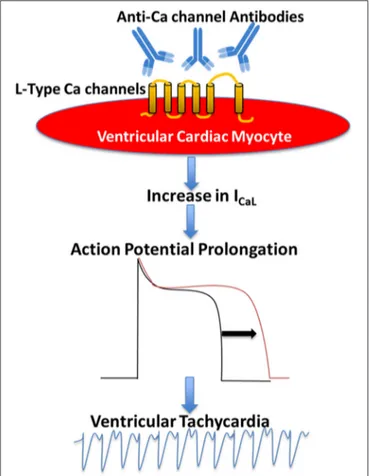
FIGURE 1 | Proposed mechanism of the pathogenic role of anti-Ca channels autoantibodies in Dilated Cardiomyopathy. Anti-Ca channels autoantibodies target L-type Ca channels in the ventricular myocyte resulting in an increase in L-type Ca current (ICaL) which in turn leads to action potential prolongation and ventricular arrhythmias.
various degrees of AVB, with complete AVB being the most
common, for which more than 60% of affected children require
lifelong pacemakers (
81
), and carries mortality rate up to 30%
(
81
,
82
). Because anti-Ro antibodies are the most prevalent
autoantibodies in CHB (
83
–
85
), anti-La antibodies are not
discussed in this review. There are 2 subtypes of anti-Ro
autoantibodies: anti-52 and anti-60 kD SSA/Ro (collectively
termed anti-Ro antibodies in this review). Anti-Ro antibodies
result from an autoimmune response to the SSA-Ro antigen,
which is an intracellular ribonucleoprotein that is not accessible
to the circulating anti-Ro antibodies in the normal cardiac
myocyte, likely because of their large size. Anti-Ro antibodies
are more prevalent in certain autoimmune diseases including
Sjögren’s syndrome, systemic lupus erythematosus, scleroderma,
rheumatoid arthritis, systemic sclerosis, and myositis (
86
,
87
).
Intriguingly, these anti-Ro antibodies are also present in the
general healthy population (
87
–
89
). The incidence of CHB is
about 1:11,000 (
81
,
90
); however, this incidence dramatically
increases to about 5% in anti-Ro positive mothers and up to
18% in subsequent pregnancies thereby affecting the decision
to have a second child (
79
,
81
). The causal relationship of
anti-Ro antibodies to the development of CHB was reproduced
in both active and passive mice models of CHB (
81
,
91
–
93
).
Various degree of AVB developed in pups born to female
mice immunized with recombinant 52 SSA/Ro protein (active
immunization) (
81
,
93
,
94
). Transfer of anti-Ro antibodies from
mothers with CHB children (anti-Ro antibody positive IgG)
directly into timely pregnant mice also resulted in first degree
AVB and, surprisingly, sinus bradycardia in about 70% of the
pups (passive immunization) (
91
). Similarly, clinical data (
95
,
96
)
also confirmed similar sinus bradycardia in newborns of mothers
with anti-Ro antibody positive IgG, indicating that the spectrum
of CHB extends beyond AVN to also affect SAN.
Anti-Ro Antibody Positive IgG Inhibits Both α
1Cand
α
1DCa Currents
As mentioned above, the hallmark of CHB is AVB. The
conduction of the impulse through the AVN depends critically
on α
1CCa current, I
Ca−L, which activates at more positive (−40
and −30 mV) potentials (
97
). It is logical to speculate that anti-Ro
antibody positive IgG might target α
1CCa channel to disturb the
electrical conduction at AVN as seen in CHB. Anti-Ro antibody
positive IgG and affinity purified anti-52 Ro antibodies from
mothers with CHB children, but not anti-Ro antibody negative
IgG from healthy mothers, inhibited I
Ca−Lin isolated SAN,
AVN cells, Purkinje fibers and in ventricular cells by 50–59%
(
77
,
78
,
98
–
100
). In addition, anti-Ro antibody positive IgG had
no effect on K currents (the transient outward current, I
toand
the inward rectifier, I
K1), or the Na current (I
Na), indicating its
specificity toward Ca channels (
98
). To exclude the possibility
of potential contamination from other ion currents, α
1CCa
channels expressed in Xenopus oocytes were similarly inhibited
about 50% by anti-Ro antibody positive IgG (
92
,
99
,
100
).
While inhibition of α
1CI
Ca−Lcould account for the AVB seen
in CHB, the contribution of α
1CI
Ca−Lto diastolic depolarization
of the SA node is generally considered to be minor. SAN
pacemaker depolarization occurs between −60 and −40 mV;
however α
1CI
Ca−Lactivates at more positive (−40 and −30 mV)
potentials (
101
). Knockout of the α
1DCa channel, which
activates at −60 and −40 mV in mice, results in significant sinus
bradycardia and AVB (
28
,
42
,
102
), a phenotype reminiscent
to that seen in CHB. Mangoni et al. (
44
) showed I
Ca−Lin
SAN cells was decreased by 75% in α
1DCa channel knockout
mice compared with wild-type mice, which indicates that the
contribution of the α
1DCa channel to total I
Ca−Lis significant
in the mouse SA node cell. Furthermore, our previous studies
demonstrated that both α
1DCa channel transcripts and proteins
are expressed in human fetal heart and in adult rabbit SAN
(
39
,
40
). Collectively, these data suggest that α
1D, along with
α
1C, contribute to form I
Ca−L, playing a critical role in pace
making activity in SAN and are a potential target by anti-Ro
antibodies. Because there are no biophysical methods or specific
blockers to separate α
1Dfrom α
1CI
CaLin native cells, the specific
effect of anti-Ro antibodies on α
1DI
Ca−Lhas been challenging.
Initial studies were carried out in expression systems to allow
individual expression of α
1DI
Ca−Lto characterize the effect of
anti-Ro antibody positive IgG
.Anti-Ro antibody positive IgG
from mothers with CHB children inhibited α
1DI
Ca−Lby about
43% in tsA201 cells and about 33% in Xenopus oocytes (
40
,
77
,
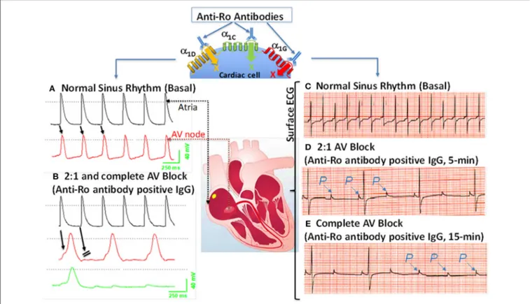
FIGURE 2 | Effects of anti-Ro antibodies from mothers of children with congenital heart block on an isolated multicellular AV nodal preparation (left) and Langendorff perfused whole heart (Right). (A) Simultaneous control action potentials from the crista terminalis (black tracing) and the AV node area (red tracing). (B) Superfusion of the preparation with positive IgG (800 µg/mL) for 10 min resulted in 2:1 AV block (indicated by the arrows) which progressed to near complete inhibition of the AV node action potential by 15 min (B), (green tracing). (C) ECG was recorded by the conventional ECG machine in lead I, except for the use of silver wires at the recording end of the leads. One lead was inserted in the atrium, the second in the left ventricle near the apex, and the third in Tyrode’s solution (ground). “P” indicates, the P wave and on the ECG. Regular sinus rhythm (horizontal scale, 50 mm/s and vertical scale: 5 mm/mV) at 300 beats/min in Tyrode’s solution. (D) After 5 min of perfusion with positive IgG (800 µg/mL), there was bradycardia associated with a 2:1 second degree AV block that degenerated into complete AV block by 15 min of IgG perfusion (E). The sectioned heart in the middle panel illustrates the location of the microelectrode recordings.
78
,
92
,
99
,
100
). To overcome this limitation of using expression
systems, our group has tested the effect of anti-Ro antibodies
on α
1DI
Ca−Lin native neonatal cardiomyocytes, in which the
α
1Cgene was effectively silenced by lentivirus. Adding anti-Ro
antibody positive IgG resulted in 35% reduction of α
1DI
Ca−Lin naïve cardiomyocytes (
103
), similar to the results seen using
expression systems.
Because anti-Ro antibodies inhibit both α
1Cand α
1DI
Ca−L, it is anticipated that anti-Ro antibodies will cause both
sinus bradycardia and AVB. Further experimental evidence
using isolated multicellular AVN preparations (Figures 2A,B)
and
Langendorff-perfused
whole
hearts
(Figures 2C–E)
demonstrated that anti-Ro antibody positive IgG resulted
in bradycardia associated with 2:1 AVB then complete
third degree AVB as recorded by surface EG. In contrast,
perfusion of the AVN preparation or whole heart with
control anti-Ro antibody negative IgG had no effect on
ECG parameters (
78
). The sinus bradycardia and AVB were
also demonstrated in Langendorff-perfused human hearts
by our group (
77
) and by others (
104
,
105
). Similar findings
were obtained using the optical mapping technique, which
allows simultaneous recording of voltage action potentials at
multiple areas of the heart including the AVN area. Perfusion
of hearts with anti-Ro antibody positive IgG revealed the
sites of conduction abnormalities at the sinoatrial junction
and AVN, thereby confirming the site of action for these
autoantibodies (
106
).
In summary, α
1Dand α
1CCa channels both contribute
to total I
Ca−Lin the heart, with α
1DCa channels playing a
more critical role in the SAN and α
1CCa channels in the
AVN. Anti-Ro antibodies inhibit I
Ca−Lemanating from both
α
1Dand α
1C, resulting in AVB and sinus bradycardia seen in
CHB. This causal relationship was confirmed by reproducing
active and passive mice CHB models by induction of
anti-Ro antibodies (active immunization) or passive transfer of
the anti-Ro positive maternal IgG into pregnant mice (passive
immunization). Altogether, anti-Ro autoantibodies’ inhibition
of Ca channels are causally related to the development
of CHB, but the low incidence of CHB children born to
anti-Ro antibodies positive mothers suggest that additional
factor(s) may be necessary to contribute to the full spectrum
of CHB.
FIGURE 3 | Schematic representation of alternative mechanism of linking anti-Ro antibodies to the development of atrioventricular block: fetal cardiomyocytes undergoing “physiological” apoptosis cause the surface translocation of the intracellular located Ro antigens. Circulating maternal anti-Ro antibodies which can cross the placenta, subsequently bind to the translocated Ro antigens at the cell surface; provoke the secretion of proinflammatory cytokines such as TGFβ from macrophages. Excessive TGFβ secretion activates fibroblasts leading to scars promoting myofibroblasts in the Atrioventricular node, resulting in atrioventricular block.
Anti-ro Antibody Positive IgG Inhibits Ca Currents by
Binding Directly to the Pore Forming Subunit of the
Ca Channels
As pointed out earlier, anti-Ro antibody positive IgG cannot cross
the sarcolemma of a normal fetal cardiac myocyte, and hence
one can suspect that its effects are not directly mediated through
its antigen, SSA/Ro, but rather via sarcolemma targets such as
Ca channels. Evidence for direct interaction between anti-Ro
antibodies and Ca channels is provided by the direct binding of
anti-Ro antibodies on the pore forming α
1subunit of VGCC,
resulting in inhibition of I
CaL. Indeed, using immunostaining and
Western blots, it was demonstrated that anti-Ro antibody positive
IgG binds directly to the Ca channels’ α
1subunit (
99
,
107
). In a
subsequent study, purified GST fusion proteins corresponding to
the extracellular loop S5–S6 of each of the four domains that form
the pore of the α
1Dsubunit were expressed and their reactivity
to anti-Ro antibody positive IgG was tested. Fourteen percent
of anti-Ro antibody positive IgG reacted specifically with the
extracellular loop S5-S6 of the first domains of the α
1Dsubunit,
as demonstrated by both ELISA and Western blots (
108
).
L-type Ca channels’ inhibition by anti-Ro antibodies is one of the
mechanisms for the electrocardiographic abnormalities seen in
CHB. The resulting formulation of the “Ca channel hypothesis”
was based on the above experimental findings and was driven
by the fact that AVN electrogenesis depends on the L-type Ca
channels. Inhibition of this channel will ultimately lead to AVB, as
seen in CHB. The “Ca channel hypothesis” states that circulating
maternal antibodies directly cross react with L-type Ca channel
pore forming protein α
1-subunit, inhibiting the currents and
leading to the development of AVB (
97
).
T-type Ca Channel and
Autoimmune-Associated Congenital Heart
Block
T-type α
1GVGCCs subtype participates with α
1Hin regulating
electrical conduction through the AVN (
18
,
27
,
31
,
34
). α
1GVGCC is highly expressed in the AVN in human hearts
(
27
,
31
,
32
). Homozygous α
1Gknockout mice exhibit
first-degree AVB and bradycardia, a phenotype seen in CHB (
25
).
These findings suggest α
1GVGCC as an additional potential
cross-reactive target with anti-Ro antibody positive IgG in
the development of CHB. Hu et al. demonstrated that
anti-Ro antibody positive IgG decreased both I
CaLand T-type
Ca current (I
Ca−T) without affecting the delayed rectifier K
current, I
K, and the funny current, I
f, in rabbit SAN cells (
98
).
The average inhibition of I
Ca−Tby anti-Ro antibody positive
IgG was 31.4% at −40 mV and 44.1% at −20 mV in rabbit
SAN cells (
98
). In addition, although anti-Ro antibody positive
IgG inhibited the α
1HI
CaTexpressed in the Xenopus oocyte
(
100
), α
1HCa channel knockout mice have no ECG changes
(
109
), likely secondary to the low level of α
1Hexpression in
the human neonatal AVN cells (
107
). These findings support
the conclusion that the α
1GCa channel is the target for
anti-Ro antibody positive IgG. Strindberg et al. demonstrated
α
1GmRNA and proteins in human fetal hearts and that
α
1GI
Ca−Trather than α
1HI
Ca−Tis the dominant current
in the AVN in newborns (
107
). Experimental data using
immunoprecipitation, Western blot and immunofluorescent
staining have demonstrated accessibility of anti-Ro antibody
positive IgG to the α
1Gepitope on the surfaces on the
cardiomyocytes in the human fetal heart (
107
). Reactivity to
α
1GT-type VGCC was significantly higher in CHB maternal
sera compared to controls. Binding epitope of anti-Ro antibody
positive IgG was mapped to the extracellular S5–S6 portion of
repeat I of α
1Gsubunit (aa305–319; designated as p305). Using
the patch-clamp technique, the authors also demonstrated that
anti-Ro antibody positive IgG inhibited I
Ca−Tin isolated mice
SAN cells (
107
). Taken together, these results indicate that
anti-Ro antibody positive IgG readily target an extracellular epitope
of α
1GT-type VGCC and inhibit the current in human fetal
cardiomyocytes, thus contributing to the development of AVB as
seen in CHB.
FIGURE 4 | Schematic illustration of the Ca channel hypothesis. Maternal anti-Ro antibodies cross react and bind to α1C(yellow), α1D(green), and α1G(red) Ca channels in the fetal human heart, inhibit all three Ca currents leading to sinus bradycardia and atrioventricular (AV) block (acute effect). Furthermore, fetal heart Ca channels are exposed chronically (chronic effect) (1) to maternal anti-Ro antibodies during pregnancy. Binding of anti-Ro antibodies to Ca channels (2), can cause cross-linking of the adjacent ion channels by the two Fab arms of IgG (3) to increase the internalization of the channel/antibody complex and thereby decrease of the channel density on the cell membrane. Internalized Ca channels are lysed by lysosomes (4). If the number of Ca channels on cell surface decreased to a critical level, then cell death will occur. Cell death, per se, could trigger inflammation subsequent to leukocytic influx resulting in damage of the surrounding healthy myocytes such as in sinoatrial node and AV node which can cause permanent sinus bradycardia and AV block.
Anti-52kD Ro antibodies are present in 80% of mothers of
children with CHB; however, the risk of having CHB children
is low, with only 1–2% in single anti-Ro antibody positive
pregnancies (
84
). Markham et al. investigated if reactivity with
p305 (anti-Ro/p305) can be used clinically to more accurately
predict CHB in anti-Ro antibody positive patients (
110
). Using
anti-Ro antibody positive IgG and with multiple control groups,
reactivity was determined and compared for binding to
Ro/p305. In mothers carrying Ro antibodies, positive
anti-Ro/p305 antibodies were detected in 3/59 (5%) CHB pregnancies,
4/30 (13%) unaffected pregnancies with a CHB-sibling, and
0/42 (0%) of unaffected pregnancies with no CHB-sibling.
Similarly, using umbilical blood from 61 CHB and 41 healthy
with CHB-sibling, in which reactivity would unambiguously
substantiate exposure to maternal antibody, no association of
anti-Ro/p305 with CHB was detected. These data indicate that
anti-Ro/p305 reactivity in pregnant anti-Ro antibody-positive
patients is not a robust maternal marker for assessing increased
risk of CHB (
110
).
As described above, it is well-recognized that maternal
anti-Ro antibody is associated with the development of the
congenial AVB, at least in part resulting from an inhibitory
cross-reaction with L- and T-type Ca channels. More recent,
studies demonstrated that 10–60% of anti-Ro-positive subjects
are at increased risk of developing QTc prolongation as a
result of anti-Ro antibodies’ interference with K channels, (
111
–
115
) resulting in complex ventricular arrhythmia, (
116
,
117
)
including Torsade’s de Pointes (TdP) (
118
,
119
). Lazzerini et al.
(
119
) recently evaluated 25 consecutive patients who experienced
TdP, where anti-Ro antibody was present in 15 out of 25
patients. Purified anti-Ro positive IgG from TdP patients
cross-reacted with the Human Ether-a-go-go-related Gene (hERG)
K channel and significantly inhibited the resulting current,
IKr. This observation indicates that anti-Ro antibodies may
represent a novel, clinically silent risk factors for TdP. To
date, studies on the association of anti-Ro antibodies and atrial
fibrillation are scarce. In our previous study (
120
), we were
able to induce atrial fibrillation in the α
1Dknockout mice but
not in the wild-type mice. One can speculate that the unique
atrial specific distribution of α
1DCa channel, together with the
documented inhibitory effect of the anti-Ro antibodies on the
α
1DCa channels, may suggest that anti-Ro positive patients
might be at increased risk of having atrial fibrillation, warranting
further investigations.
CONCLUSIONS AND FUTURE
DIRECTIONS
Cardiac Ca channels, including both L- and T-type Ca
channels, play critical roles in the impulse generation in the
SAN, the conduction through the AVN and the development
of arrhythmias. Autoantibodies targeting Ca channels have
been identified in 2 major pathologies, DCM and CHB. In
addition, several autoantibodies are directly related to sudden
death in patients with DCM, including N/K-ATPase,
anti-M2 muscarinic acetylcholine receptors, and anti-β1 receptor
antibodies, indirectly affecting the L-type VGCCs. Early risk
stratification to effectively prevent adverse outcomes in DCM
has been challenging. Recent studies confirmed the presence
of autoantibodies directly against Ca channel α
1Csubunit in
DCM, which was identified as a strong predictor for ventricular
arrhythmias and sudden cardiac death, indicating that anti-α
1CCa channel antibodies might be a valuable biomarker to predict
sudden death in DCM.
The association of anti-Ro autoantibodies with CHB
is generally accepted, but the predictive value of these
autoantibodies is still low despite overwhelming experimental
data demonstrating causality between anti-Ro antibodies and
electrocardiographic abnormalities seen in CHB (Figure 2).
This indicates that anti-Ro antibodies are necessary, but
not sufficient, for inducing the clinical electrocardiographic
phenotype. To date, two hypotheses have been proposed to
explain the molecular mechanism(s) by which maternal anti-Ro
antibodies lead to the development of CHB in the fetal hearts
(
79
,
121
). The “apoptosis hypothesis” (Figure 3) suggests that
intracellular antigens translocate to the surface of cardiomyocytes
undergoing apoptosis during physiological remodeling, thereby
exposing the antigens to the circulating maternal anti-Ro
antibodies. Binding of anti-Ro antibodies to the cell surface
antigens promotes pro-inflammatory and pro-fibrotic responses
(
122
,
123
), causing the fibrosis of the AVN, which eventually
leads to the development of the irreversible AVB (
124
,
125
).
The “Ca channel hypothesis” explained in this review is based
on molecular mimicry, whereby anti-Ro antibodies directly
cross-react and subsequently inhibit the cardiac Ca channels’
activity, thereby causing sinus bradycardia and AVB (
77
,
78
,
108
)
(Figure 4). This occurs by anti-Ro autoantibodies binding to
Ca channels and the resulting inhibition of I
CaL(Acute effect,
Figure 4
). The subsequent cross-linkage and downregulation
of Ca channels and lysis by lysosomes followed by intracellular
Ca dysregulation leads to cell death/apoptosis, inflammation,
and fibrosis of the AVN (Figure 4). The ultimate proof of
direct autoantibodies’ involvement in CHB is provided by the
identification of the site of action on the different subunits of
cardiac Ca channels (
126
–
128
), including α
1Cand α
1Dsubunits
of L-type VGCCs and α
1Gsubunit of T-type VGCCs (Figure 4).
Although autoantibodies are utilized as diagnostic or prognostic
markers in other pathologies, unfortunately, to date, there is
no specific maternal marker for assessing the increased risk of
having CHB children during an anti-Ro positive pregnancy.
It is possible that, instead of having a single CHB-inducing
antibody specificity, future studies may focus on several different
specificities that may act synergistically to induce AVB in
fetal hearts.
Peptide-based therapeutic approaches are one of the growing
classes of novel therapeutic agents. The development of short
non-immunogenic peptides and their use as decoy targets
for pathogenic autoantibodies is expected to minimize and/or
prevent autoantibody association with ion channels and their
functions. This therapeutic path awaits further development
and progress.
AUTHOR CONTRIBUTIONS
All authors listed have made a substantial, direct and intellectual
contribution to the work, and approved it for publication.
REFERENCES
1. Rougier JS, Abriel H. Cardiac voltage-gated calcium channel macromolecular complexes. Biochim Biophys Acta. (2016) 1863:1806–12.doi: 10.1016/j.bbamcr.2015.12.014
2. Betzenhauser MJ, Pitt GS, Antzelevitch C. Calcium channel mutations in cardiac arrhythmia syndromes. Curr Mol Pharmacol. (2015) 8:133–42. doi: 10.2174/1874467208666150518114857
3. Dolphin AC. Calcium channel diversity: multiple roles of calcium channel subunits. Curr Opin Neurobiol. (2009) 19:237–44. doi: 10.1016/j.conb.2009.06.006
4. Catterall WA. Signaling complexes of voltage-gated sodium and calcium channels. Neurosci Lett. (2010) 486:107–16. doi: 10.1016/j.neulet.2010.08.085 5. Mesirca P, Torrente AG, Mangoni ME. Functional role of voltage gated Ca(2+) channels in heart automaticity. Front Physiol. (2015) 6:19. doi: 10.3389/fphys.2015.00019
6. Zamponi GW, Striessnig J, Koschak A, Dolphin AC. The physiology, pathology, and pharmacology of voltage-gated calcium channels and
their future therapeutic potential. Pharmacol Rev. (2015) 67:821–70. doi: 10.1124/pr.114.009654
7. Zhang Q, Chen J, Qin Y, Wang J, Zhou L. Mutations in voltage-gated L-type calcium channel: implications in cardiac arrhythmia. Channels. (2018) 12:201–18. doi: 10.1080/19336950.2018.1499368
8. Venetucci L, Denegri M, Napolitano C, Priori SG. Inherited calcium channelopathies in the pathophysiology of arrhythmias. Nat Rev Cardiol. (2012) 9:561–75. doi: 10.1038/nrcardio.2012.93
9. Lee HC, Huang KT, Wang XL, Shen WK. Autoantibodies and cardiac arrhythmias. Heart Rhythm. (2011) 8:1788–95. doi: 10.1016/j.hrthm.2011.06.032
10. Lazzerini PE, Capecchi PL, Laghi-Pasini F, Boutjdir M. Autoimmune cardiac channelopathies: the heart of the matter. Nat Rev Cardiol. (2017) 14:566. doi: 10.1038/nrcardio.2017.111
11. Xiao H, Wang M, Du Y, Yuan J, Cheng X, Chen Z, et al. Arrhythmogenic autoantibodies against calcium channel lead to sudden death in idiopathic dilated cardiomyopathy. Eur J Heart Fail. (2011) 13:264–70. doi: 10.1093/eurjhf/hfq198
12. Durante A, Bronzato S. The increased cardiovascular risk in patients affected by autoimmune diseases: review of the various manifestations. J Clin Med Res. (2015) 7:379–84. doi: 10.14740/jocmr2122w
13. Nilius B, Hess P, Lansman JB, Tsien RW. A novel type of cardiac calcium channel in ventricular cells. Nature. (1985) 316:443–6. doi: 10.1038/316443a0 14. Bean BP. Two kinds of calcium channels in canine atrial cells. differences in kinetics, selectivity, and pharmacology. J Gen Physiol. (1985) 86:1–30. doi: 10.1085/jgp.86.1.1
15. Catterall WA. Voltage-gated calcium channels. Cold Spring Harb Perspect Biol. (2011) 3:a003947. doi: 10.1101/cshperspect.a003947
16. Dolphin AC. Voltage-gated calcium channels and their auxiliary subunits: physiology and pathophysiology and pharmacology. J Physiol. (2016) 594:5369–90. doi: 10.1113/JP272262
17. Perez-Reyes E. Molecular physiology of low-voltage-activated t-type calcium channels. Physiol Rev. (2003) 83:117–61. doi: 10.1152/physrev.00018.2002 18. Cribbs L. T-type calcium channel expression and function in the diseased
heart. Channels. (2010) 4:447–52. doi: 10.4161/chan.4.6.12870
19. Nowycky MC, Fox AP, Tsien RW. Three types of neuronal calcium channel with different calcium agonist sensitivity. Nature. (1985) 316:440–3. doi: 10.1038/316440a0
20. Mintz IM, Adams ME, Bean BP. P-type calcium channels in rat central and peripheral neurons. Neuron. (1992) 9:85–95. doi: 10.1016/0896-6273(92)90223-Z
21. Namkung Y, Smith SM, Lee SB, Skrypnyk NV, Kim HL, Chin H, et al. Targeted disruption of the Ca2+ channel beta3 subunit reduces N- and L-type Ca2+ channel activity and alters the voltage-dependent activation of P/Q-type Ca2+ channels in neurons. Proc. Natl. Acad. Sci. U.S.A. (1998) 95:12010–5. doi: 10.1073/pnas.95.20.12010
22. Lambert RC, McKenna F, Maulet Y, Talley EM, Bayliss DA, Cribbs LL, et al. Low-voltage-activated Ca2+ currents are generated by members of the CavT subunit family (alpha1G/H) in rat primary sensory neurons. J Neurosci. (1998) 18:8605–13. doi: 10.1523/ JNEUROSCI.18-21-08605.1998
23. Perez-Reyes E, Cribbs LL, Daud A, Lacerda AE, Barclay J, Williamson MP, et al. Molecular characterization of a neuronal low-voltage-activated T-type calcium channel. Nature. (1998) 391:896–900. doi: 10.1038/36110 24. Cribbs LL, Lee JH, Yang J, Satin J, Zhang Y, Daud A, et al. Cloning
and characterization of alpha1H from human heart, a member of the T-type Ca2+ channel gene family. Circ Res. (1998) 83:103–9. doi: 10.1161/01.RES.83.1.103
25. Mangoni ME, Traboulsie A, Leoni AL, Couette B, Marger L, Le Quang K, et al. Bradycardia and slowing of the atrioventricular conduction in mice lacking CaV3.1/alpha1G T-type calcium channels. Circ Res. (2006) 98:1422–30. doi: 10.1161/01.RES.0000225862.14314.49
26. Mizuta E, Shirai M, Arakawa K, Hidaka K, Miake J, Ninomiya H, et al. Different distribution of Cav3.2 and Cav3.1 transcripts encoding T-type Ca(2+) channels in the embryonic heart of mice. Biomed Res. (2010) 31:301– 5. doi: 10.2220/biomedres.31.301
27. Monteil A, Chemin J, Bourinet E, Mennessier G, Lory P, Nargeot J. Molecular and functional properties of the human alpha(1G) subunit that forms T-type calcium channels. J Biol Chem. (2000) 275:6090–100. doi: 10.1074/jbc.275.9.6090
28. Koschak A, Reimer D, Huber I, Grabner M, Glossmann H, Engel J, et al. alpha 1D (Cav1.3) subunits can form l-type Ca2+ channels activating at negative voltages. J Biol Chem. (2001) 276:22100–6. doi: 10.1074/jbc.M101469200 29. Baig SM, Koschak A, Lieb A, Gebhart M, Dafinger C, Nurnberg G, et
al. Loss of Ca(v)1.3 (CACNA1D) function in a human channelopathy with bradycardia and congenital deafness. Nat Neurosci. (2011) 14:77–84. doi: 10.1038/nn.2694
30. Ono K, Iijima T. Pathophysiological significance of T-type Ca2+ channels: properties and functional roles of T-type Ca2+ channels in cardiac pacemaking. J Pharmacol Sci. (2005) 99:197–204. doi: 10.1254/jphs.FMJ05002X2
31. Vassort G, Talavera K, Alvarez JL. Role of T-type Ca2+ channels in the heart. Cell Calcium. (2006) 40:205–20. doi: 10.1016/j.ceca.2006.04.025
32. Ferron L, Capuano V, Deroubaix E, Coulombe A, Renaud JF. Functional and molecular characterization of a T-type Ca(2+) channel during fetal
and postnatal rat heart development. J Mol Cell Cardiol. (2002) 34:533–46. doi: 10.1006/jmcc.2002.1535
33. Bohn G, Moosmang S, Conrad H, Ludwig A, Hofmann F, Klugbauer N. Expression of T- and L-type calcium channel mRNA in murine sinoatrial node. FEBS Lett. (2000) 481:73–6. doi: 10.1016/S0014-5793(00)01979-7 34. Hagiwara N, Irisawa H, Kameyama M. Contribution of two types of calcium
currents to the pacemaker potentials of rabbit sino-atrial node cells. J Physiol. (1988) 395:233–53. doi: 10.1113/jphysiol.1988.sp016916
35. Benitah JP, Gomez AM, Fauconnier J, Kerfant BG, Perrier E, Vassort G, et al. Voltage-gated Ca2+ currents in the human pathophysiologic heart: a review. Basic Res Cardiol. (2002) 97 (Suppl. 1):I11–8. doi: 10.1007/s0039502 00023
36. Striessnig J, Pinggera A, Kaur G, Bock G, Tuluc P. L-type Ca(2+) channels in heart and brain. Wiley Interdiscip Rev Membr Transp Signal. (2014) 3:15–38. doi: 10.1002/wmts.102
37. Pinggera A, Lieb A, Benedetti B, Lampert M, Monteleone S, Liedl KR, et al. CACNA1D de novo mutations in autism spectrum disorders activate Cav1.3 L-type calcium channels. Biol Psychiatry. (2015) 77:816–22. doi: 10.1016/j.biopsych.2014.11.020
38. Srivastava U, Aromolaran AS, Fabris F, Lazaro D, Kassotis J, Qu Y, et al. Novel function of alpha1D L-type calcium channel in the atria. Biochem Biophys Res Commun. (2017) 482:771–6. doi: 10.1016/j.bbrc.2016.11.109
39. Qu Y, Karnabi E, Ramadan O, Yue Y, Chahine M, Boutjdir M. Perinatal and postnatal expression of Cav1.3 alpha1D Ca(2)(+) channel in the rat heart. Pediatr Res. (2011) 69:479–84. doi: 10.1203/PDR.0b013e318217a0df 40. Qu Y, Baroudi G, Yue Y, Boutjdir M. Novel molecular mechanism
involving alpha1D (Cav1.3) L-type calcium channel in autoimmune-associated sinus bradycardia. Circulation. (2005) 111:3034–41. doi: 10.1161/CIRCULATIONAHA.104.517326
41. Marionneau C, Couette B, Liu J, Li H, Mangoni ME, Nargeot J, et al. Specific pattern of ionic channel gene expression associated with pacemaker activity in the mouse heart. J Physiol. (2005) 562:223–34. doi: 10.1113/jphysiol.2004.074047
42. Platzer J, Engel J, Schrott-Fischer A, Stephan K, Bova S, Chen H, et al. Congenital deafness and sinoatrial node dysfunction in mice lacking class D L-type Ca2+ channels. Cell. (2000) 102:89–97. doi: 10.1016/S0092-8674(00)00013-1
43. Striessnig J, Koschak A. Exploring the function and pharmacotherapeutic potential of voltage-gated Ca2+ channels with gene knockout models. Channels. (2008) 2:233–51. doi: 10.4161/chan.2.4.5847
44. Mangoni ME, Couette B, Bourinet E, Platzer J, Reimer D, Striessnig J, et al. Functional role of L-type Cav1.3 Ca2+ channels in cardiac pacemaker activity. Proc. Natl. Acad. Sci. U.S.A. (2003) 100:5543–8. doi: 10.1073/pnas.0935295100
45. Lee JH, Daud AN, Cribbs LL, Lacerda AE, Pereverzev A, Klockner U, et al. Cloning and expression of a novel member of the low voltage-activated T-type calcium channel family. J Neurosci. (1999) 19:1912–21. doi: 10.1523/JNEUROSCI.19-06-01912.1999
46. Catterall WA, Perez-Reyes E, Snutch TP, Striessnig J. International Union of Pharmacology. XLVIII. Nomenclature and structure-function relationships of voltage-gated calcium channels. Pharmacol Rev. (2005) 57:411–25. doi: 10.1124/pr.57.4.5
47. Dolphin AC, Wyatt CN, Richards J, Beattie RE, Craig P, Lee JH, et al. The effect of alpha2-delta and other accessory subunits on expression and properties of the calcium channel alpha1G. J Physiol. (1999) 519:35–45. doi: 10.1111/j.1469-7793.1999.0035o.x
48. Dubel SJ, Altier C, Chaumont S, Lory P, Bourinet E, Nargeot J. Plasma membrane expression of T-type calcium channel alpha(1) subunits is modulated by high voltage-activated auxiliary subunits. J Biol Chem. (2004) 279:29263–9. doi: 10.1074/jbc.M313450200
49. Chiang CS, Huang CH, Chieng H, Chang YT, Chang D, Chen JJ, et al. The Ca(v)3.2 T-type Ca(2+) channel is required for pressure overload-induced cardiac hypertrophy in mice. Circ Res. (2009) 104:522–30. doi: 10.1161/CIRCRESAHA.108.184051
50. Liu DQ, Zhou SS, Zhao DH, Sheng BH. Effects of furyl-dihydropyridine on action potential ventricular myocardium of rabbit in vivo and isolated guinea pig left atrium in vitro. Zhongguo Yao Li Xue Bao. (1993) 14:164–7.
51. Hirano Y, Fozzard HA, January CT. Characteristics of L- and T-type Ca2+ currents in canine cardiac Purkinje cells. Am J Physiol. (1989) 256:H1478–92. doi: 10.1152/ajpheart.1989.256.5.H1478
52. Tseng GN, Boyden PA. Multiple types of Ca2+ currents in single canine Purkinje cells. Circ Res. (1989) 65:1735–50. doi: 10.1161/01.RES.65.6.1735 53. Nilius B. Possible functional significance of a novel type of cardiac Ca
channel. Biomed Biochim Acta. (1986) 45:K37–45.
54. Nilius B, Talavera K, Verkhratsky A. T-type calcium channels: the never ending story. Cell Calcium. (2006) 40:81–8. doi: 10.1016/j.ceca.2006.04.011 55. Liao YH, Fu M. Autoimmunity in the pathogenesis of cardiomyopathy. J
Autoimmun. (2001) 16:1–2. doi: 10.1006/jaut.2000.0466
56. MacLellan WR, Lusis AJ. Dilated cardiomyopathy: learning to live with yourself. Nat Med. (2003) 9:1455–6. doi: 10.1038/nm1203-1455
57. Jahns R, Boivin V, Schwarzbach V, Ertl G, Lohse MJ. Pathological autoantibodies in cardiomyopathy. Autoimmunity. (2008) 41:454–61. doi: 10.1080/08916930802031603
58. Felker GM, Thompson RE, Hare JM, Hruban RH, Clemetson DE, Howard DL, et al. Underlying causes and long-term survival in patients with initially unexplained cardiomyopathy. N Engl J Med. (2000) 342:1077–84. doi: 10.1056/NEJM200004133421502
59. Braunwald E. Expanding indications for beta-blockers in heart failure. N Engl J Med. (2001) 344:1711–2. doi: 10.1056/NEJM200105313442210
60. Klein GJ, Krahn AD, Skanes AC, Yee R, Gula LJ. Primary prophylaxis of sudden death in hypertrophic cardiomyopathy, arrhythmogenic right ventricular cardiomyopathy, and dilated cardiomyopathy. J Cardiovas Electrophys. (2005) 16 (Suppl. 1):S28–34. doi: 10.1111/j.1540-8167.2005.50116.x
61. Xiao H, Wang M, Du Y, Yuan J, Zhao G, Tu D, et al. Agonist-like autoantibodies against calcium channel in patients with dilated cardiomyopathy. Heart Vessels. (2012) 27:486–92. doi: 10.1007/s00380-011-0176-7
62. Yu H, Pei J, Liu X, Chen J, Li X, Zhang Y, et al. Calcium channel autoantibodies predicted sudden cardiac death and all-cause mortality in patients with ischemic and nonischemic chronic heart failure. Dis Markers. (2014) 2014:796075. doi: 10.1155/2014/796075
63. Deubner N, Berliner D, Schlipp A, Gelbrich G, Caforio AL, Felix SB, et al. Cardiac beta1-adrenoceptor autoantibodies in human heart disease: rationale and design of the Etiology, Titre-Course, and Survival (ETiCS) Study. Eur J Heart Fail. (2010) 12:753–62. doi: 10.1093/eurjhf/hfq072 64. Jahns R. Autoantibodies against cardiac troponin I: friend or foe? Eur J Heart
Fail. (2010) 12:645–8. doi: 10.1093/eurjhf/hfq098
65. Heymans S, Hirsch E, Anker SD, Aukrust P, Balligand JL, Cohen-Tervaert JW, et al. Inflammation as a therapeutic target in heart failure? a scientific statement from the translational research committee of the heart failure association of the European Society of Cardiology. Eur J Heart Fail. (2009) 11:119–29. doi: 10.1093/eurjhf/hfn043
66. Sterin-Borda L, Cossio PM, Gimeno MF, Gimeno AL, Diez C, Laguens RP, et al. Effect of chagasic sera on the rat isolated atrial preparation: immunological, morphological and function aspects. Cardiovasc Res. (1976) 10:613–22. doi: 10.1093/cvr/10.6.613
67. Wallukat G, Wollenberger A. Effects of the serum gamma globulin fraction of patients with allergic asthma and dilated cardiomyopathy on chronotropic beta adrenoceptor function in cultured neonatal rat heart myocytes. Biomed Biochim Acta. (1987) 46:S634–9.
68. Magnusson Y, Marullo S, Hoyer S, Waagstein F, Andersson B, Vahlne A, et al. Mapping of a functional autoimmune epitope on the beta 1-adrenergic receptor in patients with idiopathic dilated cardiomyopathy. J Clin Invest. (1990) 86:1658–63. doi: 10.1172/JCI114888
69. Iwata M, Yoshikawa T, Baba A, Anzai T, Mitamura H, Ogawa S. Autoantibodies against the second extracellular loop of beta1-adrenergic receptors predict ventricular tachycardia and sudden death in patients with idiopathic dilated cardiomyopathy. J Am Coll Cardiol. (2001) 37:418–24. doi: 10.1016/S0735-1097(00) 01109-8
70. Magnusson Y, Wallukat G, Waagstein F, Hjalmarson A, Hoebeke J. Autoimmunity in idiopathic dilated cardiomyopathy. characterization of antibodies against the beta 1-adrenoceptor with positive chronotropic effect. Circulation. (1994) 89:2760–7. doi: 10.1161/01.CIR.89.6.2760
71. Magnusson Y, Hjalmarson A, Hoebeke J. Beta 1-adrenoceptor autoimmunity in cardiomyopathy. Int J Cardiol. (1996) 54:137–41. doi: 10.1016/0167-5273(96)02590-9
72. Christ T, Wettwer E, Dobrev D, Adolph E, Knaut M, Wallukat Get al. Autoantibodies against the beta1 adrenoceptor from patients with dilated cardiomyopathy prolong action potential duration and enhance contractility in isolated cardiomyocytes. J Mol Cell Cardiol. (2001) 33:1515– 25. doi: 10.1006/jmcc.2001.1414
73. Schulze K, Heineman FW, Schultheiss HP, Balaban RS. Impairment of myocardial calcium homeostasis by antibodies against the adenine nucleotide translocator. Cell Calcium. (1999) 25:361–70. doi: 10.1054/ceca.1999.0039
74. Liao YH. Functional analysis of autoantibodies against ADP/ATP carrier from dilated cardiomyopathy. Int J Cardiol. (1996) 54:165–9. doi: 10.1016/0167-5273(96)02594-6
75. Liao YH, Cheng LX, Dai SP, Tu YS. Autoantibodies against ADP/ATP carrier from patients with dilated cardiomyopathy increase activity of voltage-dependent Ca channels in isolated cardiac myocytes. Blood Press Supple. (1996) 3:41–4.
76. Liao YH, Yuan J. Progress in the research of targets for the molecular immunotherapy in dilated cardiomyopathy. Zhonghua yi xue za zhi. (2006) 86:1158–60. doi: 10.3760/j:issn:0376-2491.2006.17.003
77. Boutjdir M, Chen L, Zhang ZH, Tseng CE, DiDonato F, Rashbaum W, et al. Arrhythmogenicity of IgG and anti-52-kD SSA/Ro affinity-purified antibodies from mothers of children with congenital heart block. Circ Res. (1997) 80:354–62. doi: 10.1161/ 01.RES.80.3.354
78. Boutjdir M, Chen L, Zhang ZH, Tseng CE, El-Sherif N, Buyon JP. Serum and immunoglobulin G from the mother of a child with congenital heart block induce conduction abnormalities and inhibit L-type calcium channels in a rat heart model. Pediatr Res. (1998) 44:11–9. doi: 10.1203/00006450-19980700 0-00002
79. Boutjdir M. Molecular and ionic basis of congenital complete heart block. Trends Cardiovasc Med. (2000) 10:114–22. doi: 10.1016/S1050-1738(00)00059-1
80. Lazzerini PE, Capecchi PL, Guideri F, Acampa M, Selvi E, Bisogno S, et al. Autoantibody-mediated cardiac arrhythmias: mechanisms and clinical implications. Basic Res Cardiol. (2008) 103:1–11. doi: 10.1007/s00395-007-0686-8
81. Miranda-Carus ME, Boutjdir M, Tseng CE, DiDonato F, Chan EK, Buyon JP. Induction of antibodies reactive with SSA/Ro-SSB/La and development of congenital heart block in a murine model. J Immunol. (1998) 161:5 886–92.
82. Brucato A, Franceschini F, Buyon JP. Neonatal lupus: long-term outcomes of mothers and children and recurrence rate. Clin Exp Rheumatol. (1997) 15:467–73.
83. Buyon JP, Winchester RJ, Slade SG, Arnett F, Copel J, Friedman D, et al. Identification of mothers at risk for congenital heart block and other neonatal lupus syndromes in their children. comparison of enzyme-linked immunosorbent assay and immunoblot for measurement of anti-SS-A/Ro and anti-SS-B/La antibodies. Arthritis Rheum. (1993) 36:1263–73. doi: 10.1002/art.1780360911
84. Brucato A, Frassi M, Franceschini F, Cimaz R, Faden D, Pisoni MP, et al. Risk of congenital complete heart block in newborns of mothers with anti-Ro/SSA antibodies detected by counterimmunoelectrophoresis: a prospective study of 100 women. Arthritis Rheum. (2001) 44:1832–5. doi: 10.1002/1529-0131(200108)44:8<1832::AID-ART320>3.0.CO;2-C 85. Gordon P, Khamashta MA, Rosenthal E, Simpson JM, Sharland G, Brucato
A, et al. Anti-52 kDa Ro, anti-60 kDa Ro, and anti-La antibody profiles in neonatal lupus. J Rheumatol. (2004) 31:2480-7.
86. Franceschini F, Cavazzana I. Anti-Ro/SSA and La/SSB antibodies. Autoimmunity. (2005) 38:55–63. doi: 10.1080/08916930400022954 87. Satoh M, Chan EK, Ho LA, Rose KM, Parks CG, Cohn RD, et al.
Prevalence and sociodemographic correlates of antinuclear antibodies in the United States. Arthritis Rheum. (2012) 64:2319–27. doi: 10.1002/art.34380 88. Hayashi N, Koshiba M, Nishimura K, Sugiyama D, Nakamura T, Morinobu
S, et al. Prevalence of disease-specific antinuclear antibodies in general population: estimates from annual physical examinations of residents of
a small town over a 5-year period. Mod Rheumatol. (2008) 18:153–60. doi: 10.1007/s10165-008-0028-1
89. Guo YP, Wang CG, Liu X, Huang YQ, Guo DL, Jing XZ, et al. The prevalence of antinuclear antibodies in the general population of china: a cross-sectional study. Curr Ther Res Clin Exp. (2014) 76:116–9. doi: 10.1016/j.curtheres.2014.06.004
90. Siren MK, Julkunen H, Kaaja R. The increasing incidence of isolated congenital heart block in Finland. J Rheumatol. (1998) 25:1862–4. 91. Mazel JA, El-Sherif N, Buyon J, Boutjdir M. Electrocardiographic
abnormalities in a murine model injected with IgG from mothers of children with congenital heart block. Circulation. (1999) 99:1914–8. doi: 10.1161/01.CIR.99.14.1914
92. Xiao GQ, Qu Y, Hu K, Boutjdir M. Down-regulation of L-type calcium channel in pups born to 52 kDa SSA/Ro immunized rabbits. FASEB J. (2001) 15:1539–45. doi: 10.1096/fj.01-0052com
93. Salomonsson S, Sonesson SE, Ottosson L, Muhallab S, Olsson T, Sunnerhagen M, et al. Ro/SSA autoantibodies directly bind cardiomyocytes, disturb calcium homeostasis, and mediate congenital heart block. J Exp Med. (2005) 201:11–7. doi: 10.1084/jem.20041859
94. Suzuki H, Silverman ED, Wu X, Borges C, Zhao S, Isacovics B, et al. Effect of maternal autoantibodies on fetal cardiac conduction: an experimental murine model. Pediatr Res. (2005) 57:557–62. doi: 10.1203/01.PDR.0000155947.82365.E4
95. Menon A, Silverman ED, Gow RM, Hamilton RM. Chronotropic competence of the sinus node in congenital complete heart block. Am J Cardiol. (1998) 82:1119–21. doi: 10.1016/S0002-9149(98)00569-4 96. Brucato A, Cimaz R, Catelli L, Meroni P. Anti-Ro-associated
sinus bradycardia in newborns. Circulation. (2000) 102:E88–9. doi: 10.1161/01.CIR.102.11.e88
97. Mendez C, Zipes DP. Possible ionic mechanism of the action potentials of the atrioventricular node of rabbits. Bol Estud Med Biol. (1974) (Suppl 1):67–80. 98. Hu K, Qu Y, Yue Y, Boutjdir M. Functional basis of sinus bradycardia in congenital heart block. Circ Res. (2004) 94:e32–8. doi: 10.1161/01.RES.0000121566.01778.06
99. Qu Y, Xiao GQ, Chen L, Boutjdir M. Autoantibodies from mothers of children with congenital heart block downregulate cardiac L-type Ca channels. J Mol Cell Cardiol. (2001) 33:1153–63. doi: 10.1006/jmcc .2001.1379
100. Xiao GQ, Hu K, Boutjdir M. Direct inhibition of expressed cardiac l- and t-type calcium channels by IGG from mothers whose children have congenital heart block. Circulation. (2001) 103:1599–604. doi: 10.1161/01.CIR.103.11.1599
101. Takimoto K, Li D, Nerbonne JM, Levitan ES. Distribution, splicing and glucocorticoid-induced expression of cardiac alpha 1C and alpha 1D voltage-gated Ca2+ channel mRNAs. J Mol Cell Cardiol. (1997) 29:3035–42. doi: 10.1006/jmcc.1997.0532
102. Matthes J, Yildirim L, Wietzorrek G, Reimer D, Striessnig J, Herzig S. Disturbed atrio-ventricular conduction and normal contractile function in isolated hearts from Cav1.3-knockout mice. Naunyn Schmiedebergs Arch Pharmacol. (2004) 369:554–62. doi: 10.1007/s00210-004-0940-7
103. Karnabi E, Qu Y, Mancarella S, Yue Y, Wadgaonkar R, Boutjdir M. Silencing of Cav1.2 gene in neonatal cardiomyocytes by lentiviral delivered shRNA. Biochem Biophys Res Commun. (2009) 384:409–14. doi: 10.1016/j.bbrc.2009.04.150
104. Hamilton RM, Lee-Poy M, Kruger K, Silverman ED. Investigative methods of congenital complete heart block. J Electrocardiol. (1998) 30:69–74. doi: 10.1016/S0022-0736(98)80035-6
105. Garcia S, Nascimento JH, Bonfa E, Levy R, Oliveira SF, Tavares AV, et al. Cellular mechanism of the conduction abnormalities induced by serum from anti-Ro/SSA-positive patients in rabbit hearts. J Clin Invest. (1994) 93:718–24. doi: 10.1172/JCI117025
106. Restivo M, Kozhevnikov DO, Boutjdir M. Optical mapping of activation patterns in an animal model of congenital heart block. Am J Physiol Heart Circ Physiol. (2001) 280:H1889–95. doi: 10.1152/ajpheart.2001.280.4.H1889 107. Strandberg LS, Cui X, Rath A, Liu J, Silverman ED, Liu X, et al. Congenital
heart block maternal sera autoantibodies target an extracellular epitope on the alpha1G T-type calcium channel in human fetal hearts. PLoS ONE. (2013) 8:e72668. doi: 10.1371/journal.pone.0072668
108. Karnabi E, Qu Y, Wadgaonkar R, Mancarella S, Yue Y, Chahine M, et al. Congenital heart block: identification of autoantibody binding site on
the extracellular loop (domain I, S5-S6) of alpha(1D) L-type Ca channel. J Autoimmun. (2010) 34:80–6. doi: 10.1016/j.jaut.2009.06.005
109. Chen CC, Lamping KG, Nuno DW, Barresi R, Prouty SJ, Lavoie JL, et al. Abnormal coronary function in mice deficient in alpha1H T-type Ca2+ channels. Science. (2003) 302:1416–8. doi: 10.1126/science.1089268 110. Markham AJ, Rasmussen SE, Salmon JE, Martinez-Ortiz W, Cardozo TJ,
Clancy RM, et al. Reactivity to the p305 epitope of the alpha1G T-type calcium channel and autoimmune-associated congenital heart block. J Am Heart Assoc. (2015) 4:e001836. doi: 10.1161/JAHA.115.001836
111. Cimaz R, Stramba-Badiale M, Brucato A, Catelli L, Panzeri P, Meroni PL. QT interval prolongation in asymptomatic anti-SSA/Ro-positive infants without congenital heart block. Arthritis Rheum. (2000) 43:1049–53. doi: 10.1002/1529-0131(200005)43:5<1049::AID-ANR13>3.0.CO;2-X 112. Cimaz R, Meroni PL, Brucato A, Fesstova V, Panzeri P, Goulene K, et al.
Concomitant disappearance of electrocardiographic abnormalities and of acquired maternal autoantibodies during the first year of life in infants who had QT interval prolongation and anti-SSA/Ro positivity without congenital heart block at birth. Arthritis Rheum. (2003) 48:266–8. doi: 10.1002/art. 10700
113. Bourre-Tessier J, Clarke AE, Huynh T, Bernatsky S, Joseph L, Belisle P, et al. Prolonged corrected QT interval in anti-Ro/SSA-positive adults with systemic lupus erythematosus. Arthritis Care Res. (2011) 63:1031–7. doi: 10.1002/acr.20470
114. Lazzerini PE, Capecchi PL, Acampa M, Morozzi G, Bellisai F, Bacarelli MR, et al. Anti-Ro/SSA-associated corrected QT interval prolongation in adults: the role of antibody level and specificity. Arthritis Care Res. (2011) 63:1463–70. doi: 10.1002/acr.20540
115. Tufan AN, Sag S, Oksuz MF, Ermurat S, Coskun BN, Gullulu M, et al. Prolonged Tpeak-Tend interval in anti-Ro52 antibody-positive connective tissue diseases. Rheumatol Int. (2017) 37:67–73. doi: 10.1007/s00296-016-3488-1
116. Lazzerini PE, Capecchi PL, Guideri F, Bellisai F, Selvi E, Acampa M, et al. Comparison of frequency of complex ventricular arrhythmias in patients with positive versus negative anti-Ro/SSA and connective tissue disease. Am J Cardiol. (2007) 100:1029–34. doi: 10.1016/j.amjcard.2007. 04.048
117. Duke C, Stuart G, Simpson JM. Ventricular tachycardia secondary to prolongation of the QT interval in a fetus with autoimmune mediated congenital complete heart block. Cardiol Young. (2005) 15:319–21. doi: 10.1017/S1047951105000673
118. Nakamura K, Katayama Y, Kusano KF, Haraoka K, Tani Y, Nagase S, et al. Anti-KCNH2 antibody-induced long QT syndrome: novel acquired form of long QT syndrome. J Am Coll Cardiol. (2007) 50:1808–9. doi: 10.1016/j.jacc.2007.07.037
119. Lazzerini PE, Yue Y, Srivastava U, Fabris F, Capecchi PL, Bertolozzi I, et al. Arrhythmogenicity of anti-Ro/SSA antibodies in patients with torsades de pointes. Circ Arrhythm Electrophysiol. (2016) 9:e003419. doi: 10.1161/CIRCEP.115.003419
120. Mancarella S, Yue Y, Karnabi E, Qu Y, El-Sherif N, Boutjdir M. Impaired Ca2+ homeostasis is associated with atrial fibrillation in the alpha1D L-type Ca2+ channel KO mouse. Am J Physiol Heart Circ Physiol. (2008) 295:H2017–24. doi: 10.1152/ajpheart.00537. 2008
121. Izmirly P, Saxena A, Buyon JP. Progress in the pathogenesis and treatment of cardiac manifestations of neonatal lupus. Curr Opin Rheumatol. (2017) 29:467–72. doi: 10.1097/BOR.0000000000000414
122. Clancy RM, Neufing PJ, Zheng P, O’Mahony M, Nimmerjahn F, Gordon TP, et al. Impaired clearance of apoptotic cardiocytes is linked to anti-SSA/Ro and -SSB/La antibodies in the pathogenesis of congenital heart block. J Clin Invest. (2006) 116:2413–22. doi: 10.1172/JCI27803
123. Brito-Zerón P, Izmirly PM, Ramos-Casals M, Buyon JP, Khamashta MA. The clinical spectrum of autoimmune congenital heart block. Nat Rev Rheumatol. (2015) 11:301–12. doi: 10.1038/nrrheum.2015.29
124. Litsey SE, Noonan JA, O’Connor WN, Cottrill CM, Mitchell B. Maternal connective tissue disease and congenital heart block. demonstration of immunoglobulin in cardiac tissue. N Engl J Med. (1985) 312:98–100. doi: 10.1056/NEJM198501103120206
125. Ho SY, Esscher E, Anderson RH, Michaelsson M. Anatomy of congenital complete heart block and relation to maternal anti-Ro antibodies. Am J Cardiol. (1986) 58:291–4. doi: 10.1016/0002-9149(86)90064-0
126. Tonello M, Ruffatti A, Favaro M, Tison T, Del Ross T, Calligaro A, et al. Maternal autoantibody profiles at risk for autoimmune congenital heart block: a prospective study in high-risk patients. Lupus Sci Med. (2016) 3:e000129. doi: 10.1136/lupus-2015-000129
127. Reed JH, Clancy RM, Lee KH, Saxena A, Izmirly PM, Buyon JP. Umbilical cord blood levels of maternal antibodies reactive with p200 and full-length Ro 52 in the assessment of risk for cardiac manifestations of neonatal lupus. Arthritis Care Res. (2012) 64:1373–81. doi: 10.1002/acr.21704
128. Izmirly PM, Saxena A. In search of an antibody specificity highly predictive of congenital heart block. Lupus Sci Med. (2016) 3:e000154. doi: 10.1136/lupus-2016-000154
Conflict of Interest Statement: The authors declare that the research was
conducted in the absence of any commercial or financial relationships that could be construed as a potential conflict of interest.
Copyright © 2019 Qu, Lazzerini, Capecchi, Laghi-Pasini, El Sherif and Boutjdir. This is an open-access article distributed under the terms of the Creative Commons Attribution License (CC BY). The use, distribution or reproduction in other forums is permitted, provided the original author(s) and the copyright owner(s) are credited and that the original publication in this journal is cited, in accordance with accepted academic practice. No use, distribution or reproduction is permitted which does not comply with these terms.