“DAMP-ening inflammation caused by
Interferon Inducible Protein 16 (IFI16) in
systemic autoimmunity”
March 2014
PHD THESIS
PhD in Clinical & Experimental Medicine
UNIVERSITÀ DEGLI STUDI DEL PIEMONTE ORIENTALE
“AMEDEO AVOGADRO”
Dipartimento di Medicina Traslazionale
Corso di Dottorato di Ricerca in Medicina Clinica e Sperimentale ciclo XXVI
DAMP-ening inflammation caused by Interferon
Inducible Protein 16 (IFI16) in systemic autoimmunity
SSD: MED/07
Coordinatore
Prof. Marisa Gariglio
Tutor
Prof. Marisa Gariglio
Dottorando
C
ONTENTSPhD in Clinical & Experimental Medicine ... 1
1 Research Summary ... 4
2 Introduction ... 7
2.1 Autoimmunity ... 7
2.1.1 Interferons and Autoimmune Disorders ... 8
2.1.2 Systemic Sclerosis ... 10
2.1.3 Rheumatoid Arthritis ... 11
2.1.4 Other Autoimmune Diseases ... 12
2.2 Etiopathogenic Role of Type I IFNs in Systemic Autoimmunity ... 12
2.3 The Interferon-Inducible p200 (IFI-200) Family of Proteins ... 14
2.4 The Interferon Inducible 16 (IFI16) Protein ... 16
2.5 IFI16 as viral restriction factor for HCMV replication ... 28
3 Aims of the Research ... 31
4 Materials and Methods ... 33
4.1 Cell Cultures ... 33
4.2 Recombinant Proteins ... 33
4.3 Patients and determination of human extracellular IFI16 by capture ELISA. ... 34
4.4 Cell Viability Assay ... 35
4.5 Tube morphogenesis assay ... 35
4.6 Migration Assay ... 36
4.7 rIFI16-FITC membrane binding and Confocal Microscopy ... 36
4.8 Co-Culturing and Immunofluorescence ... 37
4.9 Radio-iodination of rIFI16 and binding assays ... 38
4.10 Competition and Inhibition of [125I]-rIFI16 binding ... 39
4.12 rIFI16 treatment and Quantitative real-time PCR ... 40
4.13 Transient transfection and luciferase assay ... 42
4.14 Statistical analysis ... 43
5 Results ... 44
5.1 Serum levels of IFI16 protein are increased in patients with systemic autoimmune diseases ... 44
5.2 Effects of extracellular IFI16 on different functions of primary endothelial cells ... 46
5.3 Anti-N-terminus IFI16 antibodies neutralize the cytotoxic activity of IFI16.. ... 49
5.4 Binding of extracellular IFI16 on the plasma membrane of HUVEC ... 52
5.5 Kinetics of rIFI16 binding on different cell lines ... 55
5.6 [125I]-rIFI16 binding inhibition by anti-IFI16 polyclonal antibodies ... 57
5.7 Time-dependent nuclear translocation of Nf-κB in rIFI16 treated HUVEC… ... 58
5.8 m-RNA Expression of Pro-Inflammatory Cytokines after rIFI16 treatment.. ... 61
6 Discussion ... 73
7 Bibliography... 79
1 R
ESEARCH
S
UMMARY
IFI16, a nuclear pathogenic DNA sensor induced by several pro-inflammatory cytokines, is a multifaceted protein with various functions. It is also a target for autoantibodies as specific antibodies have been demonstrated in the sera of patients affected by systemic autoimmune diseases. Following transfection of virus-derived DNA, or treatment with UVB, IFI16 delocalizes from the nucleus to the cytoplasm and is then eventually released into the extracellular milieu. In this PhD research study, using an in-house capture enzyme-linked immunosorbent assay we demonstrate that significant levels of IFI16 protein can also exist as circulating form in the sera of autoimmune patients. We also show that the rIFI16 protein, when added in-vitro to endothelial cells, does not affect cell viability, but severely limits their tubulogenesis and transwell migration activities. These inhibitory effects are fully reversed in the presence of anti-IFI16 N-terminal antibodies, indicating that its extracellular activity resides within the N-terminus. It was further demonstrated that endogenous IFI16 released by apoptotic cells bind neighboring cells in a co-culture. Immunofluorescence assays revealed existence of high-affinity binding sites on the plasma membrane of endothelial cells. Free recombinant IFI16 binds these sites on HUVEC with dissociation constant of 2.7nM, radioiodinated and unlabeled IFI16 compete for binding sites, with inhibition constant (Ki) of 14.43nM and half maximal inhibitory concentration (IC50) of
67.88nM; these data allow us to estimate the presence of 250,000 to 450,000 specific binding sites per cell. Corroborating the results from functional assays, this binding could be completely inhibited using anti-IFI16 N-terminal antibody, but not with an antibody raised against the IFI16 C-terminal. By qPCR analysis, we were able to identify the cytokine stimulating activity of IFI16. IFI16 treatment on
primary endothelial cells lead to time-dependent increased m-RNA expression of various chemokines like IL-8, CCL2, CCL5, CCL20, adhesion molecules like ICAM1, VCAM1 and TLRs such as TLR3, TLR4 and TLR9. Such pro-inflammatory cytokine expression was mediated by Nf-κB dependent pathways. Altogether, these data demonstrate that IFI16 may exist as circulating protein in the sera of autoimmune patients which binds endothelial cells causing damage and stimulates the production of pro-inflammatory cytokines, suggesting a new pathogenic, DAMP-like alarmin function through which this protein triggers the development of autoimmunity.
IFI16 is highly implicated for its role in inflammasome signaling and DNA sensing, while in turn it acts as restriction factor for viral replication. Intracellular viral DNA sensors and restriction factors are critical components of host defence, which alarm and sensitize immune system against intruding pathogens. Recently demonstrated by Gariano GR et.al. PLoS Paths 2012, that the DNA sensor IFI16 restricts HCMV replication by down-regulating viral early and late but not immediate-early mRNAs and their protein expression. However, viruses are known to evolve numerous strategies to cope and counteract such restriction factors and neutralize the first line of host defense mechanisms. Our findings as described in the attached second manuscript, that during early stages of infection, IFI16 successfully recognizes HCMV DNA. However, in late stages HCMV mislocalizes IFI16 into the cytoplasmic viral assembly complex (AC) and finally entraps the protein into mature virions. This work clarifies the mechanisms HCMV relies to overcome intracellular viral restriction, which provides new insights about the relevance of DNA sensors during HCMV infection.
The outcomes of above discussed aspects of IFI16 related research are discussed wholly in the attached manuscripts as listed below:
1. Gugliesi F*, Bawadekar M*, De Andrea M, Dell’Oste V, Caneparo V, Gariglio M and Landolfo S. Nuclear DNA Sensor IFI16 as Circulating Protein in Autoimmune Diseases Is a Signal of Damage that Impairs Endothelial Cells through High-Affinity Membrane Binding. PLoS ONE, 2013, 8(5): e63045. doi:10.1371/journal.pone.0063045 (* Contributed Equally).
2. Dell’Oste V, Gatti D, Gugliesi F, De Andrea M, Bawadekar M, Lo Cigno I, Biolatti M, Vallino M, Marschall M, Gariglio M and Landolfo S. Early stage IFI16 cytoplasmic translocation and late stage entrapment into egressing virions during HCMV infection (submitted).
2 I
NTRODUCTION
2.1 AUTOIMMUNITY
Autoimmunity is a diverse group of disorder that is characterized by the production of antibodies that react with host tissues or immune effector cells that are autoreactive to endogenous peptides. Autoimmune attack against “self” may be involved in the initiation and/or perpetuation of disease. The autoimmune processes seem to result, in certain instances, from a normal (or aberrant) immune reaction against an exogenous pathogen with subsequent “spreading” of the immune response to recognize self-tissue; this reaction can continue in the apparent absence of the initiating pathogen. Most often, however, autoimmune phenomena are simply phenomenological events (for example, false-positive autoantibody tests) without pathogenetic relevance. Autoimmune diseases are individually rare, but together they affect approximately 5 percent of the population in western countries, with the exception of more commonly occurring rheumatoid arthritis and autoimmune thyroiditis [6, 7].
Figure 1. Factors contributing for the development of Autoimmunity [4].
Many patients with autoimmune disease have increased responsiveness to type I IFNs (a/b), and therapy with these cytokines has induced or unmasked autoimmune disease in many additional patients [8].
2.1.1 Interferons and Autoimmune Disorders
Interferon (IFN) was first identified in 1957. A protective role against RNA viruses was inferred from the ability of viral infections to induce IFN production [9]. Subsequently, IFN was found to decrease tumor growth, inflammation, and angiogenesis. The existence of many different IFNs was established. IFNs are now considered to play key roles in both innate immunity (IFN-α, IFN-β) and adaptive immunity (IFN-γ). The IFN pathways are increasingly well understood. These pathways are activated in many situations such as defense mechanisms against viral and bacterial infections, solid tumors, and hematological malignancies. They are also involved in several autoimmune diseases including systemic lupus erythematosus (SLE), Sjögren’s syndrome, adult-onset rheumatoid arthritis (RA), polymyositis, and systemic sclerosis. Insights gained into the effects of IFNs, together with the ability to clone the main IFNs, have led to the development of new treatments designed either to support the IFN pathways (hepatitis C and B, adjuvant cancer treatment, treatment of some forms of multiple sclerosis) or to block the effects of IFNs (SLE). The significance of different IFNs in some common autoimmune diseases such as systemic sclerosis (SSc) and Rheumatoid Arthritis (RA) is discussed in the following sections.
Autoimmune rheumatic diseases (AIRD) affect 5-8% of the population with a female preponderance and come in two basic flavors: systemic [e.g. Systemic Lupus Erythematosus (SLE), Sjogren Syndrome (SjS)], and Systemic Sclerosis (SSc)], and organ-specific (e.g. type I diabetes, autoimmune thyroid disease, and multiple
sclerosis) diseases. SLE is a disease characterized by a broad spectrum of clinical manifestations and a multitude of laboratory abnormalities [10, 11]. In clinical practice, the diagnosis of SLE is usually made in a patient who has developed a combination of clinical and immunologic features specific to SLE. Similarly, SSc is a chronic multisystem autoimmune disease that is highly heterogeneous and has multiple overlapping and poorly defined clinical subsets. Primary SjS is considered to represent an ideal disease to study the mechanisms underlying autoimmunity because its manifestation are both organ specific and systemic in nature. Once again, there is no single test for the diagnosis of SjS, and criteria utilizing combinations of clinical and laboratory parameters are currently being used. All these diseases arise as a consequence of the breakdown in tolerance to self-antigens, and the diagnosis is also based on the detection of characteristic autoantibodies. Those autoantibodies may also be pathogenic reacting with the self-antigens in tissues and inducing an inflammatory response that results in damage and disease. Glomerulonephritis is one of the commonest and most serious manifestations of SLE [11]. Despite the overall improvement in the care of SLE in the past two decades, the prognosis of lupus nephritis (LN) remains unsatisfactory [12]. Up to 25% of patients still develop end stage renal failure 10 years after onset of renal disease. Current laboratory markers for LN such as proteinuria, urine protein-to-creatinine ratio, creatinine clearance, anti-dsDNA, and complement levels are unsatisfactory. They lack sensitivity and specificity for differentiating renal activity and damage in lupus nephritis. Besides exploring more effective but less toxic treatment modalities that will further improve the remission rate, early detection and treatment of renal activity may reduce renal damage. The investigation of novel accurate and predictive indexes of AIRD represents a primary
goal for the clinical research in this field. Early diagnosis and a better stratification of the autoimmune disease process is crucial for an early therapeutic intervention which may significantly increase the probability of disease remission and improve patient prognosis. Moreover, new diagnostic techniques need to be established that can help pathologists recognize histology patterns, correctly diagnose the disease, and identify people who are at high risk of developing lethal systemic complications. Systemic autoimmune diseases, like SLE, are characterized by the production of autoantibodies mainly directed against ubiquitous nuclear targets [13]. The skin has long been recognized as a prominent target tissue, with cutaneous disease sometimes manifesting de novo after extended exposure to sunlight, and established disease being exacerbated by such exposure. Skin lesions are present in at least 80% of patients and constitute the primary sign in about 25% of these individuals [14, 15]. The microvascular endothelium in SSc and LN is severely damaged, basal laminae are usually thickened and reduplicated, and a vast number of capillaries are missing or obliterated with the absence of new vessel formation. Microvascular endothelial cell (MVEC) injury and apoptosis is a central event in the pathogenesis of SSc and LN vasculopathy that leads to microcirculatory dysfunction and eventual organ failure. MVEC apoptosis may eventually activate the immune-inflammatory system by dendritic cells and macrophage presentation of self-antigen present in the apoptotic debris to CD4+ T cells and the subsequent triggering of autoantibodies by activated B cells.
2.1.2 Systemic Sclerosis
Type I and II IFNs inhibit collagen production both in vivo and in vitro when they are added to normal or scleroderma fibroblasts. This effect prompted a number of therapeutic trials in patients with diffuse systemic sclerosis, which met
with little success. Oddly enough, there have been several reports of systemic sclerosis induced by IFN-α therapy for hepatitis C or myeloproliferative diseases or by IFN-β therapy for multiple sclerosis. A study of the transcriptome of circulating leukocytes from patients with systemic sclerosis showed amplification of mRNAs for a few genes involved in the IFN pathway [16], although the IFN signature was less typical than in SLE. Serum from patients with systemic sclerosis and antitopoisomerase I (Scl70) antibodies induces a higher level of IFN-α production by normal peripheral blood mononuclear cells than does serum from patients with anticentromere antibody. IFN-α production is higher in patients with diffuse systemic sclerosis and in those with interstitial lung disease. Among the genes induced by IFN, IFI16 encodes a protein found in large amounts in the epidermis and inflammatory dermis of scleroderma lesions. IFI16 plays a role in endothelial cell proliferation. An immunohistochemistry study showed an infiltrate of CD123+ pDCs in scleroderma-affected skin specimens [17]. In a study of candidate genes, increased frequencies of some IRF5 allelic variants were found in patients with systemic sclerosis, and these variants were significantly associated with interstitial lung disease [18]. IRF5 encodes a transduction protein involved in the pro-inflammatory cytokine pathway.
2.1.3 Rheumatoid Arthritis
Growing evidence indicates that adult-onset RA is a syndrome. In patients who produce rheumatoid factor and anticitrullinated protein antibodies (ACPA or anti-CCP) and who carry a susceptibility allele of the PTPN22 gene and the shared epitope of the HLADRB1* alleles, the disease is often erosive and involves TNF-α as the key inflammatory cytokine. In another form of RA, there are no rheumatoid factors or erosions and the HLA phenotype is often DR3; this form is associated with
several polymorphisms of the IRF5, STAT4 genes encoding transduction proteins involved in IFN pathways [19, 20] and of TRAF1, which encodes a negative regulator of TNF-α signaling [21]. This form of RA shares similarities with SLE and Sjögren’s syndrome, including involvement of IFNs in the inflammatory response. The two forms of RA correspond to the model that contrasts two categories of autoimmune diseases, one driven by TNF-α and the other by IFN-α [22].
2.1.4 Other Autoimmune Diseases
IFNα/β exert stimulating or inhibitory effects in various autoimmune diseases that fall outside the scope of rheumatology. For instance, in type I insulin-dependent diabetes mellitus, reliable animal models indicate a deleterious role for type I IFNs. IFNs are also harmful in multiple sclerosis and its animal model of acute encephalitis, and IFN-β is used to treat relapsing/remitting forms of multiple sclerosis. Other examples include thyroiditis, some forms of autoimmune hemolytic anemia, and some forms of uveitis such as Behçet’s disease uveitis in which IFN-α is used to treat flares.
2.2 ETIOPATHOGENIC ROLE OF TYPE IIFNS IN SYSTEMIC AUTOIMMUNITY
Since their initial use in the 1980s, IFNs have become an essential component of the therapies for many diseases such as hepatitis, multiple sclerosis and some hematological malignancies. Although they have been extremely useful in conditions that pose therapeutic challenges, complications associated with their use have been widely reported, including emerging reports of several autoimmune diseases [23-25]. Many patients with autoimmune disease have increased responsiveness to type I IFNs (α/β); moreover, therapy with these cytokines has been found to induce or unmask autoimmune disease in many patients. Therefore, it is now well accepted that IFNs play a critical role in the pathogenesis of several
autoimmune diseases, including SLE, SjS, SSc, and type 1 diabetes. In particular, long-standing data indicating elevated circulating levels of IFN-α in SLE patients have recently been supplemented by gene expression analyses of patient cells studied ex vivo, and studies of the induction mechanisms of IFN-α production. Administration of recombinant IFN-α, as a therapy for malignancies or hepatitis infection, was reported to induce SLE in some cases. More importantly, immune complexes bearing anti-nuclear autoantibodies and either RNA or DNA antigens have been shown to induce IFN- production. Deregulation of IFN-α in SLE is also evident in the gene expression profile of SLE peripheral blood mononuclear cells [26-28].
Gene microarray studies have revealed the upregulation of IFN stimulated genes (ISGs) in about half of lupus patients, including genes encoding anti-viral proteins, apoptosis regulators, MHC molecules, and chemokines. This "IFN signature" is generally associated with active disease states, and renal and CNS involvement. Altogether, these findings strongly support a pathogenic role for IFN-α in SLE. Consistent with these observations, increased expression of IFN-regulated genes has been described in the salivary glands of patients affected by Sjogren’s syndrome, and plasmacytoid dendritic cells (pDCs) were identified as being the main source of IFN-β in these patients [29, 30]. Emerging data have also indicated a role for IFNs in the pathogenesis of SSc [16, 31]. Increased mRNA expression of two classical ISGs in SSc-affected skin has also been described, namely double-stranded RNA dependent protein kinase (PKR) and 2’5’-oligoadenylate synthetase (2’-5’ OAS) [17]. Our group found an enhanced expression of the IFN-inducible protein IFI16 in the epidermis and dermal inflammatory infiltrate obtained from SSc and SLE lesions [32]. Altogether, these observations identify type I IFN as the
initiator and ISGs as the executioners in the etiopathogenesis of systemic autoimmune diseases. As a whole, this finding indicates that similar etiopathogenic mechanisms may be involved in different systemic autoimmune diseases and it is likely that dysregulation of common molecular pathways will be found across the various autoimmune diseases. However, the mechanisms actually driving the autoimmune reactions remain elusive, and there are currently no validated biomarkers for disease activity or assessment of organ-specific risk.
2.3 THE INTERFERON-INDUCIBLE P200(IFI-200)FAMILY OF PROTEINS
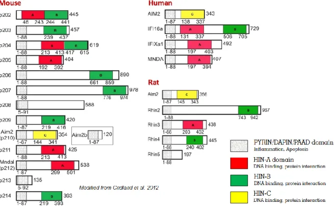
Upon binding to specific receptors, type I IFN lead to the activation of signal transduction pathways that activate a broad range of ISGs that are responsible for the antiviral, antiproliferative, pro-apoptotic, and immunomodulatory activities of type I IFN. One family of IFN-inducible genes is the HIN-200/Ifi-200 gene family, which encodes evolutionary related human (IFI16, IFIX, MNDA, and AIM2) and murine (Ifi202a, Ifi202b, Ifi203, Ifi204, Ifi205/D3, and Ifi206) proteins [33]. The human and murine gene clusters are located on syntenic genomic regions of chromosome 1 and probably stem from repeated gene duplications of one ancestral gene. The HIN-200 proteins contain at least a 200-amino acid repeat that constitutes the HIN domain, which is always near the C-terminus. The common domain architecture PYD-HIN of these protein families consists of one or two copies of the HIN domain (domains A and B) and an N-terminal PYD domain, also named PAAD, DAPIN, or Pyrin, a member of the death domain superfamily [34, 35]. The PYD domain, commonly found in cell death-associated proteins such as Pyrin, ASC, and zebrafish caspase, is present in the N-terminus of most Ifi200/HIN200 proteins, suggesting a role of these proteins in inflammation and apoptosis [36-38].
2.4 THE INTERFERON INDUCIBLE 16(IFI16)PROTEIN
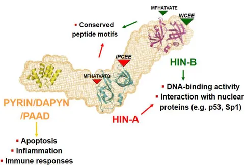
In the human IFI16 protein, the PYD domain is followed by two copies of a HIN domain (A and B respectively). These are separated by a serine-threonine-proline (S/T/P)-rich spacer region. The size of the spacer region is regulated by mRNA splicing and can contain one, two, or three copies of the highly conserved 56-aa S/T/P domain, giving rise to the isotypic variants IFI16a, IFI16b, and IFI16c respectively. A comprehensive bioinformatics analysis revealed the presence of 2 oligonucleotide/oligosaccharide binding (OB) folds in each HIN domain [39] and biophysical approaches demonstrated that the A and B domains of all family members, including IFI16, have been implicated in mediating oligonucleotide and oligosaccharide binding, protein-protein interactions and dimerization [40].
Figure 4: Predicted molecular arrangement of the Interferon Inducible 16 (IFI16) protein
For example, AIM2 binds dsDNA through its C-terminal oligonucleotide- or oligosaccharide binding domain (HIN200 domain), recruits ASC, and triggers inflammasome assembly and pro- IL-1b cleavage [41, 42]. More recently, it was demonstrated that following the stimulation of cells with transfected DNA, IFI16 binds to IFN-β stimulated viral DNA and recruits STING. Furthermore, small interfering RNA (siRNA) targeted against IFI16 inhibited DNA-induced, but not RNA-induced, activation of IRF3 and NF-kB, as well as IFN-β induction [43]. Altogether, these results demonstrate that PYD-HIN-200 proteins represent a new family of innate DNA sensors regulating the early steps of inflammation. The IFI16 nuclear phosphoprotein has been demonstrated to recognize foreign DNA and participates in the inhibition of cell cycle progression, modulation of differentiation, and cell survival [33].
Figure.5. Schematic diagram indicating the main signaling pathways triggered by IFI16 to regulate different cell functions
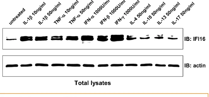
IFI16 was originally identified as a target of interferons (IFN-α/β and -γ), however additional triggers have recently been reported that include oxidative stress, cell density, and various pro-inflammatory cytokines [44]. IFI16 is expressed in hematopoietic cells and in vascular endothelial cells (EC) in blood and lymph vessels, suggesting a link between IFI16, angiogenesis and inflammation [3]. In the
skin, its expression is normally restricted to the basal proliferative layer, suggesting a possible role in the control of skin homeostasis. IFI16 has also been detected in normal salivary glands.
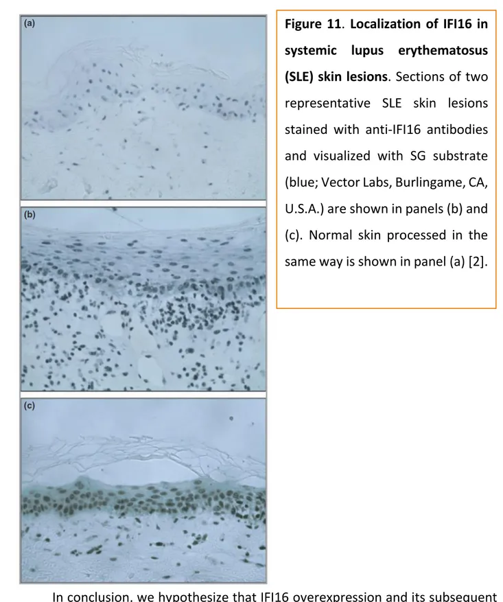
Enhanced expression levels of IFI16 were found by the proposing group in the epidermis of SLE and SSc patients and in the dermal inflammatory infiltrate obtained from patient skin lesions, detected by immunohistochemistry [32]. While IFI16 expression is restricted to the basal layer in the normal epidermis from healthy subjects, it is greatly increased and ubiquitously expressed in all layers of the epidermis in the lesional skin from both SSc (either the limited [lc-] or the diffuse [dc-] cutaneous variant) and SLE patients. Furthermore, the dermal inflammatory infiltrate shows IFI16 positive staining, indicating that the protein is expressed at a high level in lymphocytes, reactive fibroblasts, and EC. This finding raises the possibility that local tissue expression of IFI16 (or even its de-localization) in epithelial and inflammatory cells can play a role in triggering an autoimmune response against this protein. In line with the potential role of IFI16 in these autoimmune diseases, lymphocytes as well as fibroblasts and EC can be targets for Figure 6. Immunohistochemical detection of IFI16 protein in normal human tissues. (A) Exocervix. (B) Exo-endocervix junction. The epithelial cells lining the exocervix are positive, but those lining the endocervix are not (arrow). Notice that the inflammatory cells strongly expressed IFI16. Nuclei of cells not expressing IFI16 are stained blue by counterstain. (C) Vocal cord epithelium. (D) Skin. (E) High-power field of IFI16-positive cells showing nucleolar staining (arrow). (F) Strong IFI16 immunoreactivity in stromal reactive (upper arrow) and endothelial cells (lower arrow). (G) Skin stained with preimmune serum. (H) Vocal cord epithelium stained with preimmune serum. Arrows in A, C, D indicate IFI16-positive cells. A, B, C, D, G, H, 310. E, 340. F, 320 [3].
autoimmune responses in SLE and SSc, while skin is one of the main tissue targets for the clinical manifestations in these patients.
Molecular studies performed in primary endothelial cells overexpressing IFI16 demonstrated that it may be involved in the early steps of inflammation by modulating endothelial cell function, such as expression of adhesion molecules and chemokine production, cell growth, and apoptosis. Moreover, as previously demonstrated that IFI16 expression is induced by proinflammatory cytokines in primary endothelial cells [32].
On the other hand, overexpression of nuclear IFI16 protein seems to induce pro-inflammatory molecules in endothelial cells which indicates the cytokine promoting activity of endogenous nuclear IFI16 [45]. Such overexpressed IFI16 protein upregulates the production of adhesion molecules like ICAM1, VCAM1 and other chemokines like IL-8, MCP1, as shown in the Fig. 8.
Figure 7: Modulation of IFI16 protein levels by cytokines in primary human endothelial cells.
IFI16 has been much implicated on its role in inflammasome signaling. The group [43] demonstrates that IFI16 is critical for interferon-β responses upon exposure to intracellular cytoplasmic DNA and HSV-1 infection. Furthermore, IFI16 was shown to directly associate with IFN-β-inducing viral DNA motifs and the stimulator of interferon genes (STING). Early studies were unsuccessful in attempts to implicate IFI16 in the inflammasome. Specifically, an interaction between ASC and IFI16 was not found [46]. Light was shed on this in a study by [47], demonstrating that infection of endothelial cells with Kaposi sarcoma-associated herpesvirus (KSHV) led to the activation of an ASC-containing inflammasome, with a concomitant proteolytic processing of pro-IL-1β that was shown to be dependent on IFI16. In addition, the nuclear localization of pro-caspase-1 and ASC prior to infection was demonstrated. Cleaved caspase-1 (p20) and ASC were also shown to
be in the nucleus at early timepoints of infection followed by the movement of both to the cytoplasm at later times of infection with KSHV. Furthermore, by using short hairpin RNAs (shRNAs) targeting IFI16 or ASC, they demonstrated that both were required for KSHV induced processing of caspase-1 (Figure 9) [47]. IFI16 was found to be required for the induction of pro-IL-1β and IL-6 in response to HSV-1, highlighting a role for IFI16 in both transcriptional activation as well as the inflammasome [43].
Figure 9: Schematic representation of AIM2 and IFI16 inflammasome activation [1]
UVB is a well-known stimulus capable of inducing apoptosis in vitro and in vivo, and it has been associated with lupus flares [48, 49]. Aberrant IFI16 expression in endothelial cells and epithelial cells from the skin, both of which exhibit the main clinical manifestations of autoimmune diseases, indicates that IFI16 may be involved in the early steps of inflammation by modulating endothelial and keratinocyte cell function. To verify whether the induction of autoimmunity against IFI16 could indeed involve the redistribution of this nuclear protein in keratinocytes following an apoptotic stimulus like UVB, an in vitro model was developed in the laboratory of the applicant consisting of keratinocyte monolayers and human skin explants to investigate the fate of IFI16 following their irradiation with UVB [2]. In parallel, IFI16 expression and localization were analyzed in diseased skin sections from SLE patients. The results obtained clearly demonstrated that IFI16, normally restricted in the nucleus, could be induced to appear in the cytoplasm under conditions of UVB-induced cell injury. This nucleus to cytoplasm translocation was also observed in skin explants exposed to UVB and in the skin lesions from SLE patients not exposed to UVB irradiation. In addition, IFI16 was found in the supernatants of UVB-exposed keratinocytes.
Figure 10. IFI16 redistribution in ultraviolet (UV) B-irradiated human skin ex
vivo. After 24 h in culture medium, explants were UVB irradiated at 1000 J m-2
then harvested at 24 h (a) and 48 h (b) post-UVB treatment and processed for haematoxylin and eosin staining (H ⁄E) and immunohistochemical staining of IFI16 (blue); no counterstaining method was used. Arrows indicate pyknotic nuclei and hypereosinophilic cytoplasm (sunburn cells). Representative images were taken using 40· magnification. NT, not treated [2].
In conclusion, we hypothesize that IFI16 overexpression and its subsequent extranuclear appearance during the process of cell death contribute to the pathogenic mechanism through the following steps: i) induction of inflammatory
Figure 11. Localization of IFI16 in systemic lupus erythematosus (SLE) skin lesions. Sections of two representative SLE skin lesions stained with anti-IFI16 antibodies and visualized with SG substrate (blue; Vector Labs, Burlingame, CA, U.S.A.) are shown in panels (b) and (c). Normal skin processed in the same way is shown in panel (a) [2].
response in the target cells; ii) release into the extracellular milieu and induction of specific autoimmunity; and iii) triggering of proinflammatory activity via the release of extracellular free IFI16 protein.
2.5 IFI16 AS VIRAL RESTRICTION FACTOR FOR HCMV REPLICATION
IFI16 has been shown to bind to and function as a pattern recognition receptor (PRR) of virus-derived intracellular DNA, and trigger the expression of antiviral cytokines via the STING-TBK1-IRF3 signaling pathway [43, 47, 50-59]. Although many different functions have been ascribed to IFI16 (and to other proteins of the PYHIN family), its role as an antiviral restriction factor has not yet been fully described. Recent studies implicate the involvement of IFI16 in host defense against HCMV [60]. The evidence supporting such a role of IFI16 is as follows: (i) small interfering RNA (siRNA)-mediated depletion of IFI16 in primary human
Figure 12: Innate/intrinsic immune recognition and activation by herpes viruses. [5]
embryonic lung fibroblasts (HELF) significantly increases HCMV replication efficiency as a result of augmented viral DNA synthesis; (ii) similarly, viral plaque formation is enhanced in the presence of an exogenous dominant-negative IFI16 mutant that competes with the endogenous IFI16; (iii) overexpression of functional IFI16 in HCMV-infected HELFs decreases both virus yield and viral DNA copy number; and (iv) early and late, but not immediate-early viral mRNAs and proteins are strongly down-regulated under these same conditions, suggesting that IFI16 exerts its main antiviral effect at the level of viral genome synthesis. This unique defense mechanism distinguishes the activity of IFI16 from that described for ND10.
In more general terms, human viruses have to face powerful RF responses and thus have evolved a number of strategies to overcome RF attack. Viral antagonists can act through highly specialized mechanisms, such as coupling RFs to protein degradation pathways, causing their relocalization and thus down-regulating their functionality, or even by mimicking RF substrates [61]. In the case of HCMV, viral regulatory proteins (such as IE1p72, pp71, and others) mediate an efficient evasion from the antiviral state instituted by ND10, either by means of proteasomal degradation or by disrupting the host’s subnuclear structure [62, 63].
In this study (second author manuscript attached), we investigated the mechanisms used by HCMV to evade IFI16 restriction activity. We observed that starting from 72-96 hours post-infection (hpi), nuclear levels of IFI16 protein started to decrease in the nucleus and gradually increased in the cytoplasm of infected cells where it relocalized to the virus assembly complex (AC), as shown by its co-localization with the viral structural proteins gB and pp65. Finally, through the use of immunogold electron microscopy and co-precipitation experiments, we
provide evidence indicating that IFI16 eventually transits into the maturing virions embedded in the outer tegument layer. In conclusion, these data suggest that in order to overcome the restriction activity of IFI16, HCMV may stimulate its subcellular relocalization from the nucleus to the viral AC, followed by its inclusion into mature virions.
3 A
IMS OF THE
R
ESEARCH
Collectively, present data and other findings suggest the following disease model: 1) IFI16 expression is enhanced in lesional tissues as a result of abnormal type I IFN production (either endogenously produced or exogenously administered) and/or other proinflammatory stimuli, including UVB; 2) IFI16 expression triggers proinflammatory activation of endothelial cells and keratinocytes; 3) sustained IFI16 overexpression impairs cell growth and viability; 4) IFI16 is then released as a consequence of increased cell death by apoptosis/necrosis; 5) the released IFI16 protein leads to a breakdown in tolerance to self-antigens and the injury of target cells (endothelial cells and keratinocytes); 6) circulating levels of free IFI16 directly damage endothelia (via the suppression of capillary formation), skin epithelia (via the suppression of stratified epithelium formation), and glandular epithelia (via the impairment of glandular physiology), thus amplifying the inflammatory process through local tissue destruction; 7) this loss of tolerance favors the generation of specific anti-IFI16 autoantibodies that may, paradoxically, exert protective activity by inhibiting the extracellular protein from binding to its specific receptors.
To prove this disease model, the proposed research project will be developed as follows:
Aim 1: In vivo studies to analyze the presence of IFI16 in the sera of systemic autoimmune patients and co-relate its occurrence with disease condition and other types of non-autoimmune diseases like non-SLE glomerulonephritis.
Aim 2: In vitro studies aimed at elucidating the molecular actions of extracellular IFI16 on different primary endothelial cell lines. These studies will be grouped into: i) definition of the mechanisms by which extracellular IFI16, released upon cell
death, interacts with target cells (endothelial and epithelial cells), and ii) the clarification of the IFI16 receptor-stimulated molecular pathways that lead to cell damage and death. The outcome of these experiments will help us to understand the pathogenesis of the vasculopathy and the skin lesions that accompany autoimmune diseases, such as SLE, SSc, and SjS.
4
M
ATERIALS ANDM
ETHODS 4.1 CELL CULTURESPrimary human umbilical vein endothelial cells (HUVECs), pooled from multiple donors, cryopreserved at the end of the primary culture [64], were grown on 0.2% gelatin (Sigma-Aldrich, Milan, Italy) coated base in the presence of endothelial growth medium (EGM-2, Lonza-Milan) containing 2% fetal bovine serum, human recombinant vascular endothelial growth factor (rVEGF), basic fibroblast growth factor, human epidermal growth factor, IGF-1, hydrocortisone, ascorbic acid, heparin, gentamycin, amphotericin B including 1% Penicillin-Streptomycin solution (Sigma-Aldrich, Milan, Italy) which we describe as complete EGM-2. Low passage human dermal fibroblasts, HDF (ATCC), mouse fibroblasts, 3T3 (ATCC), HeLa (ATCC) and HaCaT (ATCC) cells were grown in Dulbecco's modified Eagle's medium (DMEM) (Sigma-Aldrich, Milan, Italy) supplemented with 10% fetal bovine serum and 2% Penicillin-Streptomycin solution Unless specified, all cells were grown at 37°C and 5% CO2.
4.2 RECOMBINANT PROTEINS
The entire coding sequence of the b isoform of human IFI16 was subcloned into the pET30a expression vector (Novagen, Madison, WI) containing an N-terminal histidine tag. Protein Expression and affinity purification, followed by fast protein liquid chromatography (FPLC), were performed according to standard procedures. The purity of the proteins was assessed by 10% sodium dodecyl sulfate-polyacrylamide gel electrophoresis. The FPLC purified protein was then processed with Toxin Eraser™ Endotoxin Removal Kit (Genscript, USA), while the LPS concentration of the processed product was measured using Toxin Sensor™
Chromogenic LAL Endotoxin Assay Kit which was as low as 0.005 EU/ml. the final purified rIFI16 was stored at -80°C in endotoxin free vials. As negative controls in enzyme-linked immunosorbent assays (ELISA), the polypeptide encoded by the pET30a empty vector (control peptide) was expressed and purified according to the same protocol.
4.3 PATIENTS AND DETERMINATION OF HUMAN EXTRACELLULAR IFI16 BY CAPTURE ELISA
The study groups comprised patients suffering from Systemic Sclerosis, (n = 50), Systemic Lupus Erythematosus, (n = 100), Sjogren Syndrome, (n = 49), Rheumatoid Arthritis (n = 50) and Non-SLE Glomerulonephritis (n = 46). As controls, we investigated sera from 116 sex- and age-matched healthy subjects. Written informed consent was obtained from all subjects according to the Declaration of Helsinki and approval was obtained from local ethics committees of corresponding hospital.
For the determination of circulating extracellular IFI16, a capture ELISA was employed. Briefly, polystyrene micro-well plates (Nunc-Immuno MaxiSorp; Nunc, Roskilde, Denmark) were coated with a home-made polyclonal rabbit-anti-IFI16 antibody (478-729 aa). Subsequently, plates were washed and free binding sites then saturated with PBS / 0.05% Tween / 3% BSA. After blocking, sera were added to plates in duplicate. Purified 6His-IFI16 protein was used as the standard and BSA served as the negative control. The samples were washed, monoclonal mouse anti-IFI16 antibody (Santa Cruz, sc-8023) added, and then incubated for 1h at room temperature. After washing, horseradish peroxidase-conjugated anti-mouse antibody (GE Healthcare Europe GmbH, Milan, Italy) was added. Following the addition of the substrate (TMB; KPL, Gaithersburg, MD, USA), absorbance was
measured at 450nm using a microplate reader (TECAN, Mannedorf, Switzerland). Concentrations of extracellular IFI16 were determined using a standard curve for which increasing concentrations of purified 6His-IFI16 were used.
4.4 CELL VIABILITY ASSAY
Cells were seeded at a density of 1×10⁴/well in a 96-well culture plate. After
24 hours, cells were treated with different doses (10, 25 or 50µg/ml) of recombinant IFI16 protein (IFI16), mock-treated using the same volume of vehicle as each IFI16 dose (Mock), or left untreated (NT). Where indicated, different doses (1.75µgr or 3.5µgr) of antibody against IFI16 were added. Forty-eight hours after treatment, cell viability was determined using the 3-(4,5-dimethylthiazol-2-yl)-2,5-diphenyltetrazolium bromide (MTT) (Sigma-Aldrich, Milan, Italy) method, as previously described [65].
4.5 TUBE MORPHOGENESIS ASSAY
HUVEC were seeded in complete medium in 60-mm culture dishes coated with 0.2% gelatin (Sigma-Aldrich, Milan, Italy) and were treated for 48h with different doses (10 or 25µg/ml) of recombinant IFI16 protein (IFI16). As negative controls, cells were treated with the same volumes of vehicle (Mock) used for each IFI16 dose or left untreated (NT). Where indicated, different doses (1.75µgr or 3.5µgr) of antibody against IFI16 were added. Tube morphogenesis assay was performed as described in [66]. Briefly, a 24-microwell plate, pre-chilled at -20°C, was coated with 250µl/well of Matrigel Basement Membrane (5mg/ml; Becton and Dickinson, Milan, Italy) and then incubated at 37°C for 30 min until solidified. HUVEC (8 x 10⁴ cells/500µl per well) were seeded onto the matrix and allowed to
incubate at 37°C in 5% CO2. Plates were photographed after 6h using a Leica
inverted microscope. 4.6 MIGRATION ASSAY
Twenty-four well transwell inserts with an 8µm pore size (Corning B.V. Life Sciences, Amsterdam, The Netherlands) were coated with a thin layer of gelatin (0.2%). HUVECs cultured in EGM-2 with 2% FBS and pre-treated with different concentrations of IFI16 recombinant protein or mock- or untreated for 48 hours were washed twice with PBS, trypsinized and plated into the upper chambers (4×10⁵ cells) resuspended in 200µl of EBM-2 (Lonza, Italy), 0.1% BSA (Sigma-Aldrich, Milan, Italy) plus IFI16 recombinant protein or mock solution (the same amounts as in the 48h pre-treatment). The lower chambers were filled with 600µl EGM2 containing VEGF and bFGF (as chemo-attractants) (Sigma-Aldrich, Milan, Italy), 2% FBS, and IFI16 recombinant protein or mock solution (the same amounts as in the upper chamber). The chambers were incubated for 5h at 37°C in a humidified atmosphere containing 5% CO2. After incubation, cells on the upper side of the filter were removed. The cells that had migrated to the lower side of the filter were washed twice with PBS, fixed with 2.5% glutaraldehyde (Sigma-Aldrich, Milan, Italy) for 20 min at room temperature, and stained with 0.5ml crystal violet (0.1% in 20% methyl alcohol solution) (Sigma-Aldrich, Milan, Italy). After washes, color was developed in 10% acetic acid and read in duplicate at 540nm on a microplate reader (Victor 3; Perkin-Elmer, Boston, MA).
4.7 RIFI16-FITC MEMBRANE BINDING AND CONFOCAL MICROSCOPY
HUVEC were seeded in 24-well plate in the presence of glass cover-slip and were grown overnight in presence of 1 µg/ml tunicamycin (Sigma-Aldrich, Milan,
Italy) in EGM-2 medium with 2% FBS and antibiotics. The cells were washed twice with cold PBS and incubated with increasing concentrations (10nM, 20nM, 30nM) of FITC labeled rIFI16 (FluoReporter® FITC Protein Labeling Kit by Invitrogen) for 90 minutes at 4°C. Later the cells were washed twice with cold PBS and were fixed using 2% para-formaldehyde solution for 4 minutes. The PBS wash was repeated thrice and the coverslips were mounted on glass slides using ProLong® Gold Antifade Reagent by Invitrogen. The slides were observed using Leica Confocal Microscope at 490nm excitation wavelength for FITC in one channel while trans-illuminated light in the other.
4.8 CO-CULTURING AND IMMUNOFLUORESCENCE
Co-culturing was performed with HeLa cells and HUVEC, as described in Koristka S. et.al [67]. 105 HeLa cells were seeded in 24 well-plate coated with 0.2% Gelatin in the presence of glass cover-slip and grown over-night in DMEM with 10% FCS at 37°C, 5% CO2. The cells were washed, suspended in PBS and lethally irradiated with UV-B lamp (HD 9021; Delta Ohm S.r.l., Padova, Italy). The dosage of 1000 Wm2 was counted using a UVB irradiance meter cosine corrector with spectral range of 280–319 nm (LP 9021 RAD; Delta Ohm). Followed by this, 5×10⁴ HUVEC were added in the same well, grown in EGM-2 with 2% FCS until ready. Immunofluorescence was performed after 24hr, 36hr and 48hr using a home-made anti-IFI16 polyclonal as primary antibody and Alexa488- anti-rabbit (GE Healthcare) as secondary antibody. The cells were then fixed with 2% para-formaldehyde (Sigma-Aldrich, Milan, Italy), permeabilized with 0.2% Triton X-100 (Sigma-Aldrich, Milan, Italy) and nuclear stained with 1µg/ml propidium iodide (Sigma-Aldrich, Milan, Italy). The coverslips were mounted on glass slides using ProLong® Gold
Antifade Reagent by Invitrogen and the cells were observed by Leica confocal microscope.
4.9 RADIO-IODINATION OF RIFI16 AND BINDING ASSAYS
Iodination Beads were purchased from Thermo Fischer Scientific Inc. (Rockford, IL, USA) and used according to manufacturer’s instructions. Briefly, two dry beads were washed with rIFI16 elution buffer (50mM HEPES pH 7.5; 1M NaCl) (Sigma-Aldrich, Milan, Italy), soaked dry and was incubated for 5 minutes with the solution of carrier-free 2mCi Na125I (Perkin Elmer Italia, Milan, Italy) and diluted in elution buffer. Later 200µg of rIFI16 was added and incubated for 15 minutes. The labeling reaction was passed through Zeba Spin Desalting Columns (Thermo Fischer Scientific Inc.) to remove excess Na125I or unincorporated 125I from the iodinated
protein. The concentration of the final radioiodinated [125I]-rIFI16 was calculated
using the following formula, where ‘C’ is the cpm counted, ‘V’ is volume of solution counted in ml and ‘Y’ is the specific activity of the radioligand in cpm/fmol.
Concentration of [125I]-rIFI16 in (pM) = [‘C’cpm / ‘Y’cpm/fmol] / ‘V’ml.
Binding assay was performed as described in [68, 69], 105 cells/well were seeded and attached in a 24-well plate with. Once ready, the medium was removed and the cells were washed with PBS. Further they were re-suspended with increasing concentrations of [125I]-rIFI16 (1-32nM) within different wells in the
presence of 200nM unlabeled rIFI16 for Non-Specific Binding. Separately, other wells were re-suspended with [125I]-rIFI16 (1-32nM) without any unlabeled rIFI16
for total binding. The incubation was performed at 4°C for 90 minutes. Later, the cells were washed with PBS to remove any loosely bound ligand and were then suspended in warm 1% SDS (Sigma-Aldrich, Milan, Italy) for 5 minutes. The SDS lysate of the cells was then measured on Cobra II Series Auto-Gamma Counter. All
the experiments were carried out in triplicates and the data was analyzed using non-linear regression equations from GraphPad Prism with 95% confidence intervals.
4.10 COMPETITION AND INHIBITION OF [125I]-RIFI16 BINDING
For binding competition experiments, cells were seeded into 96-well plates at a density of 104 cells/well. The medium was removed and cells were washed with
PBS. HUVEC were then incubated at 4°C for 90 minutes with unlabeled rIFI16 (10-1000nM) in the presence of 10nM [125I]-rIFI16. Binding inhibition was carried out
overnight by incubating 10nM [125I]-rIFI16 with varying concentrations
(10-1000nM) of anti-IFI16 polyclonal N-terminal (1-205 aa) or C-terminal (478-729 aa) antibodies at 4°C. This mixture was then added to 104 HUVEC and incubated for 90
minutes at 4°C. The loosely bound ligand was removed by washing twice with PBS, and cells were then detached using warm 1% SDS and the levels of [125
I]-rIFI16-binding to HUVEC assessed using a Cobra II Series Auto-Gamma Counter. The data were analyzed using non-linear regression equations in GraphPad Prism under 95% confidence intervals.
4.11 NF-ΚBIMMUNOFLUORESCENCE
Immunofluorescence was performed after 0 hr, 24 hr and 48 hr of rIFI16 treatment using 1:1000 Nf-κB p65 monoclonal antibody (F-6) sc-8008 (Santa Cruz Biotechnology, Inc., USA) as primary and 1:500 Alexa488-anti-mouse (GE Healthcare) as secondary antibody. Briefly, after treatment the cells were fixed with 2% paraformaldehyde (Sigma-Aldrich, Milan, Italy) for 20 min at 4°C, permeabilized with 0.5% Triton X-100 (Sigma-Aldrich, Italy) for 30 min at RT, incubated with primary antibody for 2 hr at 37°C in a moist chamber, incubated
with secondary antibody and nuclear stain 1:500 TO-PRO-3 (Life-Technologies, Italy) for 1 hr at RT. The glass cover slips were mounted using ProLong® Gold
Antifade Reagent (Invitrogen, Italy) and observed under Leica DM-IRE2 Confocal Microscope.
4.12 RIFI16 TREATMENT AND QUANTITATIVE REAL-TIME PCR
In the first experiment, HUVEC were grown in 6-well plate with complete/incomplete EGM-2 and treated with 50 µg/ml concentration of endo-free rIFI16 [70] and vehicle/mock for 24 hrs and gene expression levels of different pro-inflammatory cytokines as listed in the (Table 1) was assessed with quantitative real-time PCR. In another set of experiment, HUVEC were treated with 50 µg/ml rIFI16 within a time-course of 0 hr, 4 hr, 12 hr, 24 hr, 48 hr and 72 hr and activity of highly modulated genes from the first experiment was assessed. After rIFI16 treatment, HUVEC were trypsinized and m-RNA was extracted using TRI Reagent®
(Sigma-Aldrich, Italy) as described in manufacturer protocol. The resulting m-RNA was treated with DNase I Amplification Grade kit (Sigma-Aldrich, Italy) as instructed. Later, 1 µg of m-RNA was used as a template first strand c-DNA synthesis using ImProm-II™ Reverse Transcription System (Promega, Italy) and C1000 Thermal Cycler (Bio-Rad, Italy) with annealing temperature 25°C for 5 min, followed by extension at 42°C for up to one hour. The reverse transcriptase was heat inactivated at 70°C for 15 min, while the final c-DNA preparation was quantified and stored at 4°C for immediate use. A random m-RNA sample was kept as RT-
(without reverse transcriptase) to assess the presence of contaminating genomic DNA in the preparation. The quantitative real-time PCR analyses were performed using CFX96 Real-Time PCR Detection System (Bio-Rad, Italy) with SsoAdvanced™ Universal SYBR® Green Supermix (Bio-Rad, Italy) and amplification conditions as
instructed in manufacturer protocol, upto 40 cycles of PCR. Primer sequences are summarized in Table 1. The Ct values for each gene were normalized to the Ct values for GAPDH using the Ct equation. The level of target RNA, normalized to the endogenous reference and relative to the mock treated and untreated cells, was calculated by the comparative Ct method using the 2-δδCt equation.
Cytokines Forward Primer Reverse Primer
IL-1α CCGTTTTGACGACGCACTTG TTTGGCCATCTTGACTTCTTTGC
IL-1β ACGAATCTCCGACCACCACT CCATGGCCACAACAACTGAC
IL-2 GTAACCTCAACTCCTGCCACAA TGTTTCAGTTCTGTGGCCTTCT
IL-4 TGCTGCCTCCAAGAACACAAC GGTTCCTGTCGAGCCGTTTC
IL-6 GACCCAACCACAAATGCCA GTCATGTCCTGCAGCCACTG
IL-8 CTGGCCGTGGCTCTCTTG CCTTGGCAAAACTGCACCTT
IL-10 GGCGCTGTCATCGATTTCTTC GTTTCTCAAGGGGCTGGGTC
IL-12p35 GCTCCAGAAGGCCAGACAAA GGCCAGGCAACTCCCATTAG
IL-12p40 GCCCAGAGCAAGATGTGTCA CACCATTTCTCCAGGGGCAT
IL-17A AGACCTCATTGGTGTCACTGC CTCTCAGGGTCCTCATTGCG
IL-18 TGCAGTCTACACAGCTTCGG TCCAGGTTTTCATCATCTTCAGCTA
IL-33 CAAAGAAGTTTGCCCCATGT AAGGCAAAGCACTCCACAGT
CCL2 CTCTGCCGCCCTTCTGTG TGCATCTGGCTGAGCGAG CCL3 AGCTGACTACTTTGAGACGAGCA CGGCTTCGCTTGGTTAGGA CCL4 CTGCTCTCCAGCGCTCTCA GTAAGAAAAGCAGCAGGCGG CCL5 GACACCACACCCTGCTGCT TACTCCTTGATGTGGGCACG CCL7 GCACTTCTGTGTCTGCTGCT CAGCCTCTGCTTAGGGATTTT CCL8 GCCCTCCAAGATGAAGGTTT TCACGTTAAAGCAGCAGGTG CCL20 TCCTGGCTGCTTTGATGTCA TCAAAGTTGCTTGCTGCTTCTG TLR1 CTGGTATCTCAGGATGGTGTGC TTGGAGTTCTTCTAAGGGTATGTTCC Table 1: List of SYBR Green RT-PCR primers for pro-inflammatory genes
TLR2 GGCCAGCAAATTACCTGTGTG AGGCGGACATCCTGAACCT TLR3 TCCCAAGCCTTCAACGACTG TGGTGAAGGAGAGCTATCCACA TLR4 CTGCAATGGATCAAGGACCA TTATCTGAAGGTGTTGCACATTCC TLR5 TCGAGCCCCTACAAGGGAA CACTGAGACTCTGCTATACAAGCTA TLR7 TTACCTGGATGGAAACCAGCTAC TCAAGGCTGAGAAGCTGTAAGCTA TLR8 GAGAGCCGAGACAAAAACGTTC TGTCGATGATGGCCAATCC TLR9 TGGTGTTGAAGGACAGTTCTCTC CACTCGGAGGTTTCCCAGC TLR10 GAAAGGTTCCCGCAGACTTG TGGAGTTGAAAAAGGAGGTTATAG
ICAM1 GCACATTGGTTGGCTATCTTCT GCCCGAAGCGTTTACTTTGA
VCAM1 TTCCTCAGATTGGTGACTCCG AAAACTCACAGGGCTCAGGGTCAG
VEGF ATCTGCATGGTGATGTTGGA GGGCAGAATCATCACGAAGT
IRF3 CCGACCTTCCATCGTAGGAG AATCAGATCTTCCCCCGGCA
MyD88 CCACCCTTGATGACCCCTAGGACAA AC
GTCTGTTCTAGTTGCCGGATCATCTCCT GCAC
TNF-alpha ATACTGACCCACGGCTCCA GTTCGAGAAGATGATCTGACTGCC
TGF-beta1 CCCAGCATCTGCAAAGCTC GTCAATGTACAGCTGCCGCA
GM-CSF CATGTGAATGCCATCCAGGA CAGGCCCACATTCTCTCACTT
GAPDH CGGAGTCAACGGATTTGGTCGTATT GG
GCTCCTGGAAGATGGTGATGGGATTT CC
4.13 TRANSIENT TRANSFECTION AND LUCIFERASE ASSAY
The low passage (3-4) HUVECs were plated at approximately 2×10⁵ cells per
well in 6-well culture plates and incubated overnight for next day 80% confluence. Effectene Transfection Reagent (Qiagen, Italy) was optimized for 75% transfection efficiency, maintaining normal biological activity of HUVEC. Briefly, total 0.4 µg of plasmid DNA was diluted to 100 µl of EB buffer while the Enhancer and Effectene reagent was used as 4 µl and 5 µl respectively, by following the preparation conditions as directed by the manufacturer. Cells were incubated with the
transfection reagent diluted in serum/antibiotics free incomplete EGM-2 upto maximum 4 hr at 37°C with 5% CO2. Later cells were incubated overnight with
incomplete EGM-2 including 2% FCS and antibiotics, following which rIFI16, 50 µg/ml was added to the culture for next 48 hr before measurement of luciferase activity. For each transfection, the cells were co-transfected with 0.05 µg of pRL-TK (Promega) and either of 0.3 µg of pGL2-IL8-wild type, 0.3 µg of pGL2-IL8-ΔNf-κB, 0.3 µg of pGL2-IL8-ΔAP1 luciferase reporter plasmid or 0.3 µg control vector (pmaxGFP™ Vector). All plasmids were purified using the Toxin Eraser™ Endotoxin Removal Kit (Genscript, USA). Protein extracts were prepared and luciferase activity was measured using the Dual-Luciferase® Reporter Assay System (Promega, Italy) at the Victor X4 Multilabel Plate Reader (Perkin Elmer, Italy).
4.14 STATISTICAL ANALYSIS
All statistical tests were performed using GraphPad Prism version 5.00 for Windows (GraphPad, La Jolla, CA, USA). Positivity cut-off values for anti-IFI16 antibodies were calculated as the 95th percentile for the control population and the
Kruskal-Wallis test was used to measure associations. To test the effects of recombinant IFI16 protein (rIFI16) on biological functions of primary endothelial cells one-way analysis of variance (ANOVA) with Bonferroni adjustment for multiple comparisons was used. Significant upregulation of m-RNA expression was evaluated through paired t-test.
5
R
ESULTS5.1 SERUM LEVELS OF IFI16 PROTEIN ARE INCREASED IN PATIENTS WITH SYSTEMIC AUTOIMMUNE DISEASES
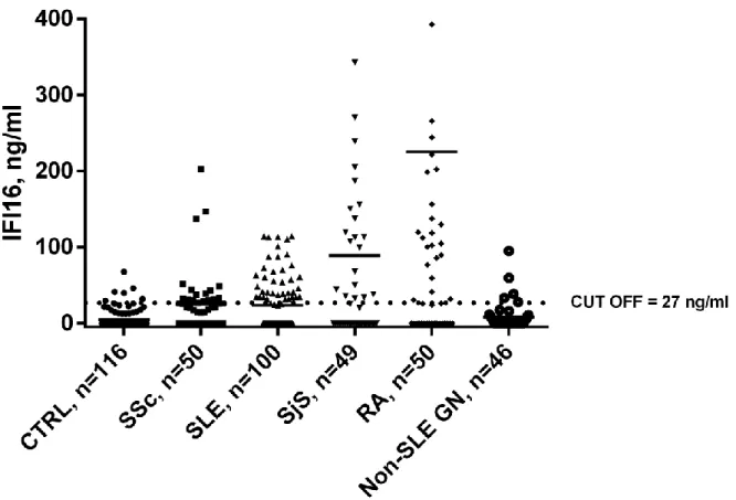
Sera were harvested from patients suffering from systemic autoimmune diseases characterized by endothelial dysfunction, including SSc, SLE, SjS, and RA. IFI16 serum levels were quantified using an in-house sandwich ELISA and compared with age- and sex-matched healthy controls. All absorbance levels were in the range of assay linearity. With the cut-off value set to the 95th percentile of the
control population (27ng/ml), mean IFI16 levels were significantly increased in patients with SSc, SLE, RA, and SjS compared to the control group (4.7ng/ml) (SSc: 25.4ng/ml, p<0.001; SLE: 23.5ng/ml, p<0.001; RA: 222ng/ml, p<0.001; SjS: 88.2ng/ml, p<0.001). Of note, the sera from RA patients displayed the highest levels of circulating free protein. IFI16 levels above the 95th percentile for control subjects
were observed in 34% of SSc, 37% of SLE, 47% of SjS, and 56% of RA patients (Figure 13). By contrast, IFI16 levels in non-SLE GN patients did not show any significant difference in comparison with healthy controls. Since the objective of this part of the study was limited to demonstrate the presence of circulating IFI16 in patients’ sera for justifying the rest of the in vitro studies, correlation with clinico-pathological parameters was behind the aim of these studies and was not performed.
Figure 13. IFI16 protein levels in patients’ and controls’ sera determined using an in-house capture ELISA. Each dot represents the concentration of IFI16 protein (expressed in ng/ml on a linear scale) in each individual subject: patients suffering from Systemic Sclerosis (SSc, n=50), Systemic Lupus Erythematosus (SLE, n=100), Sjogren’s Syndrome (SjS, n= 49), Rheumatoid Arthritis (RA, n=50), and non-SLE glomerulonephritis (non-SLE GN n=46) were investigated together with healthy controls (CTRL, n=116). The horizontal bars represent the median values. Values over the dotted line indicate the percentage of subjects with IFI16 protein levels above the cut-off value (27ng/ml) calculated as the 95th percentile
of the control population. Statistical significance: *** p < 0.001 vs. controls (Kruskall-Wallis test).
5.2 EFFECTS OF EXTRACELLULAR IFI16 ON DIFFERENT FUNCTIONS OF PRIMARY ENDOTHELIAL CELLS
Abnormalities in angiogenesis are frequently present in systemic autoimmune diseases and may lead to tissue damage and premature vascular disease [71]. To verify whether extracellular IFI16 was also involved in this pathogenic process, HUVEC were treated with increasing concentrations of recombinant IFI16 protein (rIFI16) (10, 25 or 50µg/ml), mock-treated with the same volumes of vehicle (Mock), or left untreated (NT) and then assessed for cell viability at 48 hours incubation time by MTT assay. As shown in Figure 14A, the addition of endotoxin-free rIFI16 protein did not reduce the amount of viable adherent cells when compared to mock or untreated cells at the concentration of 10 and 25µg/ml, respectively. At the highest concentration used (50µg/ml), a slight reduction in cell viability was observed, and consequently the following studies were conducted with the lower doses.
Next, to test whether the addition of rIFI16 to culture media altered other biological parameters of endothelial cells, HUVEC were treated as described for the assessment of cell viability (MTT assay) and then analyzed for their tubule morphogenesis and chemotactic activities. As shown in Figure 10B, exogenous administration of 25µg/ml rIFI16 severely limited tubulogenesis, with most cells generating incomplete tubules or aggregating into clumps. The extent of angiogenesis was quantified by counting the intact capillary-like tubules called as Lumens, which showed 75% decrease in its numerical value, as well as the number of interconnecting branch points showing 77% decrease. (Figure 14B). These effects were less pronounced when a lower dose of 10µg/ml rIFI16 was used with 22% and 15% decrease respectively. In contrast, untreated or mock-treated HUVEC
plated onto matrigel supported the formation of an extensive interconnecting polygonal capillary-like network.
Next, we evaluated the effects of rIFI16 on the migration phase of angiogenesis in a transwell migration assay routinely used to study cell migration in response to specific stimuli. HUVECs untreated, mock-treated, or incubated with rIFI16 (10 or 25µg/ml) for 48h were transferred into transwell migration chambers. As shown in Fig. 10C, only a small population of HUVECs cultured in the presence of 25µg/ml rIFI16 were able to migrate through the chamber (30% of migration), whereas mock-treatment resulted in considerable migration (95% and 87% for 10µg/ml and 25µg/ml, respectively).
Taken together, these results demonstrate the capability of extracellular rIFI16 to impair physiological functions of endothelial cells, such as the differentiation phases responsible for tube morphogenesis and migration.
Figure 14. Extracellular IFI16 affects various biological functions of primary endothelial cells. HUVEC were treated with different doses of recombinant IFI16 protein (rIFI16), the same volumes of vehicle (Mock), or left untreated (NT) for 48h. (A) Viability analysis (MTT assay); the viability of control preparations (NT) was set to 100%. (B) Capillary-like tube formation assay (Matrigel). For a quantitative assessment of angiogenesis, the number of lumens and branch points was assessed (upper panels); representative images of three independent experiments are reported (lower panels). (C) Migration analysis (Transwell assay) results are reported as the percentage of migrated cells vs. untreated HUVECs. Values represent the mean±SD of 3 independent experiments, (**p<0.01, ***p<0.001; one-way ANOVA followed by Bonferroni's multiple comparison test).
5.3 ANTI-N-TERMINUS IFI16 ANTIBODIES NEUTRALIZE THE CYTOTOXIC ACTIVITY OF IFI16
To demonstrate that the effects exerted by IFI16 protein on target cells were specific and not a consequence of cell cytotoxicity, HUVEC were treated with 25µg/ml rIFI16 in the presence or absence of specific rabbit polyclonal antibodies recognizing the N-terminal or the C-terminal domain of IFI16 protein. HUVEC incubated with rIFI16 in the presence of normal rabbit IgG were used as the positive control. As described in the previous section, rIFI16 severely affected the capability of endothelial cells to generate microtubules as well as their transwell migration activity, while the same effects were observed in presence of normal rabbit IgG (Figure 15A and 15B). In contrast, the presence of anti-N-terminus antibodies reduced the Lumens by 16%, Branch Points by 5% and migration by 1% while anti-C-terminus antibodies reduced the Lumens by 60%, Branch Points by 32% and migration by 49%. This indicates the role of anti-N-terminus antibody in inhibiting the activities of IFI16 toward endothelial cells, restoring the tube formation and migratory activities. Altogether, these results suggest that the IFI16 activity is specific and that the functional domain resides at the N-terminus, where the PYD domain is localized.
Figure 15. Anti-IFI16 antibodies restore the biological activities of extracellular IFI16. HUVEC were treated for 48h with different doses of recombinant IFI16 protein (rIFI16), the same volumes of vehicle (Mock), or left untreated (NT), alone or in combination with antibodies against IFI16. (A) Capillary-like tube formation assay (Matrigel). For a quantitative assessment of angiogenesis, the number of lumens and branch points was assessed (upper panels); representative images of three independent experiments are reported (lower panels). (B) Migration analysis (Transwell assay) results are reported as the percentage of migrated cells vs. untreated HUVECs. Values represent the mean±SD of 3 independent experiments (**p<0.01, ***p<0.001, one-way ANOVA followed by Bonferroni's multiple comparison test).
5.4 BINDING OF EXTRACELLULAR IFI16 ON THE PLASMA MEMBRANE OF HUVEC
The finding that extracellular IFI16 impairs endothelial cell functions, including tube morphogenesis and transwell migration indicates a possible alarmin function as recently demonstrated for Danger and Pathogen-associated molecular pattern molecules collectively called as DAMPs, PAMPs such as autoantigen HMGB-1 [72, 73]. Thus to find evidence in this direction it was important to evaluate the binding interaction of IFI16 on the plasma membrane of HUVEC. A series of binding experiments were conducted to verify the presence of high-affinity binding sites in the membranes of the target cells. In the previous section, it has been described that 25 µg/ml (300 nM) rIFI16 concentration is non-toxic to HUVEC, while they can still perform biological functions. Thus HUVEC were incubated with lowest concentrations (10nM, 20nM, and 30nM) of FITC-labeled rIFI16 to avoid toxicity or apoptosis and the binding was visualized by confocal microscopy. As shown in Figure 16A, binding of FITC-labeled rIFI16 was detected at least concentration of 10nM, increased at 20nM, and saturated at 30nM. To avoid the non-specific binding of rIFI16 with sugar residues on plasma membrane, HUVEC were grown in presence of tunicamycin which inhibits N-glycosylation of proteins. By contrast to above findings, human fibroblasts were negative for FITC-labeled rIFI16 at all the rIFI16 concentrations investigated (data not shown).
Furthermore, to demonstrate that the binding of rIFI16 is of physiological relevance, co-culturing experiments were organized in such a way that UV-B irradiated cells release endogenous IFI16 [2] which in turn binds to neighboring HUVEC in the same system. As shown in Figure 16B, after 24 h HUVEC were observed to be surface bound with endogenous IFI16 released from HeLa cells, while by 36 h this bound IFI16 entered the cytoplasm and by 48 h it almost
disappeared. When fibroblasts were used instead of HUVEC, the binding was not observed, while also when HUVEC were cultured with normal HeLa cells, surface presence of IFI16 was not detected (data not shown).
Figure 16. Plasma membrane binding of IFI16 to HUVEC. (A) Cells were left untreated (a, negative control) or incubated with increasing concentrations of FITC-labeled recombinant IFI16 (b, 10nM rIFI16-FITC; c, 20nM rIFI16-FITC; d, 30nM rIFI16-FITC). Binding was detected by confocal microscopy using an excitation wavelength of 490nm for FITC in one channel and trans-illuminated light in the other. Representative images of three independent experiments are shown. (B) Endogenous IFI16 released by irradiated HeLa cells binds neighboring HUVEC. UV-B irradiated HeLa cells were co-cultured with HUVEC and after 24h, 36h and 48h, dead cell debris were removed and immunofluorescence was performed on the remaining live HUVEC using a home-made anti-IFI16 polyclonal as primary antibody and Alexa-488- anti-rabbit as secondary antibody. The cells were then fixed, permeabilized, nuclear-stained using propidium iodide and analyzed by confocal microscopy. Fibroblasts were employed as negative control. Representative images of three independent experiments are shown.
![Figure 1. Factors contributing for the development of Autoimmunity [4].](https://thumb-eu.123doks.com/thumbv2/123dokorg/4812428.49946/8.918.113.399.801.1066/figure-factors-contributing-development-autoimmunity.webp)

![Figure 9: Schematic representation of AIM2 and IFI16 inflammasome activation [1]](https://thumb-eu.123doks.com/thumbv2/123dokorg/4812428.49946/24.918.128.811.439.960/figure-schematic-representation-aim-ifi-inflammasome-activation.webp)
![Figure 12: Innate/intrinsic immune recognition and activation by herpes viruses. [5]](https://thumb-eu.123doks.com/thumbv2/123dokorg/4812428.49946/29.918.114.814.595.1059/figure-innate-intrinsic-immune-recognition-activation-herpes-viruses.webp)
![Figure 17. Binding Kinetics of [ 125 I]-rIFI16 on HUVEC, HDF, and 3T3 cells](https://thumb-eu.123doks.com/thumbv2/123dokorg/4812428.49946/57.918.118.802.534.983/figure-binding-kinetics-i-rifi-huvec-hdf-cells.webp)