haematologica | 2017; 102(12)
E
DITORIALS
1
The role of SIRT6 in tumors
Vanessa Desantis, Aurelia Lamanuzzi, Angelo Vacca
Department of Biomedical Sciences and Human Oncology, Unit of Internal Medicine “G. Baccelli”, University of Bari Medical School, Policlinico, Bari, Italy
E-mail: [email protected] doi:10.3324/haematol.2017.182675
D
ysfunctional DNA-damage response and consequentgenomic instability play a pivotal role in the initiation and progression of both solid and hematologic tumors. Preservation of DNA integrity is, in fact, a key cellular function, hence several mechanisms that repair the damaged DNA need to be studied. Recent studies have focused on key players that are able to improve the DNA repair and thus may act as targets for new therapeutic approaches.
Several data have been obtained on overexpression and hyperactivity of Sirtuins (SIRTs), a family of proteins with deacylase or mono-adenosine diphosphate (ADP)-ribosyl-transferase activities that degrade nicotinamide adenine
din-ucleotide (NAD+) enzymes to enable their biological
process-es1and promote longevity.2In mammalian cells, the Sirtuin
family is composed of seven members that show different subcellular localization and functions (transcription, metabo-lism, fat mobilization, DNA repair, stress responses,
apopto-sis, tumorigenesis and aging),3,4 and conserve the catalytic
domain and the NAD+ binding site.5 In cancer and
aging-associated pathways, SIRT6 is crucial since it prevents genomic instability, maintains telomere integrity, and
regu-lates metabolic homeostasis and DNA repair.6SIRT6 can be
considered a double-edged sword in cancer because of its dual role of both tumor suppressor and oncogene (Table 1). In healthy conditions, SIRT6 either acts as a gatekeeper of DNA repair mechanisms or regulates cell survival and prolif-eration. Following the DNA damage, SIRT6 triggers the apoptotic process, hence it is down-regulated in several can-cers. However, in other cancers, it is up-regulated, corrobo-rating the idea that it can also act as oncogene.
SIRT6 as a tumor suppressor
Studies in colorectal, breast, ovarian, hepatocellular, lung, and other tumors correlate the reduction of SIRT6 expres-sion with tumor progresexpres-sion and poor clinical outcome. In the presence of DNA-damage, SIRT6 promotes apoptotic cell death, ensuring damaged cells do not proliferate.
Sebastian et al.7demonstrated in vivo that SIRT6 deficiency
favors tumor growth and invasiveness. They also showed that SIRT6 is involved in the Warburg effect, a glycolytic metabolic shift important for supporting rapid tumor growth. SIRT6 promotes both in vitro and in vivo tumor sup-pression through resup-pression of hypoxia-inducible factor
1-alpha (HIF-1α) that inhibits glycolytic metabolism in cancer
cells.7Interestingly, in mouse and human pancreatic ductal
adenocarcinoma (PDAC), the SIRT6 knockdown is due to repression of Myc-target oncofetal protein Lin28b that
nega-tively regulates the let-7 family of miRNAs.8In detail, loss of
SIRT6 triggers activation of Lin28 promoter, Myc recruit-ment, and consequent activation of Lin28b, the downstream let-7 target genes (HMGA2, IGF2BP1) and IGF2BP3 that
accelerate the PDAC progression and metastasis.8In human
colon cancer, Lin et al.9 discovered the crosstalk between
UPS10 and SIRT6 that regulates cell-cycle progression and proliferation, and showed that the dysregulated USP10 func-tion promotes tumorigenesis through SIRT6 degradafunc-tion. Lin et al. also showed an important reduction in USP10 (a deubiquitinase protein) and SIRT6 expression. Indeed, the downregulation of USP10 triggers SIRT6 instability and neg-atively controls the transcriptional activity of the c-Myc oncogene that inhibits cell-cycle progression, cancer cell
growth, and tumor formation.9 In liver cancer, the SIRT6
suppression is regulated by the c-Jun/c-Fos pathway:10c-Fos
induces SIRT6 transcription and represses survivin by
reduc-ing histone H3K9 acetylation and NF-κB activation. The
increase in SIRT6 impairs cancer development by targeting
the anti-apoptotic activity of survivin. Min et al.10identified
in human dysplastic liver nodules a specific expression pat-tern characterized by increased Jun-survivin and reduced c-Fos-SIRT6 level. In hepatocellular carcinoma (HCC),
Bhardwaj et al.11found that SIRT6 acts as a tumor suppressor
because it deacetylates nuclear pyruvate kinase M2 (PKM2) inhibiting cell proliferation and tumorigenesis via PKM2. In
ovarian cancer, Zhang et al.12 showed that SIRT6 is
down-regulated at mRNA and protein levels in tumor cells com-pared to normal cells. Moreover, SIRT6 reduces the expres-sion of neurogenic locus notch homolog protein 3 (Notch3) while the Notch3 overexpression antagonists SIRT6 exert an effect on the ovarian cell proliferation; SIRT6 thus inhibits the proliferation of ovarian tumor cells through regulation of
Notch3.12In breast cancer, the repression of SIRT6, mediated
by runt-related transcription factor 2 (RUNX2), regulates
metabolic pathways and promotes tumor development.13
More specifically, Choe et al.13showed that RUNX2
down-regulates the SIRT6 expression at both mRNA and protein levels, and that endogenous SIRT6 expression is lower in the tumor breast tissue and cell lines expressing high levels of RUNX2 regulating the metabolic pathways. In addition, Han
et al.14 demonstrated in non-small cell lung cancer (NSCLC)
that SIRT6 inhibits Twist1 expression. Twist1 is a member of basic helix-loop-helix transcription factor family that pro-motes tumor proliferation and malignant transformation. Thus, SIRT6 is able to inhibit tumor cell proliferation through Twist1 suppression. Finally, the overexpression of E2F transcription factor 1 (E2F-1) in bladder and prostate can-cer induces the downregulation of SIRT6 that closely
corre-lates with cancer progression and poor prognosis.15
SIRT6 as a tumor promoter
In contrast to these studies, several papers show that over-expression of SIRT6 in solid and in hematologic tumors can
promote oncogenic activity. Ming et al.16 demonstrated the
oncogenic role of up-regulated SIRT6 in human skin squa-mous cell carcinoma (SCC): in skin keratinocytes, SIRT6 is
increased upon exposure to ultraviolet B (UVB) light through the activation of the AKT pathway and promotes the cyclooxygenase 2 (COX-2) expression that represses the AMP-activated protein kinase (AMPK) signaling and increases proliferation and cell survival.16 Zhang et al.17
demonstrated that SIRT6 overexpression in HCC sup-presses tumor growth by blocking extracellular signal-regulated kinases (ERK) 1/2 signaling pathway. In
addi-tion, Feng et al.18and Ran et al.19showed that SIRT6 plays
an oncogenic role in HCC. In particular, the overexpres-sion of SIRT6 is required for induction of transforming
growth factor (TGF)-β1 and H2O2/HOCl reactive oxygen
species (ROS) that mediate tumorigenesis. TGF-β1
up-regulates the SIRT6 expression inducing the activation of ERK and Smad pathways, and altering the effect of these
proteins on cellular senescence.18Ran et al.19demonstrated
an oncogenic effect of SIRT6 via chromatin remodeling. At molecular level, SIRT6 induces deacetylation of H3K9 that blocks Bcl-2-associated X protein (Bax) transcription. As a consequence, it enhances p53 and E2F-1 chromatin
accessibility thus inhibiting apoptosis. Elhanati et al.20and
Lefor et al.21 correlated SIRT6 regulation to two
microRNAs (miR-) in two different cancers. At basal con-ditions, SIRT6 and miR-122 negatively regulate each other in HCC. SIRT6 down-regulates miR-122 by deacetylating H3K56 in the promoter region. The
miR-122 binds SIRT6 3′ UTR and reduces its levels, while the
loss of the negative correlation between SIRT6 and miR-122 expression is significantly associated with better
prognosis.20In addition, miR-34a plays a key role during
Editorials
2 haematologica | 2018; 103(1)
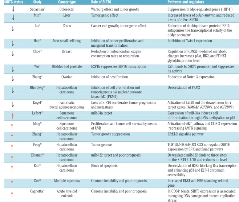
Table 1. SIRT6 expression and its role in cancer.
SIRT6 status Study Cancer type Role of SIRT6 Pathways and regulators
Sebastian7 Colorectal Warburg effect and tumor growth Suppression of Myc-regulated genes (HIF-1 )
Min10 Liver Tumorigenic effect Increased levels of c-Jun-survivin and reduced
levels of c-Fos-SIRT6
Lin9 Colon Cancer cell growth; tumorigenic effect Reduction of deubiquitinase protein USP10
antagonizes the transcriptional activity of the c-Myc oncogene
Han14 Non-small cell lung Inhibition of tumor proliferation and Inhibition of Twist1 expression
malignant transformation
Choe13 Breast Reduction of mitochondrial oxygen Regulation of RUNX2-mediated metabolic
consumption rates or respiration changes increases pAkt, HK2, and PDHK1 glycolytic protein level
Wu15 Bladder and prostate E2F1b suppresses SIRT6 transcription E2F1 binds to SIRT6 promoter and suppresses
its activity
Zhang12 Ovarian Inhibition of proliferation Reduction of Notch 3 expression
Bhardwaj11 Hepatocellular Inhibition of cell proliferation and Deacetylation of PKM2
carcinoma tumorigenesis via nuclear pyruvate
kinase M2 (PKM2)
Kugel8 Pancreatic Loss of SIRT6 accelerates tumor progression Activation of Lin28 and the downstream let-7
ductal adenocarcinomas and metastasis target genes (HMGA2, IGF2BP1, and IGF2BP3) Lefort21 Squamous miR-34a target Expression of miR-34a induces cell
cell carcinoma differentiation through DNA methylation or p53 Ming16 Squamous Proliferation and tumor cell survival by means Activation of AKT pathway and COX-2 expression
cell carcinoma of UVB repressing AMPK signaling Zhang17 Hepatocellular Tumor growth suppression ERK1/2 signaling pathway
carcinoma
Feng18 Hepatocellular Tumorigenesis TGF-β1/H2O2/HOCl ROS up-regulate SIRT6
carcinoma expression by ERK and Smad pathways Elhanati20 Hepatocellular miR-122 target and poor prognosis Deregulated miR-122 binds to three sites
carcinoma on the SIRT6 3' UTR and reduces its level Ran19 Hepatocellular Block of apoptosis Deacetylation of H3K9 blocking Bax transcription
carcinoma and enhancing p53 and E2F-1 chromatin accessibility
Cea22 Multiple myeloma Genome instability and poor prognosis Increased ELK1 and ERK signaling-related
gene
Cagnetta24 Acute myeloid Genome instability and poor prognosis In CD34+blasts, SIRT6 expression is associated
leukemia to ongoing DNA damage and intense replicative stress
UVB: ultraviolet B cancer.
↓
↓
↓
↓
↓
↓
↓
↓
↓
↓
↓
↓
↓
↓
↓
↓
↓
Editorials
haematologica | 2018; 103(1) 3
the differentiation process of HCC and SIRT6 represents one of its targets. SIRT6 downregulation induces
differen-tiation effects mediated by miR-34a.21
The role of SIRT6 is not well known in hematologic malignancies. In multiple myeloma (MM), SIRT6 is high-ly expressed as adaptive response to genomic stability, and its overexpression is associated to proliferation and
poor prognosis.22Cea et al.22demonstrated in vitro and in a
human MM xenograft model that SIRT6 down-regulates the expression of ERK signaling-related genes and sup-presses the activity of ETS-domain transcription factor (ELK1), increasing DNA repair level via Chk1 (a critical messenger of the genome integrity checkpoints involved
in the evolution of human cancer23), and conferring
resist-ance to DNA-damaging agents. In this scenario, the paper by Cagnetta et al.24 studies the biological relevance and
the genomic instability and poor prognosis associated with the mRNA upregulation of SIRT6 in the acute myeloid leukemia (AML) cells compared with low SIRT6
levels detected in normal CD34+hematopoietic
progeni-tors. SIRT6 participates in DNA double-strand break repair by deacetylation of C-terminal binding protein (CtBP), interacting protein (CtIP), poli ADP-ribosio polimerase-1 (PARP-1) and DNA-protein kinase (PK) complex. Indeed, AML cells are able to recruit SIRT6 in DNA-damaged sites and to promote deacetylation by means of DNA-PKs and CtIP. On the contrary, downreg-ulation of SIRT6 expression both in vitro and in a murine xenograft model of human AML promotes genomic insta-bility that sensitizes AML cells to daunorubicin (DNR)
and cytarabine (ARA-C). Importantly, the results from Cagnetta et al. suggest an innovative chemotherapy that may selectively target AML cells enhancing their
sensitiv-ity to DNA-damage agents (DDAs).24
In conclusion, SIRT6 fulfills a controversial role in the pathogenesis of several cancers (Figure 1). It is clear that SIRT6 plays a crucial role in the regulation of tumorigen-esis through its implication in different biological path-ways where it can act as tumor suppressor or oncogene. The pleiotropism of SIRT6 means that studies directed toward understanding the cellular mechanisms through which the Sirtuin impacts cancer are difficult to carry for-ward but tremendously exciting. As Cagnetta et al.
sug-gest,24 it is important that SIRT6 be included in the
prospective clinical trials as a novel strategy of anti-tumor therapy.
References
1. Gertler AA, Cohen HY. SIRT6, a protein with many faces. Biogerontology. 2013;14(6):629-639.
2. Longo VD, Kennedy BK. Sirtuins in aging and age-related disease. Cell. 2006;126(2):257-268.
3. Rajendran R, Garva R, Krstic-Demonacos M, Demonacos C. Sirtuins: molecular traffic lights in the crossroad of oxidative stress, chromatin remodeling, and transcription. J Biomed Biotechnol. 2011;2011: 368276.
4. Haigis MC, Sinclair DA. Mammalian sirtuins: biological insights and disease relevance. Annu Rev Pathol. 2010;5:253-295.
5. D’Onofrio N, Vitiello M, Casale R, Servillo L, Giovane A, Balestrieri ML. Sirtuins in vascular diseases: emerging roles and therapeutic potential. Biochim Biophys Acta. 2015;1852(7):1311-1322. 6. Lerrer B, Gertler AA, Cohen HY. The complex role of SIRT6 in
cinogenesis. Carcinogenesis. 2016;37(2):108-118.
7. Sebastian C, Zwaans BMM, Silberman DM, et al. The histone deacetylase SIRT6 is a tumor suppressor that controls cancer metab-olism. Cell. 2012;151(6):1185-1199.
8. Kugel S, Sebastián C, Fitamant J, et al. SIRT6 suppresses pancreatic cancer through control of Lin28b. Cell. 2016;165(6):1401-1415. 9. Lin Z, Yang H, Tan C, et al. USP10 antagonizes c-Myc transcriptional
activation through SIRT6 stabilization to suppress tumor formation. Cell Rep. 2013;5(6):1639-1649.
10. Min L, Ji Y, Bakiri L, et al. Liver cancer initiation is controlled by AP-1 through SIRT6-dependent inhibition of survivin. Nat Cell Biol. 2012;14(11):1203-1211.
11. Bhardwaj A, Das S. SIRT6 deacetylates PKM2 to suppress its nuclear localization and oncogenic functions. Proc Natl Acad Sci USA. 2016;113(5):E538-547.
12. Zhang J, Yin XJ, Xu CJ, et al. The histone deacetylase SIRT6 inhibits ovarian cancer cell proliferation via down-regulation of Notch 3 expression. Eur Rev Med Pharmacol Sci. 2015;19(5):818-824. 13. Choe M, Brusgard JL, Chumsri S, et al. The RUNX2 Transcription
Factor Negatively Regulates SIRT6 Expression to Alter Glucose Metabolism in Breast Cancer Cells. J Cell Biochem. 2015;116(10): 2210-2226.
14. Han Z, Liu L, Liu Y, Li S. Sirtuin SIRT6 suppresses cell proliferation through inhibition of Twist1 expression in non-small cell lung can-cer. Int J Clin Exp Pathol. 2014;7(8):4774-4781.
15. Wu M, Seto E, Zhang J. E2F1 enhances glycolysis through suppress-ing Sirt6 transcription in cancer cells. Oncotarget. 2015;6(13):11252-11263.
16. Ming M, Han W, Zhao B, et al. SIRT6 promotes COX-2 expression and acts as an oncogene in skin cancer. Cancer Res. 2014;74(20):5925-5933.
17. Zhang ZG, Qin CY. Sirt6 suppresses hepatocellular carcinoma cell growth via inhibiting the extracellular regulated kinase signal-ing pathway. Mol Med Rep. 2014;9(3):882-888.
18. Feng XX, Luo J, Liu M, et al. Sirtuin 6 promotes transforming growth factor-β1/H2O2/HOCl-mediated enhancement of hepatocellular car-cinoma cell tumorigenicity by suppressing cellular senescence. Cancer Sci. 2015;106(5)559-566.
19. Ran LK, Chen Y, Zhang ZZ, et al. SIRT6 overexpression potentiates apoptosis evasion in hepatocellular carcinoma via BCL2-associated X protein-dependent apoptotic pathway. Clin Cancer Res. 2016;22(13):3372-3382.
20. Elhanati S, Ben-Hamo R, Kanfi Y, et al. Reciprocal regulation between SIRT6 and miR-122 controls liver metabolism and predicts hepatocarcinoma prognosis. Cell Rep. 2016;14(2):234-242. 21. Lefort K, Brooks Y, Ostano P, et al. A miR-34a-SIRT6 axis in the
squamous cell differentiation network. EMBO J. 2013;32(16):2248-2263.
22. Cea M, Cagnetta A, Adamia S, et al. Evidence for a role of the his-tone deacetylase SIRT6 in DNA damage response of multiple myelo-ma cells. Blood. 2016;127(9):1138-1150.
23. Bartek J, Lukas J. Chk1 and Chk2 kinases in checkpoint control and cancer. Cancer Cell. 2003;3(5):421-429.
24. Cagnetta A, Soncini D, Orecchioni S, et al. Depletion of SIRT6 enzy-matic activity increases acute myeloid leukemia cells vulnerability to DNA-damaging agents. Haematologica. 2018;103(1):80-90.
Editorials
4 haematologica | 2018; 103(1)
Remission is good - relapse is bad
Paul S. GaynonChildren's Hospital Los Angeles, University of Southern California, Los Angeles, CA, USA E-mail: [email protected]
doi:10.3324/haematol.2017.182667
T
he prognostic significance of minimal residualease (MRD), or perhaps 'measurable' residual
dis-ease,1 is well-established acute and chronic
leukemia.2,3 The vast effort of European investigators in
standardizing MRD assessment by polymerase chain reaction (PCR) and flow cytometry merits recognition and
credit.4,5At present, we have several independent
quanti-tative monitoring strategies, namely, PCR on DNA tar-gets, reverse transcription (RT)-PCR on abnormal ribonu-cleic acid (RNA) transcribed from fusion genes or overex-pression of normal messenger (m)RNA, and flow cytome-try. Their relative implications remain under investiga-tion.
MRD results, whatever the target, depend on specimen quality. Marrow aspirates represent a variable mixture of marrow and peripheral blood. Sensitivity depends on the number of cells or amount of nucleic acid interrogated. Leukemia may present with uniform marrow replace-ment and remit homogeneously across the marrow. Early relapse, however, may be patchy or perhaps anatomically localized with only later dissemination. Peripheral blood may be of use, despite a consistently lower and not always predictable presence of leukemic blasts in the
peripheral blood relative to the bone marrow.6
The comparison of quantitative MRD strategies based on DNA and RNA is complex. The DNA target may per-sist from residual dying cells or in cells lacking leuke-mogenic potential, vis-à-vis the persistence of DNMT3A
mutations in acute myeloid leukemia (AML),7
represent-ing clonal hematopoiesis and not always associated with relapse. While one or two copies of DNA targets are pres-ent per cell, the expression of both the target RNA and the housekeeping genes employed as denominators can vary from patient to patient, and from cell to cell for individual patients. Interventions may affect gene expression as well as cell number. The RNA target may also be present in cells lacking leukemogenic potential. RNA is more labile than DNA.
In this issue of Haematologica, Cazzaniga et al. compare MRD monitoring by RQ-PCR of DNA-based rearranged immunoglobulin/ T-cell receptor gene rearrangements (IG/TR), and of RNA-based BCR/ABL1 fusion transcript in 90 young people with Philadelphia chromosome-positive acute lymphoblastic leukemia (PH+ ALL) who were allo-cated to imatinib on the European intergroup study of post-induction treatment of PH+ ALL (EsPhALL; EudraCT 2004-0014647-30; clinicaltrials.gov Identifier: 00287105). Of the 57 patients characterized, about 90% had the p190
transcript and 10% the p210 transcript.8 Imatinib
treat-ment was initiated after the first time point (tp1), at the completion of Induction IA at 5-7 weeks from diagnosis, and continued intermittently. Contemporary protocols for PH+ ALL begin tyrosine kinase inhibitors earlier and con-tinue them without interruption.
None of the nine patients with undetectable MRD by PCR targeting IG /TR after one month of therapy (end induction IA) relapsed. MRD positive patients had a