FACULTY OF AGRICULTURE
VITERBO – ITALY
PhD RESEARCH IN HORTICULTURE
CYCLE XXI
Scientific-disciplinary field: AGR-03
DISSERTATION
Presented for the title of
Doctor of Philosophy in Horticulture (PhD in Horticulture)
THE EFFECT OF ARBUSCULAR
MYCORRHIZA ON MICROPROPAGATED
OLIVE (Olea europaea L.) PLANT GROWTH
PhD Cordinator : Prof. Alberto Graifenberg
PhD Tutor : Prof. Eddo Rugini
DOTTORATO DI RICERCA IN
ORTOFLOROFRUTTICOLTURA
XXI CICLO
Settore scientifico – disciplinare : AGR-03
L’EFFETO DELLE MICORRIZE ARBUSCULARI
SULLA CRESCITA DI PIANTE DI OLIVO
(Olea europaea L.) MICROPROPAGATE
Cordinatore : Prof. Alberto Graifenberg
Tutore : Prof. Eddo Rugini
FINAL EXAMINATION AND DISCUSSION
PhD Research in Horticulture
Aula Blu, 27 February 2009
Faculty of Agriculture, Universita degli Studi della Tuscia
The professors commission:
Prof.ssa Irene Morone Fortunato
Dipartimento di Scienze delle Produzioni Vegetali Università degli Studi di Bari
Via Amendola, 165/A - 70126 Bari - Italy E-mail: [email protected]
Prof. Cherubino Leonardi
Dipartimento di OrtofloroArboricoltura e Tecnologie Agroalimentari Università degli Studi di Catania
Via Valdisavoia, 5 – 95123 Catania - Italy E-mail: [email protected]
Prof.ssa Catalina Egea Gilabert
Dipartimento. De Ciencia y Tecnologia Agraria, Area Fisiologia Vegetal
Universidad Politecnica de Cartagena
Paseo Alfonso XIII, 48 – 30203 Cartegena - Spain E-mail: [email protected]
Dr. Eugenio Benvenuto ENEA
Unità Biotec C.R. Casaccia
Via Anguillarese, 301 00060 ROMA E-mail: [email protected]
DEDICATION
Untuk Mama dan Papa tersayang atas cinta dan doanya For my parents for their never-ending support and love
INDEX
Page
I. INTRODUCTION ...8
1.1. History, Origin and Botany of Olive...8
1.2. Social and economic importance of olive ...12
1.3. Propagation of Olive ...14
1.4. In vitro propagation...15
1.5. Micropropagation and Mycorrhization ...15
1.6. Mycorrhiza ...17
1.7. Arbuscular Mycorrhiza ...18
1.8 The objectives ...20
II. MATERIALS and METHODS...21
2.1 Production of arbuscular mycorrhiza ...21
2.1.1 Production of arbuscular mycorrhiza in vitro by using hairy roots of carrot ...21
2.1.2 Production arbuscular mycorrhiza in pots...22
2.2 Evaluation of the stability of transgenic genes in olive plants grown in vitro, used for the experiments ...23
2.3 Identification of arbuscular mycorrhiza ...27
2.3.1 Morphological identification of arbuscular mycorrhiza ...27
2.3.2 Molecular identification of arbuscular mycorrhiza...28
PCR conditions of SSU sequences...29
PCR conditions of internal transcribed spacer regions (ITS)...30
Cloning and sequencing ...31
Restriction Analysis ...32
2.4 Micropropagation of olive...32
2.4.1 Effect dikegulac in micropropagation of olive ...32
2.4.2 Effect of three different light conditions ...33
2.5 Inoculation of arbuscular mycorrhiza in
micropropagated olive plants ... 36
III. RESULTS and DISCUSSION ... 38
3.1 Production of arbuscular mycorrhiza... 38
3.1.1 Production of arbuscular mycorrhiza in vitro by using hairy roots of carrot... 38
3.1.2 Production arbuscular mycorrhiza in pots ... 39
3.2 Evaluation of the stability of transgenic genes in 3 olive plants grown in vitro ... 40
3.3 Identification arbuscular mycorrhiza ... 42
3.3.1 Morphological identification of arbuscular mycorrhiza ... 42
3.3.2 Molecular identification of arbuscular mycorrhiza ... 48
SSU region amplification and sequencing ... 48
ITS region amplification and sequencing. ... 51
Restriction Analysis ... 56
3.4 Micropropagation of olive ... 58
3.4.1 Effect dikegulac in micropropagation of olive ... 58
3.4.2 Effect of three different light conditions ... 60
3.4.3 Multiplication and rooting in vitro... 64
3.5 Effect of arbuscular mycorrhiza on micropropagated olive ... 67
IV. CONCLUSIONS ... 75
V. ATTACHMENT ... 77
VI. REFERENCES... 85 VII. ACKNOWLEDGMENTS
INTRODUCTION
1.1. History, Origin and Botany of Olive
The olive (Olea europaea L.) is one of the most ancient domesticated fruit trees and the most extensively cultivated fruit crop in the world, covering an area of about 7.5 million hectares (Fabbri 2009). The olive is native of the Mediterranean region, tropical and central Asia and various parts of Africa. O. europaea may have been cultivated independently in two places, Crete and Syria Archaeological evidences suggest that olives were being grown in Crete as long ago as 2,500 B.C. From Crete and Syria, olives spread to Greece, Rome and other parts of the Mediterranean area (Figure.1). Olives are also commercially cultivated in California, Australia and South Africa.
Figure 1.1: Evolution through the millenniums of olive-tree production in the Near East and the Mediterranean basin (Fontanazza 2005).
The true genetic origin of today’s cultivated olive, Olea europaea L., is not known. Some scientists believe that the ‘‘European’’ olive is a hybrid between two
or more distinct species (Vossen 2007). Other scientists consider the genus Olea and species europaea to represent just one group of widely diverse plants with ‘‘ecotypes’’ or ‘‘subspecies’’ that are located in different geographic areas. In almost every location where cultivated olives grow, wild olive trees and shrubbery called
oleaster or acebuche also exist. These plants may be seedlings of cultivated varieties
spread by birds and other wildlife feeding on the fruit, or they could be more native forms of subspecies or ecotypes that already existed there before the introduction of the cultivated olive. All the species belonging to the Olea genus have the same chromosome number (2n = 46), and crosses between many of them have been successful. Most scientists now use the nomenclature of Olea europaea L. sativa to distinguish it from the wild olive subspecies oleaster (Lavee 1996).
Classification of olive: Kingdom: Plantae
Division: Magnoliophyta-flowering plants Class: Magnoliopsida-dicotyledons Order: Lamines
Family: Oleaceae Genus: Olea
Species: O. europaea
The olive tree has a wide range of adaptability: the wood resists decay, and when the top of the tree is killed by mechanical damage or environmental extremes, new growth arises from the root system. Whether propagated by seed or cuttings, the root system generally is shallow, spreading to 0.9 or 1.2 m even in deep soils. The above-ground portion of the olive tree is recognizable by the dense assembly of limbs, the short internodes, and the compact nature of the foliage. If unpruned, olives develop multiple branches with cascading limbs. The leaves are thick, leathery, and oppositely arranged; each leaf grows over a 2-year period. Leaves have stomata on their lower surfaces only. Stomata are nestled in peltate trichomes that restrict water loss and make the olive relatively resistant to drought (Martin 2009).
Figure 1.2: 19th century illustration of olive (Koehler 1887)
The flowers are born on the inflorescence and are small, yellow-white, and inconspicuous. Each contains a short, 4-segmented calyx and a short-tube corolla containing 4 lobes. The 2 stamens are opposite on either side of the 2-loculed ovary that bears a short style and capitate stigma. Two types of flowers are present each season: perfect flowers, containing stamen and pistil, and staminate flowers, containing aborted pistils and functional stamens. The proportion of perfect and staminate flowers varies with inflorescence, cultivar, and year. Large commercial
crops occur when 1 or 2 perfect flowers are present among the 15 to 30 flowers per inflorescence. As a rule, more staminate flowers than pistillate flowers are present. The perfect flower is evidenced by its large pistil, which nearly fills the space within the floral tube. The pistil is green when immature and deep green when open at full bloom. Staminate flower pistils are tiny, barely rising above the floral tube base. The style is small and brown, greenish white, or white, and the stigma is large and plumose as it is in a functioning pistil.
The olive fruit is a drupe, botanically similar to almond, apricot, cherry, nectarine, peach, and plum fruits. The olive fruit consists of carpel, and the wall of the ovary has both fleshy and dry portions. The skin (exocarp) is free of hairs and contains stomata. The flesh (mesocarp) is the tissue eaten, and the pit (endocarp) encloses the seed (Martin, 2009).
Moraiolo is Tuscany cultivar, very popular in Italy and other Mediterranean countries (http://www.pruneti.it/olio/moraiolo/moraiolo_cultivar.html), also know as Ruzzolino, Morinello, Morellino, Morello, Oriolo, Corniolo, Cimignolo, Fosco, Nostrale, Assisano, Anerina, Bucino and Carboncella. The trees have medium-to-low vigour with branches having a rising, spreading habit. The crown is gathered and has leaves, which are elliptical-lanceolate in shape, of medium dimension and gray-dark green colour. The fruit is rather small (1.5 to 2 g), rounded and spheroid in shape. Fully mature, it is purple-black in colour, but at the correct picking time it is generally purple-green. This variety has a relatively high and constant fruit production. It is self-sterile and requires specific pollinators, which are Pendolino and Maurino. It resists salty winds. The olive oil content in the fruit it’s on average between 17 and 20%, but can often be much higher. It has usually low acidity and high quantity of poliphenols. The oil is highly regarded, generally fruity with a slightly bitter aftertaste. Moraiolo is considered a rustic variety, ideal for planting in hilly zones subject to winds. Because of the small size of its fruits together with its tightly attached peduncles this cultivar is not suitable for mechanical harvest. Sensitive to cold and peacock spot (Cycloconium oleaginum).
Canino cultivar is especially widespread in Viterbo province area, although also present in other provinces. The olive oil from this cultivar has fruity flavour balanced between pleasantly bitter and pungent with pale green colour, and rather
fluid. The plant is very strong, with foliage relatively dense. The rooting adventitious is good. Leaves are medium sized, elliptical-lanceolate, light green in colour. The inflorescences are small, with 13-14 flowers, and in many cases, it has a high ovary abortion percentage (40%). The drupes have spheroid shape and they are very small (1-1.5 g), maturation is delayed and yield in oil is medium-high (18-20%). The result can be observed on a number of small Lenticels. The cultivar is auto sterile, therefore, it requires appropriate pollinators, such as Rocket, Frantoio, Crucible, Fosco, Leccino, and Moraiolo. The production is good, relatively constant, and amounts to over 20,000 tons of olives. The plant has a good tolerance to cold and flies, while it is sensitive to peacock spot (Cycloconium oleaginum) (Lombardo 2003)
Transformation techniques have been developed to olive plant, by using somatic embryogenesis, and transgenic plants, with some desirable agronomic traits, have already been generated in one cultivar. At present, field trials approved by the Italian Health Minister are conducted on transgenic rolABC, and osmotin plants. Transgenic olive plants, with rolABC genes expected plants compact vegetative habitus, smaller number of flowers per plant, and high rooting ability of cuttings. In plants over-expressing osmotin gene, higher tolerance to some fungi is expected (Rugini and Gutiérrez Pesce 2006).
1.2 Social and economic importance of olive
The traditional area of olive cultivation and production is the Mediterranean basin, which accounts for 90% of the olive orchards of the world, mainly in Spain, Italy, Portugal, Greece, Turkey, Tunisia and Morocco (Figure 1. 3) and almost 94% of the world’s olive oil are produced (IOOC 2008).
The most important reason for olive cultivation is the production of oil and table olives, although the species have other important functions, like the traditional landscape, typical of religious sites, it’s suitable in difficult areas such as calcareous, rocky and sloping soils. The importance was also due to other uses, such as lamp fuel, wool treatment, medicine and cosmetic, and soap production (Fabbri et al. 2009). Finally, its wood is of great value due to its natural durability and beauty;
hence, in some areas (like Southern Italy), it represents a secondary source of profit from olive orchards (Lambardi and Rugini 2003).
Figure. 1.3: Main producing countries in 2005 (UNCTAD) left; Olive Producing Area of Europe (Peedell et al. 2009) right.
In 2005, Italian olive production covered approximately 1.700.000 ha, 80% located in southern Italy. Puglia represents the most important region for the olive production, followed by the Calabria and Sicily. These three regions account for more than 60% of Italian olive production. In terms of olive-oil production, Italy ranks second in the world (after Spain), producing an average oil quantity over the last four years of 550 000 tonnes, mainly represented by extra-virgin and virgin olive oils. As regards olive-oil consumption, Italy is the world leader with a consumption of 650 000 tonnes, corresponding to about 12 kg per head of population (Fontanazza 2005).
The exportations of Italian olive-oil are directed towards different countries, mainly the United States of America, Japan, Canada and Australia, where the oil imported from Italy has gained a strong position in recent years in comparison with oil imported from Spain, Greece and Tunisia.
1.3 Propagation of Olive
Olives are multiplied by seed to obtain rootstock material, or for breeding to obtain new cultivars or new rootstock genotypes with deeper and stronger root systems. It has been proven that root systems grown from seeds are stronger and more consistent than root systems derived from rooting (Faiello 2009). Propagation of olive trees by seed is very frustrating, because the juvenile non-bearing phase is long (10–15 years) and the progeny very often do not even resemble the original mother tree (Vossen 2007).
Although in the past traditional propagation largely utilized a pool of rootable organs and natural formations such as stump suckers, 2- or more-year old portions of branches), at present the olive is almost entirely reproduced by cuttings and graftings (Fabbri et al. 2004). Leafy cuttings are obtained from one-year-old vigorous shoots. Cuttings (about 15 cm long, and provided with 2-3 pair of leaves in their upper part) are treated with ethanol solutions or talcum dispersions of indol-3-butyric acid (IBA), prior to being placed in a bottom-heated bench under mist conditions. Grafting propagation is a technique nowadays confined to specific areas (mainly Central and Southern Italy) where, traditionally, many olive farmers still prefer grafted to self-rooted plants when establishing olive orchards, particularly in shallow soils (Lambardi and Rugini 2003). The most common procedure makes use of scions (4-5 cm long) from one-year old branches, which, in spring, are bark grafted on potted or in-field growing olive seedlings.
In spite of recent advances in nursery technology, several problems still affect conventional vegetative propagation of olive, such: the olive cuttings root properly only if collected in two specific periods of the growing season, i.e., before blossoming (spring), and at the onset of autumn growth; adventitious rooting capacity varies greatly among cultivars; olive cultivars are made up of a combination of clones (“cultivar-populations”); hence, they can perform very differently in different nurseries, even when all the physiological, agronomic and propagation conditions are similar; and grafting propagation has been largely abandoned (except in Italian nurseries) because of the high costs of management and to the lack of specific clonal rootstocks (Lambardi and Rugini 2003).
1.4. In vitro propagation
As a consequence of the condition on in vivo propagation, the development of micropropagation procedures give much hope to modernize and to improve propagation technology, to standardize the characteristics of nursery olive plants, and to enhance their performance when in field, this technique also allows high quality production and rapid growth of the plants, which are pathogen free on the surface and in the vascular system.
Moreover, in vitro techniques allow (i) rapid propagation of cultivars which are difficult to reproduce by traditional propagation, (ii) production of disease-free plants, (iii) application of bioengineering methods, and (iv) conservation of valuable germplasm. Micropropagation of olive has already been reviewed in the past (Rugini 1984; Rugini 1987; Rugini and Fedeli 1990)
In the olive, it is possible to produce plants from either zygotic and mature tissues from different cultivars under the three micropropagation techniques: a) by axillary buds stimulation, b) by organogenesis, and c) by somatic embryogenesis. (Rugini and Gutiérrez-Pesce 2006).
Protocols for in vitro propagation of the olive cultivars were reported many years ago and the development of a specific olive medium was formulated and analysis of the main mineral elements of shoot apices from field plants, during their rapid growth (Rugini 1984). Several problems impeded the development of effective protocols of micropropagation, among which: (i) the heavy oxidation of tissues when explants (shoot tips, meristems) were collected from in-field or greenhouse plants, (ii) the difficulty of getting sterile shoots when nodal explants were used, (iii) the laboriousness of establishing shoot cultures with some cultivars. Over the last decade, many advances have been made towards the solution of these problems and the optimization of the various steps involved in olive micropropagation (Lambardi and Rugini 2003). However, today many cultivars have been established and propagated in vitro also per commercial purposes.
1.5. Micropropagation and Mycorrhization
many plant species. Micropropagation techniques allow rapid production of high quality, disease free (Raaman and Patharajan 2006) and uniform planting material in relatively short periods of time. It offers several distinct advantages not possible with conventional propagation techniques (Rajasekharan and Ganeshan 2002). Plant tissue culture relies on growing plants on nutrient rich growth substrates devoid of microbes, which results in the production of plantlets without any mutualistic symbiosis (Dolcet-Sanjuan et al. 1996). Its more widespread use is restricted by high percentage of lost or damaged plants when transferred to ex vitro conditions (greenhouse or field). After transfer from the in vitro to the ex vitro conditions the plantlets have to correct the above-mentioned abnormalities. In greenhouse, and especially in the field, irradiance is much higher and air humidity much lower than in the vessels. Even if the water potential of the substrate is higher than the water potential of media with sucharose, the plantlets may quickly wilt as water loss of their leaves is not restricted. In addition, water supply can be limiting because of low hydraulic conductivity of roots and root-stem connections. Many plantlets die during this period. Therefore, after ex vitro transplantation plantlets usually need some weeks of acclimatization with gradual lowering in air humidity (Pospisilova 1999). The transfer of plantlets cultivated in vitro to green house is one of the most important steps in the structural and physiological adaptations during preparation of plantlets. This acclimatization phase is the beginning of autotrophic existence of the plant, with the initiation of physiological processes necessary for survival. During acclimatization, the plantlets must increase absorption of water and minerals as well as the photosynthetic rate (Kapoor 2008).
Although, several techniques have been employed in the past to improve the growth and reduce the mortality rates of plantlets during transplantation, most of them basically aim at controlling environmental conditions, e.g., increasing light intensity and altering the concentration of CO2 (Kozai and Iwanami 1988) and
Laforge et al. 1990). It has been reported that biofarming of micropropagated plantlets with arbuscular mycorrhizal fungi (AMF) improves plant performance and plays a significant role in ensuring the health of plantlets (Rai 2001). Moreover, the acclimatization period of micropropagated plantlets can also be shortened by the application of AMF (Salamanca et al. 1992). The transfer of plantlets cultivated in
vitro to green house is one of the most important steps in the structural and
physiological adaptations during preparation of plantlets. This acclimatization phase is the beginning of autotrophic existence of the plant, with the initiation of physiological processes necessary for survival. During acclimatization, the plantlets must increase absorption of water and minerals as well as the photosynthetic rate (Grattapaglia and Machado 1990). Arbuscular mycorrhiza fungi are well known to increase the vigor of plants by increasing absorption of water and mineral nutrients, especially phosphorus. Moreover, AMF can protect host plants from root pathogens and mitigate the effects of extreme variations in temperature, pH and water stress. Successful AMF inoculation at the beginning of acclimatization period has been demonstrated (Branzanti et al, 1992; Estrada-Luna and Davies 2003; Rai 2001). Several examples exist to show the beneficial effects of AMF on improving the growth and development of several in vitro micropropagated plant such as coffee, pyrus and potato (Kapoor et al. 2008), plum and apple (Fortuna et al. 1996), artichoke (Morone Fortunato 2008) and oregano (Morone Fortunato and Avato 2008).
1.6. Mycorrhiza
The most important type of symbiotic plant-fungus associations are mycorrhizas, the statement that “90% of plants are mycorrhiza” has been widely presented in the literature, is not based on scientific data. The actual proportion of angiosperms known to be mycorrhizal is somewhat lower than this, i.e. 82% (Brundrett 2002). Mycorrhizal colonization of roots results in an increase in root surface area for nutrient acquisition. The extrametrical fungal hyphae can extend several cm into the soil and uptake large amounts of nutrients to the host root.
The name mycorrhizas, which literally means fungus-root, was invented by Frank (1885) for non-pathogenic symbiotic associations between roots and fungi. Symbiotic associations are essential for both the partners: fungus which is specialised for life in soils and plants, and root, or other substrate-contacting organ of a living plant, which is primarily responsible for nutrient transfer. Mycorrhizas occur in a specialised plant organ where intimate contact results from synchronised plant-fungus development (Brundrett 2004).
Physically, there are several forms of mycorrhizas, with different forms of hyphal arrangement or associated microscopic structures. At least seven different types of mycorrizhal associations have been recognised, involving different groups of fungi and host plants and distinct morphology patterns. The most common associations are: arbuscular mycorrhiza fungi (AMF), in which Zygomycete fungi produce arbuscules, hyphae, and vescicles within root cortex cells; Ectomycorrhizas (ECM): where Basidiomycetes and other fungi form a mantle around roots and Hartig net between root cells; Orchid mycorrizhas: where fungi produce coils of hyphae within roots (or stems) of orchidaceous plants; Ericoid mycorrhizas: Involving hyphal coils in outer cells of the narrow “hair roots” of plants in the plant order Ericales and the last Ectendo-mycorrhizas: arbutroid and monotropoid associations which are similar to ectomycorrhizal associations, but have specialised anatomical features (Brundrett 2004; Brundrett 1996).
1.7. Arbuscular Mycorrhiza
Arbuscular mycorrhiza are a unique example of symbiosis between two eukaryotes, soil fungi and plants. This association induces important physiological changes in each partner that lead to reciprocal benefits, mainly in nutrient supply (Balestrini e Lanfranco 2006). The AMF are ubiquitous soil microbes constituting an integral component of terrestrial ecosystems forming symbiotic associations with plant root systems of over 80% of all terrestrial plant species, including many horticultural important plants (Kapoor et al., 2008)
Mycorrhiza establishment is known to modify several aspects of plant physiology including mineral nutrient composition, hormonal balance (Harley & Smith, 1983; Azcón-Aguilar & Bago 1994; Barea et al. 2002, Taylor and Harrier 2003).
The life cycle of the AM fungus in this widespread symbiosis has a single phase in which the mycobiont is living independent of its host. During that phase, asexually produced chlamydospores are able to germinate in the soil under appropriate water and temperature conditions. (Breuninger and Requena 2004).
After spore germination, establishment of the symbiosis includes hyphal branching, appressorium development after contacting the root, colonization of the
root cortex, formation of intracellular arbuscules, and concomitantly, production of an extraradical mycelium from which spores are eventually formed (Smith and Read 1997).
Usually AM performs best in soils with low or medium fertility because under such conditions it is beneficiary for the host plant to develop symbiosis with the fungi. Due to the unique properties of AM including their very thin and high volume of hypha, which is much finer even than root hairs (this can be very beneficial in a compacted soil), mycorrhizal plants can absorb higher rate of nutrients, resulting in plant-enhanced growth. AM also enhances soil aggregate stability by its network of hypha, which may be very beneficiary in a compacted soil where the soil structure reduces considerably.
AMF are members of the Glomeromycota kingdom fungi. Initial studies of the Glomeromycota were based on the morphology of soil-borne sporocarps (spore clusters) found in or near colonized plant roots. Distinguishing features such as wall morphologies, size, shape, color, hyphal attachment and reaction to staining compounds allowed a phylogeny to be constructed. The classification based on a consensus of morphological and molecular characters are divide to three families
Glomaceae, Acaulosporaceae and Gigasporaceae (INVAM 2009).
Mycorrhiza fungi are relevant members of the rhizosphere mutualistic microsymbiont populations known to carry out many critical ecosystem functions such as improvement of plant establishment, enhancement of plant nutrient uptake, plant protection against cultural and environmental stresses and improvement of soil structure (Smith and Read 1997).
1.8 The objectives
The research described in the following pages was carried out within the PhD activity in Horticulture at Università degli Studi della Tuscia, cycle XXI.
The objective of this thesis was to improve plant quality and speed up the production of micropropagated olive, in order to increase the number of shoots during the proliferation phase and to find the best environmental conditions to induce rooting, and integrated AMF inoculation in an existing micropropagation olive plantlet.
The low percentage of plantlet survival is a problem for expanding the commercial planting of olive plants. The usefulness potential of AMF for agricultural production systems, in particular for the recovery and growth of high-value micropropagated plants, has been shown for several temperate species.
Specifically, this thesis presents data regarding:
(i) the study of production of inoculum of AMF native from Indonesia,
(ii) the evaluation of the genetic stability of transgenic olive plant grown in vitro,
(iii) the identification of AMF by morphology and molecular studies, (iv) the improvement of micropropagion of olive, and
II. MATERIALS and METHODS
2.1 Production of arbuscular mycorrhiza
2.1.1 Production of arbuscular mycorrhiza in vitro by using hairy roots of carrot
Unlike saprophytic fungi, the large-scale production of AMF fungi inoculum, due to their obligate symbiotic status, have to produce with host plants associated (Brundrett et al. 1996; Dalpé and Monreal 2004; Smith and Read 1997, Douds et al. 2004) and are difficult to locate or separated from the root tissue. This experiment was conducted in an attempt to produce AMF by using transgenic roots (hairy roots) with A. rhizogenes in vitro culture.
A local cultivar of carrot were purchased in local market and used in these experiments. Carrots were cleaned, washed, peeled and surface sterilised in 5% household bleach (Ace) for 10 min with stirred during sterilization. Further the carrots were washed three times with sterile water and than soaked for 3 min to ethanol solution. Then, carrots were washed several times and rinsed 2 times (5 min each) with sterile distilled water. Each carrot were cut 2 cm from apical and base part, and than was sliced with the disc thickness around 5 mm.
Three isolates of Agrobacterium rhizogenes (A4, 9402 and 1855) were used in this experiment. The isolate 1855 and 9402 were grown using media Yeast Manitol Broth (mannitol 10 g/l; K2HPO4 0.5 g/l; Yeast extract 0.4 g/l; MgSO4.7H2O 0.2 g/l
and NaCl 0.1 g/l), before adding agar the pH were adjusted to 7, and the media solidified with 15 g/l bacto-agar. The isolate A4 was grown using media Manitol Yeast Agar (mannitol 8 g/l; Casamino acids 0.5 g/l; yeast extract 5.0 g/l; amonium sulfate 2.0 g/l and NaCl 4.0 g/l), before adding agar the pH were adjusted to 6.6, and the media solidified with 15 g/l bacto-agar.At 280C. The plates were incubated at 25-28ºC in darkness for 36 hours before inoculation on carrot discs.
For inoculation, a loop of 36 hour old bacterial mixture was inoculated on the two side of carrot discs (each loop for each side), and plant were put in to 1% water and media with 10 mg/l AgNO3 agar media and incubated in dark at 25ºC till the
week for one-month period. The lateral transformed root were transferred to MS media with Carbenicilin (500µg/ml) and than after 1 months were sub culture to the same media, the root were inoculated by leaned and sterilized spore AMF which were washed and sterilized using 400 ppm of streptomycin sulphate.
2.1.2 Production arbuscular mycorrhiza in pots
The inoculums of AMF fungi from Indonesia M1, M2, M3 and M4 (mixed between others) were produced in pots by using Sorghum bicolor ( L.) Moench var.
sudanese (Piper) A.S. Hitchc as host plant.
One type of agronomy soil from the field trial (pH 7.940) was used in this propagation method. The medium consisted of soil and sand mixture (1:1), which was autoclaved before its utilization at 1210C, for 40 minutes. The bottom holes of pots were sealed with aluminium foil to prevent escape of the medium and poke small holes were made to allow drainage of water.
Seeds of Shorghum bicolor (l.) Moench var. sudanense (Piper) A.S. Hitchc. were surface disinfected for 10 min using a 10 % of household bleach (Ace): 1 volume of household bleach containing 5% sodium hyphochlorite (NaOCl) plus 9 volumes of water. Then they were washed three times with sterilized distilled water and transferred to a sterile plastic pot containing sterile sand. Pots were watered everyday until germination occurred.
For AMF inoculation and plant growth, five holes were made in media per each pot. Spores of AMF were put into the hole with 3 g inoculums per hole. Spores were covered with soil and the sorghum seedlings were put into the hole. The plants were watered to 12.5% weight of medium and they were grown in the glass house. Once a week, all of the plants were fertilized with a nutrient solution according to Brundtret et al. (1996).
Plants were harvested after 16 weeks of growth and after a stressing period for stimulate spores of AMF. At harvest, shoots and roots of sorghum were separated. The roots were used as inoculums with the soil, while the shoots were not used or were discarded.
Spore sieving is important to analyzed number of spores of inocula. Twenty-five g of inoculums of AMF were extracted from the soil samples by wet sieving centrifuged method. The samples is mixed in a substantial volume of tap water and decanted through a series of sieves after following heavy soil particles to settle for few seconds. The inoculums were soaked in 500 ml beaker with water for 30 min were transferred to 50 ml centrifuge tubes, 0,3v of 50% sugar solution was added into the tubes and mixed through and centrifuged at at 2500 rpm for 2 min.
The aquates part was decanted through a series of sieves (420 µm, 250 µm, 100 µm and 45 µm). In case to much debris in inoculums before added glucose solution the soil sample were centrifuged first to separated with the supernatant which contains spore of AMF. The fractions from each screen were colleted into 45
µm soil sieve, and they were washed with running water several time and poured off into a clean petridish. The number of spores for every type of AMF was counted under a dissecting microscope with scale petridish.
2.2 Evaluation of the stability of transgenic genes in olive plants grown in vitro, used for the experiments
Transgenic shoot of the olive cultivar Canino bring the eco15-5 fragment (Canino plantlets were already transformed with Agrobacterium tumefaciens strain LBA 4404 containing the rolABC gene; Rugini and Caricato 1995) and Canino AT17-2, Canino AT17-3 and Canino AT17-10 (transgenic for the osmotin gene which was delivered through A. tumefaciens strain LBA 4404 containing the osmotin gene pKYLX71 under 35S promoter; Rugini and Gutiérrez Pesce 2006) were used for this analysis. The plant material was maintained in vitro from several years at 24 ± 1 °C under a 16 h photoperiod (at 40 µmol m-2 s-1 light intensity, provided by cool white fluorescent lamps).
The DNA of transgenic olive from in vitro cultures were extracted based on Doyle and Doyle (1987) protocol. The clean fresh tissue plant was grinded firmly under liquid nitrogen with mortar and pestle, which were pre-cooled. The grinded fine powder plant was put in pre-cooled 2 ml sterile eppendeorf tube. One millilitre of extraction buffer, which consisting of 100 mM of Tris/HCl pH 8.0, 20 mM EDTA
pH 8.0, 1,4 M of NaCl, 2% w/v CTAB and 1% of polyvinylpyrrolidone were added immediately and the tube were shaken for 10-15 minutes in room temperature.
Table 2.1: Primer sequence for rolABC and npt II genes, which were used for evaluating the transgenic gene expression in transgenic olive
Primer Gene Sequenze 5’ 3’ TM
rolA (F) rolA TCT AAG CTT GTT AGG CGT GCA
rolA (R) rolA CGT ATT AAT CCC GTA GGT CTG 55°C
rol B (F) rolB GCG ACA ACG ATT CAA CCA TAT CG
rol B (R) rolB TTT ACT GCA GCA GGC TTC ATG AC 57°C
rol C (F) rolC CAG CCC ATC GAC TAA CCA TTA
rol C (R) rolC CCT GTT TCC TAC TTT GTT AAC 55°C
npt II Osmotin gene AGAGGCGGCTATGACTGG npt II Osmotin gene ATCGCCATGGGACGAGAT
55°C
The samples were incubated at 60oC for 30 min, and then were leaved to cool for another 10 min at room temperature. An equal volume (1 ml) of chloroform:isoamyl alcohol 24:1 were added and samples were shaken until an emulsion was obtained. The samples were centrifuged at maximal speed (14000 rpm) for 15 min. The centrifuged made the samples separated in three different layers: the chloroform (organic phase), the cellular debris (interphase) and the diluted genetic material (aqueous phase, which is in the top layer). The aqueous suspension, containing the nucleic acids, was removed by using micropipette into a new sterile 2 ml eppendorf tube. Ice cold isopropanol (0.7 v) was added, and incubated for about 5 min in ice in order to obtain the precipitation of the nucleic acids. A structure fiber of DNA was seen after mixed thoroughly.
The plant DNA was collected by centrifuged for 5 min at the maximal speed (14000 rpm) and the supernatant were discard. The pellet was washed by adding 0.5 ml of ethanol 76% with 0.3 M Sodium acetate and leaving it for 20 min at room temperature, then centrifuged for 1 min at maximum speed (14000 rpm): Then the pellets were washed again using ethanol 76% with 10mM Amonium Acetate for 3 min. To collect the pellet, the samples were centrifuged at max speed (14000 rpm)
for 2 min and the supernatant were carefully discarded. The pellets were dried in the heating block at 37ºC in for about 10 min. A volume of 0.2 to 0.4 ml of ddH2O was
added and the DNA was0 re-suspended in the fridge overnight.
For the RNA purification, 5 µl of RNAse (stock solution 10 mg/ml) were added for every 100 µl of DNA solution and incubated for 30 to 60 min at 37ºC. Double distillated water was added to reach a volume of 400 µl and than 1 volume of phenol:chloroform:isoamyl alcohol (PCI 25:24:1) were also was added: for 400 µl of solution, a 200 µl phenol (saturated with Tris/HCl pH 8) and a 200 µl of chloroform:isoamyl alcohol 24:1 . The samples were homogenized by shaking gently until an emulsion appeared, then they were centrifuged for 10 minutes at maximum speed (14000 rpm). The upper aqueous phase were transfer into a new 1.5 ml sterile eppendorf tube and 0.05 volume of 10 M of LiCl and 2.5 volume of ice cold absolute ethanol were added (for 400 µl of solution and 20 µl 10 M of LiCl and 1 ml of Ethanol) and then the DNA were leave to precipitated for at least 20 min at -20ºC. Precipitates were collected by centrifuging for 10 min at maximum speed (14000 rpm) and the pellet were washed with 0.5 volume 70% ice-cold ethanol. The washed proceed were repeated for two times. The pellets were dried at 37ºC for 10 min in the heating block after centrifuged for 10 min at maximum speed (14000 rpm) and the liquid was discarded.
Pellets were re-suspended gently in an appropriate volume of sterile DNA free water and the quality and concentration of the DNA were checked with spectrophotometer and electrophoresis. The readings were taken at wavelengths of 260 nm and 280 nm using quartz cuvet.
The amplification reaction for osmotin gene expression was performed in a total volume of 25 µl containing of 30 ng template DNA, 2.5 µl PCR suitable buffer, 1.5 µl of 2,5mM MgCl2, 2.5 µl of 2.5 mM dNTPs, 2.5 µl of nptII-forward, 2.5 µl of
primer nptII-reverse, 0.2 µl of Taq DNA polymerase, and 13,8 µl of sterile distillate water. Reactions were carried out in a thermal cycler machine, which was programmed as follows: at first at 94°C for 4 min, 35 cycles 94°C for 45 s, 55°C for 30 s, 72°C for 30 s, and 72°C for 7 min (Figure 2.1.)
Figure 2.1: Profile DNA amplification for the analysis of gene transgenic expression stability in olive Canino AT17-2, Canino AT17-3 and Canino AT17-10
The reaction of RT-PCR for rolA, rolB and rolC gene expression was performed in a total volume of 25 µl containing of 1 µl template DNA, 2.5 µl PCR suitable buffer 0.75 µl of 2,5mM MgCl2, 2.5 µl of 2.5 mM dNTPs, 2.5 µl of
primer-forward, 2.5 µl of primer -reverse, 0.2 µl of Taq DNA polymerase, and 13. 05µl of sterile distillate water (Figure 2.2.)
Figure 2.2: Profile DNA amplification for the analysis of the expression of the alien gene rolA, rolB and rolC, as detected in the transgenic Canino plantlets. The value of melting temperature mT was adjustedon the basis of sequencing of primers used, as reported in Table 2.1.
Following amplification, 4 µl of the PCR reaction mix was combined with 1
µl of loading buffer and loaded into the wells of the agarose 1.2% (w/v) gel
40C ~ 940C 940C 550C c 720C 720C 0.30 0:30 7:00 0:45 4:00 35cycles 40C ~ 940C 940C mT 720C 720C 1:00 1:00 7:00 1:00 4:00 30cycles
electrophoresis, which stained with Ethidium Bromide (10 µl/ µl). The Thermal Cycler was programmed as follows at first 94°C for 4 min, 30 cycles at 94°C for 1 min, mTs °C for 1 min, 72°C for 1 min, and 72°C for 7 min. The fragment of DNA were seen and photographed under UV light.
2.3 Identification of arbuscular mycorrhiza
2.3.1 Morphological identification of arbuscular mycorrhiza
The arbuscular mycorrhizal fungi were taken from an agricultural land at Lombok (8° 33′ 54″ S, 116° 21′ 3.6″ E), an island in West Nusa Tenggara province, Indonesia. The agricultural lands were dominated by tobacco. The soil samples were stored in polyethylene bags at 4 0C until processed. Soil samples, used for this identification were extracted from pot cultures, utilized for AM fungal reproduction. Single fungal productions using Sorghum bicolor (L.) Moench as host plant were produced for 4 months in a 1:1 (by volume) soil and sand in plastic pot. Three types of AMF natives from Indonesia, were named M1, M2 and M3.
Spores were collected from soil samples and were extracted by using the wet sieving method. Samples were mixed in a substantial volume of tap water and decanted through a series of sieves. Washings were repeated until the water was clear. The sieves size used were 500 µm, 100 µm and 50 µm. The fractions from each screen were put in Petri dishes. The presence of AMF spores were checked under a stereomicroscope and collected individually one by one, using micro forceps (Martin, Germany) or micropipettes (200µl), according to their morphology and colour. The spores were collected until the necessary amount for identification.
Diagnostic slides were made for AMF identification. A hundred spores were collected for diameter size measurement; and 20 spores were mounted in lactic acid reagent on each slide. Spore diameter size was measured with ocular micrometer. 25 spores were put in polyvinyl alcohol-lactic acid-glycerol (PVLG) and Meltzer’s reagent to see spore wall layers and Meltzer’s staining reaction. Spores were examined to record their morpho-taxonomic features and their identification was based on (Schenck and Perez, 1987) and INVAM (2009).
Roots were washed in tap water and cleared in 10% KOH boiling in this solution for 30 minutes. After, roots were washed with the tap water for several times and were left in 2 % HCl for 10 minutes. At last roots were stained in trypan blue solution (0.05% trypan blue in 90% lactic acid), heated for 15 minutes, and after moved to lactic acid solution
2.3.2 Molecular identification of arbuscular mycorrhiza
Two different types of AMF identified as Gigaspora albida and Gigaspoora
rosae by morphological methods, were molecularly characterised by ITS (Internal
Transcribed Spacer) region and partial SSU (Small SubUnit) ribosomal DNA (rDNA) sequences.
DNA was extracted from single spore of the isolates M1 and M3. Clean, good, fresh and healthy spores from two type of Gigaspora where taken from the same cleaning procedures for morphological identification and collected one by one in eppendorf tubes with clean distilled water (CDW) under a dissecting microscope. Spores were sonicated two times (1 min each, RF frequency 42 KHz +/- 6 %) in a B-1210 cleaner (Branson Ultrasonics, Soest, The Netherlands) and washed two times with CDW representatively and three times in sterile distilled water (SDW) on laminar air flow cabin. Cleaned spores were collected in sterile eppendorf tubes and surface disinfected with 2% (w/v) chloramine-T (siqma) and 400 ppm of streptomycin sulfate (sigma) for 20 minutes. Then were rinsed four times in SDW. Clean and sterile spores were selected again and transferred in sterile 1.5 ml eppendorf tubes before DNA extraction. Five single spores were analysed for each morphotype of Gigaspora.
According to the protocol described by Redecker et al. (1997) and Turrini et
al (2008) a single spore was crushed using a sterile glass pestle (hand made) in 2 µl
of 0.25 M NaOH (in a total volume 10µl of 0.25 M NaOH: a volume 2 µl solution were put first, in order to prevented the spore running away during the crushed process) the other 8 µl of 0.25 M NaOH were added before incubation in boiling water (1 min). Five µl of 0.5 M Tris-HCl (pH 8) and 10 µl of 0.25 M HCl were added and the microtubes were dipped again in boiling water (2 min). Each DNA extracts was diluted 10 times and 1 µl was directly used for PCR amplification.
PCR conditions of SSU sequences
DNA extracts from single spores of Gigaspora morphotypes were used to analyse partial SSU sequences. A volume of 1 µl from a 1:10 dilution of template DNA extracts were amplified in total v25 µl of PCR reaction mix using 0.625 U of AmpliTaq Gold DNA Polymerase (Applied Biosystem, Milan, Italy). The universal eukaryotic primer NS31 (10 pmol) as a forward primer (Simon et al. 1992) and the primer AM1 (10 pmol) as reverse primer (Helgason et al. 1998) were used to amplify a small portion (550 bp) of arbuscular mycorrhizal fungal SSU rDNA sequences (Douhan et al., 2005 and Turrini et al., 2008) together with 0.2 mM (each) dNTPs, 1.5 mM MgCl2 and the manufacturer’s reaction buffer. The amplifications were ran
in the thermal cycler apparatus (Eppendorf Mastercycler – Pisa University)
Figure 2.4. Scheme of the 18S gene and the region between primers NS31 and AM1 (Helgason, 1998).
The Thermal Cycler was programmed as follows: 95°C for 9 min, 10 cycles at 95°C for 1 min, 58°C for 1 min, 72°C for 1 min, 24 cycles at 95°C for 30 s, 58°C for 1 min, 72°C for 2 min, 1 cycle at 95°C for 30 s, 58°C for 1 min, and 72°C for 10 min.
PCR conditions of internal transcribed spacer regions (ITS)
The same DNA extracts which were utilized for SSU sequences analyses (a volume of 1 µl from a 1:10 dilution of template DNA) were used for analysing the internal transcribed spacer regions between the primers ITS1F (5 -CTTGGT CAT TTA GAG GAA GTA A -3 ) and ITS4 (5 -TCC TCC GCT TAT TGA TAT GC -3) (Gardes and Bruns 1993; Turrini et al. 2008). The internal transcribed spacer region between ITS1F/ITS4 regions were amplified in a total volume of 25 µl PCR reaction mix.
Figure 2.5. Scheme of ITS region between primers ITS1F and ITS4
Final concentration of PCR mix components were 0.2 mM (each) dNTPs, 1,5 mM of MgCl2, 10 pmol of each primer, 0.625 U of AmpliTaq Gold DNA
Polymerase (Applied Biosystem) and the manufacturer’s reaction buffer. Reactions were carried out in a thermal cycler machine with amplification program consisted of
an initial denaturation for 5 min at 95°C, followed by 4 cycles at 95°C for 30 s, 54°C for 30 s, 72°C for 1 min, 34 cycles at 95°C for 30 s, 53°C for 30 s, 72°C for 1 min, and a final extension step at 72°C for 10 min.
The seminested PCR reactions (Renker et al., 2003) were performed by diluting (1:100) the first PCR amplicons and using 2 µl of dilutions as template for the second reaction in a final volume of 50 µl, according to the protocol described by Turrini et al. (2008), by using the primers ITS1 (5’-TCC GCA GGT TCA CCT ACG GA-3’) and ITS4.
PCR mix components were 0.2 mM dNTPs, 2.5 mM MgCl2, 10 pmol of each primer, 0.625 U of AmpliTaq Gold DNA Polymerase and the manufacturer’s reaction buffer. Reactions were carried out following a touch down protocol to reduce the formation of spurious by-products during the amplification process. PCR reaction mix was preheated at 95°C for 5 min, then the annealing temperature was set 14°C above the expected annealing temperature (55°C) and lowered with 2°C every third cycle until 55°C, at which temperature 14 additional cycles were carried out. Cycles were performed as follows: 95°C for 30 s, annealing temperature 30 s, 72°C for 1 min. The end of the last cycle was followed by an extension step at 72°C for 10 min.
Cloning and sequencing
Three PCR products from SSU region and three semi-nested PCR products from ITS regions were purified by using Montage PCR Centrifugal Filter Devices Kit (Millipore) with a final elution volume of 20 µl. Purified products were cloned using Qiagen PCR Cloning Kit according to the manufacture’s instructions (Qiagen, Milan, Italy). Putative positive clones containing recombinant plasmid were purified by the Gen Elute Plasmid Miniprep Kit (Sigma, Milan Italy) and screened using standard SP6/T7 amplifications, followed by a PCR using NS31/AM1 (for SSU) or using ITS1/ITS4 (for ITS), as described above. NS31/AM1 amplification products were sequenced forward and reverse at BMR Genomics s.r.l. (University of Padua, Italy as connection from the Department of Agricultural Plant Biology, University of Pisa), and ITS1/ITS4 amplification products were sequenced forward and reverse at
ABI Prism 310 genetic Analyzer machine (Dipartimento di Agrobiologia ed Agrochimica, Università degli studi della Tuscia-Viterbo).
SSU and ITS sequences, which were obtained from the two morphotypes were aligned using both the Multalin program and the ClustalW program (Chenna et
al. 2003) with sequences of Gigaspora available in GenBank. After alignment, a
phylogram was generated by TREECON for Windows by using the Kimura 2 parameter (van de Peer and de Wachter 1994). The confidence of branching was assessed using 1000 bootstrap resamplings.
Restriction Analysis of PCR-Amplified ITS rDNA
The amplified ITS rDNA were digested with the restriction enzymes MboI as described by Redecker et al., (1997). Each digestion was performed separately in a sterile 1.5 ml eppendorf tube containing 1 µl of restriction enzyme MboI, 2 µl of the manufacturer’s buffer and 4 µl of the amplified rDNA, and H20 to a final volume of
20 µl. The reactions were incubated over night at 37°C. Digested fragments were separated on a 2% Metaphore gel and stained with ethidium bromide. The size of the restriction analysis were measured by using a DNA marker.
2.4 Micropropagation of olive
In attempt to improve the quality and speed up the production of micropropagated olive in vitro, some experiments have been carried out in order to increase the number of shoots during proliferation phase and to find the best environmental conditions to induce rooting.
2.4.1 Effect dikegulac in micropropagation of olive
To increase number of shoots of olive in vitro by reducing apical dominance, the effect of dikegulac in the following cultivar: Canino, Frantoio, Moraiolo, Rosciola and Piantone di Moiano have been studied. All explants were collected from in vitro shoots established some years earlier. The explants were cultured on Rugini Olive Medium (ROM) ( Table 2.2.) with 0.45 µM zeatin and 3.6% mannitol,
the pH was adjusted to 5.8 before solidified by adding plant agar (0.6% w/v) and then media were sterilized by autoclaved for 20 min at 1210C.
Two nodal shoots were excised from 40-day-old in vitro culture and placed into new jars with the same medium of multiplication plus a range of dikegulac (Sigma) with concentrations (0.0, 16.9, 33.8, 66.7, 100.5 and 133.4 µM) and dikegulac were filter-sterilized and added to the medium after autoclaving. All cultures were kept in a growth chamber at 24 ± 20C under a 16 h photoperiod (at 40 µlmol m-2 s-1 light intensity, provided by cool white fluorescent lamps). Shoot proliferation was evaluated after 40 days of culture and number, length of shoots and plant survival were recorded.
2.4.2 Effect of three different light conditions in the development of roots for the cv Canino WT and transgenic rolABC Canino
In attempt to improve rooting two type of olive plant in vitro, three types light culture condition have been studied. Olive in vitro cv Canino wt and Canino rolABC were rooted in two to three nodes stage in rooting medium consisted of ROM supplemented with concentrations of IBA (0.4mg/l) and putrescine (160 mg/l). Olive shoots were incubated in the dark at 24±2 °C for 4days and then were transferred to 24 ± 20C under a 16 h photoperiod (at 40 µlmol m-2 s-1 light intensity, provided by cool white fluorescent lamps) the explant were put in three different light condition: L1 put in dark for four days and transferred to the light in growth chamber; L2; plants were always in completely darkness from the beginning of rooting phase, and L3: were always in the light during all rooting process. The number of roots and length were recorded in the end of experiment.
2.4.3 Multiplication and rooting in vitro.
The olive cultivars used in this experiment were: Canino, Moraiolo, and transgenic Canino eco15-5, Canino AT 17-2, Canino AT 17-3 and Canino AT 17-10 for screening AMF. All explants were collected from in vitro shoots established some years earlier. The explants were cultured on Rugini Olive Medium (ROM) with 0.45 µM zeatin and 3.6% mannitol, the pH was adjusted to 5.8 before solidified by
adding plant agar (0.6% w/v) and then media were sterilized by autoclaved for 20 min at 1210C
Uninodal shoots were excised from 40-day-old in vitro culture plantlets and used for multiplication. After one culture period, the new shoots were transferred to new and the same multiplication media. Plants were continuously cultured until the number of plantlet reached for the experiment. All cultures were kept in a growth chamber at 24 ± 20C under a 16 h photoperiod (at 40 µlmol m-2 s-1 light intensity, provided by cool white fluorescent lamps)
Olive shoots were rooted in vitro at the two to three nodes stage in rooting medium consisted of ROM supplemented with concentrations of IBA (0.4mg/l) and Putrescine (160 mg/l). Olive shoots were incubated in the dark at 24±2 °C for 4-5 days and then were transferred to 24 ± 20C under a 16 h photoperiod (at 40 µlmol m-2 s-1 light intensity, provided by cool white fluorescent lamps) for 3 weeks.
Table. 2.2: Composition of basal Rugini Olive medium used for the in vitro culture of olive plants. Micro Elements CoCl2.6H2O 0.025 mg/l 0.11 µM CuSO2.5H2O 0.25 1.00 µM FeNaEDTA 36.70 0.10 mM H3BO3 12.40 0.20 mM Kl 0.83 5.00 µM MnSO4.H2O 16.90 0.10 mM Na2MoO4.2H2O 0.25 1.03 µM ZnSO4.7H2O 14.30 49.75 µM Macro Elements CaCl 2 332.16 mg/l 2.99 mM Ca(NO3) 2 416.92 2.54 mM KCl 500.00 6.71 mM KH2PO4 340.00 2.50 mM KNO3 1100.00 10.88 mM MgSO4 732.60 6.09 mM NH4NO3 412.00 5.15 mM Vitamins Biotin 0.05 mg/l 0.20 µM Folic acid 0.50 1.13 µM Glycine 2.00 26.64 µM Myo-Inositol 100.00 0.55 mM Nicotinic acid 5.00 40.62 mM Pyridoxine HCl 0.50 2.43 µM Thiamine HCl 0.50 1.48 µM
2.5 Inoculation of arbuscular mycorrhiza in micropropagated olive plants The plantlets were inoculated at time of transferring from in vitro to ex vitro environment with four different arbuscular mycorrhiza fungi M1, M2, M3 and M4 which described in Table 2.3
Table 2.3: The list of arbuscular mycorrhiza fungi used in the experiment Arbuscular mycorrhiza code
Gigaspora rosea* M1
Glomus intaradices* M2
Gigaspora albida* M3
Mixed spora inoculum M4
* The arbuscular mycorrhiza name were known after the morphological identification
Arbuscular mycorrhiza M1, M2, M3 and M4, and M0 (no inoculated) were applied to the cultivars Moraiolo, Canino, Canino AT17-2, Canino AT17-2, Canino AT17-2, Canino rolABC).
Rooted plants were transplanted into sterile pot containing sterile media 50:50 (v/v) mixture of soil and sand when they presented two to four nodes. The inoculum of AMF, containing spores, infected root and hype, was placed adjacent to and below the roots in the substrates at any plant during transplanting.
The pots where maintained in a box for 21 days with transparent polyethylene film, which was progressively removed to reduced the relative humidity inside, plants were further maintained under natural photoperiod conditions in glasshouse. Controls consisted of the remaining non-inoculated plantlets. During nursering plants were watered and fertilized with 10 ml of half Hoagland's solution/week for 6 months and 20 ml of full doses until the end of the experiment. Each treatment comprised 10 replicates, consisting of one plant per pot. The survival rate of acclimatization was calculated after one month of transferred to ex vitro environment. At the end of the experiment fresh weights (g), root length (cm), plant length (cm), shoot volume (mm3) and root volume were recorded (mm3). The plants
were firmly separated from substrate, rinsed with running water and dried with tissue paper. Fresh weights, shoot and root length as well as volume were recorded. Estimation of root colonization were carried out by washed and clean the root systems to remove soil particles and partially digested in 10% KOH (w/v) at 90°C for 30 min. After digestion, the roots were stained with 0.05% trypan blue in lactophenol (v/v) at 90°C for 15 min. Fungal structures (hyphae, vesicles, arbuscules) were stained in blue. Mycorrhizal colonization was estimated using the grid-line intersect method, described by Brundtret et al. (1996).
Plant leaf area was measured by using UTHSCSA Image Tool version 3, an image processing and analysis program provided by the University of Texas. Leaves were taken randomly from the plants starting up to the fourth node for each treatment. The quantification of chlorophyll has been done on fresh leaf discs of 0.5 cm diameter, with a variable number for each sample until a weight of 0.1 g was reached. The leaves tissue were soaked in 10 ml of DMF (N,N-Dimethylformamide) All surface of tubes were closed with aluminium foil to prevent the light come inside. The leaves were incubated at 4 ° C for 48 hours.
The supernatant were read with spectrophotometer calibrated at λ = 703 nm with DMF. The readings of the samples were made at λ = 664 nm and λ = 647 nm; following the protocol described by Moran (1982). The value of absorbance was translated into the amount of chlorophyll in accordance with the formulas below : Chlorophyll A = 12.64 (Absorbance 664) - 2.99 (Absorbance 647) Chlorophyll B = 23.26 (Absorbance 647) - 5.6 (Absorbance 664) Total chlorophyll = 7.04 (Absorbance 664) + 20.27(Absorbance 647)
Statistics
The experiments were set up according to factorial designs. The results were analysed The R Project for Statistical Computing a free software environment for statistical computing and graphics, version 2.81 (R, 2009). For all characteristics studied the statistical significance of differences between means were determined using the least significant difference (LSD).
III. RESULTS and DISCUSSION
3.1 Production of arbuscular mycorrhiza
3.1.1 Production of arbuscular mycorrhiza in vitro by using hairy roots of carrot
Carrot discs without A. rhizogenes, placed in water agar medium, after one month, showed neither root formation nor callus. On the other hand carrot discs treated with AgNO3 produced roots in a small number and short length only after 21 days in culture.
In carrot discs, treated wit A. rhizogenes, the roots started to appear after 7 days; the first ones that appeared looked very delicate, short and thin. In both media (water agar and AgNO3) the roots initiated from both sides of the discs in high number and elongated quickly (Figure 3.1).
Figure 3.1: Lateral branches of transformed roots (a, b, c). Initiation and growth of transformed roots on carrot discs inoculated with A. rhizogenes 1855 in silver nitrate medium (d) and in water agar medium (g).Control showed callus formation without root in silver nitrate medium (e) and in water agar medium (f).
a b c
e
As shown in the Figure 3.2. the highest number of root after 4 week of culture and inoculation appeared in 1% WA and strain 1855 in carrot discs. The carrot discs put in 1%WA with the strain A4 showed the best length of the roots (3.68 cm) followed by AgNO3 strain A4. Carrot disc which were put in media with AgNO3
showed the lowest number and root length; the addition of AgNO3 more likely
prevent long root transformation.
0 5 10 15 20 25 30 35 40 45
1 week 2 weeks 3 weeks 4 weeks root number a b b c d e ff 0 0,5 1 1,5 2 2,5 3 3,5 4
1 w eek 2 w eeks 3 w eeks 4 w eeks lengh of the root
c m 1%WA A0 1%WA A1 1%WA A2 1%WA A3 AgNO3 A0 AgNO4 A1 AgNO5 A2 AgNO6 A3 a a a b c c c c
Figure 3.2: Number and length of carrot roots in media 1%WA (water agar) and AgNO3 from 1-4 weeks. The letters showed the significant different in level P>0.05 at 4 week (w = week; A0 = non inoculants, A1 = A.
rhizogenes strain A4, A2 = A. rhizogenes strain 9402, A3 = strain A. rhizogenes 1885)
Although carrot is one of the most amenable species of plant for hairy root production (Danesh et al. 2006; Dhankulkar et al. 2005), there were some problems during our studied to optimizing the production of transformed hairy root, the subcultures failed for several times caused by the hairy root decay, the quality of roots and contamination. Due to those problems and the limited time that we had, the production using pot culture technique seems a good decision.
3.1.2 Production arbuscular mycorrhiza in pots
al. 2007). After 4 months, different density of spores between the different types of
AMF were observed:
Spore
0,000
400,000
800,000
1200,000
M1
M2
M3
M4
AMF
s
p
o
re
Figure 3.3: Arbuscular mycorrhiza production. Spores density among the different types of arbuscular mycorrhiza native from Indonesia
This AMF activity is very important due to the needs of inocula for screening the their activity in micropropagated olive plants. The inocula consisted of infested soil with spores, external mycelium and infected root fragments
3.2 Evaluation of the stability of transgenic genes in olive plants grown in vitro
Plant DNA from in vitro olive plants of Canino (control and transgenic) was extracted with the aim to verify the stable insertion of rolABC gene from the Ri DNA of Agrobacterium rhizogenes and the osmotin-nptII gene. PCR reactions were carried out by using specific primers for each gene like were described at material and method. After several years of in vitro cultures, the molecular analysis were been repeated on tissue samples of olive plant in vitro to analyzed the transgenic gene
stability. The olive plant DNA of Canino and transgenic Canino AT17-2, Canino AT17-3, and Canino AT17-10 were analyzed by PCR to confirm the presence of
nptII genes.
Figure 3.4: Electrophoresis profile of the amplification products of transgenic Canino AT17-2, AT17-3, AT17-10, and Canino WT with pair reverse and forward nptII primer. The transgenic cultivar of olive showed a positive fragment.
The PCR analysis showed that the fragment of the nptII primers, was present in transgenic Canino clone: AT17-2, AT17-3 and AT17-10, but not in the Canino WT (Figure 3.4.). Similar results were obtained for the transgenic plant of Canino Eco15-5 for rolABC, in which it was found the stable integration of the rol genes in shoots and stem of fresh plants after several years of in vitro culture
In Figure 3.5, it showed that the product of amplification in the tissues of leaves and stems of transgenic plants Canino Eco15-5. The products of amplification of cDNA extracted from plant tissues of wild type cultivar product is visible which indicated there are no amplification product with the pair primers rolA, rolB and
rolC.
A complete analysis of transgenic plants also involves the evaluation of the transgenic expression using standard molecular techniques such as PCR (Rugini et
al. 1999; Rugini and Gutiérrez-Pesce 2006; Flachowsky et al. 2008). This is
important due to major problems in most transformation systems, such as regeneration of escapes and chimeric shoots that emerge during transgenesis (Caboni
et al. 2000; Rugini et al. 1999)
AT17-2 AT17-3 AT17-10 Canino B Marker
Figure 3.5: Electrophoresis gel of DNA amplifications products of transgenic plants Eco 15-5 and WT of Canino using specific primers for rolA, rolB and
rolC. Lane 1-2: Canino WT; lane 3-4: transgenic Canino; lane 5: blank.
Although different studies on transgenic trees have described that the instability of the insertion of alien genes, during the in vitro subculture (Flachowsky
et al. 2008), in our cases the detection of the rol genes after subcultures clearly
indicate that the insertion is stable. In fact the PCR tests repeated many times did not show dedifferentiation during all the period of cultivation in vitro.
3.3 Identification arbuscular mycorrhiza
3.3.1 Morphological identification of arbuscular mycorrhiza
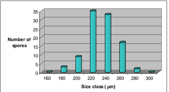
Three types of AMF native from Indonesia, named M1, M2 and M3, were identified by their morphological spore characterization mainly based on the mean of spore diameter distribution, spore colour, wall structure and hypha attachment (INVAM 2009; Schenk and Smith 1982; Schenk and Perez 1987; Nicolson and Schenk 1979; Bentivenga and Morton 1995). The observations were done to a hundred of spores of each of the three types of AMF.
Canino wt Canino rolABC Blank Leaves stem Leaves stem
330 bp 210 bp 550 bp rolB rolA rolC