SCUOLE DOTTORALE IN BIOLOGIA
SEZIONE: Biologia Applicata alla Salute dell’Uomo (BASU)
CICLO DEL CORSO DI DOTTORATO
XXV
Identification of compounds affecting
Pseudomonas aeruginosa social behaviour
and virulence
Identificazione di composti inibitori della
virulenza in Pseudomonas aeruginosa
Dottorando
Cejoice Ramachandran Pillai
Docente guida/Tutor:
Prof.ssa Livia. Leoni
Coordinatore:
Prof. Paolo Visca
CONTENTS
SUMMARY I
1. INTRODUCTION 1
1.1 Pseudomonas aeruginosa 1
1.1.1 Quorum sensing-dependent regulation of virulence
in P. aeruginosa 3
1.2 RND family efflux pumps in P. aeruginosa 7
1.2.1 RND efflux pumps and bacterial virulence 11 1.2.2 RND efflux pumps inhibitors 12
2. GENERAL RATIONALE AND AIMS 15
3. CHAPTER III
New life for an old drug: the antihelmintic drug niclosamide
inhibits Pseudomonas aeruginosa quorum sensing 17
3.1 Background 17
3.2 Results 19
3.2.1 Identification of FDA-approved compounds
inhibiting P. aeruginosa QS 19 3.2.2 Niclosamide inhibits the 3OC12-HSL-dependent QS
system of P. aeruginosa 21
3.2.3 Niclosamide represses QS-activated gene 24 3.2.4 Niclosamide strongly reduces the virulence potential
of P. aeruginosa in vitro 27
3.2.5 Niclosamide protects G. mellonella from P. aeruginosa
Infection 30
3.3 Discussion 31
4. CHAPTER IV
Beyond antibiotics: efflux pumps inhibition as anti-virulence
strategy against Pseudomonas aeruginosa. 35
4.1 Background 35
4.2 Results 36
4.2.1 In vitro anti-virulence activity of PAβN 36 4.2.2 In vivo anti-virulence activity of PAβN 43
4.3 Discussion 45
5. REFERENCES 48
6. LIST OF ABBREVIATIONS 61
I"
SUMMARY
Pseudomonas aeruginosa is the most common Gram-negative bacterium
responsible for hospital-acquired infections and is a serious threat to immune compromised individuals. P. aeruginosa infections are difficult to eradicate because this pathogen is resistant to conventional antibiotic therapies. Resistance to antibiotics is mainly due to the ability of this microorganism to form biofilm and to express efflux pumps that reduce the intracellular concentration of the drug.
Targeting the bacterial pathogenic potential rather than bacterial growth has the advantage of reducing the bacterial adaptability to the host environment and the severity of the infection without creating the selective pressure generally caused by conventional antibiotics. The use of virulence inhibitors could ultimately provide the host immune system with a better chance of clearing the infection.
The overall rationale and aim of this project has been to pave the way for the development of innovative anti-virulence therapies specifically targeting P. aeruginosa.
The objective of the study described in Chapter 3 has been the identification of compounds inhibiting P. aeruginosa quorum sensing (QS), a cell-cell communication process playing a key role in the expression of virulence factors and biofilm development.
It is well known that searching for new side activities in drugs which use in humans has already been approved is an intelligent strategy for the development of novel drugs. This strategy is expected to reduce the time and cost associated with standard drug discovery processes.
About thousand compounds already used as drugs in humans were screened for their anti-QS activity, using a biosensor developed in our laboratory. Seven compounds with anti-QS activity were identified; the most effective drug was the anthelmintic drug niclosamide.
Microarray analysis showed that niclosamide (at 20 µM) affects the transcription of 258 genes, with a high degree of target specificity towards the QS-dependent genes. 69 genes were up-regulated and 189 genes were down-regulated, 121 of the latter group were previously included within the QS regulon. Phenotypic assays demonstrated that niclosamide suppresses surface motility, production of secreted virulence factors (elastase, pyocyanin and rhamnolipids) and reduces biofilm formation. In accordance with the strong anti-virulence activity disclosed in vitro, niclosamide
II"
prevented P. aeruginosa pathogenicity in the Galleria mellonella insect model of acute infection.
To our knowledge, this is the first study in which a drug-repurposing strategy has been applied to the development of P. aeruginosa anti-virulence drugs. Besides the finding that niclosamide, an FDA-approved drug has a promising anti-virulence activity against one of the most antibiotic-resistant bacterial pathogens, this work provides a proof of concept that a lateral anti-QS activity can be detected among drugs already used in humans, validating a new approach to identify virulence inhibitors that could easily move into clinical applications.
Chapter four of this thesis describes a study aimed at investigating the inhibition of P. aeruginosa virulence via chemical inactivation of Resistance-Nodulation-Cell Division (RND) efflux pumps. Efflux pumps of this family are important for multiple drug-resistance in many pathogenic bacteria, including P. aeruginosa.
Since no close human homologues of RND transporters have been described, efflux pumps inhibitors (EPI) are considered promising scaffolds and lead compounds for the development of drugs aimed at potentiating antibiotic activity. Since in some bacterial pathogens RND efflux pumps are also involved in virulence, EPIs have been proposed as virulence inhibitors. However, no literature data are available concerning the potential anti-virulence activity of EPIs.
The main aim of this study has been to provide a first proof of concept that, apart from their role in antibiotic resistance, EPIs may act as anti-virulence drugs against P. aeruginosa, by using phenyl-arginine β-naphthylamide hydrochloride (PAβN) as model compound.
Here, we demonstrate that PAβN 6.5 µM abrogates swarming motility, an in vitro phenotype strictly related to virulence in vivo. Moreover, microarray analysis showed that PAβN affects the transcription of about 109 genes. P. aeruginosa transcriptome is affected by PAβN in a specific way, since it negatively affects the expression of particular groups of genes, mainly related to iron and phosphate acquisition, while it increases the expression of genes involved in nitrogen metabolism.
Overall, our results demonstrate that in P. aeruginosa PAβN has pleiotropic effects that go far beyond the increased susceptibility to antibiotics. In particular, PAβN decreases the transcription of virulence related genes, resulting in a reduced pathogenic potential in the G.
III"
In conclusion, the use of EPIs in therapy is a promising challenge since these compounds, besides their effect as antibiotic adjuvants, may display anti-virulence properties.
1"
1. INTRODUCTION
1.1. Pseudomonas aeruginosaPseudomonas aeruginosa is the most common Gram-negative bacterium
responsible for hospital-acquired infections, and is a serious threat to immune compromised individuals such as neutropenic, cancer, bone marrow transplant and AIDS patients. This bacterium is known to cause pneumonia, urinary tract, surgical wound, bloodstream infections, bacteremia in severe burn victims and keratitis in contact lenses users. In addition, P. aeruginosa chronic lung infection is the major cause of death in cystic fibrosis (CF) patients, a genetic disease affecting about 1/3000 newborns in the Caucasian population (Driscoll et al., 2007; Rosenthal et
al., 2012).
Antibiotics are often poorly effective against P. aeruginosa, especially in the treatment of chronic infections where this microorganism forms biofilm. Moreover, the strong selective pressure exerted by antibiotics prompts to the emergence of resistant strains. For these reasons, P.
aeruginosa infections are characterized by high morbidity and mortality
rates (Breidenstein et al., 2011; Rosenthal et al., 2012).
Overall, P. aeruginosa causes 140,000 infections in the EU region per year, with more than 10,000 deaths and around 800,000 extra hospital days costing billions of Euros. The ability of P. aeruginosa to acquire genetic determinants of antibiotic resistance results in the generation of pan-resistant strains for which currently available antibiotic therapies are no longer effective (Latifi et al., 1995), calling for the development of new anti-P. aeruginosa drugs.
The capacity of P. aeruginosa to produce diverse infections is due to an arsenal of virulence factors. These factors are collectively capable of causing extensive tissue damages, blood stream invasion and dissemination in humans and other mammals (Fig. 1; reviewed by Smith and Iglewski, 2003).
Some of the virulence factors that confer to the pathogen the ability to colonize the host are cell surface virulence factors. Actually the cell surface itself is a virulence factor because it contains very immunogenic compounds, like the lipopolysaccharide (LPS). Flagella and pili involved in different kind of motility and in chemotaxis, display a critical role in pathogenesis, by adhering to epithelial cells and stimulating an inflammatory response (Adamo et al., 2004; DiMango et al., 1995). When
2"
growth. The generally accepted definition of a biofilm is a community of cells attached to a surface or to each other, embedded in a self-made, protective matrix of extracellular polymeric substances (Kirisits and Parsek, 2006). The clinical implications of bacterial biofilms are particularly pronounced. Biofilms may form on any foreign object inserted into the human body, and also in the lungs of CF patients P. aeruginosa forms biofilm during chronic infection. In the biofilm mode the bacteria are highly tolerant to the action of several antimicrobial agents including antibiotics, disinfectants, and to the action of the immune system (Davies, 2002; Donlan and Costerton, 2002; Drenkard, 2003).
P. aeruginosa also produces several extracellular products that, after the
initial step of colonization, cause extensive tissue damage, bloodstream invasion and dissemination. Some of these extracellular products, besides having the role of favouring pathogen dissemination, also provide nutrients to P. aeruginosa by causing host tissues damage. These secreted factors are elastases, alkaline proteases, exotoxins and hemolysins that also contribute to the infection in lung disease by destroying the protective glicocalix of the respiratory epithelium (Kipnis et al., 2006). Other secreted factors are toxic compounds such as hydrogen cyanide and pyocyanin: hydrogen cyanide is a potent poison that blocks cytochrome oxidase, leading to the inhibition of mitochondrial respiration (Gallagher and Manoil, 2001). Pyocyanin is a blue pigment metabolite of P. aeruginosa that has been shown to have numerous pathogenic effects such as increasing IL-8, depressing host-response, and inducing apoptosis in neutrophils (Allen et al., 2005; Denning
et al., 1998). In animal models of acute and chronic lung infection,
pyocyanin was shown to be essential for P. aeruginosa virulence (Lau et
al., 2004). Additionally, due to its known oxidoreductive properties,
pyocyanin oxidizes glutathione and inactivates catalase in respiratory epithelial cells thus participating in oxidative-stress related damage (O’Malley et al., 2004). The need for iron in P. aeruginosa is supported by the production of the two siderophores, pyoverdine and pyochelin, that are small molecules chelating iron from the iron-poor environment encountered in the host, allowing its utilization in P. aeruginosa metabolism (Buckling
3"
Figure 1. Virulence and antibacterial resistance factors in P. aeruginosa (modified from Van
Delden and Iglewski, 1998).
1.1.1. Quorum sensing-dependent regulation of virulence in P. aeruginosa.
Many bacteria coordinate their behaviour via the secretion of specific signalling molecules in a population density-dependent manner. During growth the bacteria secrete the signal molecule that accumulates in the surrounding environment as the population density increases until a critical threshold concentration is reached, which then triggers expression of certain sets of genes (Fig. 2). This type of cell-to-cell communication is termed “quorum sensing” (QS), in order to emphasize the fact that a sufficient number of bacteria, the bacterial “quorum”, is needed to induce or repress expression of target genes (Whitehead et al., 2001).
QS is involved in the regulation of a wide variety of different bacterial processes including bioluminescence, horizontal gene transfer, antibiotic biosynthesis, motility, biofilm formation, and virulence in plant, animal and human pathogens (Càmara et al., 2002; Lazdunski et al., 2004).
4"
Figure 2. A simple representation of QS. The signal molecule is constitutively produced at a
basal level by the bacterial cells. At low-cell densities very little signal molecule is present. As cell density increases, the signal molecule accumulates until a threshold level is reached. This signal molecule concentration is responsible for a coordinate transcriptome reprogramming in the whole bacterial population.
In P. aeruginosa there are three interconnected QS systems, each one relying on different signal molecule. The first two systems, named las and
rhl, belong to the Lux-family of QS systems and employ 3-oxo-docecanoyl
homoserine lactone (3OC12-HSL) and butanoyl homoserine lactone (C4 -HSL) as signal molecules, respectively (Fig. 3). The second chemically distinct class of auto inducers are the 4-quinolones which consists of more than 50 compounds (Lepine et al., 2004) and includes the most active signal molecule 2-heptyl-3-hydroxy-4-quinolone (PQS), which is commonly referred to as the Pseudomonas quinolone signal. It is important to notice that the 3OC12-HSL-dependent QS system (i.e. the las system) positively controls the other two QS systems (Williams and Càmara, 2009).
Low cell-density High cell-density
Signal molecule
Low cell-density High cell-density
Low cell-density High cell-density
Low cell-density High cell-density
5"
Figure 3. Structure of QS molecules exploited by P. aeruginosa for cell-to-cell
communication, (modified from Piddock, 2006.).
When the bacterial population has reached a certain density, the signal molecules accumulate, bind to their respective regulator proteins, and initiate the transcription of a distinct set of genes, including own biosynthetic genes (Fig. 4). Many of the quorum sensing-regulated genes are virulence factors, and the extra cellular accumulation of acyl homoserine lactones (acyl-HSL)-signalling molecules has also been shown to impact on the structural development and stabilization of P. aeruginosa biofilms (Davies et al., 1998; Kirisits and Parsek, 2006).
The multiple extracellular pathogenic traits positively regulated by QS in P. aeruginosa include elastases, alkaline protease, exoenzyme S, neuraminidase, haemolysin, lectins, pyocyanin, rhamnolipids, hydrogen cyanide and oxidative stress-responsive enzymes catalase and superoxide dismutase (Williams and Camara, 2009). All these extracellular virulence factors are crucial for the competence of P. aeruginosa to establish and maintain the infection. Mutants defective in QS are typically compromised in their ability to establish a successful infection. QS mutants show reduced virulence in a number of mammalian and non-mammalian infection models (Cosson et al., 2002; Rahme et al., 1995; Rumbaugh et al., 1999; Tan et al., 1999; Tang et al., 1996; Wu et al., 2000). Additionally, the specific signal molecules of all three QS systems of P. aeruginosa were detected in the sputum of CF patients, thus suggesting that QS is functionally active in the CF chronic lung infections (Collier et al., 2002; Erickson et al., 2002; Middleton et al., 2002). The QS systems of P. aeruginosa contribute to its pathogenesis not only by regulating expression of virulence factors, but also by inducing host cellular responses like for instance inflammation and apoptosis (Rumbaugh, 2007; Smith et al., 2002). The rapid increase of multidrug-resistant strains is demanding novel therapeutic approaches.
6"
Given that the QS circuitry in P. aeruginosa plays an important role in controlling pathogenicity as well as biofilm formation, it represents a highly attractive target for the development of novel antimicrobial agents (Reviewed in Warren et al., 2012). It has been suggested that the use of QS inhibitors (QSI) that specifically inhibit expression of pathogenic traits without affecting growth of the bacterium has the advantage of minimizing the possibility of selecting resistant mutants. Although such mutants may eventually arise, they would not have a selective growth advantage and thus would not out-compete the parental strain. Therefore, the emergence of resistance to QSI is an unlikely event (Darch et al., 2012; Hentzer and Givskov, 2003).
Figure 4. Schematic representation of the central core of the QS network in P. aeruginosa,
with las, rhl, and pqs systems. Relevant QS-regulated virulence factors are listed (modified from Camara et al., 2002 and Diggle et al., 2003).
7"
1.2. RND family efflux pumps in P. aeruginosa
Today, the treatment of bacterial infections is severely compromised by the emergence of bacteria, including those belonging to the P. aeruginosa species, that are resistant to multiple antibiotics. The dissemination of multidrug-resistant (MDR) Gram-negative bacteria drastically impairs the efficacy of antibiotic families and limits their clinical use (Falagas and Bliziotis, 2007; Gandhi et al., 2010). The active efflux of antibiotics via efflux pumps is a major cause of the bacterial MDR phenotype. Efflux pumps have been reported to play a key role in mediating multidrug resistance in clinical isolates from varied geographic locations and populations (Martinez et al., 2009; Nikaido, 2009; Piddock, 2006; Poole, 2007).
Efflux pumps have been categorized into five families, based primarily on amino acid sequence identity, on the energy source required to drive export, and on substrate specificity (Lister et al., 2009). The families include (i) the ATP-binding cassette (ABC) family, (ii) the small multidrug resistance family (SMR), (iii) the major facilitator superfamily (MFS), (iv) the resistance-nodulation-division (RND) family, and (v) the multidrug and toxic compound extrusion family (MATE).
Although sequence analysis of the P. aeruginosa genome revealed the presence of efflux systems from all five families, the largest number of predicted pumps belong to the RND family, with a total of 12 RND systems (including two divalent metal cation transporters) (Stover et al., 2000). Unlike the primary active transporters of the ABC family, which utilize ATP hydrolysis for energy, the RND family (as well as the remaining super families) are secondary active transporters (symporters, antiporters, and uniporters) that derive the energy required for compound extrusion by proton motive force (Nikaido, 1996; Poole, 2007).
RND pumps typically exist as a tripartite system consisting of a periplasmic membrane fusion protein (MFP), an outer membrane factor (OMF), and a cytoplasmic membrane (RND) transporter (Fig. 5). This complex forms a channel spanning the entire membrane, allowing for the transportation of lipophilic and amphiphilic drugs from the periplasmic space and cytoplasm to the extracellular environment. The genes encoding these pumps are organized into operons on the P. aeruginosa chromosome (Fig. 6). Each operon is composed of at least two genes, coding for the MFP and the RND transporter. Six of the 12 operons possess an OMF gene, completing the tripartite system, while the remaining operons are devoid of an OMF gene. Several operons have an adjacent regulatory gene transcribed in the same orientation or divergently from the operon, whose product
8"
functions as a repressor or activator of pump expression. Operons may contain additional genes besides those coding for the efflux pump. For example, mexG in the mexGHI-opmD operon encodes an integral membrane protein, and PA2528-PA2527-PA2526-opmB possesses a second RND transporter gene, PA2526 (Lister et al., 2009).
The 10 RND pumps of P. aeruginosa known to be involved in antibiotic efflux are named MexAB-OprM, MexCD-OprJ, MexEF-OprN, MexXY, MexJK, MexGHI-OpmD, MexVW, MexPQ-OpmE, MexMN, and TriABC (Fig. 6). Mex is an acronym for multiple efflux, and “Tri” refers to triclosan efflux. While several of these pumps share common substrates, they are also responsible for unique resistance phenotypes inherent to their expression. Substrates of these pumps include antibiotics, biocides, dyes, detergents, organic solvents, aromatic hydrocarbons (Scweizer, 2003).
9"
Figure 5. Structure of an RND efflux pump. RND pumps typically exist in a tripartite system
consisting of an RND cytoplasmic membrane transporter (RND), a membrane fusion protein (MFP) and an outer membrane factor (OMF). The tripartite complex forms a channel spanning the entire membrane, allowing for the proton-driven transport of lipophilic and amphiphilic drugs from the cytoplasm of the cell across the cytoplasmic membrane, through the periplasmic space, across the peptidoglycan, and across the outer membrane. The RND efflux pumps can also extrude drugs from the periplasmic space before they cross the cytoplasmic membrane (Lister et al., 2009).
10"
Figure 6. Operons encoding RND efflux pumps in P. aeruginosa. Genes which encode protein
components or characterized pumps are denoted by their gene names, and genes encoding protein components of uncharacterized pumps are designated with the P. aeruginosa (PA) numbers assigned in the annotated P. aeruginosa genome sequence (www.pseudomonas.com). Genes are depicted with the following colour scheme: dark red arrow, transcriptional regulator-encoding gene; dark blue arrow, membrane fusion protein-regulator-encoding gene; light blue arrow, RND transporter-encoding gene; red arrow, outer membrane protein-encoding gene; and gold arrow, gene encoding a protein with unknown function (Schweizer, 2003).
"
11"
1.2.1. RND efflux pumps and bacterial virulence
Some bacterial efflux pumps, besides antibiotics, dyes and detergents, export antimicrobial agents and metabolites produced by the bacterium itself, including adhesins, toxins and other factors that are important for the colonization and infection of human and animal cells (Piddock, 2006).
So far, evidence of a role for the RND family pumps in pathogenicity has been obtained for some animal and plant pathogens (Piddock, 2006). For example, the Campylobacter jejuni RND family efflux pump CmeABC is essential for the colonization of 1-day-old chickens (Lin et al., 2003) and
Neisseria gonorrhoeae RND-family efflux pump MtrCDE is important for
infection of the genito-urinary tract of female mice (Jerse et al., 2003). The AcrAB–TolC RND efflux pump is important for the ability of Salmonella
typhimurium to infect BALB/c mice(Lacroix et al., 1996; Stone and Miller, 1995), to colonize chickens, to adhere to, invade and survive in mouse macrophages and to invade and survive in human embryonic intestinal cells (Baucheron et al., 2005; Buckley, 2006).
Burse and colleagues (2004a) have shown that the genome of Erwinia
amylovora (which causes the disease fire-blight in apple and pear trees)
contains an RND efflux pump homologous to AcrAB–TolC of S.
typhimurium. Disruption of E. amylovora acrB gene was found to cause a
significant impairment in the survival of E. amylovora in apple plantlets and a marked reduction in its ability to cause fire blight symptoms. Furthermore, the expression of acrAB was shown to increase two fold during growth of the bacteria in apple tissue contained antimicrobial secondary metabolites, supporting the hypothesis that AcrAB has a role in the colonization of the host plant (Burse et al., 2004 b).
Literature data are also available suggesting a role for P. aeruginosa RND-family efflux pumps in virulence. P. aeruginosa lacking the MexAB– OprM efflux pump could not invade epithelial cells [Madin–Darby canine kidney (MDCK) cells] and invasion could be restored by complementation with MexAB–OprM or by supplementation with culture supernatant obtained from MDCK cells infected with wild-type P. aeruginosa. These findings suggested that MexAB–OprM exports virulence determinants that allow P. aeruginosa to be invasive and cause infection (Hirakata et al., 2002). Further support for the hypothesis that RND efflux pumps may be involved in P. aeruginosa virulence comes from an experimental model of acute pneumonia in rats, in which, even in the absence of antibiotic treatment, isolates of P. aeruginosa that over express MexCD-OprJ and MexEF-OprN efflux pumps were identified (Join-Lambert et al., 2001). Moreover, it was also reported that MexAB-OprM participates in the efflux
12"
of 3OC12-HSL (Evans et al., 1998; Minagawa et al., 2012; Pearson et al., 1999) and that MexEF-OprN and MexGHI-OprM might be involved in transport of PQS and other 2-alkylquinolones (Lamark and Deziel, 2011).
1.2.2. RND efflux pumps inhibitors
The continuous increase in the development of multidrug resistance by many pathogens has resulted in difficult fighting of many infectious diseases. Since the majority of multidrug resistant pathogens expresses RND efflux pumps that are responsible for the extrusion of the antibiotics from inside the cells, a new direction for chemotherapeutics is the use of efflux pump inhibitors (EPIs) (Pagès and Amaral, 2009). The inhibition of the efflux pumps is promising in order to increase the intracellular drug concentration, to restore the drug activity against the resistant strains, and to minimize further development of resistant strains (Askoura et al., 2011; Zhang and Mah, 2008).
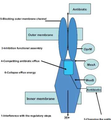
The inhibition of efflux pumps can be achieved by different mechanisms as shown in figure 7: interference with the regulatory steps needed for the expression of the efflux pump (Lister et al., 2011); chemical changes in the antibiotic structure, hence hindering its recognition as the specific substrate (Alyssa et al., 2009); disruption of the assembly of the efflux pump-components (McGowan, 2006); inhibition of the substrate (antibiotic) binding by either competitive or non-competitive binding (Hancock and Speert, 2000); blocking the outer pores responsible for the efflux of antibiotic compound (Falagas and Bliziotis, 2007); interference with the energy required for the pump activity (Pagès and Amaral, 2009).
Peptidomimetic compound molecules with phenyl-arginine β-naphthylamide hydrochloride (PAβN; also named MC-207,110) as a leading compound (Fig. 8) were discovered by MPEX pharma- ceuticals in a screen for adjuvants of levofloxacin activity against a P. aeruginosa strain that over-expressed the levofloxacin-specific efflux pump MexAB-OprM (Askoura et al., 2011). It has been demonstrated that PAβN is very potent as an efflux inhibitor against the major RND pump of P. aeruginosa (MexAB-OprM) when compared to quinoline derivatives (another class of EPIs; Davin-Regli et al., 2006). This compound also shows activity against MexCD-OprJ and MexEF-OprN (Lomovskaya et al., 2001). It was also shown that PAβN, besides quinolones, can restore the activity of other unrelated antibiotics such as chloramphenicol and macrolides; hence, it can be considered a broad spectrum efflux pump inhibitor (Lee et al., 2001; Lomovskaya and Bostian, 2006).
13"
The mechanism of action of PAβN and related compounds is believed to occur through competitive inhibition mechanism (competitively binding to the substrate site of RND efflux pump), where the efflux pumps recognize them as a substrate instead of the target antibiotics. It has been shown that PAβN can compete with certain antibiotics but not with others depending on the nature of the efflux pump, in particular of the substrate-binding site (Lee et al., 2001; Sangalang et al., 2002). In addition to the competitive binding to the RND efflux pumps, Iino and co-workers (2012) recently suggested a membrane-permeabilising effect of PAβN.
The main drawbacks associated with the PAβN-derived EPI compounds are their toxic properties hindering their clinical applications. At present, they are used in order to evaluate the different efflux mechanisms expressed by different pathogenic bacteria (Lomovskaya and Bostian, 2006; Lomovskaya et al., 2001).
Overall, PAβN-derived compounds are the most studied and developed EPIs against P. aeruginosa, though more studies concerning their structure-activity relationship, pharmacokinetics, and stability in biological fluids are required.
14"
Figure 7. Schematic illustration showing the general mechanisms of efflux pump inhibition
(and the targets that can be affected) using MexAB-OprM efflux pump as an example (Askoura
et al., 2011).
15"
2. GENERAL RATIONALE AND AIMS
P. aeruginosa was selected as model organism for this project because it
is one of the most dreaded pathogens in developed countries. Indeed, current treatments, based on traditional antibiotics that kill or inhibit the growth, are often thwarted by P. aeruginosa ability to develop resistance to all known classes of antibacterial agents (Gilbert, 2010).
The anti-virulence drugs are able to specifically inhibit the bacterial capability to establish the infection rather than bacterial growth, and represent an innovative therapeutic approach against difficult-to treat bacteria, either when used alone or in combination with antibiotics. The use of anti-virulence drugs has the advantage of reducing the bacterial adaptability to the host environment, facilitating the host immune system to clear the infection, without creating, in principle, the strong selective pressure generally caused by antibiotics (Rasko and Sperandio, 2010).
In P. aeruginosa, the QS global regulatory system is necessary for full pathogenicity and represents an ideal target to develop anti-virulence drugs (Brjansholt et al., 2010). In addition, recent studies supported the ground-breaking principle that the selection of mutants resistant to QS inhibitors (QSI) is an unlikely event. Indeed, despite the emergence of mutants resistant to QSI is possible, such mutants behave as social cheaters and are not able to enrich in a population of QSI-sensitive bacteria (Maeda et al., 2012; Mellbye and Schuster, 2011).
Overall, the state of the art knowledge strongly supports the hypothesis that targeting QS via anti-virulence drugs represents a promising strategy to prevent and/or treat P. aeruginosa infections (Bjarnsholt et al., 2010).
In addition, the notion is emerging that RND efflux pumps are involved in the secretion of endogenous bacterial products, including QS signal molecules and virulence factors (Piddock, 2006). In P. aeruginosa, efflux pump inhibitors (EPIs) have been mainly studied for their effect on antibiotic resistance, while, to our knowledge, nothing is known about their impact on QS and virulence-related phenotypes.
The general aim of this PhD work has been to gain new insights in the anti-virulence drug therapy against P. aeruginosa.
The specific aim of the work described in chapter three has been to apply a drug-repurposing approach to the identification of anti-virulence drugs targeting P. aeruginosa QS. The work described in this chapter has been recently published (Imperi et al., 2013).
16"
Chapter four describes a study investigating the potential of EPIs as anti-virulence drugs, apart from their role in antibiotic resistance, using PAβN as model compound. A manuscript concerning this study is in preparation.
17"
3. CHAPTER III
New life for an old drug: the antihelmintic drug
niclosamide inhibits Pseudomonas aeruginosa quorum
sensing
3.1. Background
The introduction of antibiotics in the clinical practise at the middle of 20th century is a milestone in the history of medicine. However, the original expectation that all bacterial infections could be one day defeated by antibiotics was soon disappointed by the emergence of antibiotic resistant strains, prompting the still ongoing race for the discovery of new antibacterial agents. While the treatment of infections sustained by antibiotic resistant bacteria has high socio-economic costs and represents a major health problem worldwide, pharmaceutical industries have dramatically reduced the investments in antibiotics research. As traditional antibiotic research appears to be helpless to cope with the emergence of antibiotic resistant strain, novel scientifically sound and cost effective approaches should be undertaken in order to identify new drugs (Gilbert, 2010).
Selective Optimization of Side Activities of drug molecules (the SOSA approach) is a smart strategy for the identification of new potential drugs (Wermuth, 2006). A limited number of highly diverse drugs, whose use in humans has already been approved, are screened for side activities against unrelated diseases. Once a hit compound has been found, this could be either tested directly in clinical studies or used as the lead for drug optimization programs. This strategy has a high probability of yielding safe and bioavailable drug-like compounds, and it is thus expected to reduce the time and cost generally associated with standard drug discovery processes (Antoniani et al., 2010; Ejim et al., 2011; Wermuth, 2000).
An innovative strategy to combat bacterial infections relies on specific inhibition of bacterial virulence, hence the ability to cause disease, rather than bacterial growth (Cegelski et al., 2008). The use of “anti-virulence drugs” could have the advantage of reducing the bacterial adaptability to the host environment, facilitating the host immune system to combat the infection and reducing the strong selective pressure exerted by conventional antibiotics (Rasko and Sperandio, 2010), although this is not yet supported by direct clinical evidence.
In many bacteria, pathogenicity is controlled and coordinated by an inter-cellular communication process named quorum sensing (QS). QS is
18"
based on the synthesis and secretion of a signal molecule that binds to a cognate receptor. The signal-activated receptor controls the expression of target genes. Since the production of the signal molecule is proportional to the bacterial growth, QS coordinates gene expression in response to the bacterial population density (Atkinson and Williams, 2009). So far, QS is considered one of the most promising targets for anti-virulence therapies (Amara et al., 2011; Njoroge and Sperandio, 2009; Rasko and Sperandio, 2009).
In this study the SOSA approach has been applied to the identification of anti-virulence drugs targeting bacterial QS, using Pseudomonas
aeruginosa as a model organism. P. aeruginosa is one of the most dreaded
Gram-negative pathogens in developed countries, being responsible for both community- and hospital-acquired infections. In addition, P. aeruginosa chronic lung infection is the major cause of death in cystic fibrosis (CF) patients, a genetic disease affecting about 1/3,000 newborns in the Caucasian population (Driscoll et al., 2007; Orsi et al., 2005; Rosenthal et
al., 2012; Talbot et al.,2000). Besides being intrinsically resistant to several
antibiotics, P. aeruginosa can easily acquire new resistance determinants, and indeed the emergence of pan-resistant strains has already been documented (Page and Heim, 2009). For these reasons, P. aeruginosa infections are generally characterized by high morbidity and mortality rates (Breidenstein et al., 2011Rosenthal et al., 2012).
The pathogenic potential of P. aeruginosa relies on the coordinated expression of a large array of virulence factors (Lee et al., 2006), the majority of which are positively controlled by QS (Williams and Càmara, 2009). The P. aeruginosa QS network consists of three different QS systems, based on the production of specific signal molecules: N-3-oxododecanoyl-homoserine lactone (3OC12-HSL), N-butanoyl-homoserine lactone (C4-HSL), and 2-heptyl-3-hydroxy-4-quinolone (PQS). P.
aeruginosa QS is hierarchically organized, since 3OC12-HSL is required for optimal production of the other QS signals (Williams and Càmara, 2009).
QS controls the expression of nearly 10% of the P. aeruginosa genome, including genes for biofilm formation, secreted virulence factors, immune-modulatory and pro-inflammatory agents (Williams and Càmara, 2009). QS signal molecules can be detected in clinical samples, proving that QS is active during P. aeruginosa infections. Moreover, QS defective mutants show strongly impaired virulence in several animal models of infection, corroborating the importance of QS for P. aeruginosa pathogenicity and its suitability as a target for the development of anti-Pseudomonas drugs (Bjarnsholt et al., 2010; Winstanley and Fothergill, 2009).
19"
We have recently developed a convenient system for the identification of compounds affecting the P. aeruginosa 3OC12-HSL-based QS system at multiple levels: (i) expression/activity of the signal receptor, (ii) expression/activity of the signal synthase, and (iii) activity/availability of the signal molecule (Massai et al., 2011). Here, screening a library of about one thousand compounds with known pharmacological activities has validated this system.
Seven hit compounds have been identified. Among these, we have focused our investigation on the anthelmintic drug niclosamide, which showed high inhibitory activity against P. aeruginosa QS and virulence both in vitro and in vivo. To the best of our knowledge, this is the first demonstration that the SOSA approach can be successfully applied to the search for anti-QS drugs.
3.2. Results
3.2.1. Identification of FDA-approved compounds inhibiting P. aeruginosa QS.
We recently developed a novel screening system for the identification of
P. aeruginosa QSI. This system is based on the co-cultivation of a biosensor
strain for 3OC12-HSL detection, PA14-R3, and a wild-type P. aeruginosa PA14 strain. The 3OC12-HSL signal synthesized by the wild-type PA14 induces bioluminescence emission by the biosensor (Massai et al., 2011). The addition of a molecule with inhibitory activity towards any process related to the 3OC12-HSL-dependent QS system, including 3OC12-HSL synthesis, transport and perception, would reduce the luminescence emitted by the biosensor with respect to a control co-culture without any compound added (Massai et al., 2011).
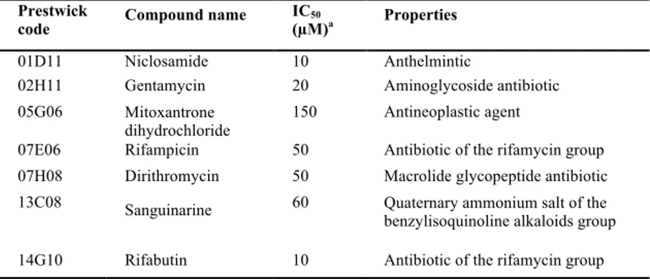
The PA14/PA14-R3 co-cultivation system was used to screen a commercial library of marketed drugs from Prestwick Chemicals (www.prestwickchemical.fr). This library contained 1,120 chemical compounds with known biological activities, selected for their high chemical and pharmacological diversity, as well as for known bioavailability and safety in humans. Each drug was tested at three different concentrations (100, 10 and 1 µg/ml) in duplicate. Criteria used for the selection of hit compounds were: (i) = 50% inhibition of bioluminescence emission and (ii) = 20% reduction of growth with respect to the untreated controls. The latter criterion was aimed at avoiding any unspecific effect of impaired growth on the QS response.
20"
which reproducibly inhibited the QS response of the PA14/PA14-R3 co-cultivation system, without affecting bacterial growth at the highest concentration tested. The seven hits were further tested in triplicate at 100, 80, 60, 40, 20, 10, 5 and 2.5 µg/ml final concentration, showing half maximal inhibitory concentration (IC50) in the range 3-77 µg/ml (corresponding to 10-150 µM; Table 1). Four of the identified compounds are antibiotics, in agreement with the well-known negative effect of sub-inhibitory concentrations of antibiotics on the P. aeruginosa QS response (Jander et al., 2000; Babic et al., 2010). The remaining three compounds corresponded to a quaternary ammonium salt, an anti-cancer drug and a teniacide for the treatment of tapeworm infections (Table 1). Among non-antibiotic drugs, the teniacide niclosamide showed the highest anti-QS activity (lowest IC50) (Table 1), and was therefore selected for further investigations.
Table 1. Hit compounds identified by screening the Prestwick Chemical Library with the
PA14/PA14-R3 QSI screening system.
Prestwick
code Compound name IC(µM)50 a Properties
01D11 Niclosamide 10 Anthelmintic
02H11 Gentamycin 20 Aminoglycoside antibiotic 05G06 Mitoxantrone
dihydrochloride
150 Antineoplastic agent
07E06 Rifampicin 50 Antibiotic of the rifamycin group 07H08 Dirithromycin 50 Macrolide glycopeptide antibiotic 13C08 Sanguinarine 60 Quaternary ammonium salt of the benzylisoquinoline alkaloids group 14G10 Rifabutin 10 Antibiotic of the rifamycin group
a The IC
50 values have been determined using the PA14/PA14-R3 co-culture grown for 4
hours at 37°C in the presence of 100, 80, 60, 40, 20, 10, 5 and 2.5 µg/ml of each compound, and then expressed as µM concentrations.
21"
3.2.2. Niclosamide inhibits the 3OC12-HSL-dependent QS system of P.
aeruginosa.
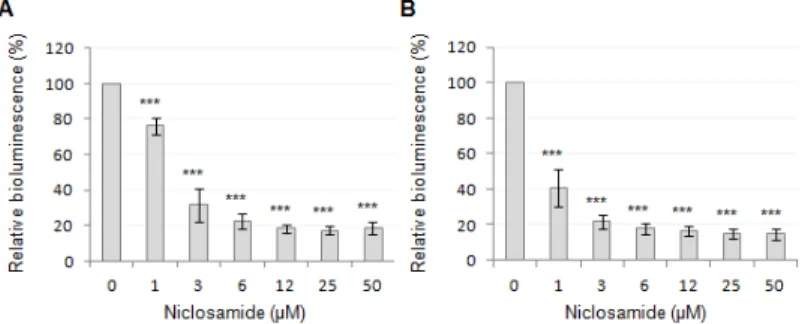
To verify the result of the screening assay, niclosamide was purchased from an alternative supplier (Sigma-Aldrich) and retested in the PA14/PA14-R3 co-cultivation system. As expected, a strong inhibition of the 3OC12-HSL-dependent QS response was observed, with an IC50 even lower than that calculated for the compound from the Prestwick library (Fig. 9A). Notably, niclosamide was also able to inhibit luminescence emission by the PA14-R3 reporter strain grown in the presence of exogenously-added synthetic 3OC12-HSL (3 µM final concentration; Fig. 9B). This result suggests that the QS inhibitory activity of niclosamide relies on its ability to hamper the response of P. aeruginosa to the signal molecule rather than to inhibit its synthesis. The possibility that the observed QS-inhibitory activity of niclosamide was due to unspecific inhibition of either bioluminescence-generating enzymes or bacterial transcription was ruled out by the observation that niclosamide had no effect on the bioluminescence emitted by a P. aeruginosa strain in which bioluminescence genes where under the control of the promoter region of the QS-independent cysB gene (data not shown), involved in cysteine metabolism and iron uptake (Imperi et al., 2010).
The effect of niclosamide on the production of 3OC12-HSL was then assessed. P. aeruginosa PA14 was grown in the absence or in the presence of 20 µM niclosamide, and 3OC12-HSL levels in culture supernatants were quantitatively determined during the whole growth curve. Niclosamide caused a significant reduction (30-60%) of 3OC12-HSL production from the late exponential growth to the entry into the stationary phase, after which 3OC12-HSL concentration fell to almost undetectable levels in both niclosamide-treated and –untreated cultures (Fig. 10A). When 3OC12-HSL levels were determined in P. aeruginosa PA14 cultures grown in the presence of different niclosamide concentrations (0-50 µM), the maximum inhibitory effect on 3OC12-HSL production (about 60% reduction) was observed at 5 µM, and higher concentrations did not further reduce 3OC12-HSL production (Fig. 10B).
Since 3OC12-HSL influences the expression of the other QS systems, the effect of niclosamide on the production of C4-HSL and PQS was tested. To this aim, C4-HSL and PQS levels in P. aeruginosa PA14 cultures treated or not with 20 µM niclosamide were determined along the growth curve. While niclosamide did not significantly affect PQS levels (data not shown), it considerably delayed the production of C4-HSL (Fig. 10C). By comparison with untreated cultures, C4-HSL levels were significantly lower
22"
in niclosamide-treated cultures during the exponential growth, while levels were comparable between treated and untreated cultures in the stationary phase (Fig. 10C). This could be due, at least in part, to the positive effect exerted by 3OC12-HSL on C4-HSL production (Williams and Càmara, 2009). However, the niclosamide-induced delay in C4-HSL production was also evident in a 3OC12-HSL-defective mutant strain inactivated in the lasI gene, encoding the 3OC12-HSL synthase (Fig. 10C), indicating that niclosamide also affects C4-HSL production independently of its inhibitory activity on 3OC12-HSL production.
Figure 9. Effect of niclosamide on the 3OC12-HSL-dependent QS of P. aeruginosa. Response
to increasing concentrations of niclosamide (0-50 µM) of (A) the PA14/PA14-R3 co-cultivation system and (B) the PA14-R3 biosensor in the presence of 3 µM exogenously-provided 3OC12-HSL. Bioluminescence emission was normalized to the cell density of the
bacterial culture (relative bioluminescence, LCPS/A600), and expressed as percentage relative to
untreated controls. Values are the mean (± SD) of at least three independent experiments. *** p < 0.001 (ANOVA).
23"
Figure 10. Effect of niclosamide on acyl-HSL production. (A) Growth curve and 3OC12-HSL
production by P. aeruginosa PA14 treated (squares) or untreated (circles) with 20 µM niclosamide. Symbols: bacterial growth (left vertical axis), grey lines and filled symbols; 3OC12-HSL levels in culture supernatants (right vertical axis), black lines and open symbols.
(B) Relative 3OC12-HSL levels (µM/A600) in culture supernatants of P. aeruginosa PA14
grown for 8 h in the presence of increasing concentrations of niclosamide (0-50 µM). (C) C4
-HSL levels in culture supernatants of P. aeruginosa PA14 (solid lines, white symbols) and PA14 lasI mutant (dashed lines, black symbols) grown in the presence (squares) or in the absence (circles) of 20 µM niclosamide. Growth curves of bacterial cultures were comparable to those reported in (A). Values are the mean ( SD) of at least three independent experiments. *** p < 0.001 (ANOVA).
24"
3.2.3. Niclosamide represses QS-activated genes.
To investigate the global effect of niclosamide on the P. aeruginosa transcriptome, the transcriptional profiles of PA14 grown to A600 = 2.5 in the presence or in the absence of 20 µM niclosamide were compared by Affymetrix high-density oligonucleotides microarray analysis. Niclosamide affected the transcription of 258 genes, 73.2% of which were repressed by this drug, including genes involved in the production of important virulence factors, such as phospholipase C, LasA protease, pyocyanin, chitinase, rhamnolipids, and LasB elastase (Tables 2 and S1). Moreover, niclosamide repressed the transcription of genes involved in adhesion and biofilm formation, such as those coding for adhesins (PA0852-cbpD and
PA2570-pa1L) and for cyclic di-GMP turnover or response proteins (PA1120-tpbB,
PA2572, and PA4781). The transcription of the mexGHI-opmD genes, encoding an efflux pump required for full virulence in rat and plant infection models (Aendekerk et al., 2005), was also strongly decreased in the presence of niclosamide (Table 2). Notably, among the 189 genes repressed by niclosamide, 96 have been identified as genes activated by 3OC12-HSL and/or C4-HSL in the main reference studies (Hentzer et al., 2003; Schuster et al., 2003; Wagner et al., 2003) (Table S1), and additional 25 genes have been suggested to be part of the QS network (Rampioni et
al., 2007; Rampioni et al., 2009;Déziel et al., 2005; Bredenbruch et al.,
2006; Lequette et al., 2006; Rampioni et al., 2010). Among the core components of the P. aeruginosa QS network (i.e. signal synthases and signal receptor genes), only the C4-HSL receptor gene rhlR was significantly repressed by niclosamide (Table 2). Similar results have been reported for other QSIs, such as furanone C-30 (Hentzer et al., 2003), iberin (Jakobsen et al., 2012) and ajoene (Jakobsen 2012). In total, 121 out of the 189 genes repressed by niclosamide (64%) can be classified as QS-regulated.
Niclosamide displayed a positive effect on the transcription of 69 genes, including two genes involved in type VI secretion pathway (PA0070-tagQ1 and PA0085-hcp1; Table 2). These genes are the only virulence-related determinants whose transcription is induced by niclosamide. Only four of the niclosamide-activated genes were previously reported to be repressed by 3OC12-HSL and/or C4-HSL (Hentzer et al., 2003; Schuster et al., 2003; Wagner et al., 2003) (Table S2), suggesting that the majority of the genes induced by niclosamide are affected via QS-independent pathway(s). This observation, together with the finding that 36% of the niclosamide-repressed genes have never been reported as QS-controlled, suggests that this drug may have additional cellular targets besides the QS network.
25"
Notably, a total of 16 putative or confirmed transcriptional regulators were identified among the genes repressed or activated by niclosamide (Tables S1 and S2). Besides rhlR, niclosamide decreased the transcription of pprB, encoding a transcriptional activator associated with biofilm formation (Giraud et al., 2011). Conversely, it positively affected the transcription of
cifR (Table 2), encoding the transcriptional repressor of the Cif toxin,
responsible for apical membrane down-regulation of the cystic fibrosis transmembrane conductance regulator (CFTR) in epithelial cells (MacEachran et al., 2008). The niclosamide-affected transcriptional factors may act as ancillary regulators, increasing the number of genes whose expression is altered by this drug beyond the QS regulon. However, a complete understanding of the niclosamide impact on P. aeruginosa physiology is partially hampered by the high percentage of niclosamide-controlled genes (~ 41%) coding for proteins still classified as hypothetical (Tables S1 and S2).
26"
Table 2. List of selected genes whose transcription is affected by niclosamide. PA
number a Gene name a Fold change b Gene product a
Virulence factors
PA0026 plcB -4.15 phospholipase C, PlcB
PA0070 tagQ1 2.73 protein secretion apparatus, type VI secretion system
PA0085 hcp1 2.99 Type VI protein secretion system component Hcp
PA1871 lasA -10.05 LasA protease precursor
PA1901 phzC1/C2 -2.22 phenazine biosynthesis protein PhzC
PA1905 phzG2 -2.19 probable pyridoxamine 5'-phosphate oxidase
PA2300 chiC -7.24 Chitinase
PA3478 rhlB -5.99 rhamnosyltransferase chain B
PA3479 rhlA -5.65 rhamnosyltransferase chain A
PA3724 lasB -2.90 elastase LasB
PA4210 phzA1/A2 -3.06 probable phenazine biosynthesis protein
PA4211 phzB1/B2 -2.42 probable phenazine biosynthesis protein
Adhesion and biofilm formation
PA0852 cbpD -3.92 chitin-binding protein CbpD precursor
PA1120 tpbB -2.19 diguanylate cyclise
PA2570 pa1L -4.08 PA-I galactophilic lectin
PA2572 --- -3.17 probable two-component response regulator
PA4781 --- -2.50 cyclic di-GMP phosphodiesterase
Gene regulation
PA2931 cifR 2.10 transcriptional regulator
PA3477 rhlR -2.56 transcriptional regulator RhlR
PA4296 pprB -3.21 two-component response regulator
Drug-efflux
PA4205 mexG -29.17 hypothetical protein
PA4206 mexH -16.94 probable RND efflux membrane fusion protein precursor
PA4207 mexI -13.87 probable RND efflux transporter
PA4208 opmD -9.12 probable outer membrane protein precursor
a PA number, gene name and gene product are from the Pseudomonas Genome Database
(http://www.pseudomonas.com). Genes previously reported to be activated by the 3OC12-HSL
and/or C4-HSL QS systems are in bold characters (Hentzer et al., 2003; Schuster et al., 2003;
Wagner et al., 2003). RND, resistance-nodulation-cell division.b Fold change in gene
expression of P. aeruginosa PA14 grown in LB supplemented with 20 µM niclosamide compared to the same strain grown in LB.
27"
3.2.4. Niclosamide strongly reduces the virulence potential of P. aeruginosa in vitro.
In order to validate the transcriptomic data at the phenotypic level, we assessed the effect of niclosamide on the production of a set of QS-regulated virulence traits. In particular, we focused on (i) the LasB elastase, which is directly regulated by the 3OC12-HSL receptor LasR at the transcriptional level (Anderson et al., 1999), (ii) pyocyanin and rhamnolipids, which are regulated by a number of different regulatory pathways and extracellular signals (Lau et al., 2004; Reis et al., 2011), and (iii) multifactorial phenotypes, such as motility and biofilm, which are crucial for the establishment and persistence of P. aeruginosa infections (Parsek and Singh, 2003; Zolfaghar et al., 2003; Andrews et al., 1982).
In accordance with microarray analysis, niclosamide had a marked inhibitory effect on the levels of QS-regulated secreted virulence factors of
P. aeruginosa PA14 (Fig. 11). Production of both pyocyanin and elastase
was dramatically reduced (85-90%) by 5-10 µM niclosamide. Likewise, the amount of rhamnolipids in supernatants of niclosamide-treated cultures was about 25% of the niclosamide-untreated control level (Fig. 11).
Regarding bacterial motility, niclosamide only slightly reduced swimming and twitching motilities of P. aeruginosa PA14 at high concentrations (= 50-100 µM), while it exerted a dramatic inhibitory effect on swarming motility (Fig. 12A-C). Swarming was completely prevented at 4 µM niclosamide concentrations, although a significant reduction was also observed at lower concentrations (Fig. 12B), which however had no effect on the 3OC12-HSL-dependent QS system (Figs. 9 and 10).
Niclosamide was also tested for its effect on P. aeruginosa biofilm formation using a standard crystal violet binding assay (Merritt et al., 2005). Niclosamide showed a significant biofilm inhibitory activity, resulting in a 2-fold reduction and 3-fold increase in the number of attached and planktonic cells, respectively (Fig. 12D). However, such inhibitory activity was only observed at = 200 µM niclosamide, i.e. at concentrations which are exceedingly higher than those active against the QS response and virulence factor production (Figs. 10-12), suggesting that the effect of niclosamide on biofilm formation is independent of its anti-QS activity.
28"
Figure 11. Effect of niclosamide on the production of QS-regulated extracellular virulence
factors. (A) LasB elastase, (B) pyocyanin and (C) rhamnolipids levels in culture supernatants of P. aeruginosa PA14 grown for 10 h (A and B) and for 24 h (C) in the presence of increasing concentrations of niclosamide (0-50 µM). Values were normalized to the cell density of the bacterial culture (relative values), and are the mean (± SD) of four independent experiments. **
29"
Figure 12. Effect of niclosamide on P. aeruginosa motility and biofilm formation. (A)
Swimming, (B) swarming and (C) twitching by P. aeruginosa PA14 in the presence of increasing concentrations of niclosamide. (D) Biofilm formation by P. aeruginosa PA14 in the presence of increasing concentrations of niclosamide, assessed as amount of attached cells (grey histograms, left axis) versus planktonic cells (black squares, right axis). Values are the mean ( SD) of at least three independent experiments. ** p < 0.01; *** p < 0.001 (ANOVA).
30"
3.2.5. Niclosamide protects G. mellonella from P. aeruginosa infection.
In order to explore the suitability of niclosamide as an anti-virulence drug against P. aeruginosa infections, we assessed the ability of this compound to inhibit the pathogenicity of P. aeruginosa in the G. mellonella insect model of infection (Jander et al., 2000).
Larvae of the wax moth G. mellonella are extremely sensitive to P.
aeruginosa injected into the hemolymph, and the PA14 strain was found to
be highly virulent in this model, with an LD50 of about one bacterial cell (Jander et al., 2000). In our study, G. mellonella larvae were inoculated with 10 µl of saline solution containing a lethal dose of P. aeruginosa PA14 (10 ± 4 cells from exponential cultures) and containing or not 750 µM niclosamide ethanolamine salt, and then incubated at 28°C for up to one week. Niclosamide ethanolamine salt was used because of its higher solubility in aqueous solutions compared with niclosamide (Andrews et al., 1982). Notably, the two niclosamide formulations displayed comparable inhibitory effects on virulence factors production, motility and QS signal molecule production (data not shown). Considering that the average weight of G. mellonella larvae was about 500 mg (see Material and Methods), and arbitrarily assuming 500 µl as the hemolymph volume of the larva, the final concentration of niclosamide in each larva was estimated to be approximately 15 µM. We have shown above that such niclosamide concentration inhibits 3OC12-HSL production and expression of 3OC12 -HSL-dependent virulence factors (Figs. 10 and 11), without affecting bacterial growth (Fig. 10A). While 100% of the larvae untreated with niclosamide died within 60 hours post-infection, niclosamide almost completely protected G. mellonella larvae from the lethal challenge with P.
aeruginosa (Fig. 13), even if incubation was prolonged for a week (data not
shown). To monitor the presence of PA14 in niclosamidetreated and -untreated larvae, at 60 h post-infection five larvae per group were homogenated in saline solution and serial dilutions of the resulting homogenates were plated on Pseudomonas isolation agar. Dead larvae contained about 7 (± 5) × 108 P. aeruginosa cells per larva, while no bacterial cells were detectable in niclosamide-treated larvae. Notably, niclosamide-treated larval hemolymph had no effect on P. aeruginosa PA14 growth in vitro (details in Materials and Methods), further confirming that the observed effect of niclosamide on P. aeruginosa pathogenicity is due to virulence inhibition rather than to growth-inhibitory effect. Overall, these findings indicate that niclosamide allowed the innate immune response of
31"
Figure 13. Efficacy of niclosamide in protecting G. mellonella larvae from P. aeruginosa
killing. Kaplan-Meier plot showing the survival of G. mellonella larvae inoculated with a lethal dose of P. aeruginosa PA14 (10 ± 4 exponentially-growing cells) in 10 µl of saline supplemented or not with 750 µM niclosamide ethanolamine salt, and then incubated at 28°C. All untreated larvae died within 60 h post-infection, while G. mellonella killing was almost completely prevented upon treatment with the niclosamide ethanolamine salt. χ2
(1) = 61.07; p =
0.0000 (Log-rank).
3.3. Discussion
The need for new anti-infective strategies based on non-antibiotic compounds together with the growing awareness of QS importance in bacterial infections has raised the interest towards the identification of QSIs endowed with anti-virulence properties (reviewed in Galloway et al., 2012).
The relationships among QS, virulence regulation and biofilm formation has most extensively been studied in P. aeruginosa. Therefore, it is not surprising that most of research on QS inhibition has been centred on this bacterium as a model system (Galloway et al., 2012).
Research on QS inhibition in Gram-negative bacteria has largely been focused on structural homologues of QS signal molecules, targeting the site of the signal receptor protein that is occupied by the natural ligand (Galloway et al., 2012). Alternatively, promising QSIs belonging to different chemical classes have been discovered, by screening random libraries of synthetic and natural compounds, and some of them proved to be effective in preventing P. aeruginosa infection in animal models (Galloway et al., 2012; Hentzer 2003; Rasmussen et al., 2005). Probably due to the high toxicity of the majority of the QSIs identified to date, garlic extract is the only QSI that has been tested in humans. Although the results were not statistically significant, a trend towards improvement of the clinical outcome of P. aeruginosa-infected CF patients after oral garlic
32"
extract administration was observed (Smyth et al., 2010). A recent study identified ajoene as the most active QSI compound in garlic extract. However, synthesized ajoene was less active than the crude garlic extract in
vitro, and was very poorly effective in an in vivo murine model of infection
(Jakobsen et al., 2012). Thus, despite the huge efforts made to date in the field of anti-QS research, clinical applications remain far away (reviewed in Galloway et al., 2012).
The main aim of this work has been to validate a new strategy for the identification of QSIs shortly deliverable to clinical use, by proving that a lateral anti-QS activity can be identified in drugs already used in humans.
By screening a library of FDA-approved chemicals, we identified some hit compounds disclosing relevant QSI activity at concentrations that did not cause substantial inhibition of P. aeruginosa growth. The salicylanilide compound niclosamide, a cestocide already approved for use in humans (Andrews et al., 1982), was characterised in detail for its anti-QS activity. At micromolar concentrations niclosamide strongly inhibited both 3OC12 -HSL and C4--HSL production, as well as production of several secreted virulence factors, such as pyocyanin, elastase and rhamnolipids (Figs. 10 and 11). As a comparative example, the previously-described QSI ajoene only inhibits C4-HSL production at very high concentrations (> 300 µM) while has no effect on 3OC12-HSL, the major QS signal produced by P.
aeruginosa (Jakobsen et al., 2012).
The large percentage of QS-regulated genes repressed by niclosamide (64%) highlights a high degree of target specificity towards C4-HSL- and 3OC12-HSL-dependent regulons and is comparable to or even higher than that disclosed by other QSIs identified so far (Hentzer 2003; Jakobsen et al., 2012; Jakobsen et al., 2012 ;Muh et al., 2006). Overall, niclosamide strongly decreased the transcription of multiple genes involved in P.
aeruginosa pathogenicity, corroborating its potential as an anti-virulence
drug.
Consistent with the strong anti-virulence activity in vitro, niclosamide suppressed P. aeruginosa pathogenicity in an acute infection model based on G. mellonella larvae (Fig. 13). P. aeruginosa can also cause chronic infections characterized by a biofilm mode of growth. Several studies using cell-flow chambers for biofilm formation coupled with confocal scanner microscopy observations have shown that a proficient QS system is required for optimal biofilm shaping and development (Kirisits and Parsek, 2006). Accordingly, biofilms treated with QSIs showed specific structural features and decreased resistance to antibiotics (Bjarnsholt et al., 2010). In this work we performed a pilot experiment using a simple biofilm model, showing