Therapeutic Advances in Medical Oncology
journals.sagepub.com/home/tam 1 Ther Adv Med Oncol 2018, Vol. 10: 1 –12 DOI: 10.1177/ 1758835918793105 © The Author(s), 2018. Article reuse guidelines: sagepub.com/journals-permissions
Progress, Opportunities and Challenges
Introduction
Recent advances in cancer immunotherapy have been achieved with antibodies that inhibit immune checkpoint receptors on immune cells and tumor cells, mainly programmed-cell death 1 (PD-1), its ligand PD-L1 and cytotoxic T lym-phocyte-associated molecule-4 (CTLA-4).1 However, most cancer patients do not respond to single checkpoint inhibition.2–4 Among the rea-sons is the lack of tumor infiltration by cytotoxic CD8+ T-cells.5 Non-T-cell-infiltrated tumors (‘cold tumors’) probably require the combination of checkpoint inhibitors with other therapies designed to attract these effector cells into the tumor microenvironment.
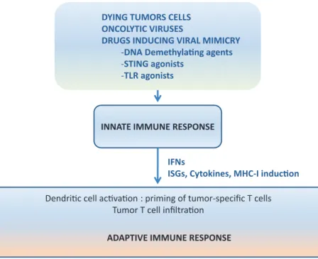
The immune system has two arms, (i) the innate immune arm that is rapidly activated after an appropriate stimulus but lacks antigen-specificity and memory, and (ii) the adaptive immune response that requires time to appear but is anti-gen-specific and long-lasting.6 Both arms of the immune system are intimately linked such that the innate arm provides the conditions for an efficient activation of the adaptive response (Figure 1). Elements of the innate defense system are physi-cal barriers, soluble factors such as complement proteins, interferons (IFNs) and IFN-stimulated proteins, and immune cells such as dendritic cells, macrophages, neutrophils and natural killer
(NK) cells. The adaptive arm of the immune sys-tem comprises T and B lymphocyte subpopula-tions that recognize pathogens in an antigen- specific way via divergent T-cell receptors and B-cell receptors, respectively. Among the latter are CD4+ T-helper and T-regulatory cells (Tregs), cytotoxic CD8+ T-cells, B-cells and antibody-producing plasma cells.6
Several virus infections including Papilloma viruses, Merkel cell polyomavirus, and hepatitis B and C viruses can induce tumors in humans. Interestingly, such tumors that are linked to virus infections seem to respond better to checkpoint inhibitors than tumors that are not virus-linked.7,8 The reason for this seems related to a more acti-vated innate immune response.9–16 Thus, like in virus infections, the innate immune system prob-ably provides a better microenvironment for the development of a potent specific antitumor response.17,18
Type I IFNs are key regulatory elements in this aspect. They are produced for example when virus components or cell-derived damage-associ-ated molecular patterns (DAMPs) bind and acti-vate pattern recognition receptors (PRRs).19 The secreted IFNs can then activate dendritic cells (DCs) in tumor-draining lymph nodes and enhance the cross-presentation of tumor- associated antigens to CD8+ T-cells,20 which
Activation of viral defense
signaling in cancer
Maria Gonzalez-Cao, Niki Karachaliou, Mariacarmela Santarpia, Santiago Viteri, Andreas Meyerhans and Rafael Rosell
Abstract: A coordinated action of innate and adaptive immune responses is required to
efficiently combat a microbial infection. It has now become clear that cancer therapies also largely benefit when both arms of the immune response are engaged. In this review, we will briefly describe the current knowledge of innate immunity and how this can be utilized to prime tumors for a better response to immune checkpoint inhibitors. Comments on compounds in development and ongoing clinical trials will be provided.
Keywords: cancer immunotherapy, innate immunity, TLRs, viral defense signaling
Received: 1 November 2017; revised manuscript accepted: 17 July 2018.
Correspondence to: Gonzalez-Cao Maria Rosell Oncology Institute (IOR), Dexeus University Hospital, Quironsalud Group, C/ Sabino Arana, 5, Barcelona 08028, Spain mgonzalezcao@ oncorosell.com
Niki Karachaliou
Rosell Oncology Institute (IOR), Sagrat Cor University Hospital, Quironsalud Group, Barcelona, Spain
Mariacarmela Santarpia
Medical Oncology Unit, Department of Human Pathology ‘G. Barresi’, University of Messina, Messina, Italy
Santiago Viteri
Rosell Oncology Institute (IOR), Dexeus University Hospital, Quironsalud Group, Barcelona, Spain Rosell Oncology Institute (IOR), Teknon Medical Center, Quironsalud Group, Barcelona, Spain
Andreas Meyerhans
Infection Biology Laboratory, Department of Experimental and Health Sciences (DCEXS), Universitat Pompeu Fabra, Barcelona, Spain Institució Catalana de Recerca i Estudis Avançats (ICREA), Barcelona, Spain
Rafael Rosell
Rosell Oncology Institute (IOR), Dexeus University Hospital, Quironsalud Group, Barcelona, Spain Rosell Oncology Institute (IOR), Sagrat Cor University Hospital, Quironsalud Group, Barcelona, Spain Catalan Institute of Oncology, Germans Trias I Pujol University Hospital, Badalona, Spain
subsequently may lead to tumor-specific CD8+ T-cell expansion and tumor destruction.
A detailed understanding of the innate immune response against viruses may provide opportuni-ties for developing more efficient treatments in the field of cancer immunotherapy. Indeed, the com-bination of checkpoint inhibitors with agents that trigger the innate immune response enhances their antitumor effect. Here, we will review the recently described means of activating innate immune responses to improve immunotherapy for cancer patients. Novel strategies that activate directly or indirectly PRRs will be commented on.
Pathogen recognition receptors (PRRs)
The innate immune response initiates with the recognition of foreign nucleic acids or other cules in host cells by PRRs. PRRs recognize mole-cules derived from pathogen-associated molecular patterns, as well as DAMPs released from endog-enous tissues that have suffered some damage.21 The understanding of the immunostimulatory as
well as pro- or antitumoral function of PRRs is necessary to exploit them for enhancing cancer immunotherapy.
Several different subtypes of PRRs are described today: Toll-like receptors (TLRs), NOD-like receptors (NLRs), c-type lectin receptors (CLRs), cytosol dsDNA sensors (CDSs) and retinol acid inducible gene 1 (RIG-1)-like receptors (RLRs).22,23 PRRs are classified according to their cellular location. They are located in cell membranes, such as TLRs, or in the cytoplasm like NLRs, CLRs, CDSs and RIG-1-like recep-tors (RLRs).21 PRR activation induces the pro-duction of type I IFNs (mainly IFN-α proteins and IFN-β).22 Subsequently, type I IFNs control the transcription of genes that are restricting viral infections (so-called ‘virus restriction factors’). In addition, type I IFNs activate NK cells, pro-mote antigen presentation24 and participate in the differentiation of specific CD8+ cytotoxic T lymphocytes (CTLs). Finally, type I IFNs have antiproliferative functions that are through TP53 gene induction.25,26
Figure 1. From stimulating innate immune responses to an adaptive antitumor response. A schematic view is given. In the tumor microenvironment, activation of PRRs is achieved by the recognition of DAMPs released by dying tumor cells and by drugs such as TLR agonists, STING agonists, DNA demethylating agents and treatments based on the use of modified oncolytic viruses. The activation of the innate immune response leads to an efficient priming by dendritic cells of T-cells in lymph nodes and to the infiltration by tumor-specific T-cells into the tumor. Details are described in the text.
DAMP, danger-associated molecular pattern; ISG, interferon-stimulated gene; PRR, pattern recognition receptor; TLR, Toll-like receptor; STING, stimulator of interferon genes complex.
TLRs constitute a receptor family that is mainly expressed on macrophages and DCs. In humans, the family has 11 members27 located on the extra-cellular membrane (TLR 1, 2, 4, 5, 6 and 11) or in the intracellular counterpart of endosomes (TLR 2, 3, 7, 8, 9 and 10). Cell membrane-bound TLRs recognize glycoproteins, while endosome-placed TLRs respond to nucleic acid molecules, in particular viral RNA.27–29
TLR4 was the first TLR identified. Activation of TLR4 signaling is preceded by binding of lipopolysaccharides produced by Gram-negative bacteria.30,31 In cancer, TLR4 activation has a dual role. Although its upregulation is associated with chemoresistance,32 metastasis and immuno-suppression33 in several tumor types, TLR4 acti-vation has also an anticancer effect. While TLR4 antagonists could help reduce metastasis, TLR4 agonists have been shown to induce antitumor immunity in patients and models of cancer.
Several TLR4 agonists, such as OM-174,34 or the Streptococcus-derived agent OK-432,35,36 Coley toxin (a mixture of killed Streptococcus pyogenes and Serratiamarcescens bacteria) and Bacillus Calmette-Guerin, have antitumoral effects.36–38 Double-stranded RNA (dsRNA) is detected via TLR3 and RLRs (RIG1 and MDA5). Among immune cells, myeloid DCs and macrophages express TLR3. TLR3 is also expressed in fibro-blasts and hepatocytes. When activated, TLR3, through TIR-domain-containing adapter-induc-ing interferon-β (TRIF), tumor necrosis factor (TNF) receptor-associated factor 6 (TRAF6) and tankyrase 1 (TANK1), activates the tran-scription factors interferon response factor 3 (IRF-3) and nuclear factor kappa B (NF-κB). This then leads to the expression of type I IFNs, mainly IFN-β39 (Figure 2). RLRs use protein adaptor mitochondrial antiviral signaling to acti-vate IRF-3, IRF-7 and NF-κB40 (Figure 2). RLRs Figure 2. Main subtypes of PRRs as targets for cancer treatment: Toll-like receptors (TLR3, TLR7/8,
TLR9), cytosol dsDNA sensors (cGAS/IFI that activate STING) and the retinol acid inducible gene RIG-1 like receptors (RIG1 and MDA5). Activation of these receptors, following detection of nucleic acids from virus, induces production of type I IFNs (IFN-α and IFN-β). dsRNA (from HERVs re-expressed after treatment with azacytidine or from exogenous infection) activates TLR3, MDA5 and RIG1; ssRNA activates TLR7/8; DNA (from pathogens or from tumor cells) activates STING.
HERV, human endogenous retrovirus; IFN, interferon; IL, interleukin; IRF-3, IFN-regulatory factor-3; NFκB, nuclear factor kappa B; PRR, pathogen recognition receptor; ssRNA, single stranded RNA; STING, stimulator of interferon genes complex; TBK1, TANK-binding kinase 1; TNF, tumor necrosis factor; TRAF6, TNF receptor-associated factor 6; TRIF, Toll/IL-1 receptor domain-containing adaptor inducing IFN-β.
are expressed in many tissues and play a promi-nent role in myeloid cells, fibroblasts, hepatocytes and central nervous system cells. While highly expressed in plasmacytoid DCs, RLRs are not essential for IFN type I production by these cells.41
The synthetic dsRNA BO-112 that activates mela-noma differentiation-associated protein 5 (MDA5) demonstrated tumor-specific immune responses with a good toxicity profile in a first in-human trial.42 A phase I clinical trial testing the combi-nation of BO-112 with anti-PD-1 antibodies is ongoing in several centers of Spain (Table 1). Other interesting antitumoral compounds in devel-opment are the synthetic analogs of dsRNA
polyinosinic:polycytidylic acid (poly I:C) that acti-vate TLR337,43–45 and RIG1/MDA546 and 5′ triphosphate small interfering RNA (ppp-siRNA) that activates RIG1 and silences specific onco-genes like BCL-2 via RNA-interference. Both types of compounds have demonstrated antitumor activities in vivo.47
The detection of single stranded RNA (ssRNA) is due to TLR7 and TLR8 which activate NF-κB, IRF-3 and IRF-7, and lead to the expression of type I IFN, TNF-α, interleukin (IL)-1 and IL-1248 (Figure 2). TLR7 is mainly expressed in plasma-cytoid DCs and B-cells, while TLR8 is expressed in myeloid DCs and Tregs. Imidazoquinolinamin derivates, such as imiquimod, approved for the Table 1. Cancer clinical trials in progress with drugs targeting innate immune response.
Drug class Drug Target In combination with Tumor type NCT number
PRR MEDI9197 TLR7/8 Durvalumab (anti-PD-L1) Solid tumors 02556463
MGN1703 TLR9 Ipilimumab (anti-CTLA-4) Solid tumors 02668770
GLA-SE TLR4 Radiotherapy Sarcoma 02180698
SD-101 TLR9 Ibrutinib, radiotherapy Pembrolizumab (anti-PD-1) Folicular lymphoma Melanoma 02927964 02521870 GSK1795091 TLR4 - Solid tumors 02798978
Poly ICLC TLR3 Durvalumab (anti-PD-L1) +
tremelimumab (anti-CTLA-4)
Solid tumors 02643303
G100 TLR4 Pembrolizumab
(anti-PD-1) Solid tumors 02501473
MIW815 (ADU-S100) STING PDR001 (anti-PD-L1) Solid tumors 03172936
BO-112 MDA5 Anti-PD-1 Solid tumors 02828098
Epigenetic Azacitidine +
entinostat
DNMT
HDAC Nivolumab (anti-PD-1) NSCLC 01928576
Azacitidine DNMT Pembrolizumab (anti-PD-1) Melanoma 02816021
Decitabine DNMT MBG453 (TRIM3)
PDR001 (anti-PD-L1) MDS, Leukemia 03066648
Decitabine DNMT Nivolumab (anti-PD-1) MDS 03259516
Decitabine DNMT Ipilimumab (anti-CTLA-4) MDS, Leukemia 02890329
Guadecitabine DNMT Durvalumab (anti-PD-L1) +
tremelimumab (anti-CTLA-4)
SCLC 03085849
CTLA-4, cytotoxic T lymphocyte-associated molecule-4; DNMT, DNA methyltransferase; HDAC, histone deacetylase; MDA, melanoma differentiation-associated protein; MDS, myelodysplastic syndromes; NCT, National Clinical Trials; NSCLC, non-small cell lung cancer; PD-1, programmed-cell death 1; PRR, pathogen recognition receptor; SCLC, small cell lung cancer; STING, stimulator of interferon genes complex; TLR, Toll-like receptor.
treatment of basal cell carcinoma, show antitu-moral effects through TLR7 and TLR8 activa-tion.49 Intratumoral resiquimod (R848), a ligand of TLR7 and TLR8, in combination with anti-OX40 in animal models induces a systemic antitu-mor effect.50 MEDI9197 (formerly 3M-052), is a novel TLR7/8 dual agonist formulated for intratu-moral injection that has been studied in patients with cutaneous tumors, demonstrating safety and immunogenicity.51
TLR9 recognizes unmethylated CpG motifs from bacteria and viruses52 and RNA:DNA hybrids.53 Its triggering activates plasmacytoid DCs and B-cells.54 Several synthetic CpG oligonucleotides have been developed as TLR9 agonists mimicking natural CpG motifs (Table 1). The combination of intratumoral CpG SD-101 with anti-OX40, which triggers a T-cell immune response, demon-strated complete responses and long-term sur-vival in animal models.50 Similarly, data from a phase I clinical trial in advanced melanoma showed objective responses in four of the five patients treated with the combination of intratu-moral CpG SD-101 and the anti-PD-1 antibody pembrolizumab.55
The CDSs cGAS and IFI16 operate via stimulator of interferon genes complex (STING) to detect free DNA in the cytosol. When cytosolic DNA is detected by cGAS it catalyzes the STING ligand cGAMP.56 STING is then activated, translocates to perinuclear endosomes and recruits TBK1 and IRF3. These molecules are phosphorylated and translocate to the nucleus with subsequent tran-scription of type I IFNs56 (Figure 2). STING is essential for the production of type I IFN signaling in DCs which enables them to present tumor anti-gens and prime CD8+ T lymphocytes leading to T-cell infiltration into the tumor.18,57 STING is located at the cytosolic site of the endoplasmic reticulum membrane56 and is activated by DNA from damaged tumor cells that reach the cytosol of DCs.58 Some intratumoral-injected compounds can activate STING in mice, like 5,6-dimethylxan-thenone-4-acetic acid (DMXAA).59 In humans, the STING agonist ML RR-S2 CDA (MIW815, ADU-S100) is in clinical development60 (Table 1).
Interferons
IFNs were discovered in the late 1950s during replication studies of influenza virus.61 They are cytokines that play a critical role in innate immune responses against viral infections and
participate in the activation of the adaptive immune response.26 Several types of IFNs have been described including type I IFNs (various IFN-α, IFN-β, and others), the type II IFNγ, and the more recently classified type III IFNs (IFN-λ). Type I IFNs are produced by most cell types: fibroblasts, DCs and hepatocytes (through RIG-1), plasmacytoid DCs (through TLR9 and TLR7/8) and macrophages and hepatocytes (through TLR3 and TLR4). Type II IFNs are mainly secreted by T-helper-1 (Th-1) lymphocytes, CD8+ lymphocytes, NK and NK T-cells.62
All the different types of IFNs signal through IFN receptors that are differently distributed among cell types. IFN-β binds to the IFN-α/β receptor (IFNAR) to further activate production of more type I IFN63 (Figure 3). IFNAR activates janus kinase (JAK) family members JAK1 and Tyk-2, and subsequently signal transducer and activator of transcription 1 (STAT1) and 2 (STAT2). STAT1/2 bind to IRF9 (p48) and form the IFN-stimulated gene factor 3 (ISGF3; Figure 3). ISGF3 initiates the transcription of several inter-feron-stimulated genes (ISGs) by binding to the promoter region of IFN-stimulated response ele-ments (ISRE; Figure 3). ISGs include PKR, IRF-1 and IRF7. When activated by TBK-1/ IKKe, ISGs regulate IFNα gene transcription.64 ISGs activate antimicrobial programs that both degrade viral proteins and inhibit cancer cell pro-liferation65 (Figure 3). Type I IFNs also stimulate the adaptive immune response. Specifically, they promote major histocompatibility complex (MHC) class I and II expression on antigen-pre-senting cells like DCs that is required for efficient T-cell stimulation. Mature DCs are then able to initiate the adaptive immune response by activat-ing antigen-specific naïve T-cells to proliferate and produce type II IFN.66,67 The type II IFN receptor is called IFNGR. It activates genes con-taining a gamma-activated sequence (GAS) through JAK1/2 and STAT1 signaling (Figure 3). Type II IFNs stimulate the adaptive immune response and activate macrophages and NK cells. The expression levels of ISGs are predictive of the response to immune checkpoint inhibitors in mel-anoma patients treated with CTLA-4 anti-bodies.68 Likewise, tumor samples from melanoma, ovarian, lung, breast or colorectal cancer can be classified according to high or low expression levels of IFN-stimulated viral defense genes for example IRF7, RIG1 STAT1, IFNB1,
IFI6 induced by the hypomethylating agent (HMA) 5-aza-cytidine (5-aza).69 Our own work shows that lung cancer and melanoma patients with high tumoral expression of IFN-γ have a better outcome with immunotherapy compared with patients with low IFN-γ expression.70 The role of DNA methylation in innate immune responses
Epigenetic modulator drugs can restore immuno-genicity and immune recognition of tumors. Therefore, there is an increasing interest in com-bined epigenetic therapy and immunotherapy.71 DNA methyltransferases (DNMTs) are impor-tant players in epigenetic modulation of the innate immune response. In non-small cell lung cancer with mesenchymal phenotype, STAT3 activates DNMT1, which methylates the promoter regions of RIG1 as well as IRF1, immunoproteasomes (PSMB8, PSMB9) and HLA molecules leading
to the reduction of their expression.72 STAT3 also inhibits STAT1 expression, a key regulator of the antigen-presentation machinery in epithe-lial cells.70,72
HMAs such as the nucleoside analogs of cytidine,5-aza and 5-aza-2-deoxycytidine (5- aza-2dc or decitabine) inhibit DNMTs and are currently approved for the treatment of hemato-logic malignancies.73,74 HMAs activate the innate immune response through PRRs, but they also have several other activities that make them a good partner to combine with immune check-point inhibitors. For instance, HMAs reactivate silenced tumor suppressor genes that encode proteins which limit the proliferative and survival capacity of a cell.75,76 HMAs induce T-cell responses by stimulating HLA I expression. For example, in brain tumors, decitabine promotes the surface presentation of tumor-associated peptides in the context of HLA I and is thus a good candidate to be combined with other Figure 3. Interferon pathway. Type I IFN binds to the IFNAR. IFNAR activates the JAK family members JAK1 and Tyk-2, with subsequent phosphorylation of signal transducer and activator of transcription 1 (STAT1) and 2 (STAT2) proteins. These proteins form the complex called ISGF3 when they bind to IRF9 (p48). ISGF3 initiates transcription of several ISGs by binding to ISREs in their promoter regions. The receptor of type II IFN is called IFNGR and also initiates induction of JAK1 and JAK2 recruitment with STAT1 homodimers that activate genes containing a GAS.
GAS, gamma-activated sequence; IFN, interferon; IFNAR, α/β receptor; ISG, interferon-stimulated gene; ISGF3, IFN-stimulated gene factor 3; ISRE, IFN-IFN-stimulated response element; JAK, Janus kinase.
immunotherapeutic regimen.77 Furthermore, treatment of tumor cell lines with the DNA methyltransferase inhibitor 5-azacytidine (AZA) enrich tumors in genes involved in immunomod-ulatory pathways, defining an ‘AZA IMmune gene set (AIMs)’ that can classify primary tumors into ‘high’ and ‘low’ AIM gene expression sub-sets (see Table 1, in Li and colleagues78). HMAs induce the expression of chemokines that ulti-mately re-educate tumor cells to become more immunogenic.71,79
Last but not least, HMAs have antitumor activi-ties by causing the re-expression of endogenous retroviruses (ERVs) in preclinical cancer mod-els. Human ERVs (HERVs) are retroviral ele-ments that have been fixed within the human genome through evolution. About 8% of the human genome is composed of these HERVs, most of which are non-functional due to the accumulation of mutations and epigenetic con-trol. Chiappinelli and colleagues showed in preclinical models that HMAs through ERV expression induce viral mimicry and IFN sign-aling to increase tumor immunogenicity and recognition.13 Other investigators have reported similar findings in colon cancer, demonstrating an IFN response through activation of MDA5 by dsRNA.80 This effect, through induction of HERVs, was synergistic with the effect of anti-CTLA-4 or anti-PD-1/PD-L1 antibodies.79,13,81 One way to activate TLRs is through induction of dsRNA from HERVs using low doses of HMAs. HERV-derived viral transcripts then increase within the cytosol leading to RIG1, MDA5 and TLR3 activation and subsequently type I IFN production.82 The HMA-induced upregulation of HERVs is synergistic with anti-PD-1/PD-L1 antibodies80 and anti-CTLA-4 antibodies.82,83
In a subset of non-small cell lung cancer patients, 20% of durable responses were observed when the anti-PD-1 antibody was given after tumor progression under low dose of HMAs, indicating that HMAs may prime tumors for a subsequent response to immunotherapy.79,84 Based on the above and other evidences, there is currently a long list of clinical trials in several types of tumors combining immune checkpoint inhibitors with epigenetic drugs (Table 1 and Table 1 in Dunn and colleagues71).
Interestingly, apart from HMAs, cyclin-depend-ent kinase 4/6 (CDK4/6) inhibitors also suppress
DNMT1 and induce viral mimicry. The combi-nation of immune checkpoint inhibitors with CDK4/6 inhibitors was found to be synergistic in vitro and in vivo85,86 and clinical trials are now ongoing with this combination [ClinicalTrials. gov identifiers: NCT02791334, NCT02079636, and NCT02779751].
Other factors in the innate immune response and antitumor therapy
NK cells play an important role against tumor cells. Cells infected by viruses, as well as cancer cells, may downregulate surface MHC class I molecules in order to avoid recognition by CD8+ T-cells.87 Paradoxically, this phenomenon makes them susceptible to NK cells, that are activated when their membrane receptors, killer-cell immu-noglobulin-like receptors (KIRs), are not bound to MHC class I molecules. Some cancer cells bypass NK control because they downregulate specific MHC class I molecules, that are needed to present peptides to CD8+ T-cells, while they still express MHC class I peptides that serve as KIR ligands.88
Viral restriction factors comprise a group of sev-eral proteins expressed by cells to suppress viral replication. These restriction factors constitute an early defense against viral infections, and are partly induced by IFNs.89 The best characterized restriction factors are: apolipoprotein B messenger RNA editing enzyme catalytic polypeptide-like3 (APOBEC3) proteins, tripartite-motif-containing 5a (TRIM5a), SAM domain and HD domain-containing protein 1 (SAMHD1), Schlafen 11 (SLFN11) and Tetherin.89 Their role in the immune response to cancer cells has not been studied, however it is known that some of them are involved in the process of somatic mutagenesis during tumor development90,91 APOBEC3 altera-tions (mutaaltera-tions or overexpression) in cancer cells have been linked to a specific hypermutation sta-tus named ‘kataegis’ that correlates with respon-siveness to immunotherapy.92 Finally, a strong correlation has been reported among APOBEC3 proteins, IFNγ and PD-L1 expression in cancer cells.92
There are several compounds in development with the aim to activate an innate immune response and direct CD8+ T-cells into tumors (Table 1). Such strategies include the combina-tion of anti-PD-1 antibodies with oncolytic viro-therapy which has shown impressive results in
early clinical trials.93 Melanoma patients treated with the combination of the anti-PD-1 antibody pembrolizumab plus talimogene laherparepvec (T-VEC) had an objective response rate of 62% with 32% of complete responses.10 Although the exact mechanism by which oncolytic viruses increase the activity of immune checkpoint block-ade is unknown, biopsies taken from patients before treatment and after 6 weeks, demonstrated an increase in CD8+ T-cell infiltration as well as increased expression of IFN-γ suggesting a role of innate immune response activation by T-VEC.10 Similarly, a hybrid of an oncolytic, nonpathogenic poliovirus (PV) and a human rhi-novirus (PVSRIPO) demonstrated an increased release of DAMPs through tumor lysis activating a type I IFN response in DCs in different cancer models.9
Conclusions and future perspectives
Innate immune responses in the form of IFN production and their subsequent effects provide the appropriate microenvironment for the effi-cient stimulation of adaptive responses that are key in fighting microbial infections. As tumors are derived from tissue cells that carry mainly self-antigens, they are expected to be purely immunogenic, and may lack innate immune acti-vation, at least initially. Subsequently, the grow-ing tumor can exploit all of the immune suppressive mechanisms that are well described in chronic infections.
Recent clinical trials and preclinical cancer mod-els now impressively demonstrate that stimuli of innate immunity in combination with other immunotherapeutic regimens can significantly augment tumor-specific responses that translate into increased response and cure rates (Figure 1). Thus, suppressive mechanisms and low immuno-genicity may be overcome at least when sufficient numbers of tumor neoantigens are present in the tissue.
While presently the PRRs agonists and HMAs stand out, other drugs and drug combinations are under study in numerous trials (Table 1). Furthermore, there are now ongoing trials that include cancer patients with persistent virus infec-tions that have previously been excluded. For example, we are currently exploring several above-mentioned factors and their relationship with innate immune responses in a phase II Spanish Lung Cancer Group clinical trial. The
anti-PD-L1 inhibitor durvalumab is given to HIV-1-infected patients with solid tumors [ClinicalTrials.gov identifier: NCT03094286]. The trial enables us to investigate the effect of immune checkpoint inhibition in reversing cancer pathways and to characterize HIV-specific T-cell functions during persistent HIV infection. The study results of this trial and of others are eagerly awaited.
It seems that after many years of stagnation, we finally face most fruitful and exciting times in tumor immunology.
Funding
AM is supported by a grant from the Spanish Ministry of Economy, Industry and Compet-itiveness and FEDER grant no. SAF2016-75505-R (AEI/MINEICO/FEDER, UE), and the ‘María de Maeztu’ Programme for Units of Excellence in R&D (MDM-2014-0370).
Work in Dr Rosell’s laboratory is partially sup-ported by a grant from La Caixa Foundation, and by a European Grant (ELBA no 765492). Conflict of interest statement
The authors declare that there is no conflict of interest.
References
1. Ribas A and Wolchok JD. Cancer
immunotherapy using checkpoint blockade. Science 2018; 359: 1350–1355.
2. Robert C, Schachter J, Long GV, et al.
Pembrolizumab versus ipilimumab in advanced melanoma. N Engl J Med 2015; 372: 2521– 2532.
3. Reck M, Rodriguez-Abreu D, Robinson AG, et al. Pembrolizumab versus chemotherapy for PD-L1-positive non-small-cell lung cancer. N Engl J Med 2016; 375: 1823–1833.
4. Motzer RJ, Escudier B, McDermott DF, et al. Nivolumab versus everolimus in advanced renal-cell carcinoma. N Engl J Med 2015; 373: 1803– 1813.
5. Tumeh PC, Harview CL, Yearley JH, et al. PD-1 blockade induces responses by inhibiting adaptive immune resistance. Nature 2014; 515: 568–571.
6. Murphy K and Weaver C. Janeway’s
immunobiology. 9th ed. New York, NY: Garland Science, 2016.
7. Colunga A, Pulliam T and Nghiem P. Merkel cell carcinoma in the age of immunotherapy: facts and hopes. Clin Cancer Res 2018; 24: 2035–2043. 8. Seiwert TY, Burtness B, Mehra R, et al.
Safety and clinical activity of pembrolizumab for treatment of recurrent or metastatic squamous cell carcinoma of the head and neck (KEYNOTE-012): an open-label, multicentre, phase 1b trial. Lancet Oncol 2016; 17: 956–965. 9. Brown MC, Holl EK, Boczkowski D, et al.
Cancer immunotherapy with recombinant poliovirus induces IFN-dominant activation of dendritic cells and tumor antigen-specific CTLs. Sci Transl Med 2017; 9: eaan4220.
10. Ribas A, Dummer R, Puzanov I, et al. Oncolytic virotherapy promotes intratumoral T cell infiltration and improves anti-PD-1 immunotherapy. Cell 2017; 170: 1109–1119. e10.
11. Chiappinelli KB, Strissel PL, Desrichard A, et al. Inhibiting DNA methylation causes an interferon response in cancer via dsRNA including
endogenous retroviruses. Cell 2017; 169: 361. 12. Gonzalez-Cao M, Iduma P, Karachaliou N, et al.
Human endogenous retroviruses and cancer. Cancer Biol Med 2016; 13: 483–488.
13. Chiappinelli KB, Strissel PL, Desrichard A, et al. Inhibiting DNA methylation causes an interferon response in cancer via dsRNA including
endogenous retroviruses. Cell 2015; 162: 974– 986.
14. Balermpas P, Martin D, Wieland U, et al. Human papilloma virus load and PD-1/PD-L1, CD8(+) and FOXP3 in anal cancer patients treated with chemoradiotherapy: rationale for immunotherapy. Oncoimmunology 2017; 6: e1288331.
15. Solomon B, Young RJ, Bressel M, et al. Prognostic significance of PD-L1+ and CD8+ immune cells in HPV+ oropharyngeal squamous cell carcinoma. Cancer Immunol Res 2018; 6: 295–304.
16. Oguejiofor K, Galletta-Williams H, Dovedi SJ, et al. Distinct patterns of infiltrating CD8+ T cells in HPV+ and CD68 macrophages in HPV- oropharyngeal squamous cell carcinomas are associated with better clinical outcome but PD-L1 expression is not prognostic. Oncotarget 2017; 8: 14416–14427.
17. Fuertes MB, Woo SR, Burnett B, et al. Type I interferon response and innate immune sensing of cancer. Trends Immunol 2013; 34: 67–73.
18. Spranger S, Dai D, Horton B, et al. Tumor-residing Batf3 dendritic cells are required for effector T cell trafficking and adoptive T cell therapy. Cancer Cell 2017; 31: 711–723.e4.
19. Dear AE. Epigenetic modulators and the new immunotherapies. N Engl J Med 2016; 374: 684–686.
20. Fuertes MB, Kacha AK, Kline J, et al. Host type I IFN signals are required for antitumor CD8+ T cell responses through CD8{alpha}+ dendritic cells. J Exp Med 2011; 208: 2005–2016.
21. Thompson MR, Kaminski JJ, Kurt-Jones EA, et al. Pattern recognition receptors and the innate immune response to viral infection. Viruses 2011; 3: 920–940.
22. Seth RB, Sun L and Chen ZJ. Antiviral innate immunity pathways. Cell Res 2006; 16: 141–147. 23. Barton GM and Medzhitov R. Toll-like receptors
and their ligands. Curr Top Microbiol Immunol 2002; 270: 81–92.
24. Altfeld M and Gale M, Jr. Innate immunity against HIV-1 infection. Nat Immunol 2015; 16: 554–562.
25. Porta C, Hadj-Slimane R, Nejmeddine M, et al. Interferons alpha and gamma induce p53-dependent and p53-inp53-dependent apoptosis, respectively. Oncogene 2005; 24: 605–615. 26. Janeway CA, Jr. and Medzhitov R. Innate
immune recognition. Annu Rev Immunol 2002; 20: 197–216.
27. Blasius AL and Beutler B. Intracellular toll-like receptors. Immunity 2010; 32: 305–315. 28. Xiao T. Innate immune recognition of nucleic
acids. Immunol Res 2009; 43: 98–108.
29. Roers A, Hiller B and Hornung V. Recognition of endogenous nucleic acids by the innate immune system. Immunity 2016; 44: 739–754.
30. Poltorak A, He X, Smirnova I, et al. Defective LPS signaling in C3H/HeJ and C57BL/10ScCr mice: mutations in Tlr4 gene. Science 1998; 282: 2085–2088.
31. Kang JY, Nan X, Jin MS, et al. Recognition of lipopeptide patterns by Toll-like receptor 2-Toll-like receptor 6 heterodimer. Immunity 2009; 31: 873–884.
32. Ran S. The role of TLR4 in chemotherapy-driven metastasis. Cancer Res 2015; 75: 2405–2410. 33. Huang L, Xu H and Peng G. TLR-mediated
metabolic reprogramming in the tumor
microenvironment: potential novel strategies for cancer immunotherapy. Cell Mol Immunol 2018; 15: 428–437.
34. Onier N, Hilpert S, Arnould L, et al. Cure of colon cancer metastasis in rats with the new lipid A OM 174. Apoptosis of tumor cells and immunization of rats. Clin Exp Metastasis 1999; 17: 299–306.
35. Okamoto M, Oshikawa T, Tano T, et al. Mechanism of anticancer host response induced by OK-432, a streptococcal preparation, mediated by phagocytosis and Toll-like receptor 4 signaling. J Immunother 2006; 29: 78–86.
36. Akeda T, Yamanaka K, Kitagawa H, et al. Intratumoral injection of OK-432 suppresses metastatic squamous cell carcinoma lesion inducing interferon-gamma and tumour necrosis factor-alpha. Clin Exp Dermatol 2012; 37: 193–194.
37. Awasthi S. Toll-like receptor-4 modulation for cancer immunotherapy. Front Immunol 2014; 5: 328.
38. Galluzzi L, Vacchelli E, Eggermont A, et al. Trial watch: experimental Toll-like receptor agonists for cancer therapy. Oncoimmunology 2012; 1: 699–716.
39. Jiang Z, Mak TW, Sen G, et al. Toll-like receptor 3-mediated activation of NF-kappaB and IRF3 diverges at Toll-IL-1 receptor domain-containing adapter inducing IFN-beta. Proc Natl Acad Sci USA 2004; 101: 3533–3538.
40. Matsumiya T and Stafforini DM. Function and regulation of retinoic acid-inducible gene-I. Crit Rev Immunol 2010; 30: 489–513.
41. Loo YM and Gale M, Jr. Immune signaling by RIG-I-like receptors. Immunity 2011; 34: 680–692.
42. Marquez Rodas I, Rodriguez-Ruiz ME, Lopez-Tarruella S, et al. First-in-human clinical trial with intratumoral BO-112 in solid malignancies: a novel immunotherapy based in double-stranded RNA (dsRNA). J Clin Oncol 2017; 35(15 Suppl.): 3082.
43. Amos SM, Pegram HJ, Westwood JA, et al. Adoptive immunotherapy combined with intratumoral TLR agonist delivery eradicates established melanoma in mice. Cancer Immunol Immunother 2011; 60: 671–683.
44. Chicoine MR, Won EK and Zahner MC. Intratumoral injection of lipopolysaccharide causes regression of subcutaneously implanted mouse glioblastoma multiforme. Neurosurgery 2001; 48: 607–614; discussion 14–5.
45. Brody JD, Ai WZ, Czerwinski DK, et al. In situ vaccination with a TLR9 agonist induces systemic lymphoma regression: a phase I/II study. J Clin Oncol 2010; 28: 4324–4332.
46. Kubler K, tho Pesch C, Gehrke N, et al.
Immunogenic cell death of human ovarian cancer cells induced by cytosolic poly(I:C) leads to
myeloid cell maturation and activates NK cells. Eur J Immunol 2011; 41: 3028–3039.
47. Poeck H, Besch R, Maihoefer C, et al.
5’-Triphosphate-siRNA: turning gene silencing and Rig-I activation against melanoma. Nat Med 2008; 14: 1256–1263.
48. Diebold SS. Recognition of viral single-stranded RNA by Toll-like receptors. Adv Drug Deliv Rev 2008; 60: 813–823.
49. Li ZJ, Sohn KC, Choi DK, et al. Roles of TLR7 in activation of NF-kappaB signaling of keratinocytes by imiquimod. PLoS One 2013; 8: e77159.
50. Sagiv-Barfi I, Czerwinski DK, Levy S, et al. Eradication of spontaneous malignancy by local immunotherapy. Sci Transl Med 2018; 10: eaan4488.
51. Gupta S, Grilley-Olson J, Hong D, et al. Safety and pharmacodynamic activity of MEDI9197, a TLR 7/8 agonist, administered intratumorally in subjects with solid tumors. Cancer Res 2017; 77: abstract CT091.
52. Ashkar AA and Rosenthal KL. Toll-like receptor 9, CpG DNA and innate immunity. Curr Mol Med 2002; 2: 545–556.
53. Rigby RE, Webb LM, Mackenzie KJ, et al. RNA: DNA hybrids are a novel molecular pattern sensed by TLR9. Embo J 2014; 33: 542–558.
54. Bafica A, Scanga CA, Feng CG, et al. TLR9 regulates Th1 responses and cooperates with TLR2 in mediating optimal resistance to Mycobacterium tuberculosis. J Exp Med 2005; 202: 1715–1724.
55. Ribas A, Gonzalez R and Kummar S. Phase 1b/2, Open-label, multicenter, dose escalation and expansion trial of intratumoral SD-101 in combination with pembrolizumab in patients with metastatic melanoma. SMR Meeting 2016. 56. Woo SR, Fuertes MB, Corrales L, et al.
STING-dependent cytosolic DNA sensing mediates innate immune recognition of immunogenic tumors. Immunity 2014; 41: 830–842. 57. Spaulding E, Fooksman D, Moore JM, et al.
STING-licensed macrophages prime type I IFN production by plasmacytoid dendritic cells in the bone marrow during severe plasmodium yoelii malaria. PLoS Pathogens 2016; 12: e1005975.
58. Corrales L and Gajewski TF. Molecular pathways: targeting the stimulator of interferon genes (STING) in the immunotherapy of cancer. Clin Cancer Res 2015; 21: 4774–4779.
59. Corrales L, Glickman LH, McWhirter SM, et al. Direct activation of STING in the tumor microenvironment leads to potent and systemic tumor regression and immunity. Cell Rep 2015; 11: 1018–1030.
60. Corrales L and Gajewski TF. Endogenous and pharmacologic targeting of the STING pathway in cancer immunotherapy. Cytokine 2016; 77: 245–247.
61. Isaacs A and Lindenmann J. Virus interference. I. The interferon. Proc R Soc Lond B Biol Sci 1957; 147: 258–267.
62. Kambayashi T, Assarsson E, Lukacher AE, et al. Memory CD8+ T cells provide an early source of IFN-gamma. J Immunol 2003; 170: 2399–2408. 63. Honda K, Yanai H, Takaoka A, et al. Regulation
of the type I IFN induction: a current view. Int Immunol 2005; 17: 1367–1378.
64. Honda K, Yanai H, Negishi H, et al. IRF-7 is the master regulator of type-I interferon-dependent immune responses. Nature 2005; 434: 772–777. 65. Zitvogel L, Galluzzi L, Kepp O, et al. Type I
interferons in anticancer immunity. Nat Rev Immunol 2015; 15: 405–414.
66. Biron CA, Nguyen KB, Pien GC, et al. Natural killer cells in antiviral defense: function and regulation by innate cytokines. Annu Rev Immunol 1999; 17: 189–220.
67. Kadowaki N, Antonenko S, Lau JY, et al. Natural interferon alpha/beta-producing cells link innate and adaptive immunity. J Exp Med 2000; 192: 219–226.
68. Snyder A, Makarov V, Merghoub T, et al. Genetic basis for clinical response to CTLA-4 blockade in melanoma. N Engl J Med 2014; 371: 2189–2199.
69. Li H, Chiappinelli KB, Guzzetta AA, et al. Immune regulation by low doses of the DNA methyltransferase inhibitor 5-azacitidine in common human epithelial cancers. Oncotarget 2014; 5: 587–598.
70. Karachaliou N, Gonzalez-Cao M, Crespo G, et al. Interferon gamma, an important marker of response to immune checkpoint blockade in non-small cell lung cancer and melanoma patients. Ther Adv Med Oncol 2018; 10: 1758834017749748.
71. Dunn J and Rao S. Epigenetics and
immunotherapy: the current state of play. Mol Immunol 2017; 87: 227–239.
72. Tripathi SC, Peters HL, Taguchi A, et al. Immunoproteasome deficiency is a feature of non-small cell lung cancer with a mesenchymal
phenotype and is associated with a poor outcome. Proc Natl Acad Sci USA 2016; 113: E1555– E1564.
73. Ley TJ, Ding L, Walter MJ, et al. DNMT3A mutations in acute myeloid leukemia. N Engl J Med 2010; 363: 2424–2433.
74. Popovic R, Shah MY and Licht JD. Epigenetic therapy of hematological malignancies: where are we now? Ther Adv Hematol 2013; 4: 81–91. 75. Kazanets A, Shorstova T, Hilmi K, et al.
Epigenetic silencing of tumor suppressor genes: paradigms, puzzles, and potential. Biochim Biophys Acta 2016; 1865: 275–288.
76. Paz MF, Wei S, Cigudosa JC, et al. Genetic unmasking of epigenetically silenced tumor suppressor genes in colon cancer cells deficient in DNA methyltransferases. Hum Mol Genet 2003; 12: 2209–2219.
77. Shraibman B, Kadosh DM, Barnea E, et al. Human Leukocyte Antigen (HLA) peptides derived from tumor antigens induced by inhibition of DNA methylation for development of drug-facilitated immunotherapy. Mol Cell Proteomics 2016; 15: 3058–3070.
78. Dietrich PA, Yang C, Leung HH, et al. GPR84 sustains aberrant beta-catenin signaling in leukemic stem cells for maintenance of MLL leukemogenesis. Blood 2014; 124: 3284–3294. 79. Wrangle J, Wang W, Koch A, et al. Alterations
of immune response of non-small cell lung cancer with azacytidine. Oncotarget 2013; 4: 2067–2079.
80. Roulois D, Loo Yau H, Singhania R, et al. DNA-demethylating agents target colorectal cancer cells by inducing viral mimicry by endogenous transcripts. Cell 2015; 162: 961–973.
81. Liu M, Ohtani H, Zhou W, et al. Vitamin C increases viral mimicry induced by 5-aza-2’-deoxycytidine. Proc Natl Acad Sci USA 2016; 113: 10238–1044.
82. Nusinzon I and Horvath CM. Positive and negative regulation of the innate antiviral response and beta interferon gene expression by deacetylation. Mol Cell Biol 2006; 26: 3106–3113. 83. Wang L, Amoozgar Z, Huang J, et al. Decitabine enhances lymphocyte migration and function and synergizes with CTLA-4 blockade in a murine ovarian cancer model. Cancer Immunol Res 2015; 3: 1030–1041.
84. Juergens RA, Wrangle J, Vendetti FP, et al. Combination epigenetic therapy has efficacy in patients with refractory advanced non-small cell lung cancer. Cancer Discov 2011; 1: 598–607.
85. Goel S, DeCristo MJ, Watt AC, et al. CDK4/6 inhibition triggers anti-tumour immunity. Nature 2017; 548: 471–475.
86. Schaer DA, Beckmann RP, Dempsey JA, et al. The CDK4/6 inhibitor abemaciclib induces a T cell inflamed tumor microenvironment and enhances the efficacy of PD-L1 checkpoint blockade. Cell Rep 2018; 22: 2978–2994.
87. Jonjic S, Babic M, Polic B, et al. Immune evasion of natural killer cells by viruses. Curr Opin Immunol 2008; 20: 30–38.
88. Guma M, Angulo A and Lopez-Botet M. NK cell receptors involved in the response to human cytomegalovirus infection. Curr Top Microbiol Immunol 2006; 298: 207–223.
89. Simon V, Bloch N and Landau NR. Intrinsic host restrictions to HIV-1 and mechanisms of viral escape. Nat Immunol 2015; 16: 546–553.
90. Henderson S and Fenton T. APOBEC3 genes: retroviral restriction factors to cancer drivers. Trends Mol Med 2015; 21: 274–284.
91. Caval V, Suspene R, Shapira M, et al. A prevalent cancer susceptibility APOBEC3A hybrid
allele bearing APOBEC3B 3’UTR enhances chromosomal DNA damage. Nat Commun 2014; 5: 5129.
92. Boichard A, Tsigelny IF and Kurzrock R. High expression of PD-1 ligands is associated with kataegis mutational signature and APOBEC3 alterations. Oncoimmunology 2017; 6:
e1284719.
93. Dummer R, Hoeller C, Gruter IP, et al. Combining talimogene laherparepvec with immunotherapies in melanoma and other solid tumors. Cancer Immunol Immunother 2017; 66: 683–695.
Visit SAGE journals online journals.sagepub.com/ home/tam